HAL Id: hal-01726226
https://hal.archives-ouvertes.fr/hal-01726226
Submitted on 8 Mar 2018
HAL is a multi-disciplinary open access archive for the deposit and dissemination of sci-entific research documents, whether they are pub-lished or not. The documents may come from teaching and research institutions in France or abroad, or from public or private research centers.
L’archive ouverte pluridisciplinaire HAL, est destinée au dépôt et à la diffusion de documents scientifiques de niveau recherche, publiés ou non, émanant des établissements d’enseignement et de recherche français ou étrangers, des laboratoires publics ou privés.
extensive white matter lesions in people age 65 years or
older: the Dijon MRI study
Phillip Tully, Sarah Qchiqach, Edwige Pereira, Stéphanie Debette, Bernard
Mazoyer, Christophe Tzourio
To cite this version:
Phillip Tully, Sarah Qchiqach, Edwige Pereira, Stéphanie Debette, Bernard Mazoyer, et al.. Develop-ment and validation of a priori risk model for extensive white matter lesions in people age 65 years or older: the Dijon MRI study. BMJ Open, BMJ Publishing Group, 2017, 7 (12), �10.1136/bmjopen-2017-018328�. �hal-01726226�
Open Access
AbstrAct
Objectives The objective was to develop and validate
a risk model for the likelihood of extensive white matter lesions (extWML) to inform clinicians on whether to proceed with or forgo diagnostic MRI.
Design Population-based cohort study and multivariable
prediction model.
setting Two representative samples from France. Participants Persons aged 60–80 years without dementia
or stroke. Derivation sample n=1714; validation sample n=789.
Primary and secondary outcome measures Volume of
extWML (log cm3) was obtained from T2-weighted images
in a 1.5 T scanner. 20 candidate risk factors for extWML were evaluated with the C-statistic. Secondary outcomes in validation included incident stroke over 12 years follow-up.
results The multivariable prediction model included
six clinical risk factors (C-statistic=0.61). A cut-off of 7 points on the multivariable prediction model yielded the optimum balance in sensitivity 63.7% and specificity 54.0% and the negative predictive value was high (81.8%), but the positive predictive value was low (31.5%). In further validation, incident stroke risk was associated with continuous scores on the multivariable prediction model (HR 1.02; 95% CI 1.01 to 1.04, P=0.02) and dichotomised scores from the multivariable prediction model (HR 1.28; 95% CI 1.02 to 1.60, P=0.03).
conclusions A simple clinical risk equation for WML
constituted by six variables can inform decisions whether to proceed with or forgo brain MRI. The high-negative predictive value demonstrates potential to reduce unnecessary MRI in the population aged 60–80 years.
IntrODuctIOn
White matter lesions (WML) are frequently observed on brain MRI in the elderly including those without overt neurological symptoms. Extensive WML (extWML) pose as a clinical risk factor for stroke,1 2
depres-sive symptoms,3 cognitive impairment4
and progression to dementia.3 5 6 Intensive
management of cardiovascular risk factors by primary care clinicians could mitigate further
risk for cerebrovascular events and neuro-cognitive disorders among persons with extWML. Specifically, identifying extWML in patients with long-standing high blood pressure (BP) can guide more aggressive BP lowering targets suggested in the PROG-RESS trial.7 Although the pathophysiology is
incompletely understood, WML are consid-ered to partly reflect ischaemic small-vessel disease and hypoperfusion. WML association with vascular factors, especially hypertension, is commonly reported.8 9 High WML load has
also been associated with many and some-times conflicting risk factors.1 10–13 Given the
breadth in possible risk factors, a challenge facing primary care clinicians is estimating the likelihood of cerebral small-vessel disease based on clinical factors alone when there is no neurological manifestation to warrant neuroimaging for dementia.14
Development and validation of a priori
risk model for extensive white matter
lesions in people age 65 years or older:
the Dijon MRI study
Phillip J Tully,1 Sarah Qchiqach,1 Edwige Pereira,1 Stephanie Debette,1 Bernard Mazoyer,2 Christophe Tzourio1
To cite: Tully PJ, Qchiqach S, Pereira E, et al. Development and validation of a priori risk model for extensive white matter lesions in people age 65 years or older: the Dijon MRI study. BMJ Open 2017;7:e018328. doi:10.1136/ bmjopen-2017-018328
►Prepublication history and additional material for this paper are available online. To view these files, please visit the journal online (http:// dx. doi. org/ 10. 1136/ bmjopen- 2017- 018328).
Received 21 June 2017 Revised 2 October 2017 Accepted 16 November 2017
1Univ. Bordeaux, Inserm,
Bordeaux Population Health Research Center, UMR 1219, CHU Bordeaux, F-33000 Bordeaux, France
2UMR5293, Groupe d’Imagerie
Neurofonctionnelle, University Bordeaux, Institut des Maladies Neurodégénératives, Bordeaux, France
correspondence to
Professor Christophe Tzourio; christophe. tzourio@ u- bordeaux. fr
Research
strengths and limitations of this study
► The study strengths include the representative population undergoing brain MRI, adherence to the transparent reporting of a multivariable prediction model for individual prognosis or diagnosis statement, examination of an exhaustive list of potential covariates and replication in an independent validation cohort.
► Limitations include the use of 1.5 T MRI which is superseded by newer generation 3 T MRI machines. ► In the absence of an accepted empirical definition,
the dichotomised threshold for extensive white matter lesions may have led to biases in the risk model development.
► The scoring system alone cannot inform the vascular aetiology of dementia nor does it replace the need for brain imaging.
► Because of limited sample size and lack of precise dementia diagnosis in the Epidemiology of Vascular Ageing study, it was not possible to take dementia heterogeneity into account.
Cerebral MRI is more sensitive to detect WML than CT15; however, it is unrealistic to perform MRI on a large
number of older patients without overt clinical indica-tions such as focal neurological symptoms.16 Moreover,
access to and usage of MRI technology can be affected by factors such as rural location, patient health insur-ance, MRI contraindications and the preference for cheaper neuroimaging techniques.17 18 Predictive
clin-ical risk scores for brain imaging are therefore of major interest to improve clinical decision-making and patient outcomes, balancing overuse and underuse of imaging. Cerebral imaging risk scores have guided the use of CT for intracranial haematoma after minor head injuries.19 20
No risk scores exist for extWML to stratify patients least likely to benefit from MRI, which could offer some guid-ance to general practitioners or at the population level of two-stage screening. Identifying a high likelihood of extWML would inform primary care management21
and reinforce patient adherence to modifiable vascular risk factors.22 The objective of this study is therefore to
develop a predictive risk score for extWML which could inform clinical decisions in primary care on whether to proceed with or forgo cerebral MRI.
MAterIAls AnD MethODs study design and sampling
This study complies with the transparent reporting of a multivariable prediction model for individual prognosis or diagnosis (TRIPOD) statement.23 Data used for this
study was obtained from the Three-City study (3C). The 3C study is a French multisite prospective cohort study investigating the determinants of dementia, coronary heart disease and stroke.24 Commencing from 1999 to
2001, the recruitment of a French population sample was sought for individuals who were 65 years or older at baseline, registered in the electoral rolls in the Dijon, Bordeaux and Montpelier catchment area, and able to provide written informed consent. Briefly, 9294 non-in-stitutionalised community-dwelling adults aged ≥65 years were recruited and underwent extensive baseline exam-inations. Serial clinic visits were scheduled at approxi-mately 2, 4, 7 and 10 years follow-up to assess cognitive function, depression, incident neurological diseases and comorbidities. Incident stroke and dementia was assessed in the greater 3C cohort of persons free from dementia and stroke at baseline (n=8023).
The development of the extWML score was obtained from a subsample of participants from the city of Dijon who underwent brain MRI and the MRI parameters are described elsewhere.25 Briefly, the 3C-Dijon MRI study is
a prospective cohort designed to study the relationship between vascular risk factors and diseases and the risk of dementia. The current analyses of extWML concern only the individuals who were eligible for MRI, aged less than 80 years and were free from dementia or stroke. The cohort included a mix of persons with mild cogni-tive impairment and cognicogni-tively intact older persons.
The study protocol has been approved by the Ethical Committee of the University Hospital of Kremlin-Bicêtre.
Cross validation of the model was conducted on an inde-pendent sample of participants from the Epidemiology of Vascular Ageing (EVA) study.12 The cohort commenced
in 1991 and was designed to investigate the risk factors for the decline in cognitive performance and the factors of progression of carotid atherosclerosis.12 At inclusion,
1389 participants were recruited among elderly persons aged 60–70 years listed on the electoral rolls of the city of Nantes, France. A cerebral examination during the second wave of follow-up was conducted for 789 partic-ipants to assess WML. At the second wave of follow-up, candidate risk factors for WML were recorded.
MrI examination
The parameters of the MRI examination in 3C have been reported previously.24 Briefly, all brain scans were
acquired using the same MRI machine (1.5 T; Siemens, Erlangen) and the same standardised image acquisi-tion protocol. Posiacquisi-tioning in the magnet was based on a common landmark for all participants—the orbitomeatal line—so that the entire brain, including cerebellum and midbrain, was contained within the field of view of acqui-sition. First, a three-dimensional (3D) high-resolution T1-weighted brain volume was acquired using a 3D inver-sion recovery fast spoiled gradient echo sequence (3D SPGR; TR: 9.7 ms; TE: 4 ms; TI: 600 ms; coronal acqui-sition). The axially reoriented 3D volume matrix size was 256×192×256, with a voxel size of 1.0×0.98×0.98 mm3.
Second, T2-weighted brain volumes were acquired using the same 2D fast spin-echo sequence with two echo times (TR: 4400 ms; TE1: 16 ms; TE2: 98 ms). T2 acquisition consisted of 35, 3.5 mm thick axial slices (with 0.5 mm spacing between slices), having a matrix size of 256×256, and an in-plane resolution of 0.98×0.98 mm2. T1 and T2
datasets were readily reconstructed, and visually checked for major artefacts before further analysis. Raw data were converted to the ACR-NEMA standard format and then transformed for analysis and storage at the Department of Neurofunctional Imaging, Caen.
Primary outcome variable: extWMl
Fully automatic image processing software was developed to detect, measure and localise white matter hyperinten-sity signals.26 With regards to location, WML located close
to the ventricular system (10 mm distance to the ventri-cles) were classified as periventricular. WML at a distance greater or equal to 10 mm to the ventricle were classified as deep. WML load was expressed as the total volume of WML normalised by the volume of the WM mask, which accounts both for head size and for the T2 image acquisi-tion actual field-of-view. During development of the stan-dardised quantification of WMLs, the automated WML software was compared and validated against a neurolo-gist visual rating26 (blinded to covariates) in a subset of
patients from 3C and EVA. This validation step used a modified version of the Scheltens scale27 which provides
Open Access
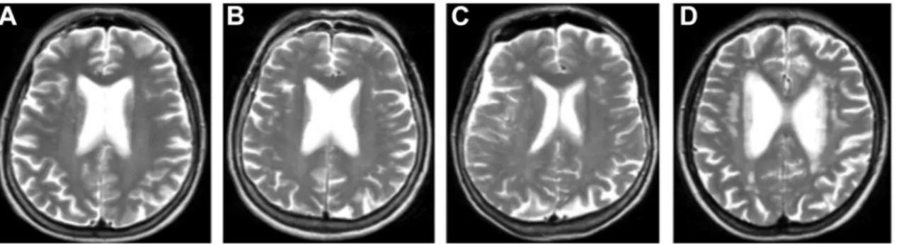
Figure 1 Four T2-weighted MRI images showing the extent of white matter lesions (WML) in each quartile of total WML volume. The T2-weighted MRI images show four separate individuals by total WML volume, (A) first quartile of WML volume (0– 25th percentile), (B) second quartile of WML volume (26–50th percentile), (C) third quartile of WML volume (51–75th percentile), (D) fourth quartile of WML volume (76–100th percentile) and group denoted as having extensive WML in the logistic models. Light grey=white matter; dark grey=grey matter.
an overall WMH grade; none, mild, moderate, severe. Figure 1 displays T2-weighted MRI images depicting each quartile of WML.
The primary outcome variable was based on WML volume greater than or equal to the upper quartile that we considered generally representative of extWML load and ensured sufficient statistical power in each class. The WML was defined as a binary variable; 0=normal to low WML load (below sex-specific upper quartile) and 1=extWML (equal or above sex-specific upper quartile; men (7.39 cm3), women (5.73 cm3). Age strata were: <70,
≥70 and <75, ≥75 years.28 secondary outcome: incident stroke
The protocol and criteria used to define prevalent and inci-dent stroke have been previously defined.29 In Bordeaux
and Montpellier, all subjects underwent a comprehen-sive neuropsychological examination and were seen by a senior neurologist. Subjects were followed up to 12 years for incident stroke (fatal and non-fatal). For persons who reported the occurrence of vascular events during follow-up, further medical data were obtained from general practitioners, specialists and hospital records. The diagnosis and classification of strokes were made by a blinded expert panel that reviewed all existing medical information including, where available, cerebral imaging. Strokes subtypes included ischaemic and haemorrhagic strokes according to International Classification of Diseases 10th revision criteria.30
statistical methods
Potential predictors of extWML were selected based on published literature and study findings of 3C.1 10–13
Other data definitions and additional statistical anal-yses are listed in the online supplementary file. The risk factors tested included: age (per 1-year increase), marital status (married (reference), not married, sepa-rated or widowed), education (bachelor degree (refer-ence), no or other education), tobacco smoking status (none (reference), former, current), body mass index <25 kg/m² (reference), 25–30 kg/m², 30 kg/m²),
systolic BP in mm Hg, diastolic BP in mm Hg, antihyper-tensive drug use for hypertension, cardiovascular disease, acute coronary event (myocardial infarction), diabetes (self-report), fasting plasma glucose, ≤6 mmol/L (refer-ence), 6.1–7.1 mmol/L, ≥7.2 mmol/L), hypercholes-terolaemia (self-report), total cholesterol (≤6.1 mmol/L (reference), 7.25 mmol/L), low-density lipoprotein cholesterol (mmol/L), high-density lipoprotein choles-terol (mmol/L), psychotropic drug use in the past month, depressive symptoms (total Center for Epidemiologi Studies Depression Scale score ≥16), history of lifetime major depression (MINI INTERNATIONAL NEURO-PSYCHIATRIC INTERVIEW diagnosis), Mini- Mental State Examination (MMSE) score, Benton Visual Reten-tion Test score, dependence in instrumental activities of daily living (IADL) (Lawton-Brody Scale), problems with balance when walking (self-report), forgetfulness, (self-report), difficulties retaining new simple informa-tion (self-report), difficulties remembering old memo-ries (self-report), difficulties with arithmetic calculations (self-report), difficulties with language or comprehen-sion (self-report), difficulties with spatial orientation (eg, in a city street).
Candidate predictors with a probabilistic likelihood of extWML P<0.25 were retained for further multivar-iate analyses. Participant age at baseline was forced into the initial multivariate model since age is strongly associated with WML.31 Thereafter age was eligible for
elimination from the model. The optimal model for clas-sifying extWML was computed with backward elimina-tion to remove covariates not significantly associated with extWML at P<0.05 level.
The probabilistic model was evaluated based on overall performance, discriminatory power and calibration. Concordance was evaluated through the construction of a receiver operating characteristics curve and the C-sta-tistic. Somer’s D provides an estimate of the difference between concordant and discordant pairs on a scale of −1 to 0, in our case between the predictive model and the MRI quantification of extWML. Model fit and cali-bration was tested with the Hosmer and Lemeshow test
(P>0.05 implies a good fit, higher numbers signify a better fit). In the derivation cohort, missing data were handled with listwise deletion. Power calculation showed that a sample size of 1542 in the derivation cohort would provide 80% power to detect an OR of 1.35 (two-sided) with multiple candidate risk factors.
Deriving the prediction score
Each predictor in the probabilistic model was converted to a point allocation system based on the methods outlined by Sullivan et al,32 described in the online
supple-mentary file 1. In brain imaging studies, the potential for discrimination with sensitivity and specificity is weighted equally given the prevalence of the disease state, the costs of diagnostic errors and likely benefit derived to the patient.33 The Youden index was used to calculate the
optimum cut-point for the score and its associated likeli-hood ratios (LR+, LR−), positive predictive value (PPV)
and negative predictive value (NPV). A bootstrapping method was used to examine the variability of the C-sta-tistic and optimism bias or overfitting to a specific sample using randomly generated samples (n=1714, 17 140 and 171 400, respectively).
cross validation
Although the sample size is large for a brain imaging study, it is relatively small for risk model testing. There-fore, cross validation was performed in the independent EVA cohort using the scoring system from the deriva-tion cohort. In EVA, participants with missing data were randomly allocated based on the proportions in 3C. Other validation
We tested whether the extWML model derived from base-line variables was associated with incident stroke events because of the close relationship between WML and brain outcomes. These analyses used the entire 3C cohort of individuals without prevalent stroke at baseline,29
irrespective of whether participants underwent MRI. Incident stroke risk was analysed with Cox proportional hazard models showing the HR and 95% CI. The highest attained age was used as the time scale and participants were censored at the date of stroke, drop-out from the study or death. Analyses were adjusted for late entry bias. results
Population characteristics
The final sample in the derivation cohort includes 1714 participants from the 3C-Dijon MRI online supplemen-tary e-figure 1. The sample was comprised 60.8% women and the median age was 72 years (IQR 69–76), 59.1% of subjects were married and 32.3% had completed at least bachelor’s level education (table 1).
White matter lesions
In the total sample, the median WML volume was 4.01 cm3
(IQR 2.75–6.37). The WML volume differed by localisa-tion and the median deep white matter hyperintensities
volume was 1.16 cm3 (IQR 0.75–1.79). By contrast, in the
periventricular region WML median volume was 2.77 cm3
(IQR 1.82–4.65). The threshold to define extWML based on the global volume of WML by highest quartile and stratified by sex was; >6.03 cm3 for men and 4.91 cm3 for
women. At this threshold of WML, the proportion of persons within each category was 168/677 men, 260/1042 women, total 428/1719.
univariate analyses of extWMl
The univariate analysis of variables and extWML are reported in the online supplementary e-table 1. Variables significantly associated with extWML P<0.05 included age, diastolic BP, antihypertensive drug use, psycho-tropic drug use, diabetes (self-reported), higher MMSE score, Benton Visual Retention Test score, dependence in IADL, calculation difficulties, forgetfulness, difficul-ties retaining new information and gait imbalance. Other candidate variables at P<0.25 included systolic BP, hyper-cholesterolaemia (self-reported), depressive symptoms on the CES-D and difficulties with language comprehension. Multivariate analyses of extWMl
The final backward deletion model from multivariate analysis predicting extWML is presented in table 2. The cardiovascular covariates associated with increased extWML risk were diastolic BP and antihypertensive drug use. Other factors that emerged in the model to increase risk for extWML were psychotropic drug use, depen-dence in IADL, forgetfulness and calculation difficulties. The Hosmer and Lemeshow test suggested an adequate model (P=0.88) and the diagnostic accuracy was in the modest range (C=0.63). The percentage of concordant and discordant pairs was high (62.8% vs 37.1%) with a small relationship between the predictive model and extWML (Somer’s D=0.26).
Derivation of the scoring equation
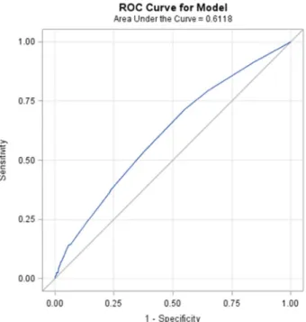
Table 3 depicts the point system to predict the likeli-hood of extWML before MRI. extWML were predicted by six clinical risk factors: diastolic BP, psychotropic drug use, antihypertensive drug use, dependence in at least one IADL, forgetfulness and difficulties with simple arithmetic calculations. The minimum and maximum scores in the 3C-Dijon MRI population were −0 and 30, respectively, (median=7, IQR 3–10). The Hosmer and Lemeshow test suggested an adequate model (P=0.97). The C-statistic=0.61 (figure 2) suggested a limited loss of predictive ability with total scores versus the raw data. A priori risk for extWML by each level of score is presented in table 4 to help determine whether to perform or forgo brain MRI to detect WMH.
A score of 7 points on the point scoring system corresponded to the optimal Youden index with sensi-tivity=63.7%, specificity 54.0%. At this score the corre-sponding LR+=1.38, LR−=0.67, PPV=31.5% and NPV=81.8%
for the given prevalence and threshold of extWML in our sample. Onlile supplementary e-table 2 describes the
Open Access Table 1 Population characteristics at baseline in the
3C-Dijon MRI cohort (N=1714)
Variable n %
Female 1042 60.8
Male 672 39.2
Median age in years, IQR 72 69–76
Married 1013 59.1 Not married 700 40.8 Bachelor degree 553 32.3 Other education 1159 67.6 BMI <25 kg/m² 841 49.0 BMI 25–30 kg/m² 677 39.5 BMI >30 kg/m² 195 11.4
Median systolic BP mm Hg, IQR 149.5 133–162.5 Median diastolic BP mm Hg, IQR 84.5 77–92 Antihypertensive drug use 730 42.6 Cardiovascular disease 71 4.1
FPG ≥7.0 mmol/L 140 8.2
Median LDL cholesterol mmol/L,
IQR 3.53 2.99–4.06
Median HDL cholesterol mmol/L,
IQR 1.61 1.37–1.90
Current tobacco smoker 97 5.7 Former tobacco smoker 559 32.6 Alcohol use (standard drinks per
week), IQR 7 1–14
Psychotropic drug use 399 23.3 Depression symptoms 221 12.9 Median MMSE score, IQR 28 27–29 Median Benton score, IQR 12 11–13 Total autonomy (living
independently)
1628 95.0 Dependence for at least one
instrumental activity of daily living 71 4.1
Gait imbalance 332 19.4
Forgetfulness 849 49.5
Difficulties retaining new simple information
681 39.7 Difficulties with simple arithmetic
calculations 275 16.1
Difficulties with language or
comprehension 1097 64.0
MRI parameters
Median no of WML, IQR 155 120–197 Median WML volume cm3, IQR 4.01 2.75–6.37
Median no of deep WML, IQR 57 42–81 Median deep WML volume cm3,
IQR 1.16 0.75–1.79 Median no of periventricular WML, IQR 95 74–121 Continued Variable n % Median periventricular WML volume cm3, IQR 2.77 1.82–4.65 BMI, body mass index; BP, blood pressure; FPG, fasting plasma glucose; HDL, high-density lipoprotein; LDL, low-density lipoprotein; MMSE, Mini-Mental State Examination; WML, white matter lesions.
Table 1 Continued
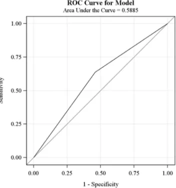
cumulative percent of participants with extWML at each level of the clinical score and the sensitivity and specificity values for reference purposes. Using a dichotomised score of 7 points only marginally reduced the C-statistic (C=0.59, figure 3), however, the percentage of concordant and discordant pairs was low (34.4% vs 16.7%) with many tied pairs (48.9%), resulting in lower Somer’s D=0.18. Internal validation with bootstrapping showed that the C-statistics varied minimally, between 0.57 and 0.59.
Because of the possibility that observed difficulties with IADL might lead to geriatrician referral and brain imaging later during the course of neurological disorders, we performed a sensitivity analysis by excluding IADL from the scoring system. The AUC value was marginally lower than the continuous score in model development (C=0.61). cross validation
Descriptive variables used in the prediction equation in the 3C and EVA cohort are shown in online supple-mentary e-table 3. Cross validation in the EVA cohort showed a much smaller range in scores, from 0 to 20 points with a median of 1 (IQR 0–7). Reproducing the scoring system in the EVA cohort and using the dichot-omised cut-point of 7 points obtained a very similar predictive value (C-statistic=0.57) suggesting high stability and reproducibility in an independent cohort online supplementary e-figure 2
Other validation
Data were available for 8023 persons and 342 inci-dent strokes in the larger cohort of 3C, incorporating Monpellier and Bordeaux participants as well as Dijon. The scoring system was predictive of incident stroke risk over 12 years using continuous (HR 1.02; 95% CI 1.01 to 1.04, P=0.02) and dichotomised values (HR 1.28; 95% CI 1.02 to 1.60, P=0.03).
DIscussIOn
Six clinical factors significantly predicted extWML detected on MRI based on sex-specific quartiles in a large population-based study of dementia-free older adults. These factors were diastolic BP, antihypertensive drug use, psychotropic drug use, dependence in IADL, forgetfulness and arithmetic difficulties. Conversion of these clinical variables into the scoring system produced modest predictive abilities for detection of extWML (C-statistic=0.57–63) but high NPV (82%). However,
Table 2 Multivariate analyses and the ORs for extensive white matter lesions (extWML) detected on MRI (n=1714)
Variable β OR 95% CI lower P value
Intercept −3.0047 – – <0.0001
Diastolic BP (per 1 mm Hg increase) 0.0156 1.016 1.006 to 1.026 0.0018
Psychotropic drug use 0.3660 1.442 1.113 to 1.868 0.0056
Antihypertensive drug use 0.5545 1.741 1.388 to 2.185 <0.0001 Dependence in at least one IADL 0.6427 1.902 1.140 to 3.171 0.0138
Forgetfulness 0.2455 1.278 1.017 to 1.607 0.0354
Difficulties with simple arithmetic calculations 0.3942 1.483 1.108 to 1.986 0.0081
The classification of extWML was based on upper quartile stratified by sex as; >6.03 cm3 for men and 4.91 cm3 for women. The regression
model used a backward stepwise deletion procedure for covariates using a threshold for inclusion of P<0.05. BP, blood pressure; IADL, instrumental activities of daily living.
sensitivity and specificity values of the scoring system here (63.7% and 54.0%, respectively) are very favour-able compared with the diagnostic predictive ability to detect extWML with CT as reported in a recent system-atic review.34 Cross validation in the EVA cohort provided
comparable findings showing high consistency, reproduc-ibility and test characteristic retention thereby pointing to the generalisability of the scoring system.
The final risk model here includes the cardiovas-cular factors diastolic BP and medication use for hyper-tension. Prior research corroborates an association between BP and periventricular WMLs, deep WMLs8
and large WML load9 supporting BP as a mechanism of
arteriolar vessel damage in the cerebral white matter. In terms of predictive models, a recent study elucidated the independent contribution of BP and other cardio-vascular risk factors associated with WML.35 However,
the previous study produced weighted scores from factor analysis which cannot be easily used by clinicians for risk stratification purposes. By contrast our scoring system can be readily used for determining extWML probabilities and might therefore serve to avoid unnec-essary and costly MRI imaging. The high NPV under-scores the clinical utility to identify persons at low risk of extWML.
Other risk factors associated with extWML here included psychotropic medication which might repre-sent the general cerebrovascular changes evident in affective and neurological disorders.3 It is controver-sial whether medications such as benzodiazepines are discrete risk factors or prodromal states preceding conversion to dementia.36 Indeed, the definition of
psychotropic drugs here was broad, incorporating anti-depressants, anxiolytics and neuroleptics which might suggest an indication bias for persons with prodromal dementia symptoms. Other risk factors in the scoring system included IADLs and it was previously docu-mented in this sample that loss of IADLs is associated with mild cognitive impairment.37 It is also possible that
some risk factors are a consequence of extWML and not a direct causative factor for white matter changes.
In terms of clinical implications, the predictive ability of the scoring system here (63.7% and 54.0%, respec-tively) was favourable compared with external reports of the diagnostic predictive ability to detect WML with CT in a pooled review of 11 non-autopsy studies (CT sensitivity=71%, specificity=55%).34 These
encour-aging findings suggest minimal loss of sensitivity and no loss of specificity using the scoring system versus CT.34 The scoring system therefore has potential to
deliver large cost savings in reducing unnecessary cere-bral imaging for the detection of extWML as evident in the high NPV. Also, testament to the clinical utility of the scoring system, bootstrapping and cross valida-tion in the EVA sample indicated high reproducibility and stability consistent with the derivation cohort. Thus the current findings will provide clinical utility as to whether clinicians should perform or forgo an MRI to detect extWML.
The study is presented with several strengths, including the large representative population size undergoing brain MRI, adherence to the TRIPOD statement,23
exam-ination of an exhaustive list of potential covariates and replication in an independent validation cohort. The limitations of our study include the use of 1.5 T MRI, similar to other cohorts,38 which is superseded by newer
generation 3 T MRI machines. A related point is that identification and quantification of WMH is commonly performed using parallel imaging and fluid-attenua-tion inversion recovery (FLAIR) sequences. As parallel imaging and FLAIR sequences were not acquired in the 3C study, the current findings may translate less readily to clinical practices or healthcare systems using such diagnostic methodologies. Moreover, in the absence of an accepted empirical definition,39 the dichotomised
threshold for extWML may have led to biases in the risk model development. However, in sensitivity analysis reducing the extWML threshold led to a loss of predic-tive ability. Future validation studies might therefore consider higher thresholds for extWML since our study was under powered to pursue higher thresholds. Future studies could grade WML according to moderate and
Open Access Table 3 The point scoring system based on regression
coefficients to derive the likelihood of extensive white matter lesions
Points allocated if positive
Diastolic blood pressure
≤79 mm Hg –1 80–84 mm Hg 0 85–89 mm Hg +1 90–94 mm Hg +2 95–99 mm Hg +3 100–104 mm Hg +4 105–109 mm Hg +5 ≥110 mm Hg +6
Psychotropic medication use
No psychotropic medication use 0 Yes psychotropic medication use +5 Antihypertensive drug use
No antihypertensive drug use 0 Yes antihypertensive drug use +7 Dependence for ≥1 instrumental activities of daily living (IADL)
Independent for IADL 0
Yes dependence for ≥1 IADL +8 Forgetfulness
No forgetfulness 0
Yes forgetfulness +3
Difficulties with simple calculations/arithmetic No difficulties with simple calculations/
arithmetic 0
Yes difficulties with simple calculations/
arithmetic +5
Psychotropic medication use is inclusive of antidepressants, mood stabilisers, anxiolytics and neuroleptics. Antihypertensive drugs only included taking antihypertensive drugs explicitly for hypertension. IADL were measured by the Lawton-Brody scale. Participants were asked whether they experienced forgetfulness (responses dichotomised as yes or no) and had difficulties performing simple arithmetic calculations (responses dichotomised as yes or no).
Figure 2 Graph showing the area under the curve for the scoring system (continuous) to predict white matter lesions (WML) in the 3C-Dijon MRI study (n=1714). Graph showing the area under the curve (AUC, sensitivity and 1-specificity) for the scoring system score to detect extensive WML. The scoring system is based on a point system derived from the regression coefficients for diastolic BP, antihypertensive drug use, psychotropic drug use, dependence in instrumental activities of daily living, forgetfulness and calculation difficulties. The dichotomisation of WML was based on sex-specific upper quartiles
(men=6.03 cm3 and women=4.91 cm3). BP, blood pressure;
ROC, receiver operating characteristic .
severe categories using Fazekas criteria as used in the Leukoaraiosis and Disability in the Elderly Study.38
Another limitation that tempers these findings is the relatively modest predictive values, sensitivity and spec-ificity, and C-statistics. This suggests that extWML are difficult to predict and may reflect the heterogeneity in extWML risk factors. Indeed, some candidate variables such as memory difficulties may be consequences of WML rather than predictive risk factors, and our study was not designed to distinguish between such poten-tial bi-directional associations. Given the complexity of
identifying risk factors for WML, the total WML load was calculated combining the periventricular and deep white matter areas whereas some risk factors are likely more specific to certain cerebral regions. However, although the scoring system did not control for location of WMLs the purpose here was to inform decisions on whether to proceed with MRI. Thus the scoring system alone cannot inform the vascular aetiology of dementia nor does it replace the need for imaging. Moreover, WML are heterogeneous and therefore their impact on cerebrovascular diseases and cognitive function may differ among individuals with normal ageing and those prone to dementia.40 41 However, because of limited
sample size and lack of precise diagnosis in the EVA study, it was not possible to take this heterogeneity into account. Another limitation to consider is that the back-ward deletion model may capitalise on chance variation within the dataset. However, the bootstrapping analysis showed consistency in the test score suggesting minimal optimism bias or overfitting to the 3C cohort. Another consideration for interpreting this model is the use of sex to derive the extWML thresholds based on previous findings of higher WML in women.11 This may
Table 4 A priori risk estimates for extensive white matter lesions (extWML) based on a scoring system from six common risk factors
Score Risk estimate Score Risk estimate
−1 14.1 17 40.1 0 15.1 18 42.0 1 16.1 19 43.9 2 17.2 20 45.9 3 18.4 21 47.8 4 19.6 22 49.8 5 20.8 23 51.7 6 22.1 24 53.7 7 23.5 25 55.6 8 24.9 26 57.5 9 26.4 27 59.4 10 28.0 28 61.3 11 29.6 29 63.1 12 31.2 30 64.9 13 32.9 31 66.7 14 34.7 32 68.4 15 36.5 33 70.0 16 38.3 34 71.6
A priori risk estimate for extWML before brain MRI based on the point scoring system described in table 3. The threshold for extWML was determined by the upper quartile stratified by sex as; >6.03 cm3 for men and 4.91 cm3 for women. The point
system is further described in table 3 and is based on six clinical risk factors; diastolic blood pressure (5 mm Hg increments), psychotropic drug use, antihypertensive drug use, dependence in at least one instrumental activities of daily living, self-reported forgetfulness and self-reported difficulties with simple arithmetic calculations.
Figure 3 Graph showing the area under the curve for a dichotomised score of 7 to predict white matter lesions (WML) in the 3C-Dijon MRI study (n=1714). Graph showing the area under the curve (AUC, sensitivity and 1-specificity) for a dichotomised score of 7 to detect extensive WML. The scoring system is based on a point system derived from the regression coefficients for diastolic BP, antihypertensive drug use, psychotropic drug use, dependence in instrumental activities of daily living, forgetfulness and calculation difficulties. The dichotomisation of WML was based on sex-specific upper quartiles
(men=6.03 cm3 and women=4.91 cm3). BP, blood pressure;
ROC, receiver operating characteristic.
as this study reports the development stage, the predic-tive ability of the scoring system requires further evalu-ation in an external implementevalu-ation study.
cOnclusIOns
A clinical risk score comprised by six factors indicated modest predictive ability to detect sex-specific extWML. Sensitivity and specificity values of the risk score were highly favourable compared with external imaging studies quantifying WML with CT.34 The C-statistics were
stable and reproducible in bootstrapping and cross vali-dation pointing to the generalisability of the scoring system. In elderly populations, the scoring system can inform clinical decisions on whether to proceed with or forgo cerebral MRI. The high-negative predictive value indicates that numerous costly MRIs could be avoided in the general population aged 65–80 years.
Acknowledgements The Three-City (3C) study is conducted under a partnership agreement between the Institut National de la Sante et de la Recherche Medicale (INSERM), the Victor Segalen-Bordeaux II University and Sanofi-Aventis.
contributors PJT wrote the statistical analysis plan, drafted and revised the paper. SQ wrote the statistical analysis plan, cleaned and analysed the data, and drafted and revised the paper. She is guarantor. EP wrote the statistical analysis plan and revised the draft paper. SD revised the draft paper. BM monitored data collection for the entire study and revised the draft paper. CT designed the study, monitored data collection for the entire study and revised the draft paper.
Funding The Fondation pour la Recherche Medicale funded the preparation and initiation of the study. The Fondation Plan Alzheimer partly funded the follow-up of the study. The 3C study is also supported by the Caisse Nationale Maladie des Travailleurs Salaries, Direction Generale de la Sante, MGEN, Institut de la Longevite, Conseils Regionaux of Aquitaine and Bourgogne, Fondation de France, and the Ministry of Research-INSERM Programme “Cohortes et collections de donnees biologiques.” The 3C study supports are listed on the study website (www. three- city- study. com).
Disclaimer The funding organisations played no role in the design and conduct of the study and were not involved in collection, management, analysis and interpretation of the data or in preparation, review or approval of the manuscript.
competing interests None declared.
ethics approval University Hospital of Kremlin-Bicêtre.
Provenance and peer review Not commissioned; externally peer reviewed.
Data sharing statement The 3C Dijon MRI study data are not on open access but are accessible through a process described on the study website (http://www. three- city- study. com/ the- three- city- study. php.
Open Access This is an Open Access article distributed in accordance with the Creative Commons Attribution Non Commercial (CC BY-NC 4.0) license, which permits others to distribute, remix, adapt, build upon this work non-commercially,
Open Access
and license their derivative works on different terms, provided the original work is properly cited and the use is non-commercial. See: http:// creativecommons. org/ licenses/ by- nc/ 4. 0/
© Article author(s) (or their employer(s) unless otherwise stated in the text of the article) 2017. All rights reserved. No commercial use is permitted unless otherwise expressly granted.
reFerences
1. Poels MM, Ikram MA, van der Lugt A, et al. Incidence of cerebral microbleeds in the general population: the Rotterdam Scan Study. Stroke 2011;42:656–61.
2. Etherton MR, Wu O, Rost NS. Recent advances in leukoaraiosis: white matter structural integrity and functional outcomes after Acute ischemic stroke. Curr Cardiol Rep 2016;18:123.
3. Gudmundsson P, Olesen PJ, Simoni M, et al. White matter lesions and temporal lobe atrophy related to incidence of both dementia and major depression in 70-year-olds followed over 10 years. Eur J Neurol 2015;22:781–e50.
4. Inaba M, White L, Bell C, et al. White matter lesions on brain magnetic resonance imaging scan and 5-year cognitive decline: the honolulu-Asia aging study. J Am Geriatr Soc 2011;59:1484–9. 5. Kandel BM, Avants BB, Gee JC, et al. White matter hyperintensities
are more highly associated with preclinical alzheimer’s disease than imaging and cognitive markers of neurodegeneration. Alzheimers Dement 2016;4:18–27.
6. Goldberg I, Auriel E, Russell D, et al. silent brain infarcts and dementia. J Neurol Sci 2012;322:250–3.
7. Arima H, Anderson C, Omae T, et al. Degree of blood pressure reduction and recurrent stroke: the PROGRESS trial. J Neurol Neurosurg Psychiatry 2014;85:1284–5.
8. Söderlund H, Nyberg L, Adolfsson R, et al. High prevalence of white matter hyperintensities in normal aging: relation to blood pressure and cognition. Cortex 2003;39:1093–105.
9. de Leeuw FE, de Groot JC, Oudkerk M, et al. A follow-up study of blood pressure and cerebral white matter lesions. Ann Neurol 1999;46:827–33.
10. Longstreth WT, Manolio TA, Arnold A, et al. Clinical correlates of white matter findings on cranial magnetic resonance imaging of 3301 elderly people. The Cardiovascular Health Study. Stroke 1996;27:1274–82.
11. van Dijk EJ, Prins ND, Vrooman HA, et al. Progression of cerebral small vessel disease in relation to risk factors and cognitive consequences: Rotterdam Scan study. Stroke 2008;39:2712–9. 12. Dufouil C, de Kersaint-Gilly A, Besançon V, et al. Longitudinal study
of blood pressure and white matter hyperintensities: the EVA MRI Cohort. Neurology 2001;56:921–6.
13. Manolio TA, Kronmal RA, Burke GL, et al. Magnetic resonance abnormalities and cardiovascular disease in older adults. The Cardiovascular Health Study. Stroke 1994;25:318–27. 14. Atri A. Imaging of neurodegenerative cognitive and behavioral
disorders: practical considerations for dementia clinical practice. In: Joseph CM, González RG, eds. Handbook of clinical neurology. The Netherlands: Elsevier, 2016:971–84.
15. Sachdev P, Kalaria R, O'Brien J, et al. Diagnostic criteria for vascular cognitive disorders: a VASCOG statement. Alzheimer Dis Assoc Disord 2014;28:206–18.
16. Scheltens P, O'Brien J. Clinical use of neuroimaging in dementia: an international perspective. Int Psychogeriatr 2011;23:3–5.
17. Del Brutto OH, Mera RM, Andrade ML, et al. Disappointing reliability of pulsatility indices to identify candidates for magnetic resonance imaging screening in population-based studies assessing prevalence of cerebral small vessel disease. J Neurosci Rural Pract 2015;6:336–8.
18. Burke JF, Kerber KA, Iwashyna TJ, et al. Wide variation and rising utilization of stroke MRI: data from eleven states. Ann Neurol 2012;71:179–85.
19. Haydel MJ, Preston CA, Mills TJ, et al. Indications for computed tomography in patients with minor head injury. N Engl J Med 2000;343:100–5.
20. Stiell IG, Wells GA, Vandemheen K, et al. The Canadian CT Head Rule for patients with minor head injury. The Lancet 2001;357:1391–6.
21. Godin O, Tzourio C, Maillard P, et al. Antihypertensive treatment and change in blood pressure are associated with the progression of white matter lesion volumes: the three-city (3C)-Dijon magnetic resonance imaging study. Circulation 2011;123:266–73.
22. Gauthier S, Patterson C, Chertkow H, et al. Recommendations of the 4th canadian consensus conference on the diagnosis and treatment of dementia (CCCDTD4). Can Geriatr J 2012;15:120–6.
23. Collins GS, Reitsma JB, Altman DG, et al. Transparent reporting of a multivariable prediction model for individual prognosis or diagnosis (TRIPOD): the TRIPOD statement. BMJ 2015;350:7594.
24. The 3C Study Group. Vascular factors and risk of dementia: design of the three-city study and baseline characteristics of the study population. Neuroepidemiology 2003;22:316–25.
25. Godin O, Dufouil C, Maillard P, et al. White matter lesions as a predictor of depression in the elderly: the 3C-Dijon study. Biol Psychiatry 2008;63:663–9.
26. Maillard P, Delcroix N, Crivello F, et al. An automated procedure for the assessment of white matter hyperintensities by multispectral (T1, T2, PD) MRI and an evaluation of its between-centre reproducibility based on two large community databases. Neuroradiology 2008;50:31–42.
27. Scheltens P, Barkhof F, Valk J, et al. White matter lesions on magnetic resonance imaging in clinically diagnosed Alzheimer's disease. evidence for heterogeneity. Brain 1992;115 :735–48. 28. Schilling S, Tzourio C, Dufouil C, et al. Plasma lipids and cerebral
small vessel disease. Neurology 2014;83:1844–52.
29. Tully PJ, Debette S, Dartigues JF, et al. Antihypertensive drug use, blood pressure variability, and Incident stroke risk in older adults: three-city cohort study. Stroke 2016;47:1194–200.
30. World Health Organization. International statistical classification of diseases and related health problems 10th revision. 2007. http:// www. who. int/ classifications/ apps/ icd/ icd10online/
31. Takami T, Yamano S, Okada S, et al. Major risk factors for the appearance of white-matter lesions on MRI in hypertensive patients with controlled blood pressure. Vasc Health Risk Manag 2012;8:169–76.
32. Sullivan LM, Massaro JM, D'Agostino RB. Presentation of multivariate data for clinical use: the framingham study risk score functions. Stat Med 2004;23:1631–60.
33. Hanley JA, McNeil BJ. The meaning and use of the area under a receiver operating characteristic (ROC) curve. Radiology 1982;143:29–36.
34. Beynon R, Sterne JA, Wilcock G, et al. Is MRI better than CT for detecting a vascular component to dementia? A systematic review and meta-analysis. BMC Neurol 2012;12:33.
35. Watts A, Honea RA, Billinger SA, et al. A combined measure of vascular risk for white matter lesions. J Alzheimers Dis 2015;45:187–93.
36. Billioti de Gage S, Moride Y, Ducruet T, et al. Benzodiazepine use and risk of Alzheimer’s disease: case-control study. BMJ 2014;349:5205. 37. Ritchie K, Ancelin ML, Beaino E, et al. Retrospective
identification and characterization of mild cognitive impairment from a prospective population cohort. Am J Geriatr Psychiatry 2010;18:692–700.
38. Poggesi A, Pantoni L, Inzitari D, et al. 2001-2011: A decade of the LADIS (Leukoaraiosis And DISability) Study: what have we learned about white matter changes and small-vessel disease? Cerebrovasc Dis 2011;32:577–88.
39. Schmidt R, Grazer A, Enzinger C, et al. MRI-detected white matter lesions: do they really matter? J Neural Transm 2011;118:673–81. 40. Gouw AA, Seewann A, van der Flier WM, et al. Heterogeneity of
small vessel disease: a systematic review of MRI and histopathology correlations. J Neurol Neurosurg Psychiatry 2011;82:126–35. 41. Barber R, Scheltens P, Gholkar A, et al. White matter lesions
on magnetic resonance imaging in dementia with Lewy bodies, Alzheimer's disease, vascular dementia, and normal aging. J Neurol Neurosurg Psychiatry 1999;67:66–72.
study
people age 65 years or older: the Dijon MRI
model for extensive white matter lesions in
Development and validation of a priori risk
Bernard Mazoyer and Christophe Tzourio
Phillip J Tully, Sarah Qchiqach, Edwige Pereira, Stephanie Debette,
doi: 10.1136/bmjopen-2017-018328
2017 7: BMJ Open
http://bmjopen.bmj.com/content/7/12/e018328
Updated information and services can be found at:
These include:
References
http://bmjopen.bmj.com/content/7/12/e018328#ref-list-1
This article cites 39 articles, 9 of which you can access for free at:
Open Access
http://creativecommons.org/licenses/by-nc/4.0/ non-commercial. See:
provided the original work is properly cited and the use is
non-commercially, and license their derivative works on different terms, permits others to distribute, remix, adapt, build upon this work
Commons Attribution Non Commercial (CC BY-NC 4.0) license, which This is an Open Access article distributed in accordance with the Creative
service
Email alerting
box at the top right corner of the online article.
Receive free email alerts when new articles cite this article. Sign up in the
Collections
Topic
Articles on similar topics can be found in the following collections (456)Neurology
Notes
http://group.bmj.com/group/rights-licensing/permissions
To request permissions go to:
http://journals.bmj.com/cgi/reprintform
To order reprints go to:
http://group.bmj.com/subscribe/