HAL Id: hal-01561318
https://hal.archives-ouvertes.fr/hal-01561318
Submitted on 5 Feb 2021
HAL is a multi-disciplinary open access archive for the deposit and dissemination of sci-entific research documents, whether they are pub-lished or not. The documents may come from teaching and research institutions in France or abroad, or from public or private research centers.
L’archive ouverte pluridisciplinaire HAL, est destinée au dépôt et à la diffusion de documents scientifiques de niveau recherche, publiés ou non, émanant des établissements d’enseignement et de recherche français ou étrangers, des laboratoires publics ou privés.
with Pittsburgh compound B or a nanobody for amyloid
plaques targeting.
Jonathan Pansieri, Marie Plissonneau, Nathalie Stransky-Heilkron, Mireille
Dumoulin, Laurence Heinrich-Balard, Pascaline Rivory, Jean-François Morfin,
Éva Tóth, Saraiva Maria Joao, Eric Allémann, et al.
To cite this version:
Jonathan Pansieri, Marie Plissonneau, Nathalie Stransky-Heilkron, Mireille Dumoulin, Laurence Heinrich-Balard, et al.. Multimodal imaging Gd-nanoparticles functionalized with Pittsburgh com-pound B or a nanobody for amyloid plaques targeting.. Nanomedicine, Future Medicine, 2017, 12 (14), pp.1675-1687. �10.2217/nnm-2017-0079�. �hal-01561318�
part of
Nanomedicine (Lond.)
Research Article 2017/06/3012
14
2017
Aim: Gadolinium-based nanoparticles were functionalized with either the Pittsburgh compound B or a nanobody (B10AP) in order to create multimodal tools for an early diagnosis of amyloidoses. Materials & methods: The ability of the functionalized nanoparticles to target amyloid fibrils made of β-amyloid peptide, amylin or Val30Met-mutated transthyretin formed in vitro or from pathological tissues was investigated by a range of spectroscopic and biophysics techniques including fluorescence microscopy. Results: Nanoparticles functionalized by both probes efficiently interacted with the three types of amyloid fibrils, with KD values in 10 micromolar and 10 nanomolar range for, respectively, Pittsburgh compound B and B10AP nanoparticles. Moreover, they allowed the detection of amyloid deposits on pathological tissues. Conclusion: Such functionalized nanoparticles could represent promising flexible and multimodal imaging tools for the early diagnostic of amyloid diseases, in other words, Alzheimer’s disease, Type 2 diabetes mellitus and the familial amyloidotic polyneuropathy. First draft submitted: 10 March 2017; Accepted for publication: 24 May 2017; Published online: 21 June 2017
Keywords: amyloid imaging • amyloidoses • gadolinium-based nanoparticles • nanobody • PIB
Amyloidoses are a set of human diseases in which a normally soluble peptide or protein aggregates into insoluble and highly orga-nized aggregates referred to as amyloid fibres. These aggregates are rich in cross-β-sheet
structures [1,2]. So far, 36 proteins are
associ-ated with either local (e.g., Type 2 diabetes mellitus [T2DM] and Alzheimer’s disease [AD]) or systemic (e.g., familial
polyneurop-athy and myeloma) amyloid depositions [3].
These diseases inflict enormous psycho-sociological and economic burdens. For example, AD and T2DM affect, respectively, approximately 44 and 415 million people
worldwide [4,5].
Symptoms of amyloidoses, especially in the cases of systemic amyloidoses, can be very subtle and vary widely from person to person and also depend on which organs are affected. The diagnosis of amyloidosis
and especially the early diagnosis, remains therefore a great challenge and often requires
several invasive examinations [6,7].
Therefore, there is a need to develop sen-sitive and minimally invasive tools to detect amyloid deposits, in order not only to
estab-lish the diagnosis of amyloidosis [8] but also
allow to follow-up the efficiency of treatment by monitoring the evolution of the amyloid
load [9]. Nowadays, a number of imaging
techniques can be used for these purposes; they include PET and MRI to detect the deposition of Aβ amyloid in the brain of AD
patients [10,11], scintigraphy to detect
V30M-TTR fibrils involved in cardiac
amyloido-sis [12], autoradiography to target islet amyloid
polypeptide (IAPP) fibrils in T2DM [13], etc.
However, each of these techniques presents some limitations. For example, while MRI allows obtaining an outstanding spatial
reso-Multimodal imaging Gd-nanoparticles
functionalized with Pittsburgh compound
B or a nanobody for amyloid plaques
targeting
Jonathan Pansieri‡,1, Marie Plissonneau‡,2,3, Nathalie Stransky-Heilkron4, Mireille Dumoulin5, Laurence Heinrich-Balard6, Pascaline Rivory6, Jean-François Morfin7, Eva Toth7, Maria Joao Saraiva8, Eric Allémann4, Olivier Tillement3, Vincent Forge1, François Lux‡,3 & Christel Marquette*,‡,1 1Laboratoire de Chimie et Biologie des Métaux, Université Grenoble Alpes, CEA Life Sciences Division, CNRS, 17 rue des Martyrs, 38054 Grenoble Cedex 9, France 2Nano-H S.A.S, 38070 Saint Quentin Fallavier, France 3Institut Lumière Matière, University of Lyon, University of Claude Bernard Lyon 1, CNRS, F-69622, Lyon, France 4School of Pharmaceutical Sciences, University of Geneva, University of Lausanne Pharmaceutical technology, Quai Ernest-Ansermet 30, 1211 Geneva, Switzerland 5Laboratory of Enzymology & Protein Folding, Centre for Protein Engineering, InBioS, University of Liege Sart Tilman, 4000 Liege, Belgium 6University of Lyon, University of Claude Bernard Lyon 1, ISPB Faculté de Pharmacie, MATEIS UMR CNRS 5510, 69373 Lyon, France. 7Centre de Biophysique Moléculaire, CNRS, UPR 4301, Université d’Orléans, Rue Charles Sadron, 45071 Orléans, France 8Instituto de Inovação e Investigação em Saúde (I3S), University of Porto, Portugal; Molecular Neurobiology Group, IBMC – Institute for Molecular & Cell Biology, University of Porto, 4150–180 Porto, Portugal *Author for correspondence: christel.marquette@cea.fr ‡Authors contributed equally
lution it strongly lacks sensitivity (1–0.01 millimolar
range) [14]. On the other hand, PET offers an excellent
sensitivity (10–1 pM range) but has a low spatial
reso-lution [15]. In order to overcome these limitations, the
development of probes allowing multimodal imaging approaches (i.e., combination of PET and MRI) will represent a breakthrough in the diagnosis of amyloido-ses [16,17]. In this context, nanoparticles have emerged
in the past decades as a very promising tool to enhance the contrast and the sensitivity of these different imag-ing techniques.
In this work, we have functionalized gadolinium (Gd) based multimodal nanoparticles (NPs), so called AGuIX with specific probes for the diagnosis of amy-loidoses. AGuIX are made of a polysiloxane network
surrounded by several DOTAGA (Gd3+) chelates [18,19].
AGuIX NPs have a diameter smaller than 5 nm and have been already efficiently used for MRI and
radio-sensitization [20]. Free ligands are available at the
sur-face of AGuIX and can be used to chelate radioisotopes
(e.g., 68Ga3+ or 111In3+), in order to perform PET or
single-photon emission computed tomography imag-ing [14,21]. Moreover, the covalent grafting of a near
infrared dye, such as cyanine5.5 (Cy5.5), is also
achiev-able for optical imaging [22]. Finally, we have recently
shown that AGuIX can be functionalized with specific Aβ peptides to selectively target and image Aβ-amyloid
fibrils [23]. However, while the results obtained clearly
demonstrate that these functionalized NPs constitute a valuable multimodal imaging tool to selectively tar-get Aβ fibrils, they displayed a low affinity (100 μM range) for their target. Therefore, the aim of the present work is to functionalize AGuIX with molecules having higher affinity for amyloid fibrils, in order to develop a sensitive tool for early diagnosis of amyloidoses.
Among the molecules binding to amyloid fibrils with relatively high affinity, thioflavin (ThS or ThT) is probably the most used probe for the detection of amy-loid aggregates, both in vitro for biochemical
charac-terization and ex vivo for histological studies [24].
Sev-eral thioflavin-derived molecules have been designed to further increase its affinity for amyloid fibrils in order
to enable PET applications [11]. One of these molecules,
the Pittsburgh compound B (PIB), shows an affinity in
the nanomolar range toward β-amyloid deposits [11,25–
26] and successfully targets senile plaques in
Alzheim-er’s patient [27]. This compound can be coupled to
metal ion such as Gd3+ for MRI or to radioisotopes
such as 111In3+ for single-photon emission computed
tomography [28] or 11C or 18F for PET. As consequence,
PIB is considered as the clinical ‘gold standard’ probe for amyloidoses including AD.
Another approach to image amyloid fibrils consists of functionalizing NPs with antibodies or antibody
fragments displaying a high specificity and affinity
for the targeted fibrils [29]. For example, an enhanced
amyloid-β plaques detection by MRI was observed within AD transgenic mouse using SuperParamagnetic Iron Oxide Nanoparticles functionalized with an
anti-AβPP antibody [30]. In this work, we have chosen to
functionalize AGuIX with the B10AP which is a chi-meric protein made of B10, a nanobody (i.e., the vari-able domain of heavy-chain antibodies) and Escherichia
coli alkaline phosphatase (AP). B10 has been selected
by phage display from a synthetic library of
nanobod-ies using Aβ(1–40) fibrils as bait [31]. It also recognizes
amyloid fibrils of several proteins (made in vitro and on tissues) including insulin, glucagon, β2-microglobulin, serum amyloid A protein and immunoglobulin light
chain (AL) [32]. AP forms homodimers and therefore
B10AP exhibits two copies of B10 within one molecule of the chimera; such a bivalent construct exhibits a
higher overall affinity for Aβ(1–42) amyloid fibrils with
a lower dissociation constant (KD: 7 nM and 475 nM
for B10AP and B10, respectively) [32]. Moreover, the
phosphatase activity of AP allows an easy and straight-forward monitoring of B10AP binding to the amyloid fibrils.
In this work, we have used PIB and B10AP as graft-ing ligands on AGuIX NPs, in order to perform active
targeting on Aβ(1–42) fibrils which deposit in the brain
of AD patients; IAPP fibrils that accumulate in Lang-erhans Islet of patients affected with T2DM; and V30M-TTR fibrils associated with familial amyloidotic polyneuropathy (FAP).
Methods NPs synthesis
AGuIX were synthesized as previously described [18,19]
(Nano-H SAS, Saint-Quentin Fallavier, France). These NPs are composed of a polysiloxane network sur-rounded by covalently grafted DOTAGA (1,4,7,10-tet-raazacyclododecane-1-glutaric
anhydride-4,7,10-triace-tic acid, CheMatech, Dijon, France) and Gd3+ chelates,
with 5 or 7% free DOTA. NPs concentrations are
stated in mol.L-1 of Gd element.
Functionalization of AGuIX NP with PIB NP
PIB derivative was synthesized as previously
described [33]. In order to functionalize AGuIX with
PIB, a polyethylene glycol bis-carboxymethyl (PEG)
was first grafted on NP as previously described [23];
after purification, the AGuIX@PEG were freeze-dried. Then, these freeze-dried NPs were dispersed at
a Gd3+ concentration of 500 mM in ultrapure water
and diluted tenfold with diethylene glycol (SDS Carlo Erba, France) at 80°C and then diluted twice with dry dimethyl sulfoxide (DMSO), leading to a final 25 mM
Gd3+. The carboxylic functions of the NPs were acti-vated by N-(3-Dimethylaminopropyl)-N0-ethylcar-bodiimidehydrochloride (EDC) and
pentafluorophe-nol (PFP) at EDC/PFP/Gd3+ molar ratio 10/20/1 for
15 min. PIB at 200 mg.ml-1 in dry DMSO was added
to the NPs suspension and the mixture was stirred
for 6 h (PIB/Gd3+ molar ratio 1/1). Finally, the NPs
were precipitated in acetone (Sodipro, France), centri-fuged and the supernatant was removed; this step was repeated three-times to allow the removal of the upmost ungrafted PIB. After redispersion of the precipitate in water, acetone was evaporated overnight at 40°C in open vials. The unreacted reagents and the degraded NPs were efficiently eliminated by repeated concentra-tion/dilution using tangential filtration cycles with a 5 kDa molecular cut-off membrane. A full experimen-tal characterization of the AGuIX@PEG@PIB samples
is described in the Supplementary Information.
Functionalization of AGuIX NP with B10APB10AP was produced in E. coli and purified as described
previ-ously [31], from the plasmid ptetB10pA6H kindly
pro-vided by Prof Horn, Jena University, Germany. Freeze-dried AGuIX with 7% of free DOTA (AGuIX@ DOTAfree7%) were first labeled with one Cy5.5 dye
mol-ecule for 1000 Gd3+ as previously described [23], and
then were dispersed at 100 mM in Gd3+ in ultrapure
water. The carboxylic acid functions of the free DOTA ligand were activated using a 100-fold excess of EDC and N-hydroxysuccinimide. After 15 min of incuba-tion, at room temperature under mild agitaincuba-tion, the activated AGuIX NPs were added in large excess (100-fold excess) to the nanobody suspension at 41 μM in phosphate-buffered saline buffer. Ungrafted AGuIX were also added to the suspension in order to reach
a Gd3+ concentration of 10 mM to stabilize grafted
NPs. After 30 min of incubation, the ungrafted NPs are removed by tangential filtration on a 50kDa cut-off membrane. Then, the AGuIX@B10AP@Cy5.5 were freeze-dried and stored away from light. A full experi-mental characterization of the AGuIX@B10AP@Cy5.5
samples is described in the Supplementary Information.
EDC, PFP, PEG, N-hydroxysuccinimide (NHS) and DMSO were purchased from Aldrich Chemical (France).
Amyloid fibril formation
Aβ(1–42) fibrils were prepared from Aβ(1–42) peptide (Genecust, Luxembourg), incubated at 100 μM, and the V30M-TTR fibrils from recombinant protein pro-duced in E. coli and were prepared at 32.3 μM, as
pre-viously described [23].
IAPP fibrils (48 μM) were prepared using lyophi-lized IAPP peptide (Genecust) dissolved in 100% 1,1,1,3,3,3-Hexafluoro-2-propanol (HFIP) and then
incubated in Tris-HCl 20 mM pH 7.4 at 37°C for
5 days, based on previous studies [34]. A full
experi-mental detail of IAPP fibrils is described in the supplementary information.
The fibrillar morphology of the aggregates formed by the three peptides or proteins was confirmed using Transmission Electronic Microscopy analysis ( JEOL1200EX/Tecnai 12).
In vitro assays & equilibrium constant determination
Dissociation constants (i.e., affinities) were obtained by partition binding tests between fibrils and
func-tionalized NPs as previously described [23]. Each
type of fibrils, were incubated at 20 or 40 μM, with the following ranges of concentrations for AGuIX@ PEG@PIB, between 0.01 and 4 μM in PIB, and for AGuIX@B10AP@Cy5.5 between 0.16 and 132 nM in B10. According to the fluorescence properties of PIB
(λex = 350 nm, λem = 420 nm) or Cy5.5 (λex = 655 nm,
λem = 685 nm), fluorescence emission spectrum of each
sample was recorded on a JASCO J-815 spectrophoto-meter. Michaelis binding model was used to calculate
the equilibrium constant affinity (KD) following the
equation:
y
=
Ym
/(
1
+
(
K
D/ )),
x
where (Ym) is the fluorescence intensity maximum and (y) is the fluorescence intensity measured for a given concentration in targeting probes within functionalized NPs (x).
Animal models
Brain sections prepared from 18 months APPswe/ PSEN1dE9 AD mouse models (kindly provided by M Dhenain, Fontenay aux Roses, France) were used to target Aβ-peptide fibrils. These models were previously
described in the literature [35,36], and a short description
is provided within Supplementary Information. Brain
sections from C57/Bl6 mice were used as control. Pancreas sections were prepared from male mice homozygous for RIPHAT transgene-expressing
human-IAPP as TDM2 models [37]. Pancreas from S/
FVB/NJ mice were used as control tissue without amy-loid burden.
Stomach tissue sections were extracted from a trans-genic mice expressing human V30M-TTR and
lack-ing endogenous mouse TTR [38]. Similar tissue from
knockdown mice for transthyretin gene was used as
control [39].
All the procedures involving animals were carried out in accordance with the European Community Council Directives. The different procedures to deeply anesthetize the animals, to extract and fix the organs/
tissues and to prepare the sections are described in
details within Supplementary Information. Tissue
sec-tions were mounted on slides for the immunostaining experiments.
Immunohistochemistry
Frozen sections were processed for immunohistochem-istry staining with AGuIX@PEG@PIB and AGuIX@ B10AP@Cy5.5 as previously described with other
func-tionalized AGuIX [23]. AGuIX@PEG@PIB were used
at (PIB) = 5 μM and AGuIX@B10AP@cy5.5 at (B10) = 81 nM. To each suspension of grafted NPs, 3 mM of ungrafted NPs were added to insure the stability of the former. In the case of PIB, fluorescence was used
to monitor the binding of AGuIX@PEG@PIB (λex =
350 nm - λem = 405 nm). TRITC-conjugated
phal-loidin (Sigma, France, #P1951) was used to detect the
actin network (λex = 545 nm-λem = 605 nm). Regarding
AGuIX@PEG@B10AP@Cy5.5, the binding to NPs to amyloid plaques was detected through either the
fluo-rescence of Cy5.5 (λex = 650 nm-λem = 670 nm) or via
the enzymatic activity of AP fused to B10AP using red
permanent as substrate (λex = 595 nm-λem = 613 nm).
In all tissue sections, amyloid burden was stained with
ThS (λex = 450 nm - λem = 488 nm) (Sigma, #T1892)
according to standard practice. Fluorescent images were recorded using either Zeiss confocal LSM800 microscope for pancreas sections or Zeiss microscope Axiovert 200 M for brain and stomach tissues. Images were analyzed using Carl Zeiss AxioVision software and Image J 1.50f software.
Results
Characterization of the grafted NPs
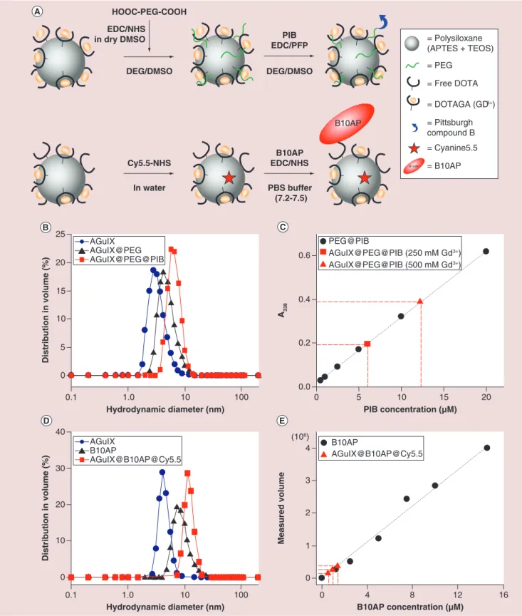
The synthesis of functionalized AGuIX NPs was achieved through a bottom-up process described
previ-ously [18,19], with around ten DOTAGA per NP. Two
different strategies were used to graft PIB and B10AP,
mainly due to their sizes (Figure 1A). The addition of
small PEG chains at the surface of AGuIX was nec-essary to guarantee the accessibility of PIB (i.e., to limit the steric hindrance due to the proximity of the NP) for the interaction with amyloid aggregates. The
hydrodynamic diameter (DH) of AGuIX increased
from 2.9 ± 0.7 nm to 4.3 ± 1.0 nm after addition of PEG chains and to 6.5 ± 1.5 nm after PIB grafting
(Figure 1B, Table 1). In addition, the relaxometric
char-acterization r1 slightly increased from naked AGuIX
to AGuIX@PEG@PIB (Supplementary Table 1), from
10 mM-1s-1 to 12.9 mM-1s-1. The number of molecules
of PIB grafted on NPs was estimated by absorption
spectroscopy (Figure 1C). A standard curve, in other
words, absorbance at 338 nm (A338) as a function of
(PIB), was first established with known solutions of
PEG@PIB. The optical properties of PIB are sensitive to the solvent polarity, thus the standard curve was
established taking into account [40,41]. Then, the PIB
concentrations were estimated for two dilutions of the
AGuIX@PEG@PIB stock solution (known [Gd3+]) to
get the ratio between PIB and Gd3+. Then, the PIB
concentrations were estimated for two dilutions of the AGuIX@PEG@PIB stock solution to get the ratio
between PIB and Gd3+: it was evaluated to one PIB for
50 Gd3+.
The strategy to graft B10AP was to use NPs display-ing about 7% of free DOTA. B10AP was then directly bound to free carboxylic acid functions of these DOTA
(Figure 1A). To allow their detection by fluorescence microscopy, Cy5.5 was grafted on the NPs before graft-ing B10AP. The hydrodynamic diameter for AGuIX@ DOTA7%free, B10AP and AGuIX@B10AP@Cy5.5 is 4.2 ± 0.7 nm, 9.2 ± 2.9 nm and 13.9 ± 2.6 nm,
respec-tively (Figure 1D). The concentration of B10AP within
the NPs suspension was estimated by SDS-PAGE. A standard curve was built with a series of known B10AP suspensions (see ‘Materials & Methods’ within the
Supplementary Information). Then, a series of three dilutions of AGuIX@B10AP@Cy5.5 stock solution was used to estimate the concentration of B10AP and
thus estimate the ratio between (B10AP) and (Gd3+).
This later was evaluated at 1 B10AP for 54 Gd3+, in
other words, 1 B10AP for 5.4 NPs (Figure 1E & Table 1).
Affinities of functionalized NPs for amyloid fibrils
The formation of Aβ(1–42), IAPP and V30M-TTR
amyloid fibrils in vitro was assessed by Transmission
Electronic Microscopy (Figure 2). NP–fibril
interac-tions were monitored by partition assays. Amyloid fibrils and NPs were incubated for 1.5 h; then, the NPs bound to amyloid fibrils were separated from the free ones by centrifugation, and the supernatants and the pellets were collected separately. At last, fluorescence spectra was recorded for the supernatants and the resuspended pellets. The fact that the sum of these two spectra was close to that of the suspension before cen-trifugation indicated that no NPs were lost during the
experiments (Figures 3A & 4A). The fluorescence
inten-sity of the pellets increased with the NP concentration at constant concentration of amyloid fibrils, showing that we were able to monitor the binding of the NPs
(Figures 3B–D & 4B–D).
In the case of AGuIX@PEG@PIB (Figure 3B–D),
the fluorescence changes at 420 nm allowed to
esti-mate KD values of these NPs for the three types of
amyloid fibrils. We were not able to use NP concentra-tions high enough to reach a plateau for the binding
Figure 1. Synthesis and characterization of AGuIX@PEG@PIB and AGuIX@B10AP@Cy5.5 a. Reaction schemes of AGuIX nanoparticles PEGylation followed by PIB grafting (upper scheme) and AGuIX cyanination followed by the grafting of B10AP (lower scheme).
(B) Dynamic light scattering measurements of AGuIX (blue circle), AGuIX@PEG (black triangle) & AGuIX@PEG@PIB nanoparticles (red
square). (C) Absorption quantification of PIB grafted on AGuIX@PEG (red markers) using standard curve of PEG@PIB (black circle) at λmax = 338 nm (A338). (D) Dynamic light scattering measurements of AGuIX@DOTAfree7% (blue circle), B10AP (black triangle) & AGuIX@ B10AP@Cy5.5 (red square). (E) SDS-PAGE quantification of B10AP in AGuIX@B10AP@Cy5.5 suspension using standard curve of B10AP. DMSO: Dimethyl sulfoxide; EDC: N-(3-Dimethylaminopropyl)-N0-ethylcarbodiimidehydrochloride; NHS: N-hydroxysuccinimide; PBS: Phosphate-buffered saline; PFP: Pentafluorophenol; PIB: Pittsburgh compound B.
(106) 0 5 10 15 20 Hydrodynamic diameter (nm) Distribution in volume (%) 1.0 0.1 10 100 25 0.0 0.2 0.4 0.6 A338 0 10 20 30 Hydrodynamic diameter (nm) Distribution in volume (%) 1.0 0.1 10 100 40 0 1 3 4 B10AP concentration (μM) Measured volume 8 0 12 16 2 4 B10AP AGuIX@B10AP@Cy5.5 PIB concentration (μM) 10 0 5 15 20 PEG@PIB AGuIX@PEG@PIB (250 mM Gd3+) AGuIX@PEG@PIB (500 mM Gd3+) AGuIX@PEG AGuIX AGuIX@PEG@PIB B10AP AGuIX AGuIX@B10AP@Cy5.5 HOOC-PEG-COOH DEG/DMSO EDC/NHS in dry DMSO DEG/DMSO PIB EDC/PFP PBS buffer (7.2-7.5) B10AP EDC/NHS In water Cy5.5-NHS B10AP = Polysiloxane (APTES + TEOS) = PEG = Free DOTA = DOTAGA (GD3+) = Pittsburgh compound B = Cyanine5.5 = B10AP B10AP
Figure 2. Transmission electron microscopy of amyloid fibrils. TEM pictures for Aβ(1–42) (A), IAPP (B) and V30M-TTR (C) fibrils.Fibrils with mean apparent diameter of respectively 5, 10 and 12 nm for Aβ(1–42), IAPP and V30M-TTR are observed. Scale bar 100 nm.
IAPP: Islet amyloid polypeptide.
model, which assumed that the binding was
noncoop-erative, allowed a fair estimation of the KD: 15 ± 3 μM
for Aβ(1–42) fibrils, 21 ± 8 μM for IAPP fibrils and 57 ±
27 μM for V30M-TTR fibrils. In the case of AGuIX@
B10AP@Cy5.5 (Figure 4B–D), the KD values are: 46 ± 7
nM for Aβ(1–42), 21 ± 4 nM for IAPP and 264 ± 51 nM
for V30M-TTR fibrils.
For all NPs tested, we checked that the binding was really due to B10AP and PIB by carrying out partition experiments with NPs but in the absence of fibrils. The results of these experiments showed that no signifi-cant amount of NPs was pelleted due to aggregation
(Supplementary Figure 2A). On the other hand, less than 5% of total fluorescence was measured in pellet for the partition experiments carried out with AGuIX@
Cy5.5 and amyloid fibrils (Supplementary Figure 2B).
These results clearly indicated that there was no inter-action between amyloid fibrils and NPs in the absence of the amyloid probes. All together, these results indi-cated that both NPs specifically bound with the three types of amyloid fibrils tested, via the amyloid probe, and that B10AP NPs had a better affinity for amyloid fibrils compared with PIB NPs.
Tissue amyloid burden targeting
Then the capacity of each NP to detect amyloid
bur-den was tested on ex vivo tissue sections from mice model of each pathology: the brain of AD mice
(Figure 5A–E, Figure 6A–E), the pancreas of T2DM
mice (Figure 5F–G, Figure 6F–G) and the stomach of
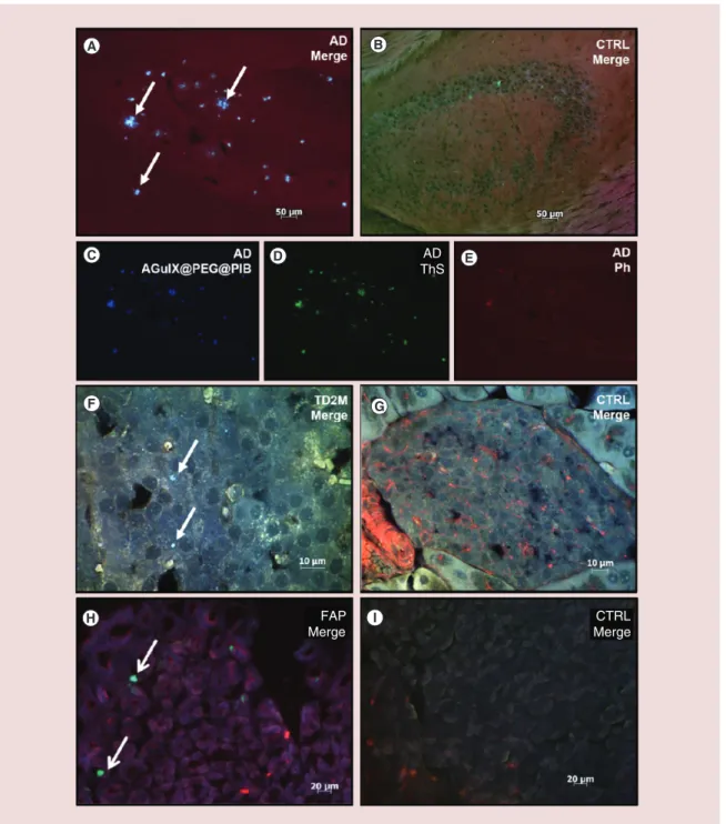
FAP mice (Figure 5H–I, Figure 6H–I). In AD mice brain,
amyloid burdens are expected mainly within the
hip-pocampus and the cortex [42]. Spots exhibiting strong
PIB fluorescence were detected within the hippocam-pus on an AD brain slice imaged with AGuIX@PEG@
PIB (Figure 5C; blue color). They co-localized with
ThS staining (Figure 5D; green color). A staining with
phalloidin was also carried out to visualize the
differ-ent structures by labeling actin networks (Figure 5E).
The merged image highlighted the co-localization of AGuIX@PEG@PIB and ThS staining, demonstrat-ing that AGuIX@PEG@PIB selectively recognized
the amyloid burden (Figure 5A). On pancreas sections
from T2DM mice, PIB and ThS signal also co-local-ized and were detected within selective and discrete areas at the level of Langherans islets, as expected for
this type of model tissue [13](Figure 5F). A similar
co-localization was also observed with stomach sections
of FAP mice (Figure 5H) where V30M-TTR deposits
have been described [43]. No fluorescence, and
there-fore no staining, was observed when similar imaging studies were carried out on control tissue sections,
Table 1. Average hydrodynamic diameter of the AGuIX, AGuIX@PEG, AGuIX@PEG@PIB, AGuIX@
DOTAfree7% and AGuIX@B10AP@Cy5.5 nanoparticles and the grafting yield of targeting agent
(Pittsburgh compound B or B10AP, grafted per nanoparticles).
AGuIX AGuIX@PEG AGuIX@PEG@
PIB AGuIX (DOTAfree7%) AGuIX@B10AP@ Cy5.5 Hydrodynamic diameter (DH) 2.9 ± 0.7 4.3 ± 1.0 6.5 ± 1.5 4.2 ± 0.7 13.9 ± 2.6 Quantification of grafting – – 1 PIB:5 NPs – 1 B10AP:5.4 NPs* PIB: Pittsburgh compound B.
Figure 3. Interaction in vitro between amyloid fibrils and AGuIX@PEG@PIB by partition experiments. (A) A total of 800 nM in PIB of AGuIX@PEG@PIB was added to 20 μM of Aβ(1–42) fibrils and the fluorescence of the suspension was measured (dotted line); then after 60 min of incubation, the suspension was centrifuged and the fluorescence of NPs bound to the amyloid fibrils (i.e., present in the pellet, solid line) and that of unbound NPs (i.e., present in the supernatant, dotted line) were measured (B, C & D). Determination of the KD using Michaelis model (1:1). The fluorescence intensity of NPs bound at approximately 420 nm is plotted as function of the total concentration of PIB functionalized on NPs, with Aβ(1–42) (B), IAPP (C) or V30M-TTR (D). The solid line represents the adjustment of the data to the Michaelis model (1:1) increasing concentration of PIB and fitted with.
IAPP: Islet amyloid polypeptide; NP: Nanoparticle; PIB: Pittsburgh compound B. 0.01 0 10 20 30 Fluorescence (a.u.) 380 400 420 440 460 Wavelength (nm) 0 50 100 150 200 Fluorescence (a.u.) AGulX@PEG@PIB: Total Free Bound 0.01 0.01 PIB concentration (μM) 40 50 0.1 1.0 10 100 KD = 21 ± 8 μM 1.0 10 100 PIB concentration (μM) 0.1 0 5 10 15 20 25 30 Fluorescence (a.u.) KD = 57 ± 27 μM 0 10 20 50 Fluorescence (a.u.) 30 40 1.0 10 100 PIB concentration (μM) 0.1 KD = 15 ± 3 μM
prepared from control mice who do not develop the
diseases (Figure 5B, G & I). All together, these results
clearly demonstrate the AGuIX@PEG@PIB selec-tively labeled amyloid plaques on tissues from the mice model of the three pathologies investigated.
Similar experiments indicated that AGuIX@ B10AP@Cy5.5 also specifically stained amyloid
depos-its on the three types of tissues (Figure 6A, F, H).
More-over, the binding of AGuIX@B10AP@Cy5.5 can be
detected through both the activity of AP (Figure 6C;
red color) and the fluorescence of Cy5.5 (Figure 6D;
yellow color). In brain sections of AD mice, these
sig-nals co-localized with ThS labeling (Figure 6E; green
color) within the hippocampus. On the merged picture
(Figure 6A), the three signals co-localized and resulted in intense yellow spots. Such co-localization of the three type of staining was also observed in pancreas tissue, and in particular in the Langherans islets of T2DM
mice (Figure 6F). Note that a labeling of the cellular
nucleus with Hoechst dye was also carried out on these later samples in order to easily identify Langherans islet (Figure 6F & G; blue color). Finally, with stomach tissue sections of FAP mice, a signal typically resulting from the co-localization of AGuIX@B10AP@Cy5.5 with ThS was also detected and assigned to amyloid depos-its (Figure 6H). Finally, the specificity of the binding of AGuIX@B10AP@Cy5.5 to amyloid was confirmed by the absence of a fluorescence signal for similar
experi-Figure 4. Interaction in vitro between amyloid fibrils and AGuIX@B10AP@cy5.5 by partition experiments. (A) A total of 11 nM in B10AP of AGuIX@B10AP@Cy5.5 was added to 40 μM of Aβ(1–42) fibrils and the
fluorescence of the suspension was measured (dotted line); then after 60 min of incubation, the suspension was centrifuged and the fluorescence of NPs bound to the amyloid fibrils (i.e., present in the pellet, solid line) and that of unbound NPs (i.e., present in the supernatant, dotted line) were measured (B, C & D). Determination of the KD using Michaelis model (1:1). The fluorescence intensity of NPs bound at approximately 685 nm (Cy5.5 fluorescence) is plotted as function of the total concentration of B10AP functionalized on NPs, with Aβ(1–42) (B), IAPP (C) or V30M-TTR (D). The solid line represents the adjustment of the data to the Michaelis model (1:1) increasing concentration of B10AP and fitted with.
IAPP: Islet amyloid polypeptide; NP: Nanoparticle. 0 100 200 300 400 500 680 700 720 740 Wavelength (nm) Fluorescence (a.u.) 0.1 1.0 10 100 B10AP concentration (nm) 0 100 200 300 Fluorescence (a.u.) AGulX@B10AP@Cy5.5: Total Free Bound 0 200 400 600 Fluorescence (a.u.) 0.1 1.0 10 100 B10AP concentration (nm) KD = 46 ± 7 nm 0 400 800 1200 1600 Fluorescence (a.u.) KD = 264 ± 51 nm 0.1 B10AP concentration (nm) 1.0 10 100 KD = 21 ± 4 nm
ments carried out on control tissues which did not
contain amyloid deposition (Figure 6B, G & I).
Discussion
The aim of this work was to develop a specific, versatile and noninvasive tool detectable by medical imaging able to specifically target amyloid deposits for a more sensitive and early diagnostic of amyloidosis.
One of the important features of functionalization of NPs is the control of the synthesis in terms of chemi-cal reaction, quality of grafting and size of the final product. The grafting of PIB to AGuIX NPs increases
their hydrodynamic diameter (Table 1). However, their
size remains homogenous (Figure 1A) and their
relaxo-metric characterization shows good relaxivity at 12.9
mM-1.s-1 allowing MRI detection. Therefore, the Gd3+
relaxation properties of the NPs are not altered by the grafting.
The affinity of AGuIX@PEG@PIB for amyloid fibrils is approximately 10 μM, close to the value that
was previously reported for PIB alone [23]. The
addi-tion of the PEG linker between PIB and the NP allows PIB to be sufficiently accessible to interact with its tar-get. One can note here, that the dissociation constants, that have been measured for PIB in this work, are sev-eral orders of magnitude higher than those reported
Figure 5. Amyloid deposit targeting with AGuIX@PEG@PIB in pathological tissue. Specific targeting of amyloid plaques with AGuIX@PEG@PIB nanoparticles on brain sections of APPswe/PSE1d9 mice (A, C–E), on pancreas sections of S/FVB/N-Tg/(ins2-hIAPP) mice (F) or stomach sections of transgenic V30M-TTR mice (H) and on respective negative control tissues: C57/Bl6 mice (B), S/FVB/NJ mice (G) and KO-TTR mice (I). Nanoparticles targeting were detected via the PIB fluorescent signal (C, blue color). Amyloid plaques were visualized using Thioflavin-S staining (D, green color). The co-localization between nanoparticles and amyloid plaques were obtained by merging PIB and ThS fluorescent staining (A, F & H, white arrows). TRITC-conjugate of phalloidin was used to label cellular actin filament (E, red color).
AD: Alzheimer’s disease; CTRL: Control; FAP: Familial amyloidotic polyneuropathy; IAPP: Islet amyloid polypeptide; NP: Nanoparticle; PIB: Pittsburgh compound B; TD2M: Type 2 diabetes mellitus; ThS: Thioflavin; TRITC: Tetramethylrhodamine. AD ThS FAP Merge CTRL Merge
in the literature [11,25–26]. The reasons for this
appar-ent discrepancy are unknown, but the conditions and
the methods used in our experiments are drastically different. As PIB has kept both its optical properties
Figure 6. Amyloid deposit targeting with AGuIX@PEG@Cy5.5 in pathological tissue. Specific targeting of amyloid plaques with AGuIX@B10AP@Cy5.5 nanoparticles on brain slices of APPswe/PSE1d9 mice (A, C–E), on pancreas sections of S/FVB/N-Tg/(ins2-hIAPP) mice (F) or stomach section of transgenic V30M-TTR mice (H) and on respective sections of negative control tissues: C57/Bl6 mice (B), S/FVB/NJ mice (G) and KO-TTR mice (I). Nanoparticles targeting were detected due to both red permanent reaction with alkaline phosphatase (C, red color) and Cy5.5 fluorescent signal (D, yellow color). Amyloid plaques were visualized using Thioflavin-S dye staining (E, green color). The co-localization between nanoparticles and amyloid plaques were obtained by merging red permanent, Cy5.5 and Thio-S fluorescent staining (A, F, H, white arrows). Cellular nucleus of the tissue were stained with Hoechst dye (F, G, blue color).
AD: Alzheimer’s disease; CTRL: Control; FAP: Familial amyloidotic polyneuropathy; TD2M: Type 2 diabetes mellitus. AD
Cy5.5
and its ability to recognize amyloid aggregates, the multimodality of the NPs has been confirmed.
A further milestone for the development of new multimodal NPs has been reached with the success-ful grafting of B10AP. This was a challenge because
of constraints implied by the use of a multidomain protein. The affinity of AGuIX@B10AP@Cy5.5 for both Aβ and IAPP fibrils is approximately 10 nM
(Figure 4B–C), and is comparable both to that measured
and to that reported in the literature [32]. These results
clearly demonstrate that B10 remains functional upon grafting to the NPs. Therefore, AGuIX NP may also constitute an interesting platform for high molecular weight molecules.
The ability of the functionalized NPs to stain amyloid fibrils on ex vivo tissues, was investigated by immunohistochemistry experiments performed on pathological tissues from mice model of each disor-der. AGuIX@PEG@PIB or AGuIX@B10AP@Cy5.5 fluorescence is observed in all tested pathological
tis-sues on distinct amyloid spots (Figures 5 & 6). In such
experiments, the co-localization of Cy5.5 fluorescence and AP activity signal in the same restricted zones was a further proof of the integrity of AGuIX@B10AP@ Cy5.5. In addition, the fact that both NPs co-localized with Thioflavin-S staining, confirms their specific targeting of amyloid burden.
The determination of the equilibrium dissocia-tion constant of these NPs is important for estimat-ing their potential as diagnosis tools. The affinity for amyloid fibrils of AGuIX@PEG@PIB is in the 10 μM range and that of AGuIX@B10AP@Cy5.5 in the 10 nanomolar range. Such values are compatible with the
use of such NP for in in vivo studies (Figures 3 & 4).
The dissociation constant of AGuIX@B10AP@Cy5.5 for V30M-TTR fibrils is significantly larger than for the other two types of fibrils. The reason for this lower affinity is unknown. However, it has been reported that B10 does not recognize all amyloid fibrils with
the same efficiency [32]; this compromises its use for
diagnostic. This work, however, clearly demonstrates that large multidomain proteins can be efficiently linked to AGuIX; this proof of concept opens the pos-sibility to functionalize these NPs with nanobodies recognizing selective epitopes of the AD at the surface of extracellular amyloid plaque or intracellular
tar-gets [44]. Moreover, small peptides with high affinity
(KD in 10 nanomolar range [45,46]) for a generic epitope
at the surface of Aβ amyloid fibrils have been recently described and could also constitute valuable probes to be grafted on AGuIX. Nevertheless, these peptides present important drawbacks since they induce cell
toxicity [45] or stabilize protofibrils [46]. Some other
peptides specifically targeting Aβ-peptide fibrils with no toxic effects have been described but their affini-ties are too low for in vivo imaging (100 micromolar
range) [23].
Conclusion
In this work, we have shown that AGuIX can be suc-cessfully functionalized with a small molecule and a large protein to specifically target amyloid fibrils. Thus, the NPs tested in this work provide complementary
tools that could be useful for in vivo diagnostic: strong affinity of AGuIX@B10AP@Cy5.5 will be helpful for early amyloid deposits detection, when the smaller size of AGuIX@PEG@PIB should diffuse more quickly, particularly interesting notably for systemic amyloi-dosis (V30M-TTR). In the case of AD diagnostic, we must take into account that such NPs have to reach their target inside the brain, implicating the crossing of the hermetic blood–brain barrier. However, the flex-ibility of AGuIX NPs may allow chemical modifica-tions for diffusion through this barrier or addition of molecules to improve active transport through specific receptor of the blood–brain barrier.
In any case, such functionalized NPs are very prom-ising to target several types of amyloid deposits in vivo, because of their very low levels of toxicity; their rapid
clearance [47]; and the divert and complementarity of
imaging techniques with great sensitivity and spatial resolution that can be used.
Acknowledgements
We thank F Daphna, M Christine and S Guy from the Elec-tron Microscopy platform of the Integrated Structural Biology of Grenoble (ISBG, UMI3265). Others TEM images were ob-tained at the electronic microscopy platform, Neurosciences Institute of Grenoble thanks to K Pernet Gallay, 38700 Greno-ble. The authors thank Horn from Jena University (Germany) for the kind gift of the plasmid ptetB10pA6H. The authors thank R Cohen for his helpful expertise in affinity experiments.
Financial & competing interests disclosure
This work was supported by EuroNanoMed ENMII JTC2012 (project 2011-ERA-002–01- Dia-Amyl) and the French Nation-al Research Agency (ANR): ANR-12-RPIB Multimage. J Pans-ieri is granted by FRM DBS2013112844. M Plissonneau and N Stransky-Heilkron were granted by ENMII JTC2012 (project 2011-ERA-002–01). M Dumoulin is an FRS-FNRS research associate. This work was also supported by the Belgian pro-gram of Interuniversity Attraction Poles administered by the Federal Office for Scientific Technical and Cultural Affairs to MD (PAI 7/44). F Lux and O Tillement have one patent to dis-close: WO2011135101. This patent protects the synthesis of the AGuIX® nanoparticles for imaging, used in this publica-tion. This work used the platforms of the Grenoble Instruct centre (ISBG; UMS 3518 CNRS-CEA-UJF-EMBL) with sup-port from FRISBI INSB-05–02) and GRAL (ANR-10-LABX-49–01) within the Grenoble Partnership for Structural Biology (PSB). The authors have no other relevant affiliations or financial involvement with any organization or entity with a financial interest in or financial conflict with the subject mat-ter or materials discussed in the manuscript apart from those disclosed. No writing assistance was utilized in the production of this manuscript.
Ethical conduct of research The authors state that they have obtained appropriate institu-tional review board approval or have followed the principles outlined in the Declaration of Helsinki for all human or animal experimental investigations. In addition, for investigations in-volving human subjects, informed consent has been obtained from the participants involved. Summary points
• To achieve the diagnostic of amyloidosis, multimodal imaging nanoparticles were functionalized with either the Pittsburgh compound B or a nanobody.
• Efficient active targeting was tested by spectroscopic and biophysics techniques on Aβ(1–42), IAPP and Val30MetTTR fibrils.
• Both nanoparticles efficiently interacted with the three types of amyloid fibrils, with KD values in 10 micromolar range for AGuIX@PIB and 10 nmolar range for AGuIX@Cya5.5@B10AP.
• Both functionalized nanoparticles are able to interact specifically with amyloid deposits localized in pathological tissue.
• These functionalized nanoparticles represent promising flexible devices for multimodal imaging in early diagnostic of amyloid plaques, in other words, Alzheimer’s disease, Type 2 diabetes mellitus and the familial amyloidotic polyneuropathy.
References
Papers of special note have been highlighted as: • of interest; •• of considerable interest
1 Knowles TP, Vendruscolo M, Dobson CM. The amyloid state and its association with protein misfolding diseases. Nat. Rev. Mol. Cell Biol. 15(6), 384–396 (2014). • Shows how much progress has been made toward
knowledge of the amyloid phenomenon providing basis for therapeutic advances.
2 Tycko R. Solid state NMR studies of amyloid fibril structure. Annu. Rev. Phys. Chem. 62, 279–299 (2011).
3 Sipe JD, Benson MD, Buxbaum JN et al. Amyloid fibril proteins and amyloidosis: chemical identification and clinical classification International Society of Amyloidosis 2016 Nomenclature Guidelines. Amyloid 23, 209–213 (2016).
4 World Alzheimer Report 2014: Dementia and Risk Reduction | Alzheimer’s Disease International. www.alz. co.uk/research/world-report-2014
5 IDF diabetes atlas – Home. www.diabetesatlas.org/
6 Perrin RJ, Fagan AM, Holtzman DM. Multimodal techniques for diagnosis and prognosis of Alzheimer’s disease. Nature 461, 916–922 (2009).
7 Gilbertson JA, Botcher NA, Rowczenio D et al. Diagnostic value of fat aspirates for amyloidosis in 950 patients. Orphanet J. Rare Dis. 10(Suppl. 1), P50 (2015).
8 Liu PP, Smyth D. Wild type transthyretin amyloid cardiomyopathy: a missed etiology of HFpEF with evolving treatment implications. Circulation 133(3), 245–247 (2015).
9 Jacobsen H, Ozmen L, Caruso A et al. Combined treatment with a bace inhibitor and anti-Aβ antibody gantenerumab enhances amyloid reduction in APPLondon mice. J. Neurosci. 34, 11621–11630 (2014).
10 Seo SW, Ayakta N, Grinberg LT et al. Regional correlations between [11C] PIB PET and post-mortem burden of amyloid-beta pathology in a diverse neuropathological cohort. NeuroImage Clin. 13, 130–137 (2017).
• Shows the utility of Pittsburgh compound B as gold standard for in vivo imaging of amyloidosis-related disease.
11 Higuchi M, Iwata N, Matsuba Y et al. 19F and 1H MRI detection of amyloid β plaques in vivo. Nat. Neurosci. 8, 527–533 (2005).
12 Rapezzi C, Quarta CC, Guidalotti PL et al. Role of 99mTc-DPD scintigraphy in diagnosis and prognosis of hereditary transthyretin-related cardiac amyloidosis. J. Am. Coll. Cardiol. Img. 4, 659–670 (2011).
13 Yoshimura M, Ono M, Watanabe H, Kimura H, Saji H. Feasibility of amylin imaging in pancreatic islets with β-amyloid imaging probes. Sci. Rep. 4, 6155 (2014).
14 Knight MJ, McCann B, Kauppinen RA, Coulthard EJ. Magnetic resonance imaging to detect early molecular and cellular changes in Alzheimer’s disease. Front. Aging Neurosci. 8, 139 (2016).
15 Moses WW. Fundamental limits of spatial resolution in PET. Nucl. Instrum. Methods Phys. Res. A 648(Suppl. 1), S236–S240 (2011).
16 Martí-Bonmatí L, Sopena R, Bartumeus P, Sopena P. Multimodality imaging techniques. Contrast Media Mol. Imaging 5, 180–189 (2010).
•• Significance on combination of various imaging techniques in diagnosis, adding functional and structural data.
17 Huang WY, Davis J. Multimodality and nanoparticles in medical imaging. Dalton Trans. 40, 6087–6103 (2011).
18 Lux F, Mignot A, Mowat P et al. Ultrasmall rigid particles as multimodal probes for medical applications. Angew. Chem. Int. Ed. Engl. 50, 12299–12303 (2011).
19 LeDuc G, Roux S, Paruta-Tuarez A et al. Advantages of gadolinium based ultrasmall nanoparticles vs molecular gadolinium chelates for radiotherapy guided by MRI for glioma treatment. Cancer Nanotechnol. 5, 4 (2014).
20 Sancey L, Lux F, Kotb S et al. The use of theranostic gadolinium-based nanoprobes to improve radiotherapy efficacy. BJR 87, 20140134 (2014).
•• Review and description of imaging properties of versatile AGuIX nanoparticles.
21 Truillet C, Bouziotis P, Tsoukalas C et al. Ultrasmall particles for Gd-MRI and 68Ga-PET dual imaging. Contrast Media Mol. Imaging 10, 309–319 (2015).
22 Bianchi A, Moncelet D, Lux F et al. Orotracheal administration of contrast agents: a new protocol for brain tumor targeting. NMR Biomed. 28, 738–746 (2015).
23 Pansieri J, Plissonneau M, Heinrich-Balard L et al. Gd-nanoparticles functionalization with specific peptides for ß-amyloid plaques targeting. J. Nanobiotechnol. 14, 60 (2016).
24 Biancalana M, Koide S. Molecular mechanism of Thioflavin-T binding to amyloid fibrils. Biochim. Biophys. Acta 1804, 1405–1412 (2010).
25 Ng S, Villemagne VL, Berlangieri S et al. Visual assessment versus quantitative assessment of 11C-PIB PET and 18F-FDG PET for detection of Alzheimer’s disease. J. Nucl. Med. 48, 547–552 (2007).
26 Maezawa I, Hong HS, Liu R et al. Congo red and
thioflavin-T analogs detect Aβ oligomers. J. Neurochem. 104, 457–468 (2008).
27 Ni R, Gillberg PG, Bergfors A, Marutle A, Nordberg A. Amyloid tracers detect multiple binding sites in Alzheimer’s disease brain tissue. Brain 136, 2217–2227 (2013).
28 Lux F, Sancey L, Bianchi A et al. Gadolinium-based nanoparticles for theranostic MRI-radiosensitization. Nanomedicine 10, 1801–1815 (2015).
29 Wang Y, Fan Z, Shao L et al. Nanobody-derived nanobiotechnology tool kits for diverse biomedical and biotechnology applications. Int. J. Nanomedicine 11, 3287–3303 (2016).
30 Sillerud LO, Solberg NO, Chamberlain R et al. SPION-enhanced magnetic resonance imaging of Alzheimer’s disease plaques in AβPP/PS-1 transgenic mouse brain. J. Alzheimers Dis. 34, 349–365 (2013).
31 Habicht G, haupt C, Friedrich RP et al. Directed selection of a conformational antibody domain that prevents mature amyloid fibril formation by stabilizing Aβ protofibrils. Proc. Natl Acad. Sci. USA 104, 19232–19237 (2007).
32 Haupt C, Bereza M, Kumar ST et al. Pattern recognition with a fibril-specific antibody fragment reveals the surface variability of natural amyloid fibrils. J. Mol. Biol. 408, 529–540 (2011).
• High-affinity nanobody B10AP for amyloid fibrils, implicated in various diseases.
33 Martins AF, Morfin JF, Kubickova A et al. PiB-conjugated, metal-based imaging probes: multimodal approaches for the visualization of β-amyloid plaques. ACS Med. Chem. Lett. 4, 436–440 (2013).
34 Cao P, Tu LH, Abedini A et al. Sensitivity of amyloid formation by human islet amyloid polypeptide to mutations at residue 20. J. Mol. Biol. 421, 282–295 (2012).
35 Borchelt DR, Davis J, Fischer M et al. A vector for expressing foreign genes in the brains and hearts of transgenic mice. Genet. Anal. 13, 159–163 (1996).
36 Borchelt DR, Thinakaran G, Eckman CB et al. Familial Alzheimer’s disease – linked Presenilin 1 variants elevate Aβ1–42/1–40 ratio in vitro and in vivo. Neuron 17, 1005–1013 (1996).
37 Janson J, Soeller WC, Roche PC et al. Spontaneous diabetes mellitus in transgenic mice expressing human islet amyloid polypeptide. Proc. Natl Acad. Sci. USA 93, 7283–7288 (1996).
38 Kohno K, Palha JA, Miyakawa K et al. Analysis of amyloid deposition in a transgenic mouse model of homozygous familial amyloidotic polyneuropathy. Am. J. Pathol. 150, 1497–1508 (1997).
39 Palha JA, Episkopou V, Maeda S, Shimada K, Gottesman ME, Saraiva MJM. Thyroid hormone metabolism in a transthyretin-null mouse strain. J. Biol. Chem. 269, 33135–33139 (1994).
40 Maskevich AA, Stsiapura VI, Kuzmitsky VA et al. Spectral properties of thioflavin T in solvents with different dielectric properties and in a fibril-incorporated form. J. Proteome Res. 6, 1392–1401 (2007).
41 Rodina NP, Sulatsky MI, Sulatskaya AI, Kuznetsova IM, Uversky VN, Turoverov KK. Photophysical properties of fluorescent probe Thioflavin T in crowded milieu. J. Spectrosc. 2017, 2365746 (2017).
42 Petiet A, Santin M, Bertrand A et al. Gadolinium-staining reveals amyloid plaques in the brain of
Alzheimer’s transgenic mice. Neurobiol. Aging 33, 1533–1544 (2012).
43 Terazaki H, Ando Y, Fernandes R, Yamamura KI, Maeda S, Saraiva MJM. Immunization in familial amyloidotic polyneuropathy: counteracting deposition by immunization with a Y78F TTR mutant. Lab. Invest. 86, 23–31 (2005).
44 Lia T, Vandesquille M, Koukoulik F et al. Camelid single-domain antibodies: a versatile tool for in vivo imaging of extracellular and intracellular brain targets. J Control. Release 243, 1–10 (2016).
45 Xiong N, Dong XY, Zheng J, Liu FF, Sun Y. Design of LVFFARK and LVFFARK-functionalized nanoparticles for inhibiting amyloid β-protein fibrillation and cytotoxicity. ACS Appl. Mater. Interfaces 7, 5650–5662 (2015).
46 Moss MA, Nichols MR, Reed DK, Hoh JH, Rosenberry TL. The peptide KLVFF-K(6) promotes beta-amyloid(1–40) protofibril growth by association but does not alter protofibril effects on cellular reduction of 3-(4,5-dimethylthiazol-2-yl)-2,5-diphenyltetrazolium bromide (MTT). Mol. Pharmacol. 64, 1160–1168 (2003).
47 Sancey L, Kotb B, Truillet C et al. Long-term in vivo clearance of gadolinium-based AGuIX nanoparticles and their biocompatibility after systemic injection. ACS Nano 9, 2477–2488 (2015).