HAL Id: hal-02391905
https://hal-univ-rennes1.archives-ouvertes.fr/hal-02391905
Submitted on 27 Apr 2020
HAL is a multi-disciplinary open access
archive for the deposit and dissemination of
sci-entific research documents, whether they are
pub-lished or not. The documents may come from
teaching and research institutions in France or
abroad, or from public or private research centers.
L’archive ouverte pluridisciplinaire HAL, est
destinée au dépôt et à la diffusion de documents
scientifiques de niveau recherche, publiés ou non,
émanant des établissements d’enseignement et de
recherche français ou étrangers, des laboratoires
publics ou privés.
Sandra E. Ghayad, Oula K. Dagher, Nada Borghol, Rene Gree, Bassam
Badran, Ali Hachem, et al.
To cite this version:
Abeer J. Ayoub, Layal Hariss, Nehme El-Hachem, Ghewa A. El-Achkar, Sandra E. Ghayad, et al..
gem-Difluorobisarylic derivatives: design, synthesis and anti-inflammatory effect. BMC Chemistry,
BMC, 2019, 13 (1), �10.1186/s13065-019-0640-5�. �hal-02391905�
synthesis and anti-inflammatory effect
Abeer J. Ayoub
1,2†, Layal Hariss
3†, Nehme El‑Hachem
4,8†, Ghewa A. El‑Achkar
1, Sandra E. Ghayad
5,
Oula K. Dagher
1, Nada Borghol
2, René Grée
6, Bassam Badran
2, Ali Hachem
3†, Eva Hamade
2†and Aida Habib
1,7*†Abstract
Introduction: New fluorinated diaryl ethers and bisarylic ketones were designed and evaluated for their anti‑inflam‑ matory effects in primary macrophages.
Methods: The synthesis of the designed molecules started from easily accessible and versatile gem‑difluoro prop‑ argylic derivatives. The desired aromatic systems were obtained using Diels–Alder/aromatization sequences and this was followed by Pd‑catalyzed coupling reactions and, when required, final functionalization steps. Both direct inhibi‑ tory effects on cyclooxygenase‑1 or ‑2 activities, protein expression of cyclooxygenase‑2 and nitric oxide synthase‑II and the production of prostaglandin E2, the pro‑inflammatory nitric oxide and interleukin‑6 were evaluated in primary murine bone marrow‑derived macrophages in response to lipopolysaccharide. Docking of the designed molecules in cyclooxygenase‑1 or ‑2 was performed.
Results: Only fluorinated compounds exerted anti‑inflammatory activities by lowering the secretion of interleu‑ kin‑6, nitric oxide, and prostaglandin E2, and decreasing the protein expression of inducible nitric oxide synthase and cyclooxygenase‑2 in mouse primary macrophages exposed to lipopolysaccharide, as well as cyclooxygenase activ‑ ity for some inhibitors with different efficiencies depending on the R‑groups. Docking observation suggested an inhibitory role of cyclooxygenase‑1 or ‑2 for compounds A3, A4 and A5 in addition to their capacity to inhibit nitrite, interleukin‑6, and nitric oxide synthase‑II and cyclooxygenase‑2 expression.
Conclusion: The new fluorinated diaryl ethers and bisarylic ketones have anti‑inflammatory effects in macrophages. These fluorinated compounds have improved potential anti‑inflammatory properties due to the fluorine residues in the bioactive molecules.
Keywords: Fluorine, Diaryl ethers, Macrophages, Cyclooxygenase, Inflammation
© The Author(s) 2019. This article is distributed under the terms of the Creative Commons Attribution 4.0 International License (http://creat iveco mmons .org/licen ses/by/4.0/), which permits unrestricted use, distribution, and reproduction in any medium, provided you give appropriate credit to the original author(s) and the source, provide a link to the Creative Commons license, and indicate if changes were made. The Creative Commons Public Domain Dedication waiver (http://creativecommons.org/ publicdomain/zero/1.0/) applies to the data made available in this article, unless otherwise stated.
Introduction
Diaryl ethers are key scaffolds present in many natu-ral or synthetic organic molecules, which are often used in medicinal chemistry [1]. Fenoprofen for instance is one of the synthetic diarylethers [2] with nonsteroidal
anti-inflammatory, analgesic and antirheumatic effects [3]. More precisely, it is a derivative of 2-aryl propanoic acids, which is an important class of nonsteroidal anti-inflammatory drugs including flurbiprofen, ibuprofen, naproxen and fenoprofen.
Moreover, benzophenone analogues, such as keto-profen, recently have been reported also as potent anti-inflammatory agents by inhibiting prostaglandin (PG) production [4, 5]. It has been shown that benzoylphenyl acetic acid for instance has anti-inflammatory activity by decreasing the volume of paw edema in treated rats [6].
On the other hand, the introduction of fluorine into organic molecules may cause profound pharmacological *Correspondence: [email protected]
†Abeer J. Ayoub, Layal Hariss and Nehme El‑Hachem contributed equally
to the work
†Ali Hachem, Eva Hamade and Aida Habib are senior authors and
contributed equally to this work
1 Department of Biochemistry and Molecular Genetics, Faculty
of Medicine, American University of Beirut, Beirut, Lebanon Full list of author information is available at the end of the article
bioactive molecules [7]. The utility of fluorine in the design of drugs results mainly from its ability to modify some functional activities, such as increasing lipophilicity [8] and extending its bioavailability [9]. Moreover, carbon forms stronger bond with fluorine (CF)n, with a higher oxidative and thermal stability than a carbon–hydrogen bond [10]. The CF2 unit for instance is generally consid-ered as a bioisostere of the oxygen atom or of a carbonyl group [11].
We therefore synthesized new gem-difluorobisarylic derivatives and evaluated their anti-inflammatory effects. We first investigated their effects on PGE2 production in mouse primary macrophages in response to lipopolysac-charide (LPS) and their anti-cyclooxygenase (COX)-1 and -2 activities. We next studied their effects on the pro-duction of the pro-inflammatory nitric oxide (NO) and interleukin (IL)-6 and the expression of NO synthase-II (NOS-II) and COX-2.
Results and discussion
Synthesis of bisarylic derivatives
Based on our previous study [12], five bisarylic com-pounds A1 to A5 were designed as indicated in Scheme 1. In our strategy, the gem-difluoro unit has been chosen as a mimic of either the ether oxygen (fenoprofen series) or of a carbonyl group (ketoprofen series). First, two phe-nylpropionic acid derivatives A1 (as a non-fluorinated
tive A2 were proposed as analogues of fenoprofen and ketoprofen. The comparison of the inhibitory activities of compounds A1 and A2 would allow establishing the impact of fluorine atom on the efficiency of these com-pounds. On the other hand, three other derivatives A3,
A4, and A5 were designed as simplified benzoic
acid-type derivatives, with three different substituents in meta position on the second aromatic ring (Scheme 1).
Synthetic procedures
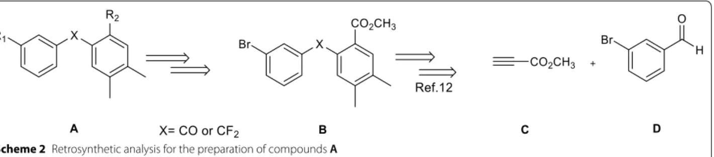
All these molecules were synthesized from bromo inter-mediates B (Scheme 2, Table 1) and were tested for their anti-inflammatory activity.
Synthesis of key intermediates 7 and 10
Addition of the lithium salt of compound 2 to 3-bro-mobenzaldehyde 3 at low temperature (− 80 °C) gave propargyl alcohol 4 in 70% yield. After oxidation with Jones reagent, propargylic ketone 5 was isolated in 80% yield. Then, Diels–Alder reaction and DDQ aromatiza-tion provided the intermediate 7 with a good yield for both steps (Scheme 3).
After treatment of ketone 5 by DAST, compound 8 was obtained in 71% yield. Similarly, Diels–Alder reac-tion and DDQ aromatizareac-tion proceeded well by giving the fluorinated intermediate 10 with excellent yields (Scheme 4). F F A3 Br O OH F F A4 O OH F F A5 O OH O A1 F F A2 O CO2Me O OH O OH CO2Me O O OH Ketoprofen NSAID O O OH Fenoprofen NSAID
Preparation of compounds A1 and A2
Starting from the key scaffolds 7 and 10, Suzuki–Miyaura couplings, with boronic acid, afforded biphenyl type compounds 11 and 12 in 92 and 94% yield, respectively.
Then, hydroboration to 13 and 14, followed by oxidation with Jones reagent led to the desired analogues A1 and
A2 in good yields (Scheme 5).
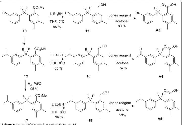
Preparation of compounds A3, A4, and A5
Starting from intermediates 10 and 12, reduction with LiBEt3H furnished alcohols 15 and 16 respectively, then oxidation by Jones reagent gave the desired acid A3. However, in the case of 16, an unexpected cleavage of the double bond occurred, affording acid A4. Using the same
gem-difluoro intermediate 12, catalytic hydrogenation
to 17, followed by reduction and oxidation afforded the desired derivative A5 in good yields (Scheme 6).
Biological activities
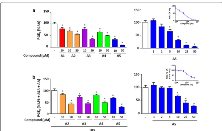
We investigated the effects of these derivatives on inflam-mation in bone marrow-derived macrophages (BMDM) by first evaluating their capacity to decrease LPS-depend-ent increase of PGE2 secretion and COX-2 expres-sion. Compound A1 (non-fluorinated) and compound
A2 (fluorinated) effects were compared to evaluate the
importance of the fluorine atom. Only compound A2 inhibited significantly in a dose-dependent manner the
Scheme 2 Retrosynthetic analysis for the preparation of compounds A
Table 1 Preparation of five bisarylic compounds
Compound X R1 R2 A1 C=O –CO2CH3 A2 CF2 –CO2CH3 A3 CF2 Br –CO2H A4 CF2 –COCH3 –CO2H A5 CF2 –CH(CH3)2 –CO2H Br H O Br OH Br O CO2Me CO2Me n-Buli 1) 2) TMSCl Jones reagent acetone THF, - 80oC 2 70 % 4 80 % 5 CO2Me Br CO2Me Br CO2Me 80oC 6 7 96% 87 % DDQ toluene O O 1 3
secretion of PGE2 (Fig. 1a) (IC50 = 16.5 ± 8.9 µM) with no effect on COX-2 expression (Fig. 1c) supporting the importance of fluorine in inhibiting PGE2 production.
In parallel, we compared the inhibitory effects of com-pounds A3, A4 and A5, which are all fluorinated but pre-sent differences in R1 group (Table 1). Compound A3 is
Br CO2Me F F Br CO2Me Br CO2Me F F F F DAST, 60oC ETOH cat. 80oC 8 9 10 71 % 95 % 91 % DDQ toluène Br O CO2Me 5
Scheme 4 Synthesis of the fluorinated key intermediate 10
the bromine intermediate obtained in the synthetic reac-tion of compound A5. Compound A4 is a ketone inter-mediate obtained unexpectedly with good yield during the synthesis of compound A5. Compounds A3, A4 and
A5 have the carboxyl group attached to the benzene ring
in the ortho position relative to CF2 group.
Figure 1b showed a dose response effect of these deriva-tives on PGE2 secretion, in which compounds A4 and A5 significantly decreased PGE2 secretion at 25 and 50 µM with IC50 of 28.1 ± 22.8 and 22.4 ± 21.5 µM, respectively. Compound A3 did not show a strong inhibition at similar concentrations. Under these conditions, only compounds
A4 and A5 significantly downregulated COX-2
expres-sion (Fig. 1d). Further analysis showed a dose-dependent
inhibitory effect on COX-2 expression for compounds
A4 and A5 (Fig. 1e and f, respectively). Thus, the nature of R groups in compounds A4 and A5 is important for their inhibitory effect on COX-2 expression and conse-quently PGE2 production. We next addressed the ques-tion whether COX activity was inhibited. We performed COX-1 activity using Human Embryonic kidney (HEK)-293 cells stably overexpressing COX-1. Cells were treated with all compounds at 10 and 50 µM and PGE2 was measured after the addition of arachidonic acid (AA). The results showed that compound A5 had the maximal inhibitory effect on COX-1 activity with more than 80% inhibition at 50 µM (Fig. 2a) with an IC50 of 5.2 µM. Scheme 6 Synthesis of gem‑diaryl derivatives A3, A4, and A5
(See figure on next page.)
Fig. 1 Effects of the gem‑difluorobisarylic derivatives on PGE2 production and COX‑2 expression in activated macrophages. BMDM were treated with 6 increasing concentrations, prior to the addition of 10 ng/mL LPS for 24 h. PGE2 secretion was measured and expressed as percentage of LPS for a compounds A1 and A2 and b compounds A3, A4 and A5. Corresponding IC50 fitting curves are shown. c, d COX‑2 and β‑actin expression in basal and LPS‑treated BMDM with 50 µM of all compounds. Results are obtained from the same blot. Protein bands for basal or LPS‑treated macrophages, in the absence of inhibitors, as shown in c and d, are identical for illustration purpose. Dose–response effect of compounds e A4 and f A5 on COX‑2 expression. β‑actin was used as loading control. Ratio of COX‑2/β‑actin was calculated after densitometry analysis using ImageJ software. Data are represented as mean ± SEM (n = 4), *p < 0.05 versus LPS (One‑way Anova followed by the Dunnett’s test)
COX-2 activity was also assessed on BMDM treated for 30 min with aspirin to inhibit basal COX activity prior to the addition of 10 ng/mL LPS for 24 h which induces COX-2. These cells were then treated with 10 and 50 µM of derivatives and further incubated with AA. PGE2 production revealed that compound A5 inhibited strongly COX-2 activity with an IC50 of 13.3 µM, whereas moderate effect was observed for compounds A2, A3 and A4 (Fig. 2b). Indeed, the assay used for COX-2 activity cannot exclude an effect on mPGES-1.
In parallel, we assessed the effect of these compounds on the production of IL-6 and NO measured by its breakdown product nitrite. Figure 3 showed a dose response inhibition of compounds A2 (Fig. 3a, c), A4
and A5 (Fig. 3b, d) for both IL-6 and NO secretion. IC50 are presented in Table 2 and were significant for com-pounds A4 and A5.
NOS-II is the inducible form of nitric oxide synthase, and is responsible for the production of the measured NO. For this, NOS-II protein expression was analyzed in LPS-stimulated BMDM, treated with 50 µM of bisarylic derivatives for 24 h. Results revealed that the fluorinated compounds A2, A4 and A5 inhibited NOS-II expression in parallel to NO production (Fig. 3e, f).
Molecular docking
We finally carried out model analysis of the inhibitors with ovine COX-1 [13] and murine COX-2 [14], to exam-ine how these compounds dock with the active sites of
Fig. 2 Effects of the gem‑difluorobisarylic derivatives on COX‑1 and COX‑2 activity. a COX‑1 activity. HEK‑293 cells overexpressing recombinant
COX‑1 were treated with 10 and 50 µM of all compounds for 45 min prior to the addition of 10 µM arachidonic acid (AA). PGE2 was measured.
b COX‑2 activity. BMDM cells were treated with 10 µM of ASA for 30 min, washed, and 10 ng/mL LPS was added for 24 h to induce COX‑2. Cells
were further incubated with 10 and 50 µM of each compound prior to the addition of 10 µM AA. PGE2 was measured. Data are represented as mean ± SEM (n = 4), *p < 0.05 versus AA for COX‑1 activity, and versus LPS + ASA + AA for COX‑2 activity (One‑way Anova followed by the Dunnett’s test)
(See figure on next page.)
Fig. 3 Effects of the gem‑difluorobisarylic derivatives IL‑6 and nitrite, and NOS‑II expression. BMDM were treated with 6 increasing concentrations
of all compounds prior to the addition of 10 ng/mL LPS for 24 h. IL‑6 and NO production was measured and expressed as percentage of LPS for a,
c compounds A1 and A2, and b, d, compounds A3, A4 and A5, respectively. Corresponding IC50 fitting curves are shown. e, f NOS‑II and β‑actin expression in basal and LPS‑treated BMDMs with 50 µM of all compounds. Results are obtained from the same blot. Protein bands for basal or LPS‑treated in macrophages in the absence of inhibitors, as shown in e and f, are identical for illustration purpose. β‑actin was used as loading control. Data are represented as mean ± SEM (n = 4), *p < 0.05 versus LPS (One‑way Anova followed by the Dunnett’s test)
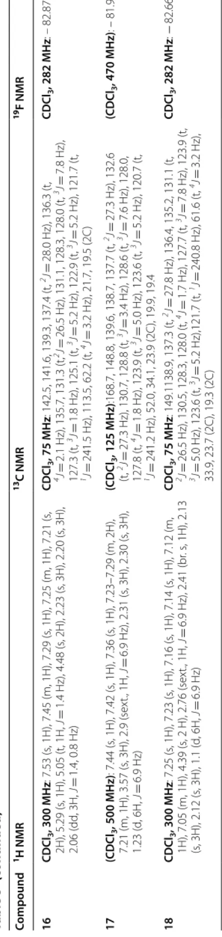
the enzymes and to determine the amino acids involved in the interaction with the compounds. Ibuprofen docked into the hydrophobic cavity of COX-2 formed by Arg121, Tyr356, Ser354, Leu353, Val350 and Tyr349, where the carboxyl group of ibuprofen interacts with Arg121 and Tyr356 by a salt bridge and a hydrogen bond. The com-pounds A3, A4 and A5 were docked near Arg121, simi-larly to ibuprofen. Compounds A3 and A4 showed interaction with Tyr356 (Fig. 4). The binding scores of
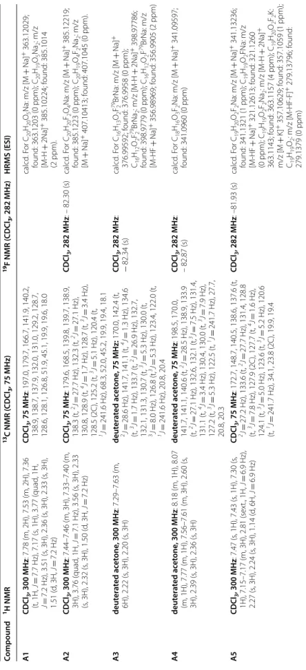
pounds A1 and A2, even though they occupy the same active pocket, interact with Arg121 through the carbox-ylate group with less binding energy, have a bulky side chains that negatively would affect the stability of these molecules in the hydrophobic pocket. For COX-1, ibu-profen docked into the hydrophobic pocket composed of the amino acids Arg120, Tyr355, Ser353, Leu352, Val349, Tyr348, Val116, Leu531, Ser530, Ala527, Gly526 and Ile523. All compounds docked in the same active hydrophobic pocket of COX-1. Only Arg120 inter-acts with the carboxylate group by a salt bridge (Fig. 5). Similarly, to ibuprofen, compounds A2, A3, A4 and A5 showed a moderate binding energy compared to ibu-profen (− 7 kcal/mol and − 7.8 kcal/mol respectively, Table 3) whereas compound A1 showed the lowest bind-ing energy, which is compatible with biological activities.
More analyses are required to fully understand the key role of the fluorine atoms on the biological activity of these molecules. However, to explain these results, it is
ND not determined a Mean ± SEM A3 60 ± 60.2 ND A4 51.2 ± 17.4 18.5 ± 2.7 A5 82.6 ± 24.0 40.9 ± 25.4 Fenoprofen 300 ± 100 43.8 ± 39.2
Fig. 4 Two‑dimensional pose of compounds A1 to A5 and Ibuprofen inside the binding pocket of mouse COX‑2 as crystallized by [13]. Ligand‑receptor interactions as highlighted by Maestro (Shrodinger, LLC). Ligands are represented in stick, and amino acids within the binding pocket are labeled. An arrow represents the H‑bonds between an amino acid and ligand groups. A line shows a potential salt bridge between two charged groups
possible that the bulky and lipophilic CF2 group could fit better in the pocket of these proteins than the carbonyl of ketoprofen or the oxygen atom of fenoprofen. Fur-ther, in the case of compounds A3, A4 and A5 it can also increase the acidity of the CO2H in ortho position. Conclusion
In conclusion, five bisarylic derivatives were prepared and tested in comparison with fenoprofen. This type of compounds is endowed with certain anti-inflammatory activities in mouse primary macrophages with a signifi-cant difference between the fluorinated analogues and the non-fluorinated one, showing the importance of the CF2 group. All fluorinated derivatives blocked PGE2, nitrite and IL-6 production in activated macrophages.
tion of COX-2 and NOS-II expression. In addition, deriv-atives A3, A4 and A5 showed better anti-inflammatory activities than the other derivatives, with compound A5 having COX-1 and COX-2 direct inhibitory activities. Molecular docking of the compounds COX-1 and COX-2 are in support of the biological activity.
Methods
Chemistry experimental part
Reactions were carried out as described previously and monitored as described by 19F NMR and by thin-layer chromatography (TLC) [15]. Yields refer to chromato-graphically and spectroscopically (1H, 13C, and 19F NMR) homogeneous materials. Nuclear magnetic resonance (NMR) spectra have been recorded as previously described [15]. Mass spectral analyses have been performed at the Centre Régional de Mesures Physiques de l’Ouest (CRMPO) in Rennes (France).
Synthesis of methyl 4-(3-bromophenyl)-4-hydroxy-but-2-ynoate 4
To a solution of methylpropiolate (2.6 mL, 29.20 mmol, 1.2 equiv.) in anhydrous THF (20 mL) cooled at − 90 °C and set under nitrogen, n-BuLi (11.4 mL, 2.5 M, 1.2 equiv.) was added dropwise. The reaction mixture was stirred for 30 min at T − 80 °C before the dropwise addition of a solution of 3-bromobenzaldehyde (4 g, 21.60 mmol)
docking data
Compound COX-1 kcal/mol COX-2 kcal/mol
A1 − 6.5 − 6.6 A2 − 7 − 6.7 A3 − 7 − 7.7 A4 − 7 − 7.7 A5 − 7 − 7.5 Ibuprofen − 7.8 − 7.7
Fig. 5 Two‑Dimensional pose of compounds A1 to A5 and Ibuprofen inside the binding pocket of human COX‑1 as crystallized by [14]. Ligand receptor interactions were evaluated for COX‑1 as described in legend for Fig. 4
dried over Na2SO4 and concentrated by evaporating the solvent. Alcohol 4 was isolated over silica gel by column chromatography.
Synthesis of methyl 4-(3-bromophenyl)-4-oxobut-2-ynoate 5
To alcohol 4 (2.1 g, 7.46 mmol) in acetone (18 mL) was added dropwise under magnetic stirring at room tempera-ture, a concentrated (5.4 M) solution of Jones reagent until disappearance of the starting material (TLC analysis). After addition of isopropanol (5 equiv.), the reaction mixture was filtered, and the filtrate was extracted with ethyl acetate. The combined organic phases were dried over Na2SO4, filtered and concentrated in vacuum. After purification by chromatography on silica gel, ketone 5 was obtained.
Synthesis of methyl 4-(3-bromophenyl)-4,4-dif-luorobut-2-ynoate 8
To propargylic ketone 5 (350 mg, 1.31 mmol) were added one drop of 95% ethanol and DAST (1.05 mL, 7.96 mmol, 6 equiv.). The reaction mixture was stirred at 60 °C for 7 h. After coming back to room temperature and hydrolysis, the reaction mixture was extracted with ethyl acetate (3 times). The organic layers were separated, washed with water (3 times), dried over Na2SO4 and concentrated under vacuum. After purification by chromatography on silica gel, fluorinated compound 8 was obtained.
Synthesis of methyl 2-(3-bromobenzoyl)-4,5-di-methylcyclohexa-1,4-dienecarboxylate 6 and methyl 2-((3-bromophenyl) difluoromethyl)-4,5-dimethylcy-clohexa-1,4-dienecarboxylate 9
Difluoro propagylic ester (1.54 mmol) and 2,3-dimethyl-1,3-butadiene (14 equiv.) were refluxed neat at nearly 80 °C. The reaction was controlled by 19F NMR after 5 h and was stopped by that time. Finally, the unreacted butadiene was evaporated. After purification by column chromatography on silica gel, cyclohexadienes 6 and 9 were isolated.
Synthesis of methyl 2-(3-bromobenzoyl)-4,5-di-methylbenzoate 7 and methyl 2-((3-bromophenyl) difluoromethyl)-4,5-dimethylbenzoate 10
A solution of the cyclohexadiene (2.18 mmol) and DDQ (1.2 equiv.) in toluene (7 mL) was stirred at 42 °C for 2 h.
benzoyl) benzoate 11 and methyl
2-(difluoro(3-(prop-1-en-2-yl) phenyl) methyl)-4,5-dimeth-ylbenzoate 12
A solution of bromo-ester (1.74 mmol), isopropenylbo-ronic acid pinacol ester (2 equiv.), palladium dichlorobis-triphenylphosphine (5% mol) and potassium carbonate (2 equiv.) in a 5/1 mixture of dioxane and water (15/3 mL) was stirred at 90 °C for 20 h. The reaction mixture was extracted by ethyl acetate (3 times). The combined organic phases were washed with water, dried over Na2SO4 and concentrated in vacuo. After purification by chromatography on silica gel, the compounds 11 and 12 were isolated.
Synthesis of methyl 2-(3-(1-hydroxypropan-2-yl) benzoyl)-4,5-dimethylbenzoate 13 and methyl 2-(difluoro(3-(1-hydroxypropan-2-yl)phenyl) methyl)-4,5-dimethylbenzoate 14
To the alkene (0.72 mmol) in anhydrous THF (5 mL) was added, dropwise under magnetic stirring and under N2 at 0 °C, a solution of BH3 in THF (5.5 equiv.). The reaction mixture was stirred overnight at room temperature. After 24 h, the mixture was oxidized by addition of H2O2 30% (4.4 equiv.) and NaOH 3 M (4.4 equiv.) and was stirred for 2 h. The organic phase was separated, while the aque-ous phase was extracted by ethyl acetate. The organic fractions were collected, dried over Na2SO4, and concen-trated in vacuo. After purification by flash chromatogra-phy on silica gel, alcohols 13 and 14 were isolated.
Synthesis of (2-((3-bromophenyl)difluoromethyl)-4,5-di-methylphenyl)methanol 15, (2-(difluoro(3-(prop-1-en-2-yl) phenyl)methyl)-4,5-dimethylphenyl)methanol 16
and (2-(difluoro(3-isopropylphenyl)methyl)-4,5-dimethyl-phenyl)methanol 18
To the ester (0.27 mmol) in anhydrous THF (4 mL) was added, dropwise under magnetic stirring and under N2 at 0 °C, a 1 M solution of LiEt3BH in THF (2.5 equiv.). The reaction mixture was stirred at 0 °C for 15 min and then quenched by addition of a saturated NH4Cl solution. The organic phase was separated, while the aqueous phase was extracted by ethyl acetate. The organic fractions were collected, dried over Na2SO4, and concentrated in vacuo. After purification by flash chromatography on silica gel, alcohols 15, 16 and 18 were isolated.
methyl)-4,5-dimethylbenzoate 17
To a solution of 12 (498 mg, 1.51 mmol) in AcOEt (15 mL), was added 50 mg of palladium-charcoal cata-lyst (10%). The mixture was stirred at room temperature under hydrogen atmosphere. After 2 h, it was filtered and compound 17 was obtained, after purification on silica gel.
The physicochemical properties and the spectral data of intermediates 4–18 are presented in the Tables 4 and
5, respectively and in Tables 6 and 7 for the synthesized
bisarylic derivatives A1 to A5.
Synthesis of gem-difluorobisarylic derivatives A1, A2, A3, A4 and A5
To alcohol in acetone was added dropwise under mag-netic stirring at room temperature, a concentrated (5.4 M) solution of Jones reagent until disappearance of the starting material (TLC analysis). After addition of
and the filtrate was extracted with ethyl acetate. The com-bined organic phases were dried over Na2SO4, filtered and concentrated in vacuum. After purification by chro-matography on silica gel, carboxylic acid was obtained.
Supporting information
Experimental details and characterization data of new compounds with copies of 1H, 13C and 19F NMR spectra are presented in the supplementary section, Additional file 1.
Evaluation of inflammation in macrophages
C57BL/6J male mice (20–25 g, 8 week-old) were obtained from Charles River (Ecully, France) and the animal facility of the American University of Beirut. Mice were housed 5 per cage in temperature- and humidity-controlled rooms, kept on a 12-h light–dark cycle, and provided with standard food and water ad lib and with enrichment
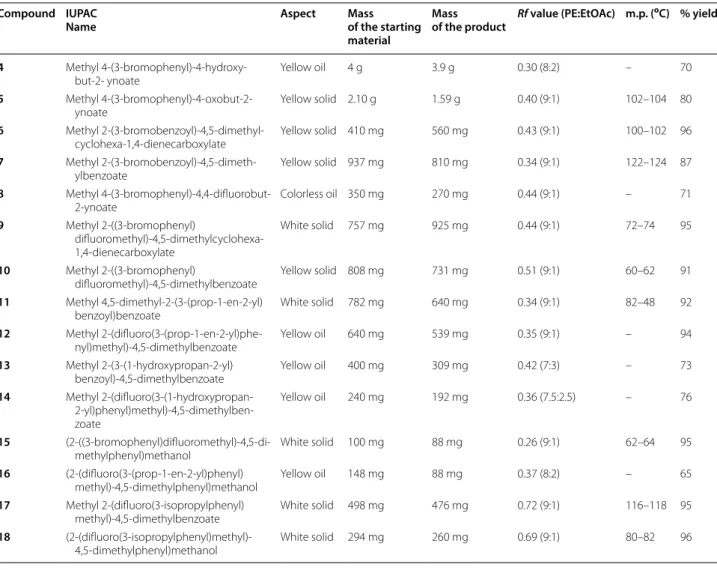
Table 4 The physicochemical properties of intermediates 4–18
Compound IUPAC
Name Aspect Mass of the starting
material
Mass
of the product Rf value (PE:EtOAc) m.p. (
oC) % yield
4 Methyl 4‑(3‑bromophenyl)‑4‑hydroxy‑
but‑2‑ ynoate Yellow oil 4 g 3.9 g 0.30 (8:2) – 70
5 Methyl 4‑(3‑bromophenyl)‑4‑oxobut‑2‑
ynoate Yellow solid 2.10 g 1.59 g 0.40 (9:1) 102–104 80
6 Methyl 2‑(3‑bromobenzoyl)‑4,5‑dimethyl‑
cyclohexa‑1,4‑dienecarboxylate Yellow solid 410 mg 560 mg 0.43 (9:1) 100–102 96
7 Methyl 2‑(3‑bromobenzoyl)‑4,5‑dimeth‑
ylbenzoate Yellow solid 937 mg 810 mg 0.34 (9:1) 122–124 87
8 Methyl 4‑(3‑bromophenyl)‑4,4‑difluorobut‑
2‑ynoate Colorless oil 350 mg 270 mg 0.44 (9:1) – 71
9 Methyl 2‑((3‑bromophenyl)
difluoromethyl)‑4,5‑dimethylcyclohexa‑ 1,4‑dienecarboxylate
White solid 757 mg 925 mg 0.44 (9:1) 72–74 95
10 Methyl 2‑((3‑bromophenyl)
difluoromethyl)‑4,5‑dimethylbenzoate Yellow solid 808 mg 731 mg 0.51 (9:1) 60–62 91
11 Methyl 4,5‑dimethyl‑2‑(3‑(prop‑1‑en‑2‑yl)
benzoyl)benzoate White solid 782 mg 640 mg 0.34 (9:1) 82–48 92
12 Methyl 2‑(difluoro(3‑(prop‑1‑en‑2‑yl)phe‑
nyl)methyl)‑4,5‑dimethylbenzoate Yellow oil 640 mg 539 mg 0.35 (9:1) – 94
13 Methyl 2‑(3‑(1‑hydroxypropan‑2‑yl)
benzoyl)‑4,5‑dimethylbenzoate Yellow oil 400 mg 309 mg 0.42 (7:3) – 73
14 Methyl 2‑(difluoro(3‑(1‑hydroxypropan‑ 2‑yl)phenyl)methyl)‑4,5‑dimethylben‑ zoate
Yellow oil 240 mg 192 mg 0.36 (7.5:2.5) – 76
15 (2‑((3‑bromophenyl)difluoromethyl)‑4,5‑di‑
methylphenyl)methanol White solid 100 mg 88 mg 0.26 (9:1) 62–64 95
16 (2‑(difluoro(3‑(prop‑1‑en‑2‑yl)phenyl)
methyl)‑4,5‑dimethylphenyl)methanol Yellow oil 148 mg 88 mg 0.37 (8:2) – 65
17 Methyl 2‑(difluoro(3‑isopropylphenyl)
methyl)‑4,5‑dimethylbenzoate White solid 498 mg 476 mg 0.72 (9:1) 116–118 95
18 (2‑(difluoro(3‑isopropylphenyl)methyl)‑
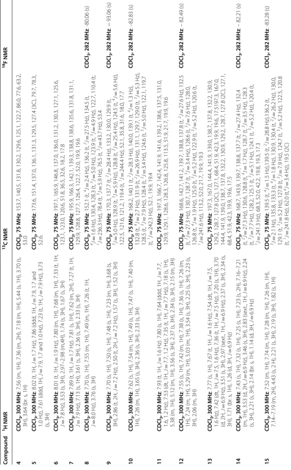
Table 5 Sp ec tr al da ta of in termedia tes 4–18 Compound 1H NMR 13C NMR 19F NMR 4 CDC l3 , 300 MH z: 7.56 (m, 1H), 7.36 (m, 2H), 7.18 (m, 1H), 5.44 (s , 1H), 3.70 (s , 3H), 3.64 (br . s , 1H) CDC l3 , 75 MH z: 153.7, 140.5, 131.8, 130.2, 129.6, 125.1, 122.7, 86.0, 77.6, 63.2, 53.0 5 CDC l3 , 300 MH z: 8.02 (t, 1H, J = 1.7 H z), 7.86 ( ddd , 1H, J = 7.9, 1.7 and 1.0 H z), 7.61 ( ddd , 1H, J = 7.9, 1.7 and 1.0 H z), 7.23 (t, 1H, J = 7.9 H z), 3.73 (s , 3H) CDC l3 , 75 MH z: 173.6, 151.4, 137.0, 136.1, 131.3, 129.5, 127.4 (3C ), 79.7, 78.3, 52.6. 6 CDC l3 , 300 MH z: 8.01 (t, 1H, J = 1.9 H z), 7.80 (m, 1H), 7.68 (m, 1H), 7.33 (t, 1H, J = 7.8 H z), 3.53 (s , 3H), 2.97–2.98 (m,4H), 1.74 (s , 3H), 1.67 (s , 3H) CDC l3 , 75 MH z: 197.1, 165.9, 146.8, 137.0, 136.0, 131.2, 130.3, 127.1, 125.6, 123.1, 123.0, 120.6, 51.8, 36.5, 32.6, 18.2, 17.8 7 CDC l3 , 300 MH z: 7.89 (t, 1H, J = 1.7 H z). 7.81 (s , 1H), 7.62 (m, 2H), 7.27 (t, 1H, J = 7.9 H z), 7.13 (s , 1H), 3.61 (s , 3H), 2.36 (s , 3H), 2.33 (s , 3H) CDC l3 , 75 MH z: 195.9, 166.3, 142.1, 139.3, 138.8, 138.6, 135.6, 131.8, 131.1, 129.9, 128.8, 127.7, 126.4, 122.7, 52.0, 19.9, 19.6 8 CDC l3 , 300 MH z: 7.70 (s , 1H), 7.55 (m, 1H), 7.49 (m, 1H), 7.26 (t, 1H, J = 8.0 H z), 3.76 (s , 3H) CDC l3 , 75 MH z: 152.1 (t, 4J = 2.4 H z), 136.2 (t, 2J = 27.5 H z), 134.5 (t, 4J = 1.6 H z), 130.4, 128.3 (t, 3J = 5.0 H z), 123.9 (t, 3J = 4.9 H z), 122.7, 110.4 (t, 1J = 236.3 H z), 78.4 (t, 3J = 5.9 H z), 76.5 (t, 2J = 43.7 H z), 53.4 CDC 9 CDC l3 , 300 MH z: 7.70 (s , 1H), 7.50 (s , 1H), 7.48 (s , 1H), 7.23 (m, 1H), 3.68 (s , 3H), 2.86 (t, 2H, J = 7.2 H z), 2.50 (t, 2H, J = 7.2 H z), 1.57 (s , 3H), 1.52 (s , 3H) CDC l3 , 75 MH z: 170.3, 137.7 (t, 2J = 28.4 H z), 133.2, 130.0, 129.9 (t, 3J = 5.0 H z), 128.9 (t, 3J = 5.9 H z), 128.8 (t, 2J = 25.4 H z), 124.5 (t, 4J = 5.6 H z), 122.5, 121.6, 121.2, 119.4 (t, 1J = 244.4 H z), 52.1, 35.8, 31.6, 18.0, 17.7 CDC 10 CDC l3 , 300 MH z: 7.62 (s , 1H), 7.54 (m, 1H), 7.49 (s , 1H), 7.47 (s , 1H), 7.40 (m, 1H), 7.26 (m, 1H), 3.65 (s , 3H), 2.36 (s , 3H), 2.33 (s , 3H) CDC l3 , 75 MH z: 168.2, 140.1 (t, 2J = 28.3 H z), 140.0, 139.1 (t, 4J = 1.3 H z), 132.8 (t, 4J = 2.7 H z), 131.9 (t, 2J = 26.9 H z), 131.1, 129.7, 129.0 (t, 3J = 5.3 H z), 128.5 (t, 3J = 7.9 H z), 128.4 (t, 3J = 3.4 H z), 124.6 (t, 3J = 5.0 H z), 122.1, 119.7 (t, 1J = 242.3 H z), 52.1, 19.9, 19.4 CDC 11 CDC l3 , 300 MH z: 7.93 (t, 1H, J = 1.6 H z). 7.80 (s , 1H), 7.64 ( ddd , 1H, J = 7.7, 1.6, 1.2 H z), 7.53 ( dt, 1H, J = 7.7, 1.2 H z), 7.35 (t, 1H, J = 7.7 H z), 7.18 (s , 1H), 5.38 (m, 1H), 5.12 (m, 1H), 3.56 (s , 3H), 2.37 (s , 3H), 2.34 (s , 3H), 2.15 (m, 3H) CDC l3 , 75 MH z: 197.3, 166.7, 142.4, 141.9, 141.6, 139.2, 138.6, 137.5, 131.0, 129.9, 129.1, 128.6, 128.3, 126.8, 125.8, 113.5, 51.9, 21.7, 19.9, 19.6 12 CDC l3 , 300 MH z: 7.55 (s , 1H), 7.42 (m, 1H), 7.38 (s , 1H), 7.36 (s , 1H), 7.26 (m, 1H), 7.24 (m, 1H), 5.29 (m, 1H), 5.03 (m, 1H), 3.54 (s , 3H), 2.25 (s , 3H), 2.23 (s , 3H), 2.06 (m, 3H) CDC l3 , 75 MH z: 168.6, 142.7, 141.2, 139.7, 138.8, 137.8 (t, 2J = 27.6 H z), 132.5 (t, 2J = 27.3 H z), 130.7, 128.7 (t, 3J = 3.4 H z), 128.6 (t, 3J = 7.6 H z), 128.0, 126.8 (t, 4J = 1.9 H z), 125.0 (t, 3J = 5.2 H z), 122.9 (t, 3J = 5.2 H z), 120.6 (t, 1J = 241.6 H z), 113.2, 52.0, 21.7, 19.9, 19.3 CDC 13 CDC l3 , 300 MH z: 7.77 (s , 1H), 7.67 (t, 1H, J = 1.6 H z), 7.54 ( dt, 1H, J = 7.5, 1.6 H z), 7.42 ( dt, 1H, J = 7.5, 1.6 H z), 7.36 (t, 1H, J = 7.5 H z), 7.20 (s , 1H), 3.70 (d , 2H, J = 6.9 H z), 3.51 (s , 3H), 2.97 (sex t., 1H, J = 6.9 H z), 2.37 (s , 3H), 2.34 (s , 3H), 1.71 (br . s , 1H), 1.26 ( d, 3H, J = 6.9 H z) CDC l3 , 75 MH z: 197.3, 167.0, 144.4, 141.9, 139.0, 138.7, 137.8, 132.2, 130.9, 129.2, 128.7, 127.8 (2C ), 127.1, 68.4, 51.9, 42.3, 19.9, 19.6, 17.5197.3, 167.0, 144.4, 141.9, 139.0, 138.7, 137.8, 132.2, 130.9, 129.2, 128.7, 127.8 (2C ), 127.1, 68.4, 51.9, 42.3, 19.9, 19.6, 17.5 14 CDC l3 , 300 MH z: 7.43 (s , 1H), 7.32 (s , 1H), 7.25 (s , 1H), 7.23 (s , 2H), 7.16–7.21 (m, 1H), 3.53 ( d, 2H, J = 6.9 H z), 3.46 (s , 3H), 2.83 (sex t., 1H, J = 6.9 H z), 2.24 (s , 3H), 2.21 (s , 3H), 2.14 (br . s , 1H), 1.14 ( d, 3H, J = 6.9 H z) CDC l3 , 75 MH z: 168.7, 143.9, 139.7, 138.8, 137.7 (t, 2J = 27.4 H z), 132.4 (t, 2J = 27.3 H z), 130.6, 128.8 (t, 4J = 1.7 H z), 128.7 (t, 3J = 3.5 H z), 128.3 (t, 3J = 7.7 H z), 128.2, 125.1 (t, 3J = 5.1 H z), 124.1 (t, 3J = 5.2 H z), 120.4 (t, 1J = 241.3 H z), 68.3, 52.0, 42.2, 19.8, 19.3, 17.3 CDC 15 CDC l3 , 300 MH z: 7.52 (m, 1H), 7.74 (m, 1H), 7.28 (m, 1H), 7.26 (m, 1H), 7.14–7.19 (m, 2H), 4.43 (s , 2 H), 2.21 (s , 3H), 2.19 (s , 3H), 1.82 (s , 1H) CDC l3 , 75 MH z: 139.5 (t, 4J = 1.5 H z),139.5 (t, 2J = 28.8 H z),136.2 (t, 3J = 2.1 H z), 135.8, 133.3 (t, 4J = 1.8 H z), 130.9, 130.4 (t, 2J = 26.2 H z), 130.0, 129.1 (t, 3J = 5.2 H z), 127.8 (t, 3J = 7.9 H z), 124.7 (t, 3J = 5.2 H z), 122.5, 120.8 (t, 1J = 241.8 H z), 62.0 (t, 4J = 3.4 H z), 19.5 (2C ) CDC
Table 5 (c on tinued) Compound 1H NMR 13C NMR 19F NMR 16 CDC l3 , 300 MH z: 7.53 (s , 1H), 7.45 (m, 1H), 7.29 (s , 1H), 7.25 (m, 1H), 7.21 (s , 2H), 5.29 (s , 1H), 5.05 (t, 1H, J = 1.4 H z), 4.48 (s , 2H), 2.23 (s , 3H), 2.20 (s , 3H), 2.06 ( dd , 3H, J = 1.4, 0.8 H z) CDC l3 , 75 MH z: 142.5, 141.6, 139.3, 137.4 (t, 2J = 28.0 H z), 136.3 (t, 4J = 2.1 H z), 135.7, 131.3 (t, 2J = 26.5 H z), 131.1, 128.3, 128.0 (t, 3J = 7.8 H z), 127.3 (t, 3J = 1.8 H z), 125.1 (t, 3J = 5.2 H z), 122.9 (t, 3J = 5.2 H z), 121.7 (t, 1J = 241.5 H z), 113.5, 62.2 (t, 4J = 3.2 H z), 21.7, 19.5 (2C ) CDC l3 , 282 MH z: – 82.87 (s) 17 (CDC l3 , 500 MH z) : 7.44 (s , 1H), 7.42 (s , 1H), 7.36 (s , 1H), 7.23–7.29 (m, 2H), 7.21 (m, 1H), 3.57 (s , 3H), 2.9 (sex t., 1H, J = 6.9 H z), 2.31 (s , 3H), 2.30 (s , 3H), 1.23 ( d, 6H, J = 6.9 H z) (CDC l3 , 125 MH z) :168.7, 148.8, 139.6, 138.7, 137.7 (t, 2J = 27.3 H z), 132.6 (t, 2J = 27.3 H z), 130.7, 128.8 (t, 3J = 3.4 H z), 128.6 (t, 3J = 7.6 H z), 128.0, 127.8 (t, 4J = 1.8 H z), 123.9 (t, 3J = 5.0 H z), 123.6 (t, 3J = 5.2 H z), 120.7 (t, 1J = 241.2 H z), 52.0, 34.1, 23.9 (2C ), 19.9, 19.4 (CDC l3 , 470 MH z) : – 81.94 (s) 18 CDC l3 , 300 MH z: 7.25 (s , 1H), 7.23 (s , 1H), 7.16 (s , 1H), 7.14 (s , 1H), 7.12 (m, 1H), 7.05 (m, 1H), 4.39 (s , 2 H), 2.76 (sex t., 1H, J = 6.9 H z), 2.41 (br . s , 1H), 2.13 (s , 3H), 2.12 (s , 3H), 1.1 ( d, 6H, J = 6.9 H z) CDC l3 , 75 MH z: 149.1138.9, 137.3 (t, 2J = 27.8 H z), 136.4, 135.2, 131.1 (t, 2J = 26.5 H z), 130.5, 128.3, 128.0 (t, 4J = 1.7 H z), 127.7 (t, 3J = 7.8 H z), 123.9 (t, 3J = 5.0 H z), 123.6 (t, 3J = 5.2 H z),121.7 (t, 1J = 240.8 H z), 61.6 (t, 4J = 3.2 H z), 33.9, 23.7 (2C ), 19.3 (2C ) CDC l3 , 282 MH z: − 82.66
environment (cotton cocoon) in the animal facility of the American University of Beirut. Body weight and food intake were monitored three times a week throughout the study period. ARRIVE guidelines were followed (Addi-tional file 2). Approval for use of animals was obtained from the Institutional Animal Care and Use Committee of the American University of Beirut (IACUC # 16-09-m379).
On the day of the procedure, 2–3 mice were euthana-tized after 3 min exposure to carbon dioxide. BMDM were isolated as previously described and were plated at 0.8 million cells per well [16]. Flow cytometry analysis was performed using F4/80 -APC antibody (BioLegend 123115) and showed 90% macrophages. BMDM were then treated for 24 h with different concentrations of the bisar-ylic derivative compounds for 30 min prior to the addition of 10 ng/mL LPS. DMSO concentration did not exceed 0.4% with no effect. The supernatants were assessed for IL-6, PGE2 and nitrite, the stable derivative of NO. Cells were washed with PBS and lysed in RIPA buffer contain-ing inhibitors of protease. Total protein concentration was determined using DC protein assay (Bio-Rad 500-0115) with BSA as standard. IL-6, nitrite and PGE2 were meas-ured as described previously [17]. Western blot of NOS-II and COX-2 was performed as previously described [17–19]. 10 µg of total protein was assessed. The primary antibodies were developed and characterized as previously described: for COX-2, mouse monoclonal antibody anti-COX-2 (clone anti-COX-214, 1/5000) [20]; for NOS-II, rabbit polyclonal antibody anti-NOS-II (dilution1/2000) [21], and mouse β-actin (dilution 1/10,000) (Sigma-Aldrich A5441). Clarity™ western ECL substrate (Bio-Rad 170-5061) was used according to the manufacturer’s instructions to reveal positive bands visualized using Bio-Rad ChemiDoc.
COX‑1 and COX‑2 activities
For COX-1 activity, human embryonic kidney (HEK)-293 cells (ATCC CRL-1573, Manassas, VA USA) stably
overexpressing human recombinant COX-1 were used [17]. Cells were treated with compounds A1 to A5 for 45 min in Hanks buffer and then 10 µM arachidonic acid (AA) were added for 30 min. PGE2 was measured and corresponded to the breakdown metabolism of PGH2 and PGE2.
For COX-2 activity, BMDM were treated with 10 μM acetylsalicylic acid (ASA) for 30 min to irreversibly inhibit COX-1 and then washed and treated with LPS 10 ng/mL for 24 h. BMDM were treated with compounds
A1 to A5 for 45 min in Hanks buffer, pH 7.4
contain-ing 1 mg/mL BSA prior to the addition of 10 µM of AA for 30 min. Supernatants were collected and PGE2 was determined.
Toxicity assay
WST-1 assay was used to determine the toxicity of the synthesized compounds (Cell proliferation WST-1 assay, Sigma-Aldrich 5015944001). Briefly, mac-rophages (50,000 cells per well) were plated in a 96 well plate in RPMI culture medium containing 10% FBS and grown for 24 h. Cells (in triplicates) were treated at 25 and 50 μM of the tested compounds. Culture medium without cells and cells without treatment were used as control. Results were expressed as percentage of cells without treatment. All compounds showed 95% viabil-ity at 50 μM.
Molecular docking
Target and small molecule preparation
All small molecules (A1 to A5) were built using Openba-bel chemical toolbox (PMID: 21982300) and subsequent low energy 3D conformations were generated using Frog2 (PMID: 20444874). Protonation state corresponded to a pH of 7. The 3D structure of the ovine COX-1 (PDBID: 4,5‑dimethylphenyl)methyl)phenyl)
propanoic acid
A3 2‑((3‑bromophenyl)difluoromethyl)‑
4,5‑dimethylbenzoic acid White solid 100 83 0.28 (6:4) 64–66 80
A4 2‑((3‑acetylphenyl)difluoromethyl)‑
4,5‑dimethylbenzoic acid White solid 103 80 0.38 (8:2) 118–120 74
A5 2‑(difluoro(3‑isopropylphenyl)methyl)‑
Table 7 Sp ec tr al da ta of syn thesiz ed bisar ylic deriv ativ es A1 t o A5 Compound 1H NMR 13C NMR (CDC l3 , 75 MH z) 19F NMR (CDC l3 , 282 MH z) HRMS (ESI) A1 CDC l3 , 300 MH z: 7.78 (m, 2H), 7.53 (m, 2H), 7.36 (t, 1H, J = 7.7 H z), 7.17 (s , 1H), 3.77 ( quad , 1H, J = 7.2 H z), 3.51 (s , 3H), 2.36 (s , 3H), 2.33 (s , 3H), 1.51 ( d, 3H, J = 7.2 H z) CDC l3 , 75 MH z: 197.0, 179.7, 166.7, 141.9, 140.2, 138.9, 138.7, 137.9, 132.0, 131.0, 129.2, 128.7, 128.6, 128.1, 126.8, 51.9, 45.1, 19.9, 19.6, 18.0 calcd . F or C20 H20 O5 Na: m/z [M + Na] + 363.12029; found: 363.1203 (0 ppm); C20 H19 O5 Na2 : m/z [M ‑H + 2Na] + 385.10224; f ound: 385.1014 (2 ppm). A2 CDC l3 , 300 MH z: 7.44–7.46 (m, 3H), 7.33–7.40 (m, 3H), 3.76 ( quad , 1H, J = 7.1 H z), 3.56 (s , 3H), 2.33 (s , 3H), 2.32 (s , 3H), 1.50 ( d, 3H, J = 7.2 H z) CDC l3 , 75 MH z: 179.6, 168.5, 139.8, 139.7, 138.9, 138.3 (t, 2J = 27.7 H z), 132.3 (t, 2J = 27.1 H z), 130.8, 128.9 (t, 4J = 1.7 H z), 128.7 (t, 3J = 3.4 H z), 128.5 (2C ), 125.2 (t, 3J = 5.1 H z), 120.4 (t, 1J = 241.6 H z), 68.3, 52.0, 45.2, 19.9, 19.4, 18.1 CDC l3 , 282 MH z: – 82.30 (s) calcd . F or C20 H20 F2 O4 Na: m/z [M + Na] + 385.12219; found: 385.1223 (0 ppm); C20 H19 O4 F2 Na2 : m/z [M + Na] + 407.10413; f ound: 407.1045 (0 ppm). A3 deut er at ed ac et one , 300 MH z: 7.29–7.63 (m, 6H), 2.22 (s , 3H), 2.20 (s , 3H) deut er at ed ac et one , 75 MH z: 170.0, 142.4 (t, 2J = 28.6 H z), 141.7, 141.1 (t, 4J = 1.3 H z), 134.6 (t, 3J = 1.7 H z), 133.7 (t, 2J = 26.9 H z), 132.7, 132.1, 131.3, 130.7 (t, 3J = 5.3 H z), 130.0 (t, 3J = 8.0 H z), 126.8 (t, 3J = 5.3 H z), 123.4, 122.0 (t, 1J = 241.6 H z), 20.8, 20.4 CDC l3 , 282 MH z: − 82.34 (s) calcd . F or C16 H13 O2 F2 79BrNa: m/z [M + Na] + 376.99592; f ound: 376.9958 (0 ppm); C16 H12 O2 F2 79BrNa 2 : m/z [M ‑H + 2Na] + 398.97786; found: 398.9779 (0 ppm); C16 H12 O2 F 79BrNa: m/z [M ‑HF + Na] + 356.98969; f ound: 356.9905 (2 ppm) A4 deut er at ed ac et one , 300 MH z: 8.18 (m, 1H), 8.07 (m, 1H), 7.77 (m, 1H), 7.56–7.61 (m, 3H), 2.60 (s , 3H), 2.39 (s , 3H), 2.36 (s , 3H) deut er at ed ac et one , 75 MH z: 198.5, 170.0, 141.7, 141.1, 140.6 (t, 2J = 28.5 H z), 138.9, 133.9 (t, 2J = 27.1 H z), 132.6, 132.1 (t, 3J = 7.5 H z), 131.4, 131.1 (t, 4J = 3.4 H z), 130.4, 130.0 (t, 3J = 7.9 H z), 127.2 (t, 3J = 5.3 H z), 122.5 (t, 1J = 241.7 H z), 27.7, 20.8, 20.3 CDC l3 , 282 MH z: – 82.87 (s) calcd . F or C18 H16 O3 F2 Na: m/z [M + Na] + 341.09597; found: 341.0960 (0 ppm) A5 CDC l3 , 300 MH z: 7.47 (s , 1H), 7.43 (s , 1H), 7.30 (s , 1H), 7.15–7.17 (m, 3H), 2.81 (sex t., 1H, J = 6.9 H z), 2.27 (s , 3H), 2.24 (s , 3H), 1.14 ( d, 6H, J = 6.9 H z) CDC l3 , 75 MH z: 172.7, 148.7, 140.5, 138.6, 137.6 (t, 2J = 27.2 H z), 133.6 (t, 2J = 27.7 H z), 131.4, 128.8 (t, 3J = 7.8 H z), 127.9 (2C ), 127.7 (t, 4J = 1.6 H z), 124.1 (t, 3J = 5.0 H z), 123.6 (t, 3J = 5.2 H z), 120.6 (t, 1J = 241.7 H z), 34.1, 23.8 (2C ), 19.9, 19.4 CDC l3 , 282 MH z: –81.93 (s) calcd . F or C19 H20 O2 F2 Na: m/z [M + Na] + 341.13236; found: 341.1321 (1 ppm); C19 H19 O2 FNa: m/z [M ‑HF + Na] + 321.12613; f ound: 321.1260 (0 ppm); C19 H19 O2 F2 Na2 : m/z [M ‑H + 2Na] + 363.1143; f ound: 363.1157 (4 ppm); C19 H20 O2 F2 m/z [M + K] + 357.10629; f ound: 357.1059 (1 ppm); C19 H19 O2 : m/z [M ‑HF ‑F] + 279.13796; f ound: 279.1379 (0 ppm)
oatoms were removed from the structure. Chain A was retained including ibuprofen and heme group. Hydro-gen atoms were added, atom typing, and partial charges were assigned using Amber forcefield in Chimera (PMID: 15264254). Corresponding ligand-receptor binding ener-gies were estimated in kcal/mol and averaged for best poses that recapitulate ibuprofen binding. A single inter-acting conformation was retained after visual inspection in Maestro (Schrödinger, LLC).
Data analysis
IL-6, nitrite and PGE2 measurement were determined from 3 to 5 independent experiments and expressed as percentage of LPS alone and expressed as mean ± SEM. COX-1 activity for the compounds were expressed as percentage of PGE2 measured in cells exposed to vehicle and AA, and as percentage of PGE2 in cells treated with LPS, ASA and AA for COX-2. Curve fitting and calcula-tion of the IC50 values were done using Grafit7 Software (Erithacus software, Staines, UK) and GraphPad Prism 6. Images of western blot were analyzed using ImageJ Software (version 1.52a, NIH, MA). Ratio of COX-2 to β-actin was determined and the results are expressed as fold of LPS signal.
Statistical analysis was performed using one-way ANOVA, followed by Dunnett’s multiple comparisons test. Differences were considered significant when p < 0.05 (GraphPad Prism 6 Software, La Jolla, CA, USA).
Supplementary information
Supplementary information accompanies this paper at https ://doi.
org/10.1186/s1306 5‑019‑0640‑5.
Additional file 1. Proton (H), carbon (C) and fluorine (F) NMR spectra for
intermediates 4 to 18 and for compounds A1 to A5.
Additional file 2. ARRIVE Check list. Abbreviations
AA: arachidonic acid; ASA: acetylsalicylic acid; BMDM: bone marrow‑derived macrophages; COX: cyclooxygenase; FBS: fetal bovine serum; IL‑6: interleu‑ kin‑6; LPS: lipopolysaccharide; NO: nitric oxide; NOS‑II: nitric oxide synthase‑II; NSAIDs: non‑steroidal anti‑inflammatory drugs; PGE2: prostaglandin E2.
NH performed the molecular docking; analysis and interpretation of data (AA, LH, SG, GEA, OD, NB, BB, AHachem, EH, AH); IC50 analysis and fitting (OD
and AA); drafting of the manuscript (AA, LH, GEA, AHachem, EH, AA); critical revision of the manuscript for important intellectual content (AA, LH, GEA, RG, AHachem, EH, AH); statistical analysis (AA, AH); study supervision (AHachem, EH, AH). AHachem, EH and AH provided financial support. All authors read and approved the final manuscript.
Funding
This work was supported by the Research Grant Program at the Lebanese University, Lebanon (Ali Hachem and Eva Hamade) and the Medical Practice Plan and the University Research Board of the American University of Beirut (Aida Habib) (Grant No. 320024).
Data availability
The data used to support the findings of this study are available from the cor‑ responding author upon request.
Competing interests
The authors declare that they have no competing interests.
Author details
1 Department of Biochemistry and Molecular Genetics, Faculty of Medicine,
American University of Beirut, Beirut, Lebanon. 2 Laboratory of Cancer Biol‑
ogy and Molecular Immunology, Faculty of Sciences I, Lebanese University, Hadath, Beirut, Lebanon. 3 Laboratory for Medicinal Chemistry and Natural
Products, Faculty of Sciences I and PRASE‑EDST Lebanese University, Beirut, Lebanon. 4 Integrative Systems Biology, Institut de Recherches Cliniques de
Montréal, Montreal, QC, Canada. 5 Department of Biology, Faculty of Sciences
II, EDST, Lebanese University, Fanar, Lebanon. 6 Université de Rennes, CNRS,
ISCR (Institut des Sciences Chimiques de Rennes) UMR 6226, 35000 Rennes, France. 7 Université de Paris, Centre de Recherche sur l’Inflammation (CRI),
INSERM, UMR1149, CNRS, ERL 8252, 75018 Paris, France. 8 Present Address:
Department of Electrical and Computer Engineering, American University of Beirut, Beirut, Lebanon.
Received: 12 July 2019 Accepted: 3 October 2019
References
1. Özbey F, Taslimi P, Gülçin İ, Maraş A, Göksu S, Supuran CT (2016) Synthesis of diaryl ethers with acetylcholinesterase, butyrylcholinesterase and carbonic anhydrase inhibitory actions. J Enzyme Inhib Med Chem 31(sup2):79–85
2. Jung N, Bräse S (2009) Synthesis of natural products on solid phases via copper‑mediated coupling: synthesis of the aristogin family, spiraformin a, and hernandial. Eur J Org Chem 26:4494–4502
3. Moore RA, Derry S, McQuay HJ, Wiffen PJ (2011) Single dose oral analgesics for acute postoperative pain in adults. Cochrane Database of Systematic Reviews 9:CD008659
4. Khanum SA, Begum BA, Girish V, Khanum NF (2010) Synthesis and evalu‑ ation of benzophenone‑N‑ethyl morpholine ethers as anti‑inflammatory agents. Int J Biomed Sci 6(1):60–65
5. Terrazas PM, de Souza Marques E, Mariano LN, Cechinel‑Filho V, Niero R, Andrade SF, Maistro EL (2013) Benzophenone guttiferone A from Garcinia achachairu Rusby (Clusiaceae) presents genotoxic effects in different cells of mice. PLoS ONE 8(11):e76485
•fast, convenient online submission
•
thorough peer review by experienced researchers in your field
• rapid publication on acceptance
• support for research data, including large and complex data types
•
gold Open Access which fosters wider collaboration and increased citations maximum visibility for your research: over 100M website views per year
•
At BMC, research is always in progress. Learn more biomedcentral.com/submissions
Ready to submit your research? Choose BMC and benefit from: of some novel biphenyl‑4‑carboxylic acid 5‑(arylidene)‑2‑(aryl)‑4‑oxothia‑
zolidin‑3‑yl amides. Acta Pol Pharm 67(1):63–67
7. Thuronyi BW, Chang MC (2015) Synthetic biology approaches to fluori‑ nated polyketides. Acc Chem Res 48(3):584–592
8. Muller K, Faeh C, Diederich F (2007) Fluorine in pharmaceuticals: looking beyond intuition. Science (New York, NY) 317(5846):1881–1886 9. Shaughnessy MJ, Harsanyi A, Li J, Bright T, Murphy CD, Sandford G (2014)
Targeted fluorination of a nonsteroidal anti‑inflammatory drug to pro‑ long metabolic half‑life. ChemMedChem 9(4):733–736
10. Dang H, Whittaker AM, Lalic G (2016) Catalytic activation of a single C‑F bond in trifluoromethyl arenes. Chem Sci 7(1):505–509
11. Gillis EP, Eastman KJ, Hill MD, Donnelly DJ, Meanwell NA (2015) Applica‑ tions of fluorine in medicinal chemistry. J Med Chem 58(21):8315–8359 12. Khalaf A, Grée D, Abdallah H, Jaber N, Hachem A, Grée R (2011) A new
flexible strategy for the synthesis of gem‑difluoro‑bisarylic derivatives and heterocyclic analogues. Tetrahedron 67(21):3881–3886 13. Selinsky BS, Gupta K, Sharkey CT, Loll PJ (2001) Structural analysis of
NSAID binding by prostaglandin H2 synthase: time‑dependent and time‑independent inhibitors elicit identical enzyme conformations. Biochemistry 40(17):5172–5180
14. Orlando BJ, Lucido MJ, Malkowski MG (2015) The structure of ibuprofen bound to cyclooxygenase‑2. J Struct Biol 189(1):62–66
15. Hariss L, Ibrahim R, Jaber N, Roisnel T, Grée R, Hachem A (2018) A general approach to various five‑ and six‑membered gem‑difluoroheterocycles: application to the synthesis of fluorinated analogues of sedamine. Eur J Org Chem 2018(27–28):3782–3791
Le Gall M, Letteron P, Pilard N et al (2019) Inhibition of monoacylglycerol lipase, an anti‑inflammatory and antifibrogenic strategy in the liver. Gut 68:522–532
17. El‑Achkar GA, Jouni M, Mrad MF, Hirz T, El Hachem N, Khalaf A, Hammoud S, Fayyad‑Kazan H, Eid AA, Badran B et al (2015) Thiazole derivatives as inhibitors of cyclooxygenases in vitro and in vivo. Eur J Pharmacol 750:66–73
18. Habib A, Shamseddeen I, Nasrallah MS, Antoun TA, Nemer G, Bertoglio J, Badreddine R, Badr KF (2007) Modulation of COX‑2 expression by statins in human monocytic cells. FASEB J 21(8):1665–1674
19. Mouawad CA, Mrad MF, Al‑Hariri M, Soussi H, Hamade E, Alam J, Habib A (2013) Role of nitric oxide and CCAAT/enhancer‑binding protein transcription factor in statin‑dependent induction of heme oxygenase‑1 in mouse macrophages. PLoS ONE 8(5):e64092
20. Creminon C, Frobert Y, Habib A, Maclouf J, Pradelles P, Grassi J (1995) Immunological studies of human constitutive cyclooxygenase (COX‑ 1) using enzyme immunometric assay. Biochimica Biophysica Acta 1254(3):333–340
21. Habib A, Bernard C, Lebret M, Creminon C, Esposito B, Tedgui A, Maclouf J (1997) Regulation of the expression of cyclooxygenase‑2 by nitric oxide in rat peritoneal macrophages. J Immunol 158(8):3845–3851
Publisher’s Note
Springer Nature remains neutral with regard to jurisdictional claims in pub‑ lished maps and institutional affiliations.
![Fig. 4 Two‑dimensional pose of compounds A1 to A5 and Ibuprofen inside the binding pocket of mouse COX‑2 as crystallized by [13]](https://thumb-eu.123doks.com/thumbv2/123doknet/14508411.529240/10.892.87.436.171.334/dimensional-compounds-ibuprofen-inside-binding-pocket-mouse-crystallized.webp)
![Fig. 5 Two‑Dimensional pose of compounds A1 to A5 and Ibuprofen inside the binding pocket of human COX‑1 as crystallized by [14]](https://thumb-eu.123doks.com/thumbv2/123doknet/14508411.529240/11.892.85.437.170.309/dimensional-compounds-ibuprofen-inside-binding-pocket-human-crystallized.webp)