HAL Id: inserm-02263536
https://www.hal.inserm.fr/inserm-02263536
Submitted on 5 Aug 2019
HAL is a multi-disciplinary open access archive for the deposit and dissemination of sci-entific research documents, whether they are pub-lished or not. The documents may come from teaching and research institutions in France or abroad, or from public or private research centers.
L’archive ouverte pluridisciplinaire HAL, est destinée au dépôt et à la diffusion de documents scientifiques de niveau recherche, publiés ou non, émanant des établissements d’enseignement et de recherche français ou étrangers, des laboratoires publics ou privés.
lymphocytes in response to mycobacterial antigens: A
help for early diagnosis of peritoneal tuberculosis in a
low TB incidence country
Sophie Henrard, Véronique Corbière, Liliane Schandené, Martine Ducarme,
Anne van Praet, Emmanuelle Petit, Mahavir Singh, Camille Locht, Violette
Dirix, Françoise Mascart
To cite this version:
Sophie Henrard, Véronique Corbière, Liliane Schandené, Martine Ducarme, Anne van Praet, et al.. Proportions of interferon-γ-producing ascites lymphocytes in response to mycobacterial antigens: A help for early diagnosis of peritoneal tuberculosis in a low TB incidence country. PLoS ONE, Public Library of Science, 2019, 14 (4), pp.e0214333. �10.1371/journal.pone.0214333�. �inserm-02263536�
Proportions of interferon-γ-producing ascites
lymphocytes in response to mycobacterial
antigens: A help for early diagnosis of
peritoneal tuberculosis in a low TB incidence
country
Sophie Henrard1☯, Ve´ronique Corbière2☯, Liliane Schandene´3, Martine Ducarme3, Anne Van Praet2, Emmanuelle Petit4,5,6,7, Mahavir Singh8, Camille Locht4,5,6,7, Violette Dirix2, Franc¸oise MascartID2,3*
1 Immunodeficiencies Treatment Unit, Hoˆ pital Erasme, Universite´ Libre de Bruxelles (U.L.B.), Brussels, Belgium, 2 Laboratory of Vaccinology and Mucosal Immunity, Universite´ Libre de Bruxelles (U.L.B.), Brussels, Belgium, 3 Immunobiology Clinic, Hoˆpital Erasme, Universite´ Libre de Bruxelles (U.L.B.), Belgium,
4 INSERM, U1019, Lille, France, 5 CNRS, UMR8204, Lille, France, 6 Universite´ de Lille, Lille, France,
7 Institut Pasteur de Lille, Centre d’Infection et d’Immunite´ de Lille, Lille, France, 8 Lionex Diagnostics and Therapeutics, Braunschweig, Germany
☯These authors contributed equally to this work.
*fmascart@ulb.ac.be
Abstract
Background
Peritoneal tuberculosis (TB) remains difficult to diagnose because of its non-specific clinical features and the lack of efficient microbiological tests. As delayed diagnosis is associated with high mortality rates, new diagnostic tools are needed.
Methods and findings
We investigated for 24 patients prospectively enrolled with a possible diagnosis of peritoneal TB, the diagnostic value of the analysis of IFN-γproduction by peritoneal fluid lymphocytes in response to a short in vitro stimulation with mycobacterial antigens. The patients were classified in two groups: non-TB and confirmed or highly probable TB. Diagnosis of TB was based on microbiological and histopathological criteria and/or a favorable response to anti-TB treatment. The IFN-γproduction by peritoneal CD4+T lymphocytes was analyzed by flow cytometry after an overnight in vitro stimulation with three different mycobacterial anti-gens, purified protein derivative (PPD), heparin-binding haemagglutinin (HBHA) or early-secreted-antigen-target-6 (ESAT-6). The percentages of PPD-, HBHA- or ESAT-6-induced IFN-γ-producing peritoneal fluid CD4+T lymphocytes were higher in the TB group than in the non-TB group (p = 0.0007, p = 0.0004, and p = 0.0002 respectively). Based on cut-off values determined by ROC curve analysis of the results from TB and highly probable TB compared to those of non-TB patients, the sensitivity of these three tests was 100% with a specificity of 92%. a1111111111 a1111111111 a1111111111 a1111111111 a1111111111 OPEN ACCESS
Citation: Henrard S, Corbière V, Schandene´ L, Ducarme M, Van Praet A, Petit E, et al. (2019) Proportions of interferon-γ-producing ascites lymphocytes in response to mycobacterial antigens: A help for early diagnosis of peritoneal tuberculosis in a low TB incidence country. PLoS ONE 14(4): e0214333.https://doi.org/10.1371/ journal.pone.0214333
Editor: Katalin Andrea Wilkinson, University of
Cape Town, SOUTH AFRICA
Received: December 28, 2018 Accepted: March 11, 2019 Published: April 4, 2019
Copyright:© 2019 Henrard et al. This is an open access article distributed under the terms of the
Creative Commons Attribution License, which permits unrestricted use, distribution, and reproduction in any medium, provided the original author and source are credited.
Data Availability Statement: All relevant data are
within the manuscript.
Funding: This work was supported by: F.M., Fonds
National de la Recherche Scientifique (FNRS) – PDR T.0147.13; F.M., the European Community within the Horizon2020 program TBVAC2020-grant agreement 643381; and F.M., the “Re´gion de Bruxelles-Capitale – Innoviris". The funders had no role in study design, data collection and analysis,
Conclusions
The analysis of mycobacterial-induced IFN-γproduction by peritoneal lymphocytes is a promising tool to reliably and rapidly diagnose peritoneal TB. Further studies should be per-formed on larger cohorts of patients in high-TB-incidence countries to confirm the clinical value of this new diagnostic approach for peritoneal TB.
Introduction
Tuberculosis (TB) is one of the world’s most lethal infectious diseases and peritoneal TB result-ing from the growth ofMycobacterium tuberculosis complex bacteria in the peritoneum, is the sixth most frequent site of extra-pulmonary involvement. Peritoneal TB represents 1% to 2% of all clinical manifestations of TB and 31% to 58% of the abdominal TB, and is associated with pulmonary TB in 3.5% of cases [1]. Complications of septicemia, acute intestinal occlusion, infertility in women, are frequent [1], and its mortality rate ranges from 15% to 31% represent-ing thus a significant public health problem, especially in endemic areas [2,3].
Peritoneal TB most commonly arises following the reactivation of latentM. tuberculosis infection from bacterial foci that have resided within the peritoneum after a hematogenous spread ofM. tuberculosis, either from a primary lung focus or during miliary TB. Much less frequently, the organisms enter the peritoneal cavity after transmural migration from the infected small intestine or contiguously from TB salpingitis, and ascites develops secondary to the exudation of proteinaceous fluid from peritoneal tubercles [2,3]. Peritoneal TB is a sub-acute disease with unspecific symptoms evolving over a period of several weeks to months, so that the clinical presentation is rather insidious [1–3]. The most frequent clinical signs are asci-tes, abdominal pain, night sweats, fever and abdominal distension. TB peritonitis frequently occurs in patients with severe underlying medical conditions, such as end-stage renal or liver disease, further adding to the diagnostic difficulty [2,3]. Laboratory blood tests, such as a mod-erate inflammatory syndrome and elevated carbohydrate antigen-125 concentrations, provide unspecific results. High ascites protein concentrations and elevated cellularity with a predomi-nance of lymphocytes are informative but also unspecific. These laboratory results may easily be mis-interpreted for a malignant etiology [1–3]. The gold-standard for the diagnosis of peri-toneal TB is a positive culture of mycobacteria from periperi-toneal fluids, but the sensitivity of this method is only 35% [2], so that diagnosis of peritoneal TB remains a real challenge. More sen-sitive diagnosis depends on a highly invasive procedure, which is the peritoneal biopsy per-formed by laparoscopy providing a positive culture in 92% to 98% of cases [3]. However, this procedure is associated with a significant risk for the patient, is costly and not available in resource-poor countries. Yet, early diagnosis is important, as delayed initiation of anti-TB therapy is associated with high mortality [2,4].
A rapid, non-invasive, sensitive diagnostic test is urgently needed to guide decisions about the need for invasive laparoscopic examination or the initiation of empirical anti-TB treat-ment. Different approaches have already been evaluated, including various immunological tests. Among them, the most widely used remains the tuberculin skin test (TST), followed by the interferon-γ release assay (IGRA) performed on blood samples. However, these tests are not intended for the diagnosis of active TB for which they have a poor specificity, as they can-not distinguish active from latent TB and are can-not sensitive enough to diagnose peritoneal TB [5,6]. Another approach is based on the determination of adenosine deaminase (ADA) activity in ascites, that although not specific for TB, was reported to provide a high diagnostic accuracy
decision to publish, or preparation of the manuscript.
Competing interests: The authors have declared
of peritoneal TB [7]. More recently, new diagnostic approaches to diagnose extra-pulmonary TB have been described, based on the production of IFN-γ by lymphocytes collected from the site of infection. This approach is promising for pleural TB but also for pulmonary TB, as it provides a high degree of TB suspicion within 24 hours [8–10]. We report here on the diagnos-tic accuracy for rapid diagnosis of peritoneal TB of the percentages of IFN-γ-containing-CD4+
T lymphocytes after a shortin vitro stimulation with purified protein derivative (PPD) or two different purified mycobacterial antigens, the heparin-binding haemagglutinin (HBHA) [11] and the early-secreted antigen target-6 (ESAT-6) [12].
Material and methods
Ethics statement
This study was approved by the ethics committee ULB-Hoˆpital Erasme (P2007/115, P2011/ 113 and P2016/252) and all the participants signed an informed consent form.
Patients
Twenty-four patients living in Belgium, a low TB incidence country, hospitalized for fever and/or abdominal pain and with demonstrated ascites by abdominal ultrasound or computer-ized tomography scanning, were prospectively included in the study between 2010 and 2017. One patient (n˚6) had also a bilateral pleural effusion. As all the patients were suspected to present active TB, peritoneal fluid was collected and sent to the laboratory as part of the classi-cal diagnostic procedure, and the remaining fluid was tested for IFN-γ production by perito-neal CD4+T lymphocytes in response to mycobacterial antigens.
Mycobacterial induction of IFN-
γ production
Peritoneal fluids were filtered and centrifuged as described for other fluids and red blood cells were lysed when necessary [10]. The cell recovery from peritoneal fluids was 2.2.106cells/ml for patients suffering from TB (median; 25th- 75thinter-quartiles: 1.5. 106–4.6. 106), and 0.019.106mononuclear cells /ml for non-TB patients (median; 25th- 75thinter-quartiles: 0.002. 106–0.096. 106). The peritoneal cell recovery was thus significantly different between the 2 groups of patients (p = 0.0047). The cell viability assessed by Trypan blue coloration was always > 90% and we considered that a sample cannot be further processed in case of cell mor-tality >50%. Five samples were discarded, as the number of lymphocytes was too small to per-form the tests (less than 3.106). The lymphocytes (2x10⁶cells/ml, 500μl/condition) were incubated overnight with 4μg/ml PPD (Staten Serum Institute, Copenhagen, Denmark), 10μg/ml native HBHA purified as described (13)[13], 10μg/ml ESAT-6 (Lionex Diagnostics & Therapeutics GmbH, Braunschweig, Germany), 0.5μg/ml staphylococcal enterotoxin B (SEB, Sigma-Aldrich, Bornem, Belgium) or left unstimulated, as described [10]. Brefeldin A (10μg/ml, Sigma-Aldrich) was added during the last 4 hours of incubation. After incubation and washing as described [10] the cells were stained with anti-CD3, anti-CD4 and anti- CD8 monoclonal antibodies (BD Biosciences, Erembodegem, Belgium) for 30 min. at 4˚C, fixed and permeabilized using Caltag Fix and Perm reagent (ThermoFisher Scientific, Waltham, MA, USA) according to the manufacturer’s instructions. The cells were then labeled with an anti-IFN-γ PE antibody (BD Biosciences) for 30 min. at room temperature and acquired on a Navios flow cytometer (Beckman Coulter). The data were analyzed using the Kaluza software 1.5a (Beckman Coulter). The lymphocytes were gated based on the forward vs side scatter parameters (FSC, SSC), the dead cells and cell doublets were excluded before gating on the var-ious cell subsets of interest, and a minimum of 100.000 CD4+T lymphocytes was acquired.
When blood was available (6 patients with TB and 8 classified as non-TB), an IFN-γ release assay was performed on peripheral blood mononuclear cells (PBMC)in vitro stimulated dur-ing 24 hrs with ESAT-6 as previously described [14].
Statistical analysis
Data were analyzed with GraphPad Prism software, version 7.03 for Windows (GraphPad Soft-ware, La Jolla, CA,www.graphpad.com). The significance of the differences between two groups was determined using the non-parametric Mann-Whitney U test or the Wilcoxon test when paired values were compared, and a value ofP < 0.05 was considered to be significant. Receiver Operating Characteristic (ROC) curves were established for each antigen by compar-ing results obtained for the patients with a confirmed or highly suspected diagnosis of TB to those from the patients with an alternative diagnosis, and cut-off values were determined to obtain optimal sensitivity and specificity. The significance of correlations was analyzed by the non- parametric Spearman test.
Results
Patient’s diagnosis based on classical criteria
The main demographic and clinical data from the individuals included in the final analysis (n = 19) and the main laboratory results are reported in Tables1and2, respectively. The BCG vaccination status of the patients was mostly unknown, and the patients did not received anti-biotics prior to sample collection, except patient n˚16 who received amoxicilline/clavulanate. The classical laboratory analysis of all peritoneal fluids suggested a possible diagnosis of TB (high protein concentrations and mostly high percentages of lymphocytes). However, micro-scopic examination on a single smear was negative forM. tuberculosis for all ascites samples, and the culture was positive forM. tuberculosis only for one patient (n˚ 3). For two other patients, the culture was positive on the peritoneal biopsy, one forM. tuberculosis, and the other one forM. bovis, so that the diagnosis of peritoneal TB was microbiologically confirmed only for 3 patients (n˚ 1 to 3). Four other patients were treated for TB based on high clinical suspicion (n˚4 to 7), and, as they all had a favorable clinical evolution with disappearance of the ascites, they were classified as highly probable TB patients. Both TB patients and highly probable TB patients were however grouped for further analysis. The other 12 patients were finally considered as non-TB cases, as they all had alternative diagnosis compatible with ascites (n˚ 8 to 19).
PPD-, HBHA-, and ESAT-6-induced IFN-
γ production by ascites T
lymphocytes
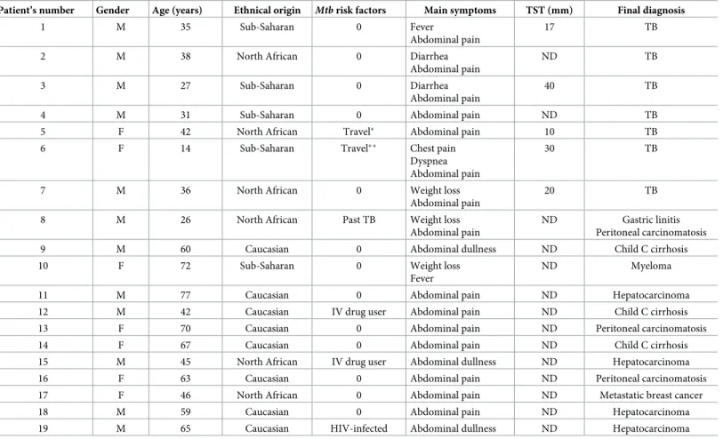
The peritoneal fluids from TB patients contained approximately 6 times more non-stimulated IFN-γ-producing CD4+
T lymphocytes than to those of non-TB patients (medians 0.630% and 0.105%,Fig 1, upper and lower panels, TB and non-TB respectively), whereas the proportion of IFN-γ-containing CD8+
T lymphocytes was similar in the two groups of patients (medians 0.09% and 0.08% for TB and non-TB). Afterin vitro incubation with PPD, HBHA or ESAT-6, the proportions of the IFN-γ-containing-CD4+
T lymphocytes from all TB patients were sig-nificantly enhanced compared to the non-stimulated cells (p<0.05, Figs1and2, upper panels). In contrast, for the non-TB patients, a rise in the proportions of peritoneal IFN-γ-producing CD4+T lymphocytes was only observed for some of them afterin vitro stimulation with PPD, HBHA or ESAT-6 (Figs1and2, lower panels). Among CD8+T lymphocytes, there was a trend for higher proportions of PPD, HBHA, ESAT-6-induced IFN-γ-containing cells but the
differences between stimulated and non-stimulated cells were not significant, neither for TB patients, nor for non-TB patients. The proportions of PPD, HBHA, ESAT-6-induced IFN-γ-containing CD8+and CD4+T lymphocytes were however correlated (Fig 3).
Diagnostic potential of the measurement of IFN-
γ-producing ascites
lymphocyte proportions in response to mycobacterial antigens
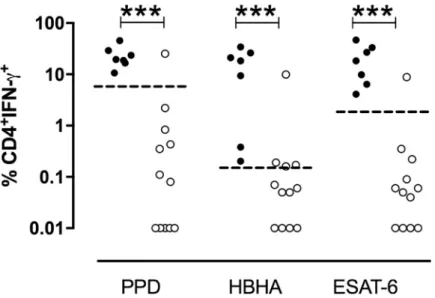
TB patients had significantly higher proportions of IFN-γ-containing ascites CD4+T lympho-cytes afterin vitro stimulation with mycobacterial antigens than non-TB patients (p = 0.0007 in response to PPD, p = 0.0003 in response to HBHA, and p = 0.0002 in response to ESAT-6, Fig 4). The percentages of cells shown inFig 4are those obtained after subtraction of the values obtained in the absence of antigen stimulation from those obtained in response to the myco-bacterial antigens. The values obtained for the two groups of patients for each mycomyco-bacterial antigen were further compared by receiver operator characteristics (ROC) curve analysis to define the most discriminant cut-off values and evaluate the diagnostic potential of these tests for peritoneal TB (Fig 5). The areas under the curve (AUC) were of 0.94, 0.96, and 0.98 for
Table 1. Demographic and clinical characteristics of included patients.
Patient’s number Gender Age (years) Ethnical origin Mtb risk factors Main symptoms TST (mm) Final diagnosis
1 M 35 Sub-Saharan 0 Fever
Abdominal pain
17 TB
2 M 38 North African 0 Diarrhea Abdominal pain
ND TB
3 M 27 Sub-Saharan 0 Diarrhea
Abdominal pain
40 TB
4 M 31 Sub-Saharan 0 Abdominal pain ND TB
5 F 42 North African Travel� Abdominal pain 10 TB
6 F 14 Sub-Saharan Travel�� Chest pain
Dyspnea Abdominal pain
30 TB
7 M 36 North African 0 Weight loss Abdominal pain
20 TB
8 M 26 North African Past TB Weight loss Abdominal pain
ND Gastric linitis Peritoneal carcinomatosis 9 M 60 Caucasian 0 Abdominal dullness ND Child C cirrhosis 10 F 72 Sub-Saharan 0 Weight loss
Fever
ND Myeloma 11 M 77 Caucasian 0 Abdominal pain ND Hepatocarcinoma 12 M 42 Caucasian IV drug user Abdominal pain ND Child C cirrhosis 13 F 70 Caucasian 0 Abdominal pain ND Peritoneal carcinomatosis 14 F 67 Caucasian 0 Abdominal pain ND Child C cirrhosis 15 M 45 North African IV drug user Abdominal dullness ND Hepatocarcinoma 16 F 63 Caucasian 0 Abdominal pain ND Peritoneal carcinomatosis 17 F 46 North African 0 Abdominal pain ND Metastatic breast cancer 18 M 59 Caucasian 0 Abdominal pain ND Hepatocarcinoma 19 M 65 Caucasian HIV-infected Abdominal dullness ND Hepatocarcinoma M: male; F: female; TST: tuberculin skin test; TB: tuberculosis; IV: intravenous; ND: not done
�Recent travel to Marocco
��Recent arrival in Belgium (<12 months)
PPD, HBHA, and ESAT-6, respectively. Optimal cut-off values providing 100% of sensitivity for the identification of patients with peritoneal TB or highly suspected peritoneal TB, were 6.44%, 0.20% and 2.23% of IFN-γ+
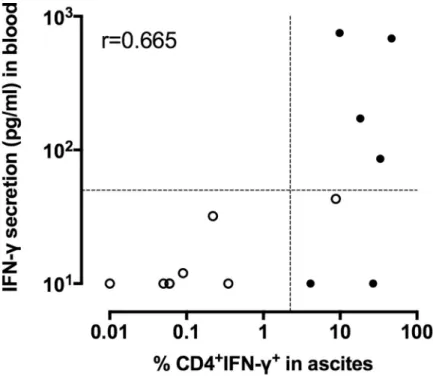
CD4+T lymphocytes for PPD, HBHA and ESAT-6, respec-tively. They were all associated with a specificity of 92%. The values obtained in response to ESAT-6 provided the largest difference between TB and non-TB patients and, when blood was available, they were compared to the concentrations of IFN-γ released by PBMC from the same patientsin vitro stimulated with ESAT-6. The results shown onFig 6indicated that only two thirds of the TB patients had a blood IFN-γ response to ESAT-6 above the cut-off value. In contrast, all but one patient classified as non-TB were negative for both the index case on asci-tes lymphocyasci-tes and for the blood IFN-γ release in response to ESAT-6. This patient (n˚15) classified as non-TB showed in fact values on ascites lymphocytes above the cut-off for all three antigens. However, this patient was a diabetic intravenous drug user suffering from cir-rhosis with an hepato-carcinoma and presenting with chronic renal disease, so that a perito-neal biopsy was contra-indicated. As both the blood ESAT-6-induced interferon-γ-release assay (Fig 6), and the QuantiFERON test performed on his blood (Table 2, patient n˚15) were positive in addition to the positive results obtained on ascites lymphocytes, this suggests that this patient was infected withM. tuberculosis and may thus have been initially mis-classified by the clinician.
Table 2. Main biological results.
Blood Peritoneal fluid Peritoneal biopsy
Patient’s Number CRP Conc. (N<10mg/L) QFT Ag TB-Nil (N<0.35IU/ml) Prot. Conc. (N<30g/L) Lymphos (%) Mtb Smear Mtb Cult. Mtb Smear PCR Mtb Cult. 1 7.2 1.12 60 32 - - - - + 2 11.0 ND� 60 73 - - - - + M. bovis 3 29.0 1.77 60 51 - + ND ND ND 4 25.0 2.66 58 7 - - ND ND ND 5 62.0 ND�� 48 ND - - ND ND ND 6 53.0 0.11 67 54 - - ND ND ND 7 29.0 3.32 68 61 - - ND ND ND 8 53.0 1.08 45 45 - - ND ND ND 9 38.0 ND 23 6 - - ND ND ND 10 14.0 ND� 4 31 - - ND ND ND 11 19.0 ND� 14 48 - - ND ND ND 12 16.0 0.00 4 81 - - ND ND ND 13 63.0 ND� 12 5 - - ND ND ND 14 45.0 ND 37 25 - - ND ND ND 15 41.0 0.88 33 23 - - ND ND ND 16 31.0 ND� 5 6 - - ND ND ND 17 33.0 ND 13 19 - - ND ND ND 18 16.0 0.00 38 65 - - ND ND ND 19 74.0 0.00 35 ND - - ND ND ND
�As the QFT test was not routinely available at the beginning of the study, a home-made interferon-γ-release assay in response to ESAT-6 was performed for some
patients as described [14] and was negative (�) or positive (��) for patient n˚5 with 172 pg/ml interferon-γ.
CRP: C-reactive protein; N = Normal value; QFT: QuantiFERON TB Gold in-Tube; conc: concentration; lymphos: lymphocytes; PCR: polymerase chain reaction;Mtb: Mycobacterium tuberculosis; ND: not done
Discussion
Peritoneal TB is associated with a high mortality rate and its diagnosis is often delayed due to unspecific clinical symptoms and low bacillary burden resulting often in negative acid-fast bacilli smear examination and negative culture [3]. In the present study conducted in a low TB incidence country, we report the diagnostic accuracy of the analysis of the IFN-γ production by peritoneal fluid CD4+T lymphocytes in response to a shortin vitro stimulation with myco-bacterial antigens. Although in the absence of stimulation by mycomyco-bacterial antigens, TB patients had already higher proportions of IFN-γ-producing CD4+
ascites lymphocytes than the non-TB patients, this difference was not statistically significant and had therefore a low dis-criminatory power between TB and non-TB patients. CD8+T lymphocytes also produced IFN-γ- in response to an in vitro stimulation with mycobacterial antigens, but the differences between the results obtained with or without antigenic stimulation were not significant with therefore low diagnostic relevance of these results.
IFN-γ production by fluid lymphocytes in response to an in vitro stimulation with myco-bacterial antigens has already been suggested as a potential diagnostic aid for pulmonary TB, pleural TB and TB meningitis [8–10,15]. Two independent case reports also suggested a diag-nostic potential of such tests for peritoneal TB [16,17], whereas a large study performed in a high TB incidence country reported no added value of the T-SPOT.TB test (Oxford
Fig 1. Percentages of IFN-γ+
CD4+T lymphocytes among non-stimulated (NS) compared to mycobacterial antigens-stimulated ascites lymphocytes. The percentages of IFN-γ-containing CD4+lymphocytes were measured by
flow cytometry after an overnightin vitro culture of the ascites lymphocytes left non-stimulated (NS) or stimulated
with PPD (left panels), HBHA (middle panels) or ESAT-6 (right panels) for TB (upper panels) and non-TB patients (lower panels). Individual values obtained for each patient for lymphocytes left unstimulated and those stimulated with a mycobacterial antigen are linked. NS, non-stimulated. Results obtained in the presence or absence of antigens were compared by the Wilcoxon test�,P<0.05.
Immunotec) performed on ascites lymphocytes over the diagnostic accuracy provided by mea-suring the peritoneal fluid ADA [18]. However, only a sensitivity of 82% with a specificity of 79% was achieved by ADA measurements for the diagnosis of peritoneal TB in this study. Therefore, the authors proposed a two-step algorithm based on both T cell-based assays and
Fig 2. Representative dot-plots of the flow cytometry analyses for one TB patient (upper panel) and one non-TB patient (lower panel). Ascites lymphocytes were in vitro cultured overnight in the absence (medium) or the presence of PPD, HBHA or ESAT-6, as indicated. IFN-γ-producing CD4+T cells are shown after a sequential gating of Forward Scatter (FSC)/Side scatter (SSC), single cells and CD3+T lymphocytes. The percentages of IFN-γ+
T lymphocytes in each condition are indicated.
https://doi.org/10.1371/journal.pone.0214333.g002
Fig 3. Correlations between the percentages of IFN-γ-containing CD4+and CD8+T lymphocytes induced by the
mycobacterial antigens. Ascites lymphocytes werein vitro cultured overnight in the absence (medium) or the
presence of PPD, HBHA or ESAT-6, and the percentages of IFN-γ-containing CD4+
and CD8+T lymphocytes were analyzed by flow cytometry. The values shown are obtained by subtracting the values obtained in the absence of antigen stimulation from those obtained in response to mycobacterial antigens. Filled circles represent values from patients suffering from TB, whereas open circles represent values from non-TB patients. r: Spearman coefficient of correlation.
ADA determinations to reach a correct classification of 67% of the patients [18]. ADA deter-minations are not available as a clinical setting in Belgium and we have chosen here to analyze the IFN-γ production by ascites lymphocytes using flow cytometry. This allowed us to focus on the CD4+T lymphocytes, the major T lymphocyte subset producing IFN-γ in response to a shortin vitro stimulation with mycobacterial antigens. By focusing on these cells, we observed extremely high proportions of CD4+ascites T lymphocytes from TB patients producing IFN-γ in response to mycobacterial antigens, up to 47%. These extremely elevated responses may be due at least partially to the inhibition by ADA, known to be elevated in TB ascites, of CD4+ regulatory T cells that normally tend to inhibit extreme degrees of activation of CD4+effector T lymphocytes [19]. Based on these results, we propose this new test as a valuable adjunct for the diagnosis of peritoneal TB. We have tested the inter-laboratory reproducibility of the assay
Fig 4. Percentages of IFN-γ-containing CD4+
ascites T lymphocytes induced by PPD, HBHA or ESAT-6. Ascites
lymphocytes werein vitro cultured overnight in the absence or presence of PPD, HBHA ESAT-6, and the percentages
of IFN-γ-containing CD4+T lymphocytes were analyzed by flow cytometry. The values shown are obtained by
subtracting the values obtained in the absence of antigen stimulation from those obtained in response to mycobacterial antigens. Filled circles represent values from patients suffering from TB, whereas open circles represent values from non-TB patients. Dotted horizontal lines represent the cut-offs determined by ROC analysis comparing the values for TB patients with those for non-TB patients. Values obtained for TB and non-TB patients were compared by Mann-Whitney U test.���,P<0,001.
https://doi.org/10.1371/journal.pone.0214333.g004
Fig 5. Receiver operator characteristics (ROC) curve analysis of the percentages of ascites IFN-γ-containing CD4+
lymphocytes obtained for TB and non-TB patients. The percentages of PPD-, HBHA-, and ESAT-6- induced IFN- γ-containing CD4+lymphocytes were assessed by ROC analysis to define the most discriminant cut-off values between
TB and non-TB patients. Cut-off values associated with optimal sensitivity and specificity were determined by the likelihood ratio analysis and are indicated by the black circles.
by dividing some samples into two aliquotsand processing them in two independent laborato-ries for flow cytometry analysis. The percentages of CD4+IFN-γ-containing T lymphocytes reported by the two laboratories were strongly correlated for all 3 antigens, with r values of 0.90, 0.71 and 0.81 for PPD, HBHA and ESAT-6, respectively (Spearman correlation test).
As results can be obtained within only 24 hours, and as flow cytometry is available in most clinical laboratories, this test provides very quickly useful data to guide decisions about the need for invasive laparoscopic examination and for the initiation of empirical anti-TB treat-ment. It should be particularly helpful for rapid differential diagnosis with confounding etiolo-gies, such as ovarian cancer [20], and may also help to diagnose peritoneal TB sometimes associated with cancer [21]. This help cannot be obtained by blood tests like the Quanti-FERON that can be negative in patients with peritoneal TB [6], as patient n˚6 in this study, or other forms of TB [5], even with the QuantiFERON-TB Gold Plus [22]. On the contrary, the QuantiFERON test may be positive in subjects with latent TB presenting with ascites not con-sidered as related to aM. tuberculosis infection (patients n˚ 8 and n˚15), as this test cannot dis-tinguish active from latent TB [5].
High risk populations for peritoneal TB include patients with AIDS or with other severe immunosuppressive conditions, including cirrhosis and continuous ambulatory peritoneal dialysis [23]. Diagnosis of peritoneal TB is further complicated for these patients, as peritoneal biopsy performed by laparoscopy may be contra-indicated, such as for patient n˚15 of our study. The recent case report of a peritoneal TB being the cause of ascites in a patient with cir-rhosis [24] lead us to consider that patient n˚15 could in fact have been a miss-classification. If
Fig 6. Comparison of the peripheral blood and peritoneal cell IFN-γ-responses to ESAT-6. Ascites lymphocytes
werein vitro cultured overnight in the absence (medium) or the presence of ESAT-6, and the percentages of
IFN-γ-containing CD4+T lymphocytes were analyzed by flow cytometry. Peripheral blood mononuclear cells werein vitro
cultured overnight in the absence or the presence of ESAT-6, and the concentration of IFN-γ-released was measured by ELISA. The values obtained in the absence of antigen stimulation were subtracted from those obtained in response to mycobacterial antigens for both the flow cytometry and the ELISA results. Filled circles represent values from patients suffering from TB, whereas open circles represent values from non-TB patients. r: Spearman coefficient of correlation.
we consider patient n˚15 as a patient with TB, this brings the specificity of the determination of the percentages ofM. tuberculosis specific IFN-γ-containing ascites CD4+T lymphocytes to 100% for the diagnosis of peritoneal TB.
The main limitation of this study is the relatively low number of patients included with a final diagnosis of peritoneal TB, a limitation inherent to the fact that the study was performed in a low TB incidence country. Moreover, only 3 cases of peritoneal TB were confirmed by positive microbiological results as, except in case of peritoneal biopsies obtained by laparos-copy and associated with a significant risk for the patient, only 35% of the peritoneal TB are confirmed by a positiveM. tuberculosis culture on ascites [2]. However, the detailed clinical characterization of the patients allowed the clinicians to be confident in their classification of the patients as TB or not TB, and the good clinical and biological responses to anti-TB treat-ment confirmed that the patients classified as “highly probable TB” were indeed most likely suffering from a peritoneal TB. Only one patient may have been misclassified as non-TB as dis-cussed above.
In conclusion, our study indicates that the analysis of the IFN-γ response of ascites CD4+
T lymphocytes to the mycobacterial antigens HBHA and ESAT-6 is a promising tool for rapid and reliable diagnosis of peritoneal TB, with a high degree of sensitivity and specificity. This diagnostic method may be very useful, especially in challenging situations, where pulmonary infection is absent. Further investigation with a larger cohort especially in a high TB-incidence country is warranted to confirm the results.
Acknowledgments
This work was supported by the Fonds National de la Recherche Scientifique (FNRS–PDR T.0147.13), by the European Community within the Horizon2020 program TBVAC2020 (grant agreement 643381), and by the “Re´gion de Bruxelles-Capitale–Innoviris”. We are grate-ful to Dr F Vandergheynst, Dr S Allard, Dr J Coussement, Dr F Vermeulen and Dr JL Van Laethem for their important help in the patient’s inclusions, and to J Tresnie, MC Termonia, A Godefroid and S Islane for their excellent technical assistance.
Author Contributions
Conceptualization: Ve´ronique Corbière, Violette Dirix, Franc¸oise Mascart.
Data curation: Sophie Henrard, Ve´ronique Corbière, Anne Van Praet, Violette Dirix, Fran-c¸oise Mascart.
Formal analysis: Sophie Henrard, Ve´ronique Corbière, Liliane Schandene´, Violette Dirix, Franc¸oise Mascart.
Funding acquisition: Franc¸oise Mascart.
Investigation: Sophie Henrard, Ve´ronique Corbière, Franc¸oise Mascart.
Methodology: Ve´ronique Corbière, Liliane Schandene´, Martine Ducarme, Anne Van Praet, Emmanuelle Petit, Mahavir Singh, Camille Locht, Violette Dirix, Franc¸oise Mascart.
Project administration: Franc¸oise Mascart. Resources: Franc¸oise Mascart.
Supervision: Violette Dirix, Franc¸oise Mascart.
Validation: Sophie Henrard, Ve´ronique Corbière, Liliane Schandene´, Violette Dirix, Fran-c¸oise Mascart.
Writing – original draft: Sophie Henrard, Violette Dirix, Franc¸oise Mascart.
Writing – review & editing: Ve´ronique Corbière, Liliane Schandene´, Martine Ducarme, Anne Van Praet, Emmanuelle Petit, Mahavir Singh, Camille Locht, Violette Dirix, Franc¸oise Mascart.
References
1. Guirat A, Koubaa M, Mzali R, Abid B, Ellouz S, Affes N, et al. Peritoneal tuberculosis. Clin Res Hepatol Gastroenterol. janv 2011; 35(1):60–9.https://doi.org/10.1016/j.gcb.2010.07.023PMID:21215540
2. Chow KM, Chow VCY, Hung LCT, Wong SM, Szeto CC. Tuberculous peritonitis-associated mortality is high among patients waiting for the results of mycobacterial cultures of ascitic fluid samples. Clin Infect Dis. 15 aouˆt 2002; 35(4):409–13.https://doi.org/10.1086/341898PMID:12145724
3. Sanai FM, Bzeizi KI. Systematic review: tuberculous peritonitis—presenting features, diagnostic strate-gies and treatment. Aliment Pharmacol Ther. 15 oct 2005; 22(8):685–700.https://doi.org/10.1111/j. 1365-2036.2005.02645.xPMID:16197489
4. Weledji EP, Pokam BT. Abdominal tuberculosis: Is there a role for surgery? World J Gastrointest Surg. 27 aouˆt 2017; 9(8):174–81.https://doi.org/10.4240/wjgs.v9.i8.174PMID:28932351
5. Metcalfe JZ, Everett CK, Steingart KR, Cattamanchi A, Huang L, Hopewell PC, et al. Interferon-γ
release assays for active pulmonary tuberculosis diagnosis in adults in low- and middle-income coun-tries: systematic review and meta-analysis. J Infect Dis. 15 nov 2011; 204 Suppl 4:S1120–1129.
6. Bourgain G, Sbai W, Luciano L, Massoure MP, Brardjanian S, Goin G, et al. Hepato-peritoneal tubercu-losis with negative interferon gamma assay (QuantiferonTM) in an immunocompetent patient: A case report. Clin Res Hepatol Gastroenterol. sept 2016; 40(4):e44–45.https://doi.org/10.1016/j.clinre.2015. 11.006PMID:26774362
7. Tao L, Ning H-J, Nie H-M, Guo X-Y, Qin S-Y, Jiang H-X. Diagnostic value of adenosine deaminase in ascites for tuberculosis ascites: a meta-analysis. Diagn Microbiol Infect Dis. mai 2014; 79(1):102–7.
https://doi.org/10.1016/j.diagmicrobio.2013.12.010PMID:24629577
8. Losi M, Bossink A, Codecasa L, Jafari C, Ernst M, Thijsen S, et al. Use of a T-cell interferon-gamma release assay for the diagnosis of tuberculous pleurisy. Eur Respir J. de´c 2007; 30(6):1173–9.https:// doi.org/10.1183/09031936.00067307PMID:17715165
9. Jafari C, Thijsen S, Sotgiu G, Goletti D, Domı´nguez Benı´tez JA, Losi M, et al. Bronchoalveolar lavage enzyme-linked immunospot for a rapid diagnosis of tuberculosis: a Tuberculosis Network European Trialsgroup study. Am J Respir Crit Care Med. 1 oct 2009; 180(7):666–73.https://doi.org/10.1164/rccm. 200904-0557OCPMID:19590020
10. Place S, Verscheure V, de San N, Hougardy J-M, Schepers K, Dirix V, et al. Heparin-binding, hemag-glutinin-specific IFN-gamma synthesis at the site of infection during active tuberculosis in humans. Am J Respir Crit Care Med. 15 sept 2010; 182(6):848–54.https://doi.org/10.1164/rccm.201001-0083OC
PMID:20508213
11. Menozzi FD, Rouse JH, Alavi M, Laude-Sharp M, Muller J, Bischoff R, et al. Identification of a heparin-binding hemagglutinin present in mycobacteria. J Exp Med. 1 sept 1996; 184(3):993–1001. PMID:
9064359
12. Andersen P, Andersen AB, Sørensen AL, Nagai S. Recall of long-lived immunity to Mycobacterium tuberculosis infection in mice. J Immunol. 1 avr 1995; 154(7):3359–72. PMID:7897219
13. Masungi C, Temmerman S, Van Vooren J-P, Drowart A, Pethe K, Menozzi FD, et al. Differential T and B cell responses against Mycobacterium tuberculosis heparin-binding hemagglutinin adhesin in infected healthy individuals and patients with tuberculosis. J Infect Dis. 15 fe´vr 2002; 185(4):513–20.https://doi. org/10.1086/338833PMID:11865404
14. Wyndham-Thomas C, Corbière V, Dirix V, Smits K, Domont F, Libin M, et al. Key role of effector mem-ory CD4+ T lymphocytes in a short-incubation heparin-binding hemagglutinin gamma interferon release assay for the detection of latent tuberculosis. Clin Vaccine Immunol. mars 2014; 21(3):321–8.
15. Thomas MM, Hinks TSC, Raghuraman S, Ramalingam N, Ernst M, Nau R, et al. Rapid diagnosis of Mycobacterium tuberculosis meningitis by enumeration of cerebrospinal fluid antigen-specific T-cells. Int J Tuberc Lung Dis. juin 2008; 12(6):651–7. PMID:18492332
16. Lorenz R, Wu¨rl P, Haerter G, Cammerer G, Barth T, Hausladen S, et al. Interferon-gamma release assay in the ascites: Early hint for diagnosis of abdominal tuberculosis. Infection. fe´vr 2010; 38(1):69– 72.https://doi.org/10.1007/s15010-009-9469-5PMID:19904487
17. Mu¨ller C, Puttinger H, Winnicki W, Winkler H-M, Vychytil A, Winkler S. Rapid T-cell-based immunodiag-nosis of tuberculous peritonitis in a peritoneal dialysis patient. Scand J Urol Nephrol. aouˆt 2012; 46 (4):314–6.https://doi.org/10.3109/00365599.2012.659206PMID:22339389
18. Lee JY, Kim S-M, Park S-J, Lee S-O, Choi S-H, Kim YS, et al. A rapid and non-invasive 2-step algorithm for diagnosing tuberculous peritonitis using a T cell-based assay on peripheral blood and peritoneal fluid mononuclear cells together with peritoneal fluid adenosine deaminase. J Infect. avr 2015; 70 (4):356–66.https://doi.org/10.1016/j.jinf.2014.09.012PMID:25305499
19. Naval-Macabuhay I, Casanova V, Navarro G, Garcı´a F, Leo´n A, Miralles L, et al. Adenosine deaminase regulates Treg expression in autologous T cell-dendritic cell cocultures from patients infected with HIV-1. J Leukoc Biol. fe´vr 2016; 99(2):349–59.https://doi.org/10.1189/jlb.3A1214-580RRPMID:26310829
20. Lataifeh I, Matalka I, Hayajneh W, Obeidat B, Al Zou’bi H, Abdeen G. Disseminated peritoneal tubercu-losis mimicking advanced ovarian cancer. J Obstet Gynaecol. avr 2014; 34(3):268–71.https://doi.org/ 10.3109/01443615.2013.870140PMID:24476396
21. Alshahrani AS, Lee IS. Gastric Cancer with Peritoneal Tuberculosis: Challenges in Diagnosis and Treatment. J Gastric Cancer. juin 2016; 16(2):111–4.https://doi.org/10.5230/jgc.2016.16.2.111PMID:
27433397
22. Horne DJ, Jones BE, Kamada A, Fukushima K, Winthrop KL, Siegel S a. R, et al. Multicenter study of QuantiFERON®-TB Gold Plus in patients with active tuberculosis. Int J Tuberc Lung Dis. 1 juin 2018; 22(6):617–21.https://doi.org/10.5588/ijtld.17.0721PMID:29862944
23. Vaid U, Kane GC. Tuberculous Peritonitis. Microbiol Spectr. 2017; 5(1).https://doi.org/10.1128/ microbiolspec.TNMI7-0006-2016PMID:28185616
24. Vaz AM, Peixe B, Ornelas R, Guerreiro H. Peritoneal tuberculosis as a cause of ascites in a patient with cirrhosis. BMJ Case Rep. 14 juill 2017;2017.