HAL Id: hal-01274954
https://hal.archives-ouvertes.fr/hal-01274954
Submitted on 17 Feb 2016
HAL is a multi-disciplinary open access
archive for the deposit and dissemination of
sci-entific research documents, whether they are
pub-lished or not. The documents may come from
teaching and research institutions in France or
abroad, or from public or private research centers.
L’archive ouverte pluridisciplinaire HAL, est
destinée au dépôt et à la diffusion de documents
scientifiques de niveau recherche, publiés ou non,
émanant des établissements d’enseignement et de
recherche français ou étrangers, des laboratoires
publics ou privés.
Involvement of the serotonin 5-HT2B receptor in cardiac
hypertrophy linked to sympathetic stimulation: control
of interleukin-6, interleukin-1beta, and tumor necrosis
factor-alpha cytokine production by ventricular
fibroblasts.
Fabrice Jaffré, Jacques Callebert, Alexandre Sarre, Nelly Etienne, Canan G
Nebigil, Jean-Marie Launay, Luc Maroteaux, Laurent Monassier
To cite this version:
Fabrice Jaffré, Jacques Callebert, Alexandre Sarre, Nelly Etienne, Canan G Nebigil, et al..
In-volvement of the serotonin 5-HT2B receptor in cardiac hypertrophy linked to sympathetic
stimu-lation: control of interleukin-6, interleukin-1beta, and tumor necrosis factor-alpha cytokine
produc-tion by ventricular fibroblasts.. Circulaproduc-tion, American Heart Associaproduc-tion, 2004, 110 (8), pp.969-974.
�10.1161/01.CIR.0000139856.20505.57�. �hal-01274954�
Hypertrophy Linked to Sympathetic Stimulation
Control of Interleukin-6, Interleukin-1
, and Tumor Necrosis Factor-␣
Cytokine Production by Ventricular Fibroblasts
Fabrice Jaffré, MS; Jacques Callebert, PharmD, PhD; Alexandre Sarre, MS; Nelly Etienne, MS;
Canan G. Nebigil, PharmD, PhD; Jean-Marie Launay, PharmD, PhD;
Luc Maroteaux, PhD; Laurent Monassier, MD, PhD
Background—The serotonergic 5-HT2Breceptor regulates cardiomyocyte development and growth. A putative
contribu-tion of this receptor to fibroblast-dependent cardiac funccontribu-tion has not been identified.
Methods and Results—By mimicking sympathetic stimulation with chronic isoproterenol perfusion in vivo, we found that
mice developed a cardiac hypertrophy, which was prevented by exposure to the 5-HT2Breceptor antagonists SB206553
or SB215505 or in 5-HT2Breceptor– knockout mice. The isoproterenol-induced hypertrophy was associated with an
increase in the plasma levels of interleukin-1 and tumor necrosis factor-␣ but not interleukin-6. In contrast, the plasma isoproterenol-induced cytokine increase was not observed in either 5-HT2Breceptor–mutant or wild-type mice perfused
with isoproterenol⫹SB206553. We demonstrated that stimulation of wild-type cardiac fibroblasts by isoproterenol markedly increased the production of the interleukin-6, interleukin-1, and tumor necrosis factor-␣ cytokines. Strikingly, we found that this isoproterenol-induced cytokine production was abolished by SB206553 or in 5-HT2B
receptor– knockout fibroblasts. Serotonin also stimulated production of the 3 cytokines in wild-type fibroblasts, which was effectively reduced in 5-HT2Breceptor– knockout fibroblasts.
Conclusions—Our results demonstrate for the first time that 5-HT2B receptors are essential for isoproterenol-induced
cardiac hypertrophy, which involves the regulation of interleukin-6, interleukin-1, and tumor necrosis factor-␣ cytokine production by cardiac fibroblasts. (Circulation. 2004;110:969-974.)
Key Words: fibroblasts 䡲 hypertrophy 䡲 interleukins 䡲 nervous system, sympathetic 䡲 remodeling
M
yocardial hypertrophy has long been considered an adaptive process to increased wall stress in the heart.1Sustained cardiac hypertrophy is often maladaptive and leads to significant ventricular dysfunction and failure.2This
phe-nomenon involves not only an increase in size and mass of the cardiomyocytes but equally, the activation of cardiac fibro-blasts.3 Recent studies have emphasized the role of G
protein– coupled receptors in the initiation of processes that play crucial roles in myocyte hypertrophy4 and fibroblast
stimulation.5 Moreover, it was demonstrated that G q/G11
proteins activated after stimulation of either AT1angiotensin
II (AGII),␣1-adrenergic (AR), or ETAendothelin 1 receptors
are key regulators of these hypertrophic responses.6Although
the action of these mediators on cardiomyocytes is direct, it also implicates autocrine and paracrine factors. For example, AR stimulation has been demonstrated to induce the release of the hypertrophic cytokines interleukin-6 (IL-6),
interleukin-1 (IL-1), and tumor necrosis factor-␣ (TNF-␣) from cardiac fibroblasts,7which are the predominant source
of cytokines in myocardium.8 In vivo, the 3 cytokines are
produced after either myocardial infarction or a cardiac remodeling process9 triggered by sympathetic
overstimula-tion. An increase in serotonin (5-HT) levels has been identi-fied in normal and failing heart,10 and its release could be
associated with sympathetic overstimulation,11 contributing
to myocardial remodeling in left ventricular dysfunction. The effects of 5-HT are mediated by actions on numerous cognate receptors belonging to the G protein– coupled recep-tors and ionotropic receprecep-tors. Activation of the 5-HT2Gq/G11
-coupled subtypes participates in cell proliferation.12We have
previously shown that 5-HT, via the 5-HT2B receptor
(5-HT2BR), regulates cardiac embryonic development and adult
functions; gene targeting of the 5-HT2BR gene by homologous
recombination leads to a dilated cardiomyopathy without
Received October 29, 2003; de novo received January 24, 2004; revision received March 16, 2004; accepted March 23, 2004.
From the Laboratoire de Neurobiologie et de Pharmacologie Cardiovasculaire, INSERM E333, Faculté de médecine, Strasbourg (A.S., L. Monassier); Institut de Génétique et de Biologie Moléculaire et Cellulaire, CNRS, INSERM, Université L. Pasteur de Strasbourg, Illkirch (F.J., N.E., C.G.N., L. Maroteaux); and Centre de Recherches Claude Bernard, Service de Biochimie, Hôpital Lariboisière, Paris (J.C., J.-M.L.), France.
Correspondence to Laurent Monassier, MD, PhD, Laboratoire de Neurobiologie et de Pharmacologie Cardiovasculaire, INSERM E333, Faculté de médecine, 11 rue Humann, 67085 Strasbourg, France. E-mail laurent.monassier@medecine.u-strasbg.fr
© 2004 American Heart Association, Inc.
Circulation is available at http://www.circulationaha.org DOI: 10.1161/01.CIR.0000139856.20505.57
hypertrophy.12,13 Alternatively, the selective overexpression
of the 5-HT2BR in cardiomyocytes induces a myocardial
hypertrophy,14and a direct survival effect of 5-HT in
cardio-myocytes was placed in evidence.15 Therefore, the G q
-coupled 5-HT2BR could be implicated in trophic responses of
the myocardium by acting directly on cardiomyocytes or indirectly on noncardiomyocytes through the release of para-crine factors.
The aims of the present work were to study whether, on -adrenergic stimulation, the 5-HT2BR could (1) modify the
cardiac hypertrophic responses in vivo and (2) contribute to IL-6, IL-1, and TNF-␣ cytokine production by ventricular fibroblasts.
Methods
5-HT2BR–Knockout MiceTargeted mutagenesis has been described previously.12 Animal
experimentation was performed in accordance with institutional guidelines. Protocols were approved by the French Animal Care Committee in accordance with European regulations. 129/PAS (genetic background of KO mice) served as wild-type (WT) controls.
Induction of Cardiac Hypertrophy by Isoproterenol
In 11-week-old mice, vehicle (saline), isoproterenol (ISO) (30g · g⫺1 · d⫺1) alone, or ISO associated with the 5-HT2BR antagonist
SB206553 or SB215505 (1 mg · kg⫺1 · d⫺1) (all chemicals from Sigma) was delivered by osmotic minipumps (1007D, Alzet Corpo-ration) implanted subcutaneously under anesthesia (sodium pento-barbital, 40 mg/kg IP). After 5 days, the heart was excised and weighed, and the apex was quickly frozen. The remaining left ventricle was fixed in 4% paraformaldehyde PBS solution. For planimetric quantification of cardiomyocyte area, at least 40 cardio-myocytes were measured on 1 midventricular section. Heart rate was recorded by the tail-cuff method (Letica Model 5002).13 For
tele-metric measurements, mice anesthetized with ketamine (100 mg/kg) and xylazine (0.4 mg/kg) were fitted with the TA11PA-C20 sensor model (Data Sciences) placed in the right common carotid artery.16
Seven days later, basal parameters were recorded every 5 minutes over a period of 24 hours before implantation of an osmotic minipump. The mean values of systolic and diastolic blood pressures and heart rate were calculated for the whole of this period. Similarly, the animals were recorded over a period of 24 hours after 7 days of treatment. Transthoracic echocardiograms were performed with a 15-MHz linear transducer on a Sonos 5500 (Philips) in anesthetized mice (sodium pentobarbital, 30 mg/kg IP).13 This anesthetic was
selected to exemplify, and reveal, any cardiodepressive property of SB206553 better than echocardiography in the conscious state.
5-HT2R Expression in Fibroblasts
Semiquantitative reverse transcription–polymerase chain reaction was performed on 2g of total RNA.13The following primers were
used: 5⬘-AAGCCTCGAACTGGACAATTGATG-3⬘ (5-HT2AR
for-ward), 5⬘-AATGATTTTCAGGAAGGCTTTGGTT-3⬘ (5-HT2AR
re-verse), 5⬘-ACAACTTCTGAGCACATTTTAAG-3⬘ (5-HT2BR
for-ward), 5⬘-AATTAACCATACCACTGTAATC-3⬘ (5-HT2BR reverse),
5⬘-CCCTTATTGACCTCAACTACATGGT-3⬘ (GAPDH forward), and 5⬘-GAGGGGCCATCCACAGTCTTCTG-3⬘ (GAPDH reverse).
Adult Cardiac Fibroblast Primary Culture
Cultures of fibroblasts were obtained from the ventricles of adult (10- to 12-week-old) mice using a modification of a previously described protocol.17After anesthesia, the heart was excised and the
ventricles, free from the atria and atrioventricular valves, were minced and incubated with 0.1 mg/mL type IV collagenase and 1 mg/mL pancreatin (Sigma) at 37°C. Cells were plated in Dulbecco/
Ham F12 medium with 10% calf serum and gentamicin. After a 2-hour incubation period at 37°C in 5% CO2/95% air, the unattached
cardiomyocytes were removed, and the attached cells (mostly fibro-blasts) were grown. All experiments were performed using cells of the first passage. Cardiac fibroblasts were identified by characteristic morphology and positive staining with antibody to vimentin (Sig-ma).17 One day before the experiments, cells were transferred to
serum-free medium.
Dosage of Cytokines
Cultured fibroblasts corresponding to each time point were plated in 6-well plates. Supernatants and cells were collected at 0, 2, 4, 8, 12, and 24 hours after onset of stimulation. This was performed after stimulation with ISO (10mol/L) in the absence or presence of the 5-HT2BR antagonist SB 206553 (100 nmol/L). Cells were stimulated
with 5-HT (1mol/L) and BW723C86 (BW) (100 nmol/L) (Sigma). The plasma cytokine quantification was performed after centrifuga-tion (2000g, 10 minutes) of 1 mL total blood. Concentracentrifuga-tions of IL-6, IL-1, and TNF-␣ were measured in plasma and cell culture supernatants by ELISA kits (DY 406, DY 401, and DY 410, R&D).
Binding Assays
Radioligand binding studies of1- and2-ARs in WT and KO mice
hearts were performed according to Molenaar et al.18-ARs were
labeled with (⫺)-[125I]-cyanopindolol (Amersham). The proportions
of 1- and 2-ARs were determined by use of CGP 20712A
(1-selective antagonist) and ICI 118,551 (2-selective antagonist)
(Sigma). Kdand Bmaxwere obtained from competition curves.
Data Analysis and Statistics
All results are expressed as mean⫾SEM. Different groups were compared by 1-way ANOVA followed by a Newman-Keuls test. With 2 groups, the comparison was performed using a Student’s t test. All calculations were made with the GraphPad Prism program (San Diego).
Results
Role of 5-HT2BRs in ISO-Mediated Cardiac
Hypertrophy In Vivo
We used the classic chronic-AR agonist ISO perfusion (30 g · kg⫺1· d⫺1) as a model of cardiac hypertrophy.19In WT
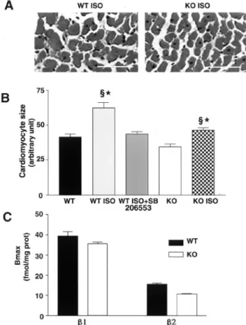
mice, 5 days of perfusion produced a marked increase in the ratio of cardiac mass to tibia length (CMTL) (59⫾17%, ISO versus WT) (Figure 1A). The ISO perfusion induced an important tachycardia (62⫾4% in WT-ISO versus WT) (Figure 1B). The increase in cardiac mass was primarily because of the hypertrophy of cardiomyocytes (Figure 2, A and B). The ISO-induced increase in CMTL was completely prevented by the 5-HT2B/2CR antagonist SB206553 (1 mg ·
kg⫺1 · d⫺1) (Figures 1A and 2B) but not the ISO-induced tachycardia (Figure 1B). Similarly, the selective 5-HT2BR
antagonist SB215505 (1 mg · kg⫺1 · d⫺1) prevented the ISO-induced increase in CMTL but not the tachycardia. In 5-HT2BRKO mice, the ISO perfusion produced a slight
increase (19%) in CMTL, which was significantly lower than WT ISO-treated mice (Figures 1A and 2B). However, in 5-HT2BRKO mice, ISO induced a tachycardia similar to that
observed in WT mice (Figure 1B). The 1- and 2-AR
expression levels (Bmax) were not significantly different in
5-HT2BRKO mice compared with control animals (Figure
2C); the respective affinity constants (Kd) were also not
modified (data not shown). The putative cardiovascular effects of SB206553 (1 mg · kg⫺1· d⫺1) were investigated in WT mice by telemetry and echocardiography. Chronic
sure to this drug modified neither the cardiac function nor the blood pressure in WT mice (Table). The ISO-induced hyper-trophy observed in the WT mice is thus dependent on 5-HT2BRs.
Role of 5-HT2BRs in ISO-Mediated Increase in
Cytokine Plasma Levels
Five days of ISO perfusion in WT mice increased the plasma levels of TNF-␣ significantly (2-fold over basal) and IL-1 (2-fold) (Figure 3, B and C). These increases were prevented by SB206553. In 5-HT2BRKO, the basal values of these 2
cytokines were not different from those of WT mice, and ISO was unable to increase their plasma concentrations (Figure 3, B and C). In WT mice, the perfusion of ISO, either alone or in combination with SB206553, did not significantly modify IL-6 plasma concentrations, nor did it do so in 5-HT2BRKO.
However, basal IL-6 plasma concentration was approxi-mately twice as high in 5-HT2BRKO mice as in WT mice
(Figure 3A). Therefore, this ISO increase of TNF-␣ and IL-1 plasma levels observed in the WT mice was prevented entirely by pharmacological or genetic ablation of 5-HT2BRs.
Role of 5-HT2BRs on IL-6, IL-1, and TNF-␣
Cytokine Production by Cardiac Fibroblasts Stimulated by ISO
We focused on cardiac fibroblasts, the main source of hypertrophic cytokines that are involved in the cardiac remodeling processes.17 Supernatants of primary cultures
displayed no difference in basal levels of IL-6, TNF-␣, and IL-1 between WT and 5-HT2BRKO. In WT fibroblasts, ISO
(10 mol/L) induced a peak of cytokine production (IL-6, 12-fold over basal; TNF-␣, 4-fold; and IL-1, 2-fold) be-tween 4 and 8 hours of stimulation (Figure 4, A through C). In WT cells treated with SB206553 (100 nmol/L) or in 5-HT2BRKO fibroblasts, a prevention of the ISO-induced
production of IL-6, TNF-␣, and IL-1 was observed. There-fore, 5-HT2BRs appear necessary for the induction by ISO of
the 3 cytokines in cardiac fibroblasts.
Role of 5-HT2BRs on Cytokine Production by
Cardiac Fibroblasts Stimulated by 5-HT
Adult WT cardiac ventricular fibroblasts constitutively ex-press 5-HT2A and 5-HT2BRs (Figure 5A). 5-HT (1 mol/L)
markedly increased IL-6 (6-fold over basal), TNF-␣ (12-fold) and IL-1 (4-fold) levels in WT fibroblasts at 4 hours after agonist exposure (Figure 5, B through D). In WT fibroblasts, the 5-HT–induced IL-1 production was mimicked by stim-ulation with the 5-HT2BR–selective agonist BW (100 nmol/
L). BW reproduced 66% of the IL-6 response observed with 5-HT in WT. The response to 5-HT of KO fibroblasts reached only 30% of the WT IL-6 response. For TNF-␣, BW induced a peak significantly smaller than the response to 5-HT in WT (⬇50%), although similar to that in 5-HT2BRKO fibroblasts.
These findings indicate a role for 5-HT2BRs in the production of
these 3 cytokines by cardiac fibroblasts in response to 5-HT.
Discussion
This work demonstrates for the first time that 5-HT2BR
blockade in vivo can reduce cardiac hypertrophy caused by
Figure 1. In vivo cardiac effects of chronic ISO perfusion.
ISO-induced effects were evaluated by measuring heart weight:tibia length (mg/mm) (A) and assessing heart rate in beats per minute (bpm) by tail-cuff method (B) in WT and in 5-HT2BRKO (KO) mice
in absence or presence of 5-HT2BR antagonists SB206553 (n⫽6)
or SB215505 (n⫽3). *P⬍0.05 vs KO, §P⬍0.05 vs WT.
Figure 2. 5-HT2BR blockade reduces cardiomyocyte
hypertro-phy. Representative sections from adult heart left ventricles of WT ISO-treated and KO ISO-treated mice (A) were measured by planimetric quantification of cardiomyocyte transversal surface (B) (n⫽6 each). *P⬍0.05 vs KO, §P⬍0.05 vs WT. Bar⫽50m. Observed hypertrophy is independent of modifications in1
-and2-AR levels of expression assessed by binding studies (C);
values are mean⫾SEM of 3 independent experiments performed in triplicate.
-AR stimulation. Moreover, 5-HT2BRs are required for the
in vivo regulation of TNF-␣ and IL-1 cytokine production. Our in vitro results indicate that the 5-HT2BR is a key
regulator for IL-6, TNF-␣, and IL-1 production by myocar-dial fibroblasts in response to 5-HT and equally to -AR activation, which explains, at least in part, our in vivo findings.
A role for 5-HT2Rs in cardiac hypertrophy linked to
hypertension and left ventricular dysfunction was suggested
by earlier reports on the antihypertrophic effects of the 5-HT2
antagonist ketanserin.20More recently, sarpogrelate, a
non-selective 5-HT2 receptor antagonist, was reported to reduce
the hypertrophic responses in cultured cardiomyocytes, alone or in combination with fibroblasts.21-ARs contribute to the
cardiac hypertrophy linked to catecholamine overstimula-tion22and to the activation of cardiac fibroblasts in chronic
pressure overload.23 Moreover, in rats, the chronic -AR
stimulation by ISO induces myocardial generation of IL-6, TNF-␣, and IL-1.7In WT, we demonstrate that the cardiac
hypertrophy induced by continuous ISO perfusion is completely prevented by the 5-HT2BR antagonists SB206553 or SB215505
at doses that were previously shown to prevent the BW-induced hyperphagia in freely feeding rats.24 The markedly reduced
ISO-induced cardiac hypertrophy in 5-HT2BRKO mice
demon-strates the selectivity of the SB compounds. The small residual cardiac hypertrophy to ISO in KO mice could result from unknown compensatory mechanisms to the dilated cardiomyop-athy of KO. Such a difference was previously described in
Figure 3. In vivo effects of chronic ISO perfusion on plasma
cy-tokines. Plasma levels of IL-6 (A), TNF-␣ (B), and IL-1 (C) were evaluated by ELISA in WT and KO mice after 5 days of ISO treatment in absence or presence of SB206553. WT, n⫽5; KO, n⫽4; KO ISO, n⫽6; WT ISO, n⫽7; WT ISO⫹SB206553, n⫽7; §P⬍0.05 vs WT.
Cardiac Function and Blood Pressure
Basal SB206553 (7 d) MBP, mm Hg 108⫾1 104⫾0.6 HR, bpm 556⫾8 551⫾17 LVM, mg 63⫾6 63⫾4 FS, % 37⫾1 34⫾2 CO, mL/min 36⫾11 30⫾6 E/A 1.5⫾0.2 1.8⫾0.1 IVRT, ms 17.3⫾2.0 17.3⫾0.6
Mean blood pressure关MBP⫽diastolic BP⫹(1/3)(systolic BP⫺diastolic BP)兴 and heart rate (HR) were obtained by telemetry. Echocardiographic parameters: left ventricular mass {LVM⫽1.055⫻关(Sd⫹PWd⫹LVEDD)3⫺(LVEDD)3兴, where Sd
indicates septal thickness in diastole; PWd, posterior wall thickness in diastole;
and LVEDD, left ventricular end-diastolic diameter}, left ventricular fractional shortening 关FS⫽(LVEDD⫺LVESD/LVEDD)⫻100; LVESD, left ventricular end-systolic diameter兴, cardiac output 关CO⫽(AoD/2)2⫻⫻VTI⫻HR, where AoD is
the aortic diameter and VTI the velocity time integral of the aortic flow兴, ratio of the early rapid peak mitral filling wave velocity (E) to the late filling velocity caused by atrial contraction (A) (E/A), and the isovolumetric relaxation time (IVRT).
Figure 4. Cytokine production by cardiac fibroblasts in
response to ISO. Production of IL-6 (A), TNF-␣ (B), and IL-1 (C) was measured in supernatant of cardiac fibroblast primary cul-tures after various times of exposure to ISO, n⬎4. *P⬍0.05, WT vs KO ISO; #P⬍0.05 vs WT ISO⫹SB206553.
angiotensin II 1a receptor– knockout mice (AT1KO) in which the cardiac hypertrophy was not completely prevented after transverse aortic constriction25 or myocardial infarction,26
whereas the AT1 antagonist losartan suppressed entirely the
hypertrophic response of WT in the same models.27,28
Further-more, strong alterations of the AR signaling pathway in 5-HT2BRKO mice are unlikely to explain the reduction of
ISO-induced hypertrophic effect, as suggested by the similarities in the number of-adrenergic cardiac binding sites and ISO-induced tachycardia in both WT and KO mice.
The ISO-induced increase in TNF-␣ and IL-1 plasma levels, which is observed in WT, is completely prevented by pharmacological (SB) or genetic ablation of the 5-HT2BR,
supporting the in vivo role of this receptor in cardiac IL-6, IL-1, and TNF-␣ cytokine production. Similar to rats,7the
increase in plasma IL-6 was not found in response to ISO in either WT or KO mice. The high resting level of plasma IL-6 in KO mice could be of noncardiac origin, because we were unable to detect its mRNA expression in their myocardium (data not shown). Moreover, no inflammatory cells were identified in the myocardium of these animals.13
In ISO-induced cardiac hypertrophy, fibroblasts constitute a main source of hypertrophic cytokines. As expected, we found that ISO increased the production of IL-6, TNF-␣, and IL-1 in WT fibroblasts. According to our in vivo results, this increase is prevented by the 5-HT2B/2Cantagonist SB206553 at
100 nmol/L,29 which, here, can be considered as a pure
5-HT2BR antagonist, 5-HT2CRs not being expressed either in
heart30or in vasculature.31Although changes in IL-6 plasma
levels could not be detected in vivo, our in vitro results confirm that at least part of the ISO-induced plasma cytokines originate from cardiac fibroblasts.
Our work reveals for the first time that 5-HT markedly increased the production of IL-6, TNF-␣, and IL-1 in WT cardiac fibroblasts, which was mimicked by the 5-HT2BR
preferential agonist BW. BW exhibits a significantly higher
affinity at 5-HT2BR than 5-HT2AR.24Our work demonstrates
that in adult mice cardiac fibroblasts, the 5-HT–induced production of IL-1 involves only 5-HT2BRs, because the
IL-1 production is eliminated in 5-HT2BRKO fibroblasts,
whereas BW provoked a response superimposable to 5-HT in WT cells. The 5-HT2BR is also a main contributor of 5-HT–
induced IL-6 and TNF-␣ cytokine production. BW only partially reproduced the maximum 5-HT–induced TNF-␣ or IL-6 responses in WT fibroblasts, with different kinetics for TNF-␣. In KO cells, these maximum responses are reduced but not eliminated. These findings indicate that stimulation of 5-HT2BRs constitutes an essential trigger for the complete
production of TNF-␣ and IL-6 in combination with other 5-HTRs. This result is in agreement with observations indi-cating that 5-HT stimulates IL-6 production in vascular smooth muscle cells.32Taken together, these data suggest that
5-HT participates in in vivo cardiac hypertrophy.
The mechanisms by which 5-HT2BRs control ISO-induced
IL-6, IL-1, and TNF-␣ cytokine production remain to be elucidated. Crosstalk between Gq-coupled 5-HT2BR and Gs
-coupled -AR signaling pathways could be suggested, be-cause a recent report demonstrated a dual transinhibition of AT1and-AR by an antagonist targeting a single receptor33;
the effects of valsartan are obtained in the absence of angiotensin II. These findings support our result showing that the 5-HT2BR antagonist SB206553 blocks the ISO-induced
IL-6, IL-1, and TNF-␣ cytokine production by cardiac fibroblasts in the absence of 5-HT. The possible existence of complexes formed between-ARs and 5-HT2BRs that would
explain these interactions is under investigation. Whether hypertrophic responses to agents such as angiotensin II or endothelin 1 could also be affected by 5-HT2B receptor
blockade will be examined in subsequent studies.
In conclusion, the lack of a cardiodepressive property of the 5-HT2BR antagonist, together with the essential role of
5-HT2BRs in cardiac hypertrophy triggered by-AR
stimu-Figure 5. Cytokine production by cardiac
fibroblasts in response to 5-HT. Reverse transcription–polymerase chain reaction reveals expression of 5-HT2AR and
5-HT2BR subtypes in cardiac ventricular
fibroblasts with GAPDH as internal con-trol (A); M, molecular weight marker. Pro-duction of IL-6 (B), TNF-␣ (C), and IL-1 (D) was measured in supernatant of car-diac fibroblast primary cultures after vari-ous times of exposure to 5-HT or BW, n⬎4. *P⬍0.05, WT vs KO 5-HT; †P⬍0.05 vs WT BW.
lation, suggests that 5-HT2BR antagonists could constitute
new opportunities to prevent or reduce myocardial remodel-ing associated with left ventricular dysfunction and sympa-thetic overactivity.
Acknowledgments
This work has been supported by Centre National de la Recherche Scientifique, Institut National pour la Recherche Me´dicale, Hoˆpitaux Universitaires de Strasbourg, and Universite´ Louis Pasteur and by grants from Fondation de France, Fondation pour la Recherche Me´dicale, Association pour la Recherche Me´dicale, and the French Ministry of Research, Action Concerte´e Incitative. F. Jaffré is supported by a fellowship from the Groupe de Réflexion pour la Recherche Cardiovasculaire—Pfizer and Dr Nebigil by the Lefoulon-Delalande Fondation. We thank P. Hickel, L. El Fertak, and A. Guimond for technical assistance and Dr S. Brooks for English corrections.
References
1. Chien KR. Stress pathways and heart failure. Cell. 1999;98:555–558. 2. Francis GS. Pathophysiology of chronic heart failure. Am J Med. 2001;
110(suppl 7A):37S– 46S.
3. Weber KT. Fibrosis and hypertensive heart disease. Curr Opin Cardiol. 2000;15:264 –272.
4. Akhter SA, Luttrell LM, Rockman HA, Iaccarino G, Lefkowitz RJ, Koch WJ. Targeting the receptor-Gq interface to inhibit in vivo pressure overload myocardial hypertrophy. Science. 1998;280:574 –577. 5. Colombo F, Noel J, Mayers P, Mercier I, Calderone A.-Adrenergic
stimulation of rat cardiac fibroblasts promotes protein synthesis via the activation of phosphatidylinositol 3-kinase. J Mol Cell Cardiol. 2001;33: 1091–1106.
6. Wettschureck N, Rutten H, Zywietz A, Gehring D, Wilkie TM, Chen J, Chien KR, Offermanns S. Absence of pressure overload induced myo-cardial hypertrophy after conditional inactivation of Galphaq/Galpha11 in cardiomyocytes. Nat Med. 2001;7:1236 –1240.
7. Murray DR, Prabhu SD, Chandrasekar B. Chronic-adrenergic stimu-lation induces myocardial proinflammatory cytokine expression.
Circu-lation. 2000;101:2338 –2341.
8. Yin F, Li P, Zheng M, Chen L, Xu Q, Chen K, Wang YY, Zhang YY, Han C. Interleukin-6 family of cytokines mediates isoproterenol-induced delayed STAT3 activation in mouse heart. J Biol Chem. 2003;278: 21070 –21075.
9. Ono K, Matsumori A, Shioi T, Furukawa Y, Sasayama S. Cytokine gene expression after myocardial infarction in rat hearts: possible implication in left ventricular remodeling. Circulation. 1998;98:149 –156. 10. Sole MJ, Shum A, Van Loon GR. Serotonin metabolism in the normal
and failing hamster heart. Circ Res. 1979;45:629 – 634.
11. Singh S, Johnson PI, Javed A, Gray TS, Lonchyna VA, Wurster RD. Monoamine- and histamine-synthesizing enzymes and neurotransmitters within neurons of adult human cardiac ganglia. Circulation. 1999;99: 411– 419.
12. Nebigil CG, Choi DS, Dierich A, Hickel P, Le Meur M, Messaddeq N, Launay JM, Maroteaux L. Serotonin 2B receptor is required for heart development. Proc Natl Acad Sci U S A. 2000;97:9508 –9513. 13. Nebigil CG, Hickel P, Messaddeq N, Vonesch JL, Douchet MP,
Monassier L, Gyorgy K, Matz R, Andriantsitohaina R, Manivet P, Launay JM, Maroteaux L. Ablation of serotonin 5-HT2Breceptors in mice leads to
abnormal cardiac structure and function. Circulation. 2001;103: 2973–2979.
14. Nebigil CG, Jaffre F, Messaddeq N, Hickel P, Monassier L, Launay JM, Maroteaux L. Overexpression of the serotonin 5-HT2Breceptor in heart
leads to abnormal mitochondrial function and cardiac hypertrophy.
Cir-culation. 2003;107:3223–3229.
15. Nebigil CG, Etienne N, Messaddeq N, Maroteaux L. Serotonin is a novel survival factor of cardiomyocytes: mitochondria as a target of 5-HT2B receptor signaling. FASEB J. 2003;17:1373–1375.
16. Butz GM, Davisson RL. Long-term telemetric measurement of cardio-vascular parameters in awake mice: a physiological genomics tool.
Physiol Genomics. 2001;5:89 –97.
17. Burger A, Benicke M, Deten A, Zimmer HG. Catecholamines stimulate interleukin-6 synthesis in rat cardiac fibroblasts. Am J Physiol. 2001;281: H14 –H21.
18. Molenaar P, Sarsero D, Arch JR, Kelly J, Henson SM, Kaumann AJ. Effects of (⫺)-RO363 at human atrial beta-adrenoceptor subtypes, the human cloned beta 3-adrenoceptor and rodent intestinal beta 3-adrenoceptors. Br J Pharmacol. 1997;120:165–176.
19. Kudej RK, Iwase M, Uechi M, Vatner DE, Oka N, Ishikawa Y, Shannon RP, Bishop SP, Vatner SF. Effects of chronic beta-adrenergic receptor stimulation in mice. J Mol Cell Cardiol. 1997;29:2735–2746. 20. Cobo C, Alcocer L, Chavez A. Effects of ketanserin on left ventricular
hypertrophy in hypertensive patients. Cardiovasc Drugs Ther. 1990; 4(suppl 1):73–76.
21. Ikeda K, Tojo K, Tokudome G, Hosoya T, Harada M, Nakao K. The effects of sarpogrelate on cardiomyocyte hypertrophy. Life Sci. 2000;67: 2991–2996.
22. Engelhardt S, Hein L, Wiesmann F, Lohse MJ. Progressive hypertrophy and heart failure in beta1-adrenergic receptor transgenic mice. Proc Natl
Acad Sci U S A. 1999;96:7059 –7064.
23. Grimm D, Huber M, Jabusch HC, Shakibaei M, Fredersdorf S, Paul M, Riegger GA, Kromer EP. Extracellular matrix proteins in cardiac fibro-blasts derived from rat hearts with chronic pressure overload: effects of beta-receptor blockade. J Mol Cell Cardiol. 2001;33:487–501. 24. Kennett GA, Ainsworth K, Trail B, Blackburn TP. BW 723C86, a
5-HT2B receptor agonist, causes hyperphagia and reduced grooming in rats. Neuropharmacology. 1997;36:233–239.
25. Harada K, Komuro I, Zou Y, Kudoh S, Kijima K, Matsubara H, Sugaya T, Murakami K, Yazaki Y. Acute pressure overload could induce hyper-trophic responses in the heart of angiotensin II type 1a knockout mice.
Circ Res. 1998;82:779 –785.
26. Nakamura Y, Yoshiyama M, Omura T, Yoshida K, Kim S, Takeuchi K, Iwao H, Yoshikawa J. Transmitral inflow pattern assessed by Doppler echocardiography in angiotensin II type 1A receptor knockout mice with myocardial infarction. Circ J. 2002;66:192–196.
27. Rockman HA, Wachhorst SP, Mao L, Ross J Jr. Ang II receptor blockade prevents ventricular hypertrophy and ANF gene expression with pressure overload in mice. Am J Physiol. 1994;266:H2468 –H2475.
28. Patten RD, Aronovitz MJ, Einstein M, Lambert M, Pandian NG, Men-delsohn ME, Konstam MA. Effects of angiotensin II receptor blockade versus angiotensin-converting-enzyme inhibition on ventricular remod-elling following myocardial infarction in the mouse. Clin Sci (Lond). 2003;104:109 –118.
29. Grignaschi G, Fanelli E, Scagnol I, Samanin R. Studies on the role of serotonin receptor subtypes in the effect of sibutramine in various feeding paradigms in rats. Br J Pharmacol. 1999;127:1190 –1194.
30. Nebigil CG, Launay JM, Hickel P, Tournois C, Maroteaux L. 5-Hydroxy-tryptamine 2B receptor regulates cell-cycle progression: cross-talk with tyrosine kinase pathways. Proc Natl Acad Sci U S A. 2000;97:2591–2596. 31. Ullmer C, Schmuck K, Kalkman HO, Lubbert H. Expression of serotonin
receptor mRNAs in blood vessels. FEBS Lett. 1995;370:215–221. 32. Ito T, Ikeda U, Shimpo M, Yamamoto K, Shimada K. Serotonin increases
interleukin-6 synthesis in human vascular smooth muscle cells.
Circu-lation. 2000;102:2522–2527.
33. Barki-Harrington L, Luttrell LM, Rockman HA. Dual inhibition of
-adrenergic and angiotensin II receptors by a single antagonist: a
func-tional role for receptor-receptor interaction in vivo. Circulation. 2003; 108:1611–1618.