HAL Id: inserm-00319921
https://www.hal.inserm.fr/inserm-00319921
Submitted on 19 Mar 2012HAL is a multi-disciplinary open access archive for the deposit and dissemination of sci-entific research documents, whether they are pub-lished or not. The documents may come from teaching and research institutions in France or abroad, or from public or private research centers.
L’archive ouverte pluridisciplinaire HAL, est destinée au dépôt et à la diffusion de documents scientifiques de niveau recherche, publiés ou non, émanant des établissements d’enseignement et de recherche français ou étrangers, des laboratoires publics ou privés.
Localization of aurora A and aurora B kinases during
interphase: role of the N-terminal domain.
Yoann Rannou, Marie-Bérengère Troadec, Clotilde Petretti, Fabienne Hans,
Stéphanie Dutertre, Stefan Dimitrov, Claude Prigent
To cite this version:
Yoann Rannou, Marie-Bérengère Troadec, Clotilde Petretti, Fabienne Hans, Stéphanie Dutertre, et al.. Localization of aurora A and aurora B kinases during interphase: role of the N-terminal domain.: Interphase localization of Aurora kinases A and B. Cell Cycle, Taylor & Francis, 2008, 7 (19), pp.3012-20. �10.4161/cc.7.19.6718�. �inserm-00319921�
1
LOCALIZATION OF AURORA A AND AURORA B KINASES DURING
INTERPHASE: ROLE OF THE N-TERMINAL DOMAIN.
Yoann Rannou1§, Marie-Bérengère Troadec1§, Clotilde Petretti1, Fabienne Hans3, Stéphanie Dutertre2, Stefan Dimitrov3 and Claude Prigent1*
1 - CNRS UMR 6061 Institut de Génétique et Développement de Rennes, Université de Rennes 1, IFR140, Rennes, France.
2 - IFR140 Génétique Fonctionnelle Agronomie et Santé, Université de Rennes 1, 35043 Rennes, France.
3 - CRI INSERM U823 Université Joseph Fourier, Institut Albert Bonniot, Site Santé La Tronche, BP170, 38042 Grenoble cedex 9, France.
Running title: Interphase localization of Aurora kinases A and B
§ - these authors contributed equally to the work
Address correspondence to : Claude Prigent, IGDR UMR6061 CNRS UR1, 2 avenue du Pr. Léon Bernard, CS 34317, 35043 Rennes cedex, FranceTel : (33) (0)2 23 23 46 93, Fax : (33) (0)2 23 23 44 78, claude.prigent@univ-rennes1.fr
2
AKNOWLEDGMENTS
We wish to thank Tim Hunt for Xenopus Aur-B cDNA and Diane M Ward for proofreading. The images were taken on the Fluorescence Microscopy Platform of the University of Rennes (GIS EUROPIA, IFR140). Y Rannou and M-B Troadec were fellows of the Ligue Nationale Contre le Cancer, Y Rannou was also a fellow of the Association pour La recherché sur le Cancer. C Petretti was a fellow of the Region Bretagne. S Dimitrov was supported the Agence Nationale de la Recherche (Project R05075CC) and Rhône-Alpes Canceropôle CLARA (EpiProNetwork). C Prigent was
supported by the Canceropôle Grand Ouest and the CNRS. S Dimitrov and C Prigent were both financed by the “Ligue Nationale Contre le Cancer” (équipes Labellisées).
ABBREVIATIONS
The abbreviations used are: APC/C, Anaphase Promoting Complex/Cyclosome; GFP, Green Fluorescence Protein; INCENP, INner CENtromer Protein, MCAK, Mitotic centromere-associated kinesin; TACC; Transforming Acidic Coiled Coil protein, TPX2 Targetting Protein for Xklp2,
3
ABSTRACT
Aurora kinases possess a conserved catalytic domain (CD) and a N-terminal domain (ND) that varies in size and sequence. We have previously reported that the N-terminal domain of AuroraA (AurA) participates in the localization of the kinase to the centrosome in interphase. AuroraB (AurB) is a chromosome passenger protein and its N-terminal domain is not necessary for its localization or function during mitosis. Using various combinations of GFP-AurA and AurB protein domains we show that in interphase, AurB N-terminal domain is required for nuclear localization in Xenopus XL2 cells. In human cells, however, we found both AurA and AurB kinases in the nucleus, AurA being mainly cytoplasmic and AurB mainly nuclear. Both proteins are actively excluded from the nucleus by a CRM1 dependent pathway. Interestingly, at a functional level, in interphase, every combination of Aurora kinase domains (ND-CD) rescues histone H3 Serine10 phosphorylation defect induced by AurB knockdown. This clearly indicates the presence of a functional AurA in the nucleus. Additionally, the chimera ND-AurA/CD-AurB was much more efficient than the ND-AurB/CD-AurA to rescue multinucleation also induced by AurB knockdown. This indicates that the catalytic domain of AurB is required to fulfill specific functions during mitosis that cannot be fulfilled by the catalytic domain of AurA, probably for localization reasons during mitosis.
4
INTRODUCTION
The Aurora serine/threonine kinases are essential for normal mitosis to proceed. Mammalian cells possess three different Aurora kinases named AurA, -B and -C (AurA, AurB, AurC) 1. AurA is a
centrosome kinase while AurB and C are chromosome passenger proteins 2 3. All three Aurora
kinases are overexpressed in numerous cancers 4 5 6, but only AurA transforms NIH3T3 or Rat1
cells when overexpressed, revealing an oncogenic potential 7 8. AurA activity is required for
centrosome maturation, and mitotic spindle assembly and stabilization by phosphorylating proteins such as TACC, TPX2 and Eg5 9 10 11 12. The kinase also phosphorylates CDC25B
contributing to the G2/M transition 13. AurB activity is required during the metaphase checkpoint
to resolve synthelic attachments of chromosome kinetochores through phosphorylation of MCAK 14 15. The kinase might also be necessary for chromosome condensation by
phosphorylating histone H3 16 and for cytokinesis by phosphorylating vimentin, MgcRacGAP
and the kinesin Mklp1 17 18 19 20. AurB forms at least two different mitotic protein complexes,
one with INCENP and one with INCENP, survivin, and Borealin 21. The function of AurC is still
unknown, however the protein overexpressed in HeLa cells behaves like a chromosome passenger protein 22 and rescues AurB deficiency 23. Aurora kinases possess three domains; a large catalytic
domain located in the C-terminal half of the protein, a variable N-terminal non-catalytic domain and a very short carboxy-terminal domain 24. Aurora catalytic domains show a high degree of
identity, whereas, the N-terminal non-catalytic domains are different in sequence and length (Figure 1A and B). Xl-AurA and Xl-AurB are 407 and 361 amino acids proteins respectively and they share 52% identity (Figure 1C). Their catalytic domains (CD) are very conserved in length and sequence (69% identity), while their non-catalytic domains (ND) show only 18% identity (Figure 1C). The length of these non-catalytic domains is also different: 138 amino acids in length for Xl-AurA and 92 amino acids in length for Xl-AurB 25. Interestingly, these two kinases
localized differently in interphase cells. AurA begins to localize on duplicated centrosomes in late S-phase then stays associated with the centrosomes until early G1-phase 26. At this cell cycle
stage, AurA is targeted by the APC/C (Anaphase Promoting Complex/Cyclosome) and degraded by the proteasome in a Cdh1 dependent manner 27 28. The protein is then resynthesized during
5
the following S-phase and delocalizes to duplicated centrosomes. AurB is a chromosome passenger protein 29. It appears only in late G2-phase in the nucleus on chromatin condensation foci and the
protein is also degraded by the proteasome in a Cdh1 dependent manner 30. In early mitosis, AurB
localization is restrained to the kinetochores on condensed chromosomes. At the metaphase anaphase transition AurB moves from kinetochores to the central spindle and decorates the mid-body in telophase cells 31. AurB localization during mitosis depends only on the presence of a
short sequence in the C-terminal domain of the protein. The N-terminal domain of the kinase is not required for the mitotic localization or for its mitotic function 32. We previously showed that
the non-catalytic N-terminal domain of Xl-AurA was able to target the GFP (Green Fluorescent protein) to the centrosome mimicking interphase AurA localization 25. We further demonstrated
that this domain participates to the kinase localization to the centrosome using an active mechanism that depends on the presence of microtubules 25. The catalytic domain can also
localize the GFP to the centrosome but with a much lower efficiency and independently of the presence of microtubules. This secondary localization mechanism was proposed to be a passive one due to the interaction between Xl-AurA and its substrates already localized on the centrosome. The N-terminal domain of the Xl-AurA then plays an essential role in increasing the efficiency of the localization of the kinase, at the centrosome 25. AurC is the shortest kinase of
the family (without N-terminal domain), the kinase is also closely related to AurB, at the sequence level but also at the functional level, AurC can rescue AurB RNAi 23. AurC is like an
AurB without any N-terminal domain. In a previous work we have reported that AurC, in contrast to AurB, does not enter the nucleus during G2 33. In this report we asked whether the N-term
domain of AurB participates to the localization of the kinase during interphase. We found that the non-catalytic N-terminal domain of Xl-AurB fused to GFP enters the nucleus in G2 whereas the catalytic domain does not. We also show that by switching the N-terminal domains we were able to localize Xl-AurB-ND fused to Xl-AurA-CD in the nucleus suggesting that the presence of the N-terminal domain of AurB correlates with an increase efficiency in the nuclear localization of the kinase. Further analysis of Aurora kinases nuclear localization in human cells showed that both AurA and AurB can enter the nucleus. AurA is, however, readily exported in the cytoplasm. We also found that proteins containing the catalytic domain of AurA rescue the histone H3
6
Ser10 phosphorylation defect due to AurB RNA interference, but they do not rescue the cytokinesis defect.
RESULTS
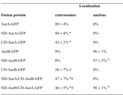
The N-terminal domain of Xl-AurA and both catalytic domains of Xl-AurA and Xl-AurB localiz e the GFP to centrosome in interphase cells. In order to check whether the N-terminal of Xl-AurB, like for Xl-AurA, plays any role in the localization processes of Aurora B protein during interphase we transfected XL2 Xenopus cells with various Aurora protein domains tagged with GFP. As a control, GFP protein alone was also expressed. Xl-AurA is a centrosome protein and Xl-AurB a nuclear protein, therefore, we particularly analyzed the protein localization at these structures.
We started by analyzing the centrosomes localization using γ-tubulin indirect immunofluorescence as control (Figure 2, panels B1 to B9). When the GFP protein alone was expressed, the whole cell (nucleus and cytoplasm) was fluorescent (Figure 2, panels A1 and C1) and the centrosomes stained with anti-γ-tubulin were not decorated with GFP (Figure 2, panels B1 and C1). As we previously reported 25, the AurA-GFP (Figure 2, panels A2 to D2) and the
ND-AurA-GFP proteins (Figure 2, panels A3 to D3) localized to the centrosome when expressed in XL2 cells. Eighty-nine ± 4% of interphase cells transfected with AurA-GFP and 80 ± 4% transfected with ND-AurA-GFP contained GFP decorated centrosomes (Table 1). Likewise, the CD-AurA-GFP protein localized to centrosomes in XL2 interphase cells (Figure 2, panels A4 to D4), but with a weaker efficiency, 43 ± 2% of transfected interphase cells contained GFP stained centrosomes (Table 1). Therefore, as already reported, we confirm that the ND of Xl-AurA participates in the centrosome localization of the protein 25. When the AurB-GFP protein (Figure
2, panels A5 to D5) or the ND-AurB-GFP protein (Figure 2, panels A6 to D6) were expressed in XL2 cells, the fusion protein was never observed at the centrosomes in interphase cells. Interestingly, the CD-AurB targeted the GFP to centrosomes in interphase cells (Figure 2, panels A7 to D7) with efficiency in the same range than CD-AurA. Fifty-six ± 7% of interphase cells expressing CD-AurB-GFP and 43 ± 2% for CD-AurA-GFP exhibited GFP-decorated centrosomes
7
(Table 1). In addition, when the ND-AurA/CD-AurB-GFP protein was expressed in XL2 cells, the fusion protein localized to centrosomes in 47 ± 7% of transfected interphase cells (Figure 2, panels A8 to D8; table1). ND-AurB/CD-AurA targeted the GFP on centrosomes with the same efficiency: 46 ± 5% of transfected cells in interphase (Figure 2, panels A9 to D9; Table 1). Although ND-AurA is highly efficient to localize the GFP to centrosome, it loses its efficiency when fused to CD-AurB whereas it gains in efficiency when fused to CD-AurA. This suggested that there is cooperation between ND-AurA and CD AurA that does not occur between ND-AurA and CD-AurB, at the contary. This is also in agreement with the fact that both Aurora-A domains (N-terminal and catalytic-domain) contain sequences involved in centrosome localization of the kinase 34. To summarize, in interphase all the chimera proteins containing either Xl-AurA
N-terminal domain or catalytic-domain localized the GFP to the centrosomes while Xl-AurB was found in the centrosome only in the absence of its own N-terminal domain.
The N-terminal domain of Xl-AurB targets all the fusion proteins in the nucleus. Because the Xl-AurA N-terminal domain plays a role in localizing Xl-AurA in interphase we asked whether the Xl-AurB N-terminal domain would also play a role in the localization of Xl-AurB in interphase. All the fusion proteins localized in the cytoplasm, especially AurA-GFP, ND-AurA-GFP and CD-AurA-GFP (data not shown). When the AurB-GFP was expressed, 96 ± 1% of transfected interphase cells displayed a GFP labeling in the nucleus (Table1). This localization was consistent with the previous AurB localization described in interphase 20. Ninety-seven% ± 2% of interphase cells expressing ND-AurB-GFP exhibited a
nuclear GFP staining with the same efficiency than AurB-GFP expressing cells (Table1). The CD-AurB-GFP never localized in the nucleus (Table1). These results strongly suggest that without its N-terminal domain, Xl-AurB can no longer localize to the nucleus. This implies that the Xl-AurB non-catalytic domain plays a role in the localization of the protein in interphase XL2 cells. Interestingly, none of the AurA domain was able to target the GFP to the nucleus. We then asked whether the Xl-AurB non-catalytic domain would be sufficient to target the Xl-AurA catalytic domain in the nucleus. When the ND-AurB/CD-AurA-GFP was expressed in XL2 cells, the fusion protein was readily found in the nucleus of 96 ± 1% of these cells (Table1). In contrast, the
ND-8
AurA/CD-AurB-GFP was never found in the nucleus. All these data strongly confirm that the Xl-AurB N-terminal domain is essential to localize the protein kinase in the nucleus in XL2 cells.
Both human Aurora A and B kinases are found in the nucleus, but the N-terminal domain of Hs-AurB increases the nuclear localization efficiency. Xenopus XL2 cells are known to be resistant to drug and are also difficult to perform RNA interference on, therefore, we decided to study AurA and AurB localization in human Hela cells during interphase. AurA and AurB are two proteins highly conserved between Xenopus and Human (Figure 1), therefore this study gave us the opportunity to analyze the conservation of localization mechanisms from Xenopus to Human.
We analyzed the nuclear localization of human proteins AurA and AurB tagged with GFP. In Human cells we decided to discriminate three kinds of localization : (1) mainly nuclear, (2) nuclear and cytoplasmic and (3) mainly cytoplasmic (figure 3A). In agreement with our results in XL2 cells, we found that in 74.1 ± 6.1% of the human HeLa cells Hs-AurB-GFP was mainly nuclear, whereas in 25.6 ± 6.1% it was both cytoplasm and nuclear, we didn’t see any cell with only cytoplasmic AurB-GFP (Figure 3A). Surprisingly in HeLa cells, Hs-AurA-GFP was equally distributed between the cytoplasm and the nucleus in 78.7 ± 3.8% of the cells. In 18.3 ± 2.6% of the cells AurA-GFP was nuclear and in only 3% ± 1.6% of the cells the kinase was exclusively cytoplasmic.
We then analyzed the localization of ND-AurB, ND-AurA/CD-AurB and ND-AurB/CD-AurA tagged with HA (Figure 3C). We changed the tag for practical reasons; those constructs have been previously used, controlled and tested 32.
We first checked whether the localization of the ND of human AurB in human cells was the same than the localization of the ND of Xenopus AurB in Xenopus cells. We found that in almost 100% of the cells expressing HA-ND-AurB the protein was found in the nucleus, those cells were distributed as followed 13.7% ± 1.4% showed a localization mainly nuclear, and 84.9% ± 1.2% showed an homogenous distribution nucleus and cytoplasm and only 1.4% ± 0.4% a localization mainly cytoplasmic (Figure 3C).
9
We then analyzed the localization of chimera proteins. The human chimera HA-BA (ND-AurB/CD-AurA tagged with HA) localized in the nucleus (72.5% ± 3.4%) like AurB-GFP (Figure 3B). The human chimera HA-AB (ND-AurA/CD-AurB) was also found in the nucleus (28.8% ± 6.2%) but with a majority of cells (51.4% ± 5.3%) showing a homogeneous staining within the cytoplasm and the nucleus and in 19.8% ± 6.3 of them a localization mainly cytoplasmic (Figure 3C). These results suggest that the N-terminal domain of Hs-AurB increases the nuclear localization efficiency. However it is also clear that to do so the N-terminal of AurB must be associated to an Aurora catalytic domain. Indeed AurB is as efficient as HA-BA chimera to localize in the nucleus, whereas HA-AB is 2.5 less efficient with a large part (around 20%) being now mainly cytoplasmic (Figure 3C).
AurA and AurB are actively exported from the nucleus. AurA and Hs-AurB proteins have been found in the nucleus of human cells. We examined whether Hs-AurA and Hs-AurB exited the nucleus in a CRM1 dependent manner. We used Leptomycin B (LMB) to inhibit CRM1 dependent nucleus-to-cytoplasm export. The percentage of cells showing a nuclear GFP-staining restricted to the nucleus was indeed enhanced in cell exposed to LMB compared to control cells. In the presence of LMB almost 100% (96.4% ± 1.7%) of cells showed AurB-GFP exclusively in the nucleus whereas in the case of AurA-GFP it was only 50% (48.4% ± 1.6%) (Figure 3D). This means that the whole population of AurB proteins goes to the nucleus whereas a subpopulation of AurA never does. The chimera protein HA-BA behaved exactly like the full length AurB in the presence or absence of LMB, almost all the cells show nuclear localization (Figure 3D and E). At the contrary, the chimera protein HA-AB was not as efficient to localize in the nucleus; in this chimera the N-terminal domain of AurB as been replaced by the N-terminal domain AurA (Figure 3E). These data indicated that, although the N-terminal domain of AurB alone does not preferentially localize to the nucleus, when it is associated with an Aurora kinase domain it increases the nuclear localization of the protein (Figure 3C), and plays an important role to keep the protein in the nucleus (Figure 3E).
10
The ND-AurB is not required to rescue the multinucleated phenotype induced by AurB knock-down. We found that proteins containing the N-terminal domain of AurB were more efficiently localized in the nucleus. We then asked whether the presence of this domain was sufficient to rescue the loss of histone H3 phosphorylation on S10 in interphase cells and the appearance of multinucleated cells due to cytokinesis defects after AurB knock-down by RNA interference (Figure 4). AurB was eliminated using small hairpin RNA interference produced by pSuper-shAurB as described in Scrittori et al. 2004 (Figure 4B) and the rescue was performed by co-transfection of a vector encoding mRNA for either AurA-GFP, AurB-GFP, the HA-tagged chimera HA-AB and HA-BA. All constructs were resistant to RNA interference. AurB is responsible for the mitotic phosphorylation of the serine 10 of histone H3 16 35. During
interphase 8‰ of the cells were positive for histone H3 on S10 phosphorylation (H3-S10) (Figure 4 A and C). These cells correspond to late G2 cells containing nuclear AurB. After AurB knock-down by RNA interference the amount of interphase cells showing H3-S10 phosphorylation falls down to 1.6‰. Overexpression of AurA, AurB, HA-AB or HA-BA in AurB silenced cells restore H3-S10 phosphorylation during interphase to a level between 3.8 to 4.6‰. These data suggest that overexpression of any Aurora kinase is able to phosphorylate H3-S10 in the nucleus during interphase in the absence of endogenous AurB. AurB is also required for cytokinesis 36 20 37. As expected, the elimination of AurB induces the appearance of 13.2% of multinucleated cells (Figure 4 D). We found that the expression of AurB and HA-AB rescued cytokinesis to about 5%. On the contrary HA-BA was not efficient to rescue polyploidy since 11.6% of the cells remain multinucleated. This result suggests that the presence of the catalytic domain of AurB is necessary to avoid multinucleated cell formation seen in AurB knockdown cells.
DISCUSSION
Aurora kinases (A, B and C) are critical at multiple stages of mitosis insuring accurate cell division. They regulate centrosome maturation, bipolar spindle assembly and stability, chromosome condensation and segregation, spindle checkpoint and cytokinesis 2 6 38. AurA
11
regulates events linked to centrosome/microtubule behavior while AurB and AurC regulate interplay between chromosomes and microtubules and also cytokinesis. Aurora kinases are key mitotic players that fulfill different function and localize differently during mitosis. The main difference between Aurora kinases structures lies in the length and sequence of their N-terminal domains; AurA possesses the longest domain and AurC the shortest. It was then tempting to suggest that this domain could be involved either in the localization and/or function of the kinase during mitosis 25, however, neither AurA nor AurB really need its N-terminal domain to insure its
localization or its function during mitosis 32. In addition, AurC that possesses only a very short
N-terminal domain can rescue AurB deficiency in mitosis 23. Furthermore a chimera protein
associating the N-terminal domain of AurA fused to the C-terminal domain of AurB is able to rescue AurB mitotic function when depleted by RNA interference 23. All these data, strongly
suggest that the N-terminal domain of AurB is not required for the kinase function during mitosis. So why do Aurora kinases possess a different terminal domain? What is the function of this N-terminal domain?
We wonder whether the N-terminal domain of AurB could be involved in the localization of the kinase during interphase. We were indeed intrigued by the difference in localization of the two Aurora kinases A and B during interphase. AurA localizes to the centrosome while AurB never does, and AurB is seen in the nucleus at the end of G2 whereas AurA is not.
We have also previously reported that the non-catalytic domain of AurA increases the centrosome localization efficiency during interphase 25. Others have found that a sequence within
the AurA catalytic domain also participated to centrosome localization together with a sequence in the N-terminal 34. So does the non-catalytic domain of AurB play a role in the localization of
the kinase during interphase?
We first addressed the question in Xl2 Xenopus cells. We repeated the centrosome localization analysis of AurA and included AurB domains and chimera proteins designed with AurA and AurB N-terminal and catalytic domains. As previously reported, we found that the N-terminal domain and the catalytic domain of AurA localized to the centrosome but the presence of ND-AurA increases the efficiency of the centrosome localization. In the case of AurB, the CD-AurB also possesses information for centrosome localization; its efficiency to localize to centrosome is
12
comparable to CD-AurorA. In a recent study, we also reported that in human cells GFP-AurC localized to centrosome at the end of G2 33. AurC is the smallest Aurora kinase, because it carries
the smallest N-terminal domain. It seems that every Aurora catalytic domain carries localization information sufficient to target GFP to centrosomes. Only the full length AurB and the terminal domain of AurB were never found at the centrosome. We then asked whether AurB N-terminal domain would also play a role in localizing the kinase in the nucleus during G2, while AurA localizes to centrosomes AurB localizes to foci in the nucleus. We decided to analyze the nuclear localization of the same proteins in XL2 cells. Only the proteins containing the N-terminal domain of AurB were seen in the nucleus. The data obtained in Xenopus cell lines were then pretty clear, the N-terminal domain of AurB was required to localize the kinase in the nucleus.
We then extended the study to human cell and analyzed the nuclear localization of Aurora kinases. We found that although the ND of AurB is important for the presence of the kinase in the nucleus, it is not required to enter the nucleus. Indeed, AurB, AurA and all chimera constructs accumulate in the nucleus in the presence of LMB, an inhibitor of nuclear to cytoplasm transport. The AurB full-length protein and the chimera ND-AurB/CD-AurA show exactly the same behavior, about 75% of the cells show nuclear localization of the protein, that increased to about 95% in the presence of LMB indicating that the protein can use CRM1 to out the nucleus. AurA also accumulates in the nucleus in the presence of LMB, but to a much lesser extent indicating that a population of AurA remains exclusively cytoplasmic. These data also clearly indicate that like AurB, AurA can enter the nucleus. Once in the nucleus both kinases are exported back to the cytoplasm by the CRM1 dependent pathway. In the absence of LMB, a large majority of AurB stays in the nucleus presumably because it binds to chromosomes and participates to chromosome passenger complexes 3. In contrast, AurA has no affinity for chromosomes and is readily
translocated to the cytoplasm where it binds to the centrosomes. Finally, in this work, we found that the N-terminal domain of the Aurora kinase contributes to the different behavior of the protein in interphase, the AurA-ND increases the centrosome localization efficiency while the AurB-ND contributes to its nuclear localization. We found slight differences between Xenopus and human cells, in Xenopus we did not find any construction that enter the nucleus without
Xl-ND-13
AurB, while in Human we found that Hs-ND-AurB only increases nuclear localization efficiency. At a functional level, AurA, B and all chimeras can rescue H3 S10 phosphorylation defect induced by AurB RNAi. However proteins containing the CD of AurA did not rescue multinucleation. Even if NT domains help proper localization of Aurora kinases, specific functions are associated with their catalytic domains, functions that cannot be fulfilled by a different overexpressed Aurora kinase. This is probably a consequence of a localization problem revealed in Scrittori et al, 2005 32, the CD of AurB possesses a small sequence that is not present in the CD of AurA that is
required for correct localization during mitotis. This fact has consequences for design of drugs to be used in cancer treatment, Aurora kinases being privileged targets 39.
MATERIALS AND METHODS
Cell culture and transfections- Xenopus XL2 cells line 40 were cultured in Leibovitz medium (L15; Life Technologies, Inc.) containing 5% fetal calf serum (FCS). Cells were incubated at 25°C. For transfection of the various constructs, 5.105 Xenopus XL2 cells were plated out on glass coverslips in 12-well plate. Cells were transfected with FuGENE 6 transfection reagent (Roche) following the manufacturer’s instructions.
Human HeLa cells were maintained in Dulbecco’s modified Eagle’s medium (DMEM; Invitrogen) supplemented with 10% foetal calf serum (FCS) and penicillin-streptomycin (Invitrogen). For transfection experiments HeLa cells were seeded at 2.105 on glass coverslips in 12-well plate. Twenty
four hours later cells were transfected with 2µg of DNA construct with Lipofectamine2000 reagent (Invitrogen) following the manufacturer's instructions.
Both XL2 and HeLa transfected cells were observed 48h after transfection.
DNA constructs- The sequence of the full-length AurB was amplified by PCR using phy22-GST-AurB vector as a template reference (gift from Tim Hunt) and the following primers containing the restriction sites 5’- TTT TAC CGC TAG CAT ATG GAG TAC-3’ (NheI) and 5’-GGA CAG CTA GCA TGT TTT GAT TGG 3’ (NheI). The sequences of the AurB N-terminal domain (ND) and
14
AurB catalytic domain (CD) were also amplified by PCR using vector Phy22-GST-AurB as a template and the following primers containing the restriction sites 5’-TTT TAC CGC TAG CAT ATG GAG TAC-3’ (NheI), 5’-CAA TGG GTA CCG TCA TCG ATG GT-3’ (KpnI) and 5’-TTC ACC GGA TCC AAT GGA CTT TGA C-3’ (BamHI), 5’GGACAGCTAGCATGTTTTGATTGG3’ (NheI) respectively. The resulting PCR fragments were cloned into pGEM-T Easy vector (Promega). The PCR fragments encoding full-length (NheI/NheI fragment) and CD-domain (NheI/BamHI fragment) of AurB were then subcloned in a modified MCS of pEGFP-N3 (Clontech). The PCR fragment corresponding to the ND-domain of AurB was subcloned in the MCS of the pEGFP-N3 using EcoRI and KpnI restriction sites. All fusion proteins have the GFP fused to the carboxy-terminal. All these constructs were checked by sequencing.
Aurora fusion proteins- We used the three Xenopus AurA constructs previously prepared (Giet and Prigent 2001) that encoded Xl-AurA N-terminal domain (ND-AurA-GFP), Xl-AurA catalytic domain (CD-AurA-GFP) and the full length Xl-AurA (AurA-GFP). The same three AurB constructs were prepared that encoded Xl-AurB N-terminal domain (ND-AurB-GFP), Xl-AurB catalytic domain (CD-AurB-GFP) and a full length AurB ((CD-AurB-GFP). Two chimera constructs were also used: a AurA N-terminal domain fused to AurB catalytic domain ([ND-AurA/CD-AurB] -GFP) and Xl-AurB Nterminal domain fused to Xl-AurA catalytic domain ([ND-Xl-AurB/CD-AurA]-GFP).
Expression vectors for various human Aurora constructs (HsAurA-GFP, HsAurB-GFP, Hs-HA-ND-AurB, Hs-HA-ND-AurA/CD-AurB and Hs-HA-ND-AurB/CD-AurA were kindly provided by S. Dimitrov. These constructs have been previously described in Scrittori et al., 2005 32.
ShRNA transfection- HeLa cells were seeded at 2.105 on glass coverslips in 12-well plate. Twenty four
hours later cells were transfected with 2µg of pSuper-AuroraB construct with Lipofectamine2000 reagent (Invitrogen) following the manufacturer's instructions. Cells were fixed and stained 48h after transfection for observation.
15
Inhibition of CRM1 dependant pathways- Twenty four hours after DNA transfection, HeLa cells were treated with Leptomycin B (5 ng/mL; Sigma). Cells were fixed and stained 24h after drug treatment. At the same time, control cells were treated with methanol (0.7 ‰).
Immunofluorescence- For analysis of XL2 GFP-transfected cells, these were fixed for 10 min at room temperature with 75% methanol, 3.7% formaldehyde in 0.5X PBS and permeabilized for 2 min at RT with 0.1% Triton X-100 in PBS. Regarding HeLa transfected cells, they were fixed for 10 min at room temperature with 1 M PHEM buffer, 3.7% formaldehyde, 0.2% Triton. Immunostaining of centrosomes was performed using a monoclonal anti-γ-tubulin antibody (1/500 in PBS, 3% BSA; clone GTU-88; Sigma). Immunostaining of microtubules was performed using a monoclonal anti-α-tubulin antibody (1/1000 in PBS, 3% BSA; clone YL1/2; Chemicon). Immunostaining of HA-tag Aurora protein was performed with a monoclonal mouse anti-HA antibody (1/1000 in PBS, 3% BSA; Clone 16B12; Covance).
Samples were observed under a Leica DMIRE2 inverted confocal microscope or Leica DMRXA2 fluorescent microscope with a 63x (N.A. 1.32) lens and equipped with standard fluorescence filters. Images were acquired with a CoolSnapHQ camera (Roper Scientific) using Metamorph software (Universal Imaging) and prepared as single sections or maximum intensity projections before being processed in Photoshop 7.0 (v. 7.0; Adobe).
REFERENCES
1. Nigg EA. Mitotic kinases as regulators of cell division and its checkpoints. Nat Rev Mol Cell Biol 2001;2(1):21-32.
2. Ducat D, Zheng Y. Aurora kinases in spindle assembly and chromosome segregation. Exp Cell Res 2004;301(1):60-7.
16
2003;4(11):842-54.
4. Mahadevan D, Bearss DJ, Vankayalapati H. Structure-based design of novel anti-cancer agents targeting aurora kinases. Curr Med Chem Anticancer Agents 2003;3(1):25-34.
5. Katayama H, Brinkley WR, Sen S. The Aurora kinases: role in cell transformation and tumorigenesis. Cancer Metastasis Rev 2003;22(4):451-64.
6. Meraldi P, Honda R, Nigg EA. Aurora kinases link chromosome segregation and cell division to cancer susceptibility. Curr Opin Genet Dev 2004;14(1):29-36.
7. Bischoff JR, Anderson L, Zhu Y, Mossie K, Ng L, Souza B, Schryver B, Flanagan P, Clairvoyant F, Ginther C, Chan CS, Novotny M, Slamon DJ, Plowman GD. A homologue of Drosophila aurora kinase is oncogenic and amplified in human colorectal cancers. Embo J 1998;17(11):3052-65.
8. Zhou H, Kuang J, Zhong L, Kuo WL, Gray JW, Sahin A, Brinkley BR, Sen S. Tumour amplified kinase STK15/BTAK induces centrosome amplification, aneuploidy and transformation. Nat Genet 1998;20(2):189-93.
9. Giet R, McLean D, Descamps S, Lee MJ, Raff JW, Prigent C, Glover DM. Drosophila Aurora A kinase is required to localize D-TACC to centrosomes and to regulate astral microtubules. J Cell Biol 2002;156(3):437-51.
10. Tsai MY, Wiese C, Cao K, Martin O, Donovan P, Ruderman J, Prigent C, Zheng Y. A Ran signalling pathway mediated by the mitotic kinase Aurora A in spindle assembly. Nat Cell Biol 2003;5(3):242-8.
11. Eyers PA, Erikson E, Chen LG, Maller JL. A novel mechanism for activation of the protein kinase Aurora A. Curr Biol 2003;13(8):691-7.
12. Giet R, Uzbekov R, Cubizolles F, Le Guellec K, Prigent C. The Xenopus laevis aurora-related protein kinase pEg2 associates with and phosphorylates the kinesin-related protein XlEg5. J Biol Chem 1999;274(21):15005-13.
13. Dutertre S, Cazales M, Quaranta M, Froment C, Trabut V, Dozier C, Mirey G, Bouche JP, Theis-Febvre N, Schmitt E, Monsarrat B, Prigent C, Ducommun B. Phosphorylation of CDC25B by
17
Aurora-A at the centrosome contributes to the G2-M transition. J Cell Sci 2004;117(Pt 12):2523-31. 14. Lan W, Zhang X, Kline-Smith SL, Rosasco SE, Barrett-Wilt GA, Shabanowitz J, Hunt DF, Walczak CE, Stukenberg PT. Aurora B phosphorylates centromeric MCAK and regulates its localization and microtubule depolymerization activity. Curr Biol 2004;14(4):273-86.
15. Andrews PD, Ovechkina Y, Morrice N, Wagenbach M, Duncan K, Wordeman L, Swedlow JR. Aurora B regulates MCAK at the mitotic centromere. Dev Cell 2004;6(2):253-68.
16. Hsu JY, Sun ZW, Li X, Reuben M, Tatchell K, Bishop DK, Grushcow JM, Brame CJ, Caldwell JA, Hunt DF, Lin R, Smith MM, Allis CD. Mitotic phosphorylation of histone H3 is governed by Ipl1/aurora kinase and Glc7/PP1 phosphatase in budding yeast and nematodes. Cell 2000;102(3):279-91.
17. Minoshima Y, Kawashima T, Hirose K, Tonozuka Y, Kawajiri A, Bao YC, Deng X, Tatsuka M, Narumiya S, May WS, Jr., Nosaka T, Semba K, Inoue T, Satoh T, Inagaki M, Kitamura T. Phosphorylation by aurora B converts MgcRacGAP to a RhoGAP during cytokinesis. Dev Cell 2003;4(4):549-60.
18. Guse A, Mishima M, Glotzer M. Phosphorylation of ZEN-4/MKLP1 by aurora B regulates completion of cytokinesis. Curr Biol 2005;15(8):778-86.
19. Goto H, Yasui Y, Kawajiri A, Nigg EA, Terada Y, Tatsuka M, Nagata K, Inagaki M. Aurora-B regulates the cleavage furrow-specific vimentin phosphorylation in the cytokinetic process. J Aurora-Biol Chem 2003;278(10):8526-30.
20. Terada Y, Tatsuka M, Suzuki F, Yasuda Y, Fujita S, Otsu M. AIM-1: a mammalian midbody-associated protein required for cytokinesis. Embo J 1998;17(3):667-76.
21. Gassmann R, Carvalho A, Henzing AJ, Ruchaud S, Hudson DF, Honda R, Nigg EA, Gerloff DL, Earnshaw WC. Borealin: a novel chromosomal passenger required for stability of the bipolar mitotic spindle. J Cell Biol 2004;166(2):179-91.
22. Li X, Sakashita G, Matsuzaki H, Sugimoto K, Kimura K, Hanaoka F, Taniguchi H, Furukawa K, Urano T. Direct association with inner centromere protein (INCENP) activates the novel chromosomal passenger protein, Aurora-C. J Biol Chem 2004;279(45):47201-11.
18
23. Sasai K, Katayama H, Stenoien DL, Fujii S, Honda R, Kimura M, Okano Y, Tatsuka M, Suzuki F, Nigg EA, Earnshaw WC, Brinkley WR, Sen S. Aurora-C kinase is a novel chromosomal passenger protein that can complement Aurora-B kinase function in mitotic cells. Cell Motil Cytoskeleton 2004;59(4):249-63.
24. Giet R, Prigent C. Aurora/Ipl1p-related kinases, a new oncogenic family of mitotic serine-threonine kinases. J Cell Sci 1999;112 ( Pt 21):3591-601.
25. Giet R, Prigent C. The non-catalytic domain of the Xenopus laevis auroraA kinase localises the protein to the centrosome. J Cell Sci 2001;114(Pt 11):2095-104.
26. Roghi C, Giet R, Uzbekov R, Morin N, Chartrain I, Le Guellec R, Couturier A, Doree M, Philippe M, Prigent C. The Xenopus protein kinase pEg2 associates with the centrosome in a cell cycle-dependent manner, binds to the spindle microtubules and is involved in bipolar mitotic spindle assembly. J Cell Sci 1998;111 ( Pt 5):557-72.
27. Castro A, Arlot-Bonnemains Y, Vigneron S, Labbe JC, Prigent C, Lorca T. APC/Fizzy-Related targets Aurora-A kinase for proteolysis. EMBO Rep 2002;3(5):457-62.
28. Littlepage LE, Ruderman JV. Identification of a new APC/C recognition domain, the A box, which is required for the Cdh1-dependent destruction of the kinase Aurora-A during mitotic exit. Genes Dev 2002;16(17):2274-85.
29. Adams RR, Eckley DM, Vagnarelli P, Wheatley SP, Gerloff DL, Mackay AM, Svingen PA, Kaufmann SH, Earnshaw WC. Human INCENP colocalizes with the Aurora-B/AIRK2 kinase on chromosomes and is overexpressed in tumour cells. Chromosoma 2001;110(2):65-74.
30. Stewart S, Fang G. Destruction box-dependent degradation of aurora B is mediated by the anaphase-promoting complex/cyclosome and Cdh1. Cancer Res 2005;65(19):8730-5.
31. Adams RR, Wheatley SP, Gouldsworthy AM, Kandels-Lewis SE, Carmena M, Smythe C, Gerloff DL, Earnshaw WC. INCENP binds the Aurora-related kinase AIRK2 and is required to target it to chromosomes, the central spindle and cleavage furrow. Curr Biol 2000;10(17):1075-8.
32. Scrittori L, Skoufias DA, Hans F, Gerson V, Sassone-Corsi P, Dimitrov S, Margolis RL. A small C-terminal sequence of Aurora B is responsible for localization and function. Mol Biol Cell
19
2005;16(1):292-305.
33. Dutertre S, Hamard-Peron E, Cremet JY, Thomas Y, Prigent C. The absence of p53 aggravates polyploidy and centrosome number abnormality induced by Aurora-C overexpression. Cell Cycle 2005;4(12):1783-7.
34. Stenoien DL, Sen S, Mancini MA, Brinkley BR. Dynamic association of a tumor amplified kinase, Aurora-A, with the centrosome and mitotic spindle. Cell Motil Cytoskeleton 2003;55(2):134-46.
35. Giet R, Glover DM. Drosophila aurora B kinase is required for histone H3 phosphorylation and condensin recruitment during chromosome condensation and to organize the central spindle during cytokinesis. J Cell Biol 2001;152(4):669-82.
36. Schumacher JM, Golden A, Donovan PJ. AIR-2: An Aurora/Ipl1-related protein kinase associated with chromosomes and midbody microtubules is required for polar body extrusion and cytokinesis in Caenorhabditis elegans embryos. J Cell Biol 1998;143(6):1635-46.
37. Honda R, Korner R, Nigg EA. Exploring the functional interactions between Aurora B, INCENP, and survivin in mitosis. Mol Biol Cell 2003;14(8):3325-41.
38. Giet R, Petretti C, Prigent C. Aurora kinases, aneuploidy and cancer, a coincidence or a real link? Trends Cell Biol 2005;15(5):241-50.
39. Mountzios G, Terpos E, Dimopoulos MA. Aurora kinases as targets for cancer therapy. Cancer Treat Rev 2007.
40. Anizet MP, Huwe B, Pays A, Picard JJ. Characterization of a new cell line, XL2, obtained from Xenopus laevis and determination of optimal culture conditions. In Vitro 1981;17(4):267-74.
FIGURE LEGENDS
N-20
terminal domain of AurA and AurB of both Xenopus and human are weakly conserved whereas (B) their catalytic domains are well conserved. For instance (C), for Xenopus, AurA and AurB shared 18% and 69% of homology between N-terminal and C-terminal domains respectively.
Fig. 2: Centrosomal localization of Xl-AurA-GFP and CD-AurB-GFP proteins in interphase Xl2 cells. This panel shows (column (A): overlay, (B) gamma-tubulin immunofluorescence, (C) GFP and (D) DNA staining with DAPI within Xl2 cells transfected with the indicated vectors. Original magnification x63, scale bar is 10 µm.
Fig. 3: Localisation of GFP-fusion Hs-AurA and Hs-AurB and chimera AurA/AurB in interphase Hela cells. (A) Representative images of immunofluorescence used to count cells. Left panel: GFP direct observation or HA indirect immunostaining, Right panel: overlay of nucleus staining (DAPI in blue), GFP or HA in green and alpha-tubulin in red. Bar is 10 µm, original magnification x63. (B) Percentage of GFP positive cells showing GFP-AurA and GFP-AurB staining localized as indicated (C) Percentage of HA positive cells showing HA-ND-AurB, HA-AB and HA-BA staining localized as indicated (D) Percentage of cells treated with Leptomycin for 24 h and showing AurA and GFP-AurB staining localized as indicated (E) Percentage of cells treated with Leptomycin for 24 h and showing HA-ND-AurB, HA-AB and HA-BA staining localized as indicated. (B)(C) Statistical tests (Mann-Whitney) were performed, for each vector, by comparison between each localization classes, *p<0.05. (D)(E) Mann-Whitney test was calculated compared to the situation without Leptomycin B *p<0.05, ns non statistically significant. Black is for mainly nuclear, grey for nuclear and cytoplasmic, and white for mainly cytoplasmic. HA-AB: AurA/CD-AurB; HA-BA: HAtag-ND-AurB/CD-AurA.
Fig. 4: Functional rescue of Hs-AurB knock-down in Hela cells. (A) Representative images of H3-phosphoS10 immunofluorescence in interphase cells. Left panel: H3-H3-phosphoS10 staining, right panel: merge with nucleus staining (DAPI) Original magnification x40. (B) Western Blot detecting AurB and
21
β-tubulin in HeLa cells after AurB depletion by shRNA. (C) Count of H3-phosphoS10 positive cells in interphase. (D) Count of multinucleated cells. Mann-Whitney test was calculated compared to pSuper-shAurB transfected cells or as indicated by bracket. *p<0.05, HA-AB: HAtag-ND-AurA/CD-AurB; HA-BA: HAtag-ND-AurB/CD-AurA.
22
TABLES
Table 1 Localisation of GFP-fusion proteins in Xl2 interphase cells Localization
Fusion protein centrosomes nucleus
AurA-GFP 89 ± 4% 0% ND-AurA-GFP 80 ± 4% * 0% CD-AurA-GFP 43 ± 2% * 0% AurB-GFP 0% 96 ± 1% ND-AurB-GFP 0% 97 ± 2% ns CD-AurB-GFP 56 ± 7% # 0% ND-AurA/CD-AurB-GFP 47 ± 7% *# 0% ND-AurB/CD-AurA-GFP 46 ± 5% *# 96 ± 1% ns
Table 1: Localisation of GFP-fusion proteins in Xl2 interphase cells.
*p<0.05 compared to full length AurA-GFP protein, #p<0.05 compared to full length AurB-GFP protein, ns non statistically significantcompared to full length AurB-GFP protein, Mann-Whitney test, n=3.