HAL Id: hal-02263752
https://hal-amu.archives-ouvertes.fr/hal-02263752
Submitted on 10 Sep 2019
HAL is a multi-disciplinary open access archive for the deposit and dissemination of sci-entific research documents, whether they are pub-lished or not. The documents may come from teaching and research institutions in France or abroad, or from public or private research centers.
L’archive ouverte pluridisciplinaire HAL, est destinée au dépôt et à la diffusion de documents scientifiques de niveau recherche, publiés ou non, émanant des établissements d’enseignement et de recherche français ou étrangers, des laboratoires publics ou privés.
parasites
Louise Basmaciyan, Derrick Robinson, Nadine Azas, Magali Casanova
To cite this version:
Louise Basmaciyan, Derrick Robinson, Nadine Azas, Magali Casanova. (De)glutamylation and cell death in Leishmania parasites. PLoS Neglected Tropical Diseases, Public Library of Science, 2019, 13 (4), pp.e0007264. �10.1371/journal.pntd.0007264�. �hal-02263752�
(De)glutamylation and cell death in Leishmania
parasites
Louise Basmaciyan1,2¤
, Derrick R. Robinson3, Nadine Azas1,2, Magali Casanova ID1,2* 1 Aix Marseille Univ, IRD, AP-HM, SSA, VITROME, Marseille, France, 2 IHU-Me´diterrane´e Infection,
Marseille, France, 3 Laboratoire MFP CNRS UMR-5234, France
¤ Current address: UMR PAM A, Valmis team, France
*magali.casanova@univ-amu.fr
Abstract
Trypanosomatids are flagellated protozoan parasites that are very unusual in terms of cyto-skeleton organization but also in terms of cell death. Most of the Trypanosomatid cytoskele-ton consists of microtubules, forming different substructures including a subpellicular corset. Oddly, the actin network appears structurally and functionally different from other eukaryotic actins. And Trypanosomatids have an apoptotic phenotype under cell death conditions, but the pathways involved are devoid of key mammal proteins such as caspases or death receptors, and the triggers involved in apoptotic induction remain unknown. In this article, we have studied the role of the post-translational modifications, deglutamylation and glutamylation, in Leishmania. We have shown that Leishmania apoptosis was linked to poly-glutamylation and hypothesized that the cell survival process autophagy was linked to deglutamylation. A balance seems to be established between polyglutamylation and deglu-tamylation, with imbalance inducing microtubule or other protein modifications characteriz-ing either cell death if polyglutamylation was prioritized, or the cell survival process of autophagy if deglutamylation was prioritized. This emphasizes the role of post-translational modifications in cell biology, inducing cell death or cell survival of infectious agents.
Author summary
Leishmania are unique unicellular organisms in terms of cytoskeleton organization and
mechanisms of cell death. For example, the major cytoskeletal components of these para-sites are microtubules, which form a subpellicular corset. In terms of cell death, an apopto-tic phenotype has been characterized inLeishmania but the pathways remain unknown,
being devoid of key mammal cell death proteins. In a previous article, we demonstrated that the cytoskeleton of this parasite is extensively glutamylated but, paradoxically, overex-pression or inhibition of polyglutamylase exoverex-pression have limited visible cellular conse-quences. In this manuscript, we have highlighted the link between polyglutamylation and
Leishmania cell death, suggesting the importance of the
polyglutamylation/deglutamyla-tion balance in this parasite. Further, we have identified, for the first time inLeishmania,
deglutamylases, among which one that, in an original manner, deglutamylates glutamates at branching points but also long glutamate side chains. This work emphasizes the role of
a1111111111 a1111111111 a1111111111 a1111111111 a1111111111 OPEN ACCESS
Citation: Basmaciyan L, Robinson DR, Azas N,
Casanova M (2019) (De)glutamylation and cell death in Leishmania parasites. PLoS Negl Trop Dis 13(4): e0007264.https://doi.org/10.1371/journal. pntd.0007264
Editor: Armando Jardim, McGill university,
CANADA
Received: November 14, 2018 Accepted: February 26, 2019 Published: April 24, 2019
Copyright:© 2019 Basmaciyan et al. This is an open access article distributed under the terms of theCreative Commons Attribution License, which permits unrestricted use, distribution, and reproduction in any medium, provided the original author and source are credited.
Data Availability Statement: All relevant data are
within the manuscript and its Supporting Information files.
Funding: This work was supported by the French
Government under the « Investissements d’avenir » (Investments for the Future) program managed by the Agence Nationale de la Recherche (National Agency for Research)(reference: Me´diterrane´e Infection 10-IAHU-03) (MC, NA). The funders had no role in study design, data collection and analysis, decision to publish, or preparation of the manuscript.
post-translational modifications as essential regulators of protein function, not only of mammal cells such as neurons or ciliated/flagellated cells, but also of infectious agents. This work suggests an important and discernible “live or die”—“cell death or autophagy” balance pathway and the conceptual mechanism that is involved in cellular decision making.
Introduction
Microtubules are key components of the eukaryotic cytoskeleton that dynamically assemble from heterodimers ofα- and β-tubulin, and whose structure and protein sequence are highly conserved in evolution. Microtubules are involved in intracellular transport, organelle posi-tioning, cell shape, mitosis or cell mobility. Two different mechanisms can generate microtu-bule diversity, explaining their large variety of cellular functions: the expression of different α-andβ-tubulin genes, referred to as tubulin isotypes, and the generation of post-translational modifications (PTM) on their C-termini (acetylation, phosphorylation, polyglutamylation, polyglycylation, palmitoylation, polyamination and detyrosination) [2,3]. PTMs mark subpop-ulations of microtubules and selectively affect downstream microtubule-based functions [4]. In this way, the tubulin modifications generate a “code” called the “tubulin code”, linked to the nature, length and spacing patterns of these modifications, that can be read by microtubule-associated proteins in a manner analogous to how the histone code directs diverse chromatin functions [4]. Among microtubule modifications, polyglutamylation has recently been docu-mented. It generates glutamate side chains of variable length on the gamma-carboxyl group of glutamate residues within the primary sequence of the target protein, essentiallyα- and β-tubulins [5]. Polyglutamylation may help stabilise or conversely destabilise microtubules; it may also affect processes such as the interaction of microtubules with kinesins, microtubule-associated proteins or microtubule-severing factors through a modulation of affinity depend-ing on the polyglutamate chain length and positiondepend-ing [2,6–9]. Polyglutamylation is generated by members of the Tubulin Tyrosine Ligase-Like (TTLL) family [10], while deglutamylation is mediated by members of the cytosolic carboxypeptidase (CCP) family [11,12]. Each polygluta-mylase displays defined reaction preferences, for modifying theα- or β-tubulin, for generating short or long side chains and for initiating or elongating the chain [12,13]. Polyglutamylases can also modify many other substrates than tubulins, such as nucleocytoplasmic shuttling pro-teins [14].
Leishmania are kinetoplastids and are flagellated parasitic protozoa of the Trypanosomatid
family. Microtubules are highly abundant constituents of the Trypanosomatid cytoskeleton [15]. They are present in four sub-structures: the mitotic spindle, the flagellar axoneme, the basal body of the flagellum and the sub-pellicular “corset”. This corset is exclusively made of a dense network of microtubules that are cross-linked to each other and to the plasma mem-brane, forming a helical pattern along the long axis of the cell [16]. The cytoskeleton is respon-sible for cell shape and plays a major role in events such as positioning of organelles, mitosis and cytokinesis [17]. Our published data demonstrated thatLeishmania microtubules are
intensely glutamylated at all stages of the cell cycle and identified four proteins which appeared to be involved in microtubule polyglutamylation, usingin vitro activity assays: LmTTLL4A
and LmTTLL6B that proved clearly to be active enzymes, whereas LmTTLL4C and
LmTTLL6A had only slight activity on the substrates tested [18]. The results from that work underline that, paradoxically, in view of the importance of tubulins in these organisms, and of their extensive glutamylation, the inhibition of most TTLL has no effect on cell growth or cell Competing interests: The authors have declared
cycle ofTrypanosoma brucei procyclic forms, a parasite from the same Trypanosomatid family.
Furthermore, for the moment, no deglutamylase has been identified in Trypanosomatids. Under a variety of stress stimuli including nitric oxide or reactive oxygen species produced by the host, hydrogen peroxide or leishmanicidal drugs such as amphotericin B, curcumin, miltefosine or pentamidine, apoptosis-like morphological and biochemical features have been described inLeishmania, among which growth inhibition, cell rounding up, cell shrinkage,
mitochondrial depolarization or TUNEL-positivity [19–25]. Since apoptosis is defined by its morphology [26], we can talk about apoptosis in this parasite. InLeishmania, it has been
dem-onstrated that cell death is paradoxically essential for successful survival of the population and for parasite infectivity [27]. Indeed, apoptosis allows regulating the parasite cell density in the host to avoid hyperparasitism [27]. It allows the fittest cells to survive and to be selected, unfit cells being eliminated [28]. It also modulates host immunity [27]. Despite the evidence for apo-ptosis inLeishmania, very little is known about the cell death pathways and the implicated
exe-cutioner proteins. Indeed, essential proteins involved in mammalian apoptosis, such as death receptors and caspases, are apparently not encoded in the genome ofLeishmania [29] and the existence of pro-apoptotic molecules is still controversial [30].
The work presented in this article aims at defining the link betweeen PTMs, deglutamyla-tion and polyglutamyladeglutamyla-tion, and cell death inLeishmania. We demonstrated that
polyglutamy-lases were overexpressed during cell death and that overexpression of some polyglutamypolyglutamy-lases inducedLeishmania apoptosis. Conversely, overexpression of deglutamylases inhibited Leish-mania regulated cell death (RCD). We hypothesized that autophagic stimuli such as serum
deprivation induce deglutamylases overexpression and soLeishmania survival through
autop-hagy, rendering the balance between polyglutamylation/deglutamylation essential for Leish-mania homeostasis: imbalance induces either cell death or cell survival. This work
corroborates the importance of PTM as cytoskeleton regulators, already identified in several pathologies, but here emphasized in an infectious disease.
Methods
Parasites
L. major ‘Friedlin’ promastigotes (MHOM/IL/81/Friedlin) were grown in Schneider’s Dro-sophila medium (Life Technologies, Saint-Aubin, France) supplemented with 100U/mL
peni-cillin, 100μg/mL streptomycin, 2mM glutamin and 20% heat inactivated fetal calf serum (FCS) (Life Technologies) at 26˚C.
Molecular constructs
The gene encoding the deglutamylases CCP5A (LmjF.34.2810) and CCP5B (LmjF.36.4030) were
PCR-amplified fromL. major genomic DNA. The PCR products were cloned into pGEM-T-Easy
(Promega, Madison, WI, USA) before digestion by MfeI and HpaI restriction enzymes and inser-tion into the expression vectors pTH6cGFPn and pTH6nGFPc previously digested by the same enzymes (kind gift from Patrick Bastien, Montpellier University)[31]. These constructions allowed, afterLeishmania transfection, the episomal expression of CCP5A or CCP5B fused to
the Green Fluorescent Protein (GFP) in N-terminal (pTH6cGFPn vector) or C-terminal
(pTH6nGFPc vector). The reading frame of the recombinant protein was checked by sequencing.
Transfection procedure
LogarithmicL. major promastigotes were harvested by centrifugation at 600xg for 10min,
Nucleofector solution (Lonza, Basel, Switzerland). Cells were transferred to Amaxa electropo-ration cuvettes maintained at 4˚C containing 10μg of DNA. Cells were then electroporated with the program U-033 on the Nucleofector machine (Amaxa GmbH, Cologne, Germany). Following electroporation, cells were incubated overnight in their culture medium and trans-fectants were selected with 30μg/mL hygromycin B (Life Technologies).
Induction of cell death and autophagy
Cell death was induced by harvesting logarithmicL. major cells by centrifugation at 600xg for
10min and incubating cells at 107cells/mL in culture medium with 40μM miltefosine (Santa Cruz Biotechnology, Dallas, TX, USA) or 50μM curcumin (Sigma-Aldrich, Saint-Louis, MO, USA) for 24h.
For nutrient deprivation, logarithmicL. major cells, after harvesting, were washed once
with sterile PBS and incubated at 107cells/mL in a serum-deprived medium. Cell concentra-tion was evaluated using a Thoma counting chamber.
Determination of miltefosine and curcumin IC50
In order to determine the miltefosine and curcumin IC50, a MTT assay was carried out. Briefly, promastigotes in log-phase were incubated at an average density of 106parasites/mL in sterile 96-well plates with various concentrations of miltefosine dissolved in water or curcumin dissolved in ethanol (final concentration less than 0.5% v/v) incorporated in triplicate. Appro-priate controls without any drug and with ethanol were added to each set of experiments. After a 72h incubation period at 26˚C, parasite metabolic activity was determined. After the addition of MTT (0.5mg/ml in PBS, 20μl/well), plates were incubated for 4 h at 26˚C. The reac-tion was stopped and the pellet dissolved by addireac-tion of 100μL of 10% SDS + 50% isopropanol. The absorbance was measured in a plate reader at 570nm. Inhibitory concentration 50% (IC50) was defined as the concentration of drug required to inhibit by 50% the metabolic activ-ity ofLeishmania compared to the control.
For determination of the optical density, the same protocol has been used. Indeed, 20μL of MTT 0.5mg/mL was added to 100μL of each sample in triplicate. It was incubated for 4h at 26˚C before addition of 100μL of SDS/isopropanol and absorbance measure at 570nm in a plate reader.
Immunofluorescence imaging
For cytoskeleton preparation, cells were washed in PBS, gently resuspended in PIPES 100mM pH 6.9, MgCl21mM, Nonidet P-40 0.25%, washed in PBS and fixed in 4%
parafor-maldehyde (PFA) (4˚C, 30 min). In the other cases, cells were directly fixed in PFA. Cells were then air-dried on microscope fluorescence slides after a PBS wash and the slides were mounted with SlowFade Gold antifade mountant with DAPI (Life Technologies). For immu-nofluorescence, cells were permeabilized 10min using 0.2% Triton X-100 in PBS after fixa-tion, washed in PBS and incubated with the GT335 (1:10,000, Adipogen, San Diego, CA, USA), the PolyE (1:10000, kind gift from Carsten Janke, Curie Institure, Paris-Sud 11 Uni-versity) or anti-α-tubulin (12G10, 1:500, kind gift from Carsten Janke) antibodies for 1h, fol-lowed by 45min with a goat anti-mouse Texas Red antibody (1:500, Life Technologies). After PBS wash, slides were mounted. Observations were done using a BX51 fluorescence micro-scope (Olympus, Rungis, France) and images acquired using the fluorescence imaging sys-tem CellA(Olympus). The maximum of GT335 and PolyE fluorescence was quantified using the Image J software.
Immuno-electron microscopy
A mid-log phaseL. major GFP-tagged CCP5A cell culture (5mL) was harvested, 1,000xg for
10min, washed in PBS (1,000xg) and resuspended in 500μL PBS. The cell suspension was placed on parafilm strips on a flat surface and glow-discharged, carbon and formvar coated, G200 nickel EM grids were floated onto the droplets for 5 min RT to adhere the cells to the grids. The droplets were then transferred onto 1% IGEPAL CA-630 (Sigma-13021) in PEME buffer (10min, RT)(2 mM EGTA, 1 mM MgSO4, 0.1 mM EDTA, 0.1 M piper-azine-N,N = -bis(2-ethanesulfonic acid)–NaOH (PIPES-NaOH), protease inhibitor cocktail, pH 6.9) and washed four times in PEME buffer. Grids were transferred to droplet containing 4% PFA in PBS for 10min. Fixed cytoskeletons were then neutralised 2 x 10min in 100mM glycine in PBS. Cytoskeletons were incubated with rabbit anti-GFP (Clontech, Saint-Germain-en-Laye, France), 1:100 in PBS+0.1% Tween 2h at RT. Grids were washed 3 x 10min in PBS and then incubated in a 50:50 mixture A and G 10nm gold particles (Electron Microscopy Sciences, Hatfield, PA, USA) diluted 1:20 in PBS. Grids were washed 3 x 10min in PBS, then fixed in 2.5% glutaraldehyde in PBS for 5min, washed in PBS 2 x 5min, air dried and negatively stained in 5μL Nanovan. Images were viewed and recorded on a Philips Technai 12 TEM.
TUNEL
To detect DNA double-strand breaks, we applied the TUNEL test using thein situ cell death
detection kit, fluorescein (Roche, Meyla, France). Cells were fixed with PFA 4%, adhered onto an immuno-slide and permeabilized with a 0.1% triton X-100 and 0.1% sodium citrate solu-tion. The reaction solution from the kit was then added, before addition of SlowFade Gold antifade mountant with DAPI (Life Technologies) and observation with a BX51 fluorescence microscope (Olympus). Bright field and fluorescence images were acquired using the fluores-cence imaging system CellA(Olympus).
Reverse transcription quantitative PCR (RT-qPCR)
For RNA extraction, the RNeasy Plus mini kit was used (Qiagen, Courtaboeuf, France). Cells were harvested by centrifugation at 600xg for 10min and lysed with the RLT-Plus solution. After passing through a gDNA eliminator column, cells were washed with ethanol 70%, RW1 and RPE buffers. The concentration of the eluated RNAs was evaluated using a NanoVue Plus spectrophotometer (GE Healthcare, Ve´lizy-Villacoublay, France) before being aliquoted and conserved at -80˚C. One-step reverse transcription was performed using the high capacity cDNA reverse transcription kit (Applied Biosystems, Foster City, CA, USA). RNA (10μL) was added to an equal volume of RT-PCR mix containing RT buffer, dNTPs, random primers and the multiscribe reverse transcriptase. Reverse transcription was performed using the following cycling conditions: 10min at 25˚C, 120min at 37˚C and 5min at 85˚C. For quantita-tive PCR, 5μL of cDNA were added to 20μL of PCR mix containing Sybr Green I (Roche, France) and placed in a Light Cycler 480 with the following cycling conditions: Taq polymer-ase activation at 95˚C for 10min and 45 cycles of amplification of 15sec at 95˚C and 60sec at 60˚C. Thekmp11 (Kinetoplastid Membrane Protein 11) gene was used as control, having the
same level of expression in all the conditions used. Ratios of gene of interest/kmp11 expression
were calculated using the Pfaffl method where: ratio = (effgene)ΔCgene(control-treated)/
(effkmp11)ΔCqkmp11(control-treated) with “eff” the efficiency, “control” the WT condition
and ‘treated’ the death or autophagic condition. The PCR efficiency of the different oligonucle-otide pairs was determined using the serial dilution method on the basis of a linear regression slope.
Statistical analyses
For statistics, unpaired Student t-tests or Mann Whitney tests were done. Results were consid-ered statistically significant whenp<0.05. For significant differences,�means p<0.05,��
p<0.01 and���p<0.001.
Results
Apoptotic drugs induce ovexpression of polyglutamylase genes
Four polyglutamylases have been identified as active inL. major: TTLL4A, TTLL4C, TTLL6A
and TTLL6B [18]. In order to gain insight into the relationship between cell death and the PTM polyglutamylations, we monitored their expression by RT-qPCR in normal and death conditions. To induceLeishmania cell death, we added anti-Leishmania drugs previously
described as regulated cell death-inducing drugs: miltefosine and curcumin [19,25,32]. These drugs notably induce growth inhibition, decrease in metabolic activity, cell rounding, cell shrinkage, calcein-positivity and TUNEL-positivity [19]. As shown inFig 1A, the apoptotic drug miltefosine induced overexpression of thettll4a, ttll4c and ttll6a genes, expression of
these genes being 1.5 to 2.2 times higher than the expression of the housekeeping genekmp11
in death conditions in comparison to normal conditions. We note that thettll6b gene is
expressed at very high levels inL. major, as previously evaluated by Northern blot [18] and RNAseq [33], which could explain the difficulty to identify increased levels of expression dur-ing miltefosine-inducedLeishmania cell death. Curcumin induced overexpression of the four
genes coding for active polyglutamylases (expression 1.5 to 1.9 times higher for thettll genes
than for thekmp11 gene) (Fig 1A). This indicates that polyglutamylase genes were overex-pressed duringLeishmania miltefosine- and curcumin-induced cell death.
Overexpression of polyglutamylases induces
Leishmania regulated cell
death
We transfectedL. major cells independently with vectors containing one each of the four active
polyglutamylases, allowing the episomal expression of recombinant GFP-proteins and so over-expression of the corresponding TTLL. This overover-expression induced no change concerning cell proliferation or cell survival in the absence of drugs, as shown on the growth curves in the S1 Fig. We carried out an MTT assay in order to determine the miltefosine and curcumin IC50 for each cell line, that is to say the drug concentration for which 50% of the cells are dead in comparison with control cells. As seen inFig 1B, the miltefosine IC50 was significantly lower in cells overexpressing the polyglutamylases TTLL4C or TTLL6B, in comparison with the WT cells. Additionally, the curcumin IC50 was significantly lower in TTLL4A-, TTLL4C-and TTLL6B-overexpressing cells. Therefore, the overexpression of these polyglutamylases induced a higher sensitivity to miltefosine and curcumin.
In order to define the type of cell death process induced in TTLL overexpressing cells, we measured the percentage of apoptotic cells in each cell line, after miltefosine cell death induc-tion. For this, we carried out a TUNEL assay. This technique, that evaluates DNA fragmenta-tion, clearly identifiesLeishmania apoptosis while calcein cannot be used in GFP-fluorescent
cells [19]. We observed that TTLL4C overexpression induced a significant increase in the per-centage of TUNEL-positive cells after the addition of miltefosine for 24 h (Fig 1C). However, no significant differences in the percentage of dead cells could be detected when the other three active polyglutamylases were overexpressed (S2 Fig). We also measured the Forward Scatter (FSC) by flow cytometry, an increase in FSC indicating cell shrinkage, which is a hall-mark ofLeishmania apoptosis [19]. As shown inFig 1D, a significant increase in FSC was
observed after miltefosine addition when any of the four different polyglutamylases was over-expressed, while the empty plasmid (pTH6cGFPn) induced no change in FSC. The fact that overexpression of all TTLL induced FSC increase after treatment with miltefosine while only TTLL4C appeared involved inL. major apoptosis according to the TUNEL assay could be
explained by the fact that flow cytometry (for evaluating FSC) is more sensitive than fluores-cence microscopy used for the TUNEL assay.
CCP5A and CCP5B are deglutamylases that induce a flagellum length
decrease and cell cycle defects
Tubulin deglutamylases are members of the M14 zinc carboxypeptidase protein family. By usingin silico GeneDB database (www.genedb.org), we identified two proteins: LmjF.34.2810 and LmjF.36.4030, that we named, respectively, CCP5A and CCP5B for their homology with the mammal CCP5 [18]. The study of their localization after episomal fusion with the green fluorescent protein (GFP) indicated that CCPP5A-GFP labelled filament-like structures in the cell body as visualized by fluorescence (Figs2Aand4AandS3). These filament-like structures were often seen in rounded cells, as shown inS3 Fig. Immuno-electron microscopy indicated Fig 1.Ttll genes are overexpressed after cell treatment with miltefosine and/or curcumine and overexpression of polyglutamylases increased Leishmania
miltefosine- and/or curcumin-induced cell death. (A) RT-qPCR quantification of the mRNA expression ofkmp11 (Kinetoplastid Membrane Protein, used as
a control), and the four active polyglutamylases:ttll4a, ttll4c, ttll6a and ttll6b, after the addition of 40 μM of the pro-apoptotic drug miltefosine or 50 μM of
curcumin for 24 hours. The expression was normalized to the expression in control conditions (without drug). Means± sd from three independent
experiments. (B) Miltefosine and curcumin IC50 for the WT cells and the cells overexpressing the polyglutamylases. The number of independent experiments (n) is mentioned in the figure. (C) Percentage of WT and TTLL4C overexpressing cells that are TUNEL-positive after the addition of 40μM of miltefosine for 24 hours (means± sd from three independent experiments). (D) Mean FSC median measured by flow cytometry of the WT cell line, the cells expressing the empty plasmid (pTH6nGFPc) and the cells overexpressing the polyglutamylases TTLL4A, TTLL4C, TTLL6A and TTLL6B, all treated with 40μM of miltefosine (n = 25 for WT and n = 6 for the five other cells). The significant increase in FSC median indicates cell shrinkage, a hallmark of apoptosis. Mann Whitney test: ns: not significant,�: p<0.05,��: p<0.01,���: p<0.001.
that the overexpression of CCP5A by transfection with CCP5A-GFP induced the appearance of a darker filament-like structure when negatively stained with Nanovan compared to the rest of the cell and that sometimes showed increased immunolabelling within the cell (Fig 2B and 2C). The filament-like structures were always present after cytoskeleton extraction, as seen by fluorescence microscopy (Fig 2D). Interestingly, when the CCP5A protein was taggedin situ
by fusion of the endogene with the mNeonGreen sequence by CRISPR/Cas9, no filament-like structure was observed. On the contrary, CCP5B localized in the whole cell (Fig 2E).
CCP5B-GFP was also found on the flagellum and at the base of the flagellum as shown after cytoskeleton extraction (Fig 2F).
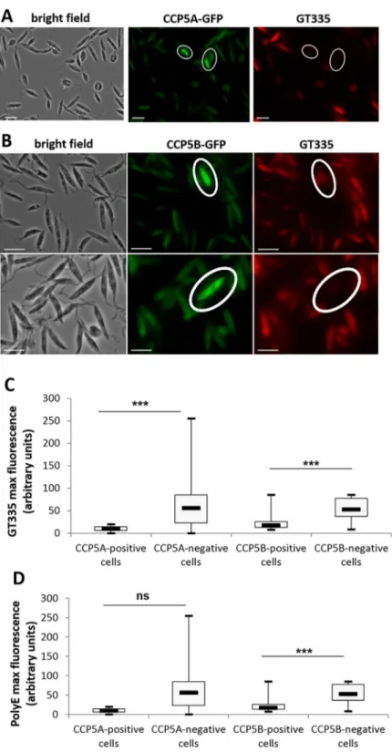
To confirm the enzymatic activity of the CCP proteins, we carried out an immunofluores-cence assay with GT335, a monoclonal antibody that recognizes all forms of polyglutamylated tubulin independently of the length of the polyglutamate side chain [34]. As previously dem-onstrated, inLeishmania, microtubules are intensely glutamylated at all stages of the cell cycle
[18]. However, cells expressing CCP5A-GFP or CCP5B-GFP (circled in white inFig 3A and 3B) were not labelled with the glutamylation specific antibody GT335 (Fig 3A and 3B, respec-tively). This deglutamylation in cells highly expressing CCP was confirmed by quantifying the maximum of GT335 fluorescence in CCP-positively and negatively stained cells: the maximum of GT335 fluorescence was significantly lower in CCP5A or CCP5B highly labelled cells in comparison to non-labelled cells (Fig 3C). To evaluate whether CCP5A and CCP5B remove one glutamate at the branching point or long side chains of glutamates, we carried out an immunofluorescence assay with PolyE, a polyclonal antibody that recognizes side chains of at Fig 2. Cellular localization of CCP5A and CCP5B. (A) Fluorescence microscopy showing localization of CCP5A
after fusion of the protein with GFP. CCP5A-GFP labeled filament-like structures in the cell body (bar = 5μm). (B) Immuno-electron microscopy of a cell expressing CCP5A-GFP. Filament-like structures were labeled with sometimes increased labeling within the cell. The lower panel is a magnification of the above square. (C) Another immuno-electron microscopy of a cell expressing CCP5A-GFP, showing strong label of a filament-like structure. (D) Localization of CCP5A-GFP after cytoskeleton extraction. The GFP-positive filament-like structure was still observed (bar = 5μm). (E) Localization of CCP5B after fusion of the protein with GFP, showing labelling of the whole cell body (bar = 5μm). (F) CCP5B-GFP fluorescence after cytoskeleton preparation. CCP5B was localized at the flagellum and at the base of the flagellum (bar = 5μm).
Fig 3. CCP5A and CCP5B are deglutamylases. (A) Immunofluorescence assay: CCP5A-GFP fluorescence in green
and GT335 immunofluorescence in red (Texas Red). GT335 is a monoclonal antibody that recognizes all forms of polyglutamylated tubulin independently of the length of the polyglutamate side chain [34]. Here we show that cells overexpressing CCP5A (circled cells) were poorly stained with GT335, indicating CCP5A deglutamylase activity (bar = 10μm). (B) Immunofluorescence assay: CCP5B-GFP fluorescence (green) and GT335 immunofluorescence (red). The deglutamylase activity of CCP5B was shown by the poor GT335 labelling of cells clearly overexpressing CCP5B (circled cells) (bar = 10μm). (C) Quantification of the maximum of GT335 fluorescence, in arbitrary units, in CCP-positively (n = 17) and negatively (n = 151 for CCP5A and n = 99 for CCP5B) labeled cells. (D) Quantification of the maximum of PolyE fluorescence, PolyE being a polyclonal antibody recognizing long glutamate side chains. t-test: ns = not significant,���: p<0.001.
least three glutamates long [35].Fig 3Dshows that the maximum of PolyE fluorescence was significantly lower in CCP5B highly labelled cells in comparison to non-labelled cells, suggest-ing that CCP5B removes glutamates at branchsuggest-ing points and also from long side chains. On the contrary, the absence of significant difference in the PolyE labelling between cells highly expressing or not CCP5A suggests that CCP5A does not remove long glutamate side chains.
We noted that the overexpression of CCP5A and CCP5B, due to the episomal expression of the corresponding protein fused to the GFP, induced a significant decrease of flagellum length (Fig 4A). Furthermore, the overexpression of CCP5A induced severe cell cycle defects with the appearance of abnormal cells, as compared to the classical dividingLeishmania forms
described by Ambitet al. [36], including about 20% of multinucleated cells apparently unable to terminate cytokinesis as exemplified by the description “cytokinesis block” inFig 4B. Such abnormal cells are shown inFig 4Cand inS3 Fig, the filament-like structures being often found in cells blocked in cytokinesis. On the contrary, overexpression of CCP5B did not induce mitotic abnormalities (Fig 4B).
Overexpression of deglutamylases inhibits regulated cell death
Overexpression of CCP5A and CCP5B, owing to the episomal expression of the recombinant GFP-CCP protein, induced significant changes in the growth curve when cells were cultivated with 40μM of miltefosine, while the growth was similar to WT cells in the absence of drug. Fig 4. Overexpression of CCP5A and CCP5B induced flagellum length decrease and cell cycle defects. (A)
Box plots representing flagellum length of WT, CCP5A overexpressing and CCP5B overexpressing cells. Minimum 40 cells were analyzed. The thick line inside each box represents the median value; the lower and upper edge of each box indicate the 25thand 75thpercentiles, respectively; the lower and upper whiskers (ends of the box arms) represent the minimum and maximum, respectively. t-test:�: p<0.05,��: p<0.01,���: p<0.001 (B) Cell cycle configuration of WT cells and cells overexpressing CCP5A or CCP5B. The lower panel corresponds to the configuration of the abnormal (other) cells. When CCP5A was overexpressed, we noted the appearance of abnormal cells, consisting essentially of cells blocked in cytokinesis (about 20% of the total population). The overexpression of CCP5B induced no obvious cell cycle defect (K = Kinetoplast; K�= Kinetoplast in replication or in G2 phase prior to segregation; N = Nucleus; F = Flagellum). (C) Fluorescence microscopy showing the localization of CCP5A as filament-like structures in a cell blocked in cytokinesis (bar = 5μm).
Indeed, CCP5A and CCP5B overexpressing cells had a significantly reduced death rate when cultivated with miltefosine (Fig 5A). This growth difference was linked to a decrease in the per-centage of TUNEL-positive cells, compared to WT cells (Fig 5B). The reduction in the percent-age of apoptotic cells when CCP were overexpressed was also observed in the presence of curcumin (Fig 5B). As a consequence, overexpression of the deglutamylases inhibited miltefo-sine and curcumin-induced RCD.
Deglutamylase genes are overexpressed during autophagy
Since autophagy is a process allowing the cell surviving nutrient depletion, that is closely linked to RCD [37], we have studied the relationships between autophagy and (de)glutamyla-tion. By carrying out RT-qPCR experiments, we observed that theccp5a and ccp5b genes were
overexpressed when the cells were cultivated in a serum-deprived medium, therefore in autop-hagic conditions: the expression of these genes was 2 to 6 times higher than expression of the Fig 5. Overexpression of CCP5A and CCP5B inhibited RCD. (A) Growth curve of WT, CCP5A and CCP5B
overexpressing cells after the addition of 40μM of miltefosine (means from a minimum of three independent experiments). The growth of the overexpressing cells was compared to the growth of the WT cells, in death conditions. (B) Percentage of dead (TUNEL-positive and anucleated) WT, CCP5A and CCP5B overexpressing cells after the addition of 40μM of miltefosine or 50 μm of curcumin: means ± sd from three independent experiments. The percentage of dead cells significantly decreased in each deglutamylase overexpressing cells in death conditions. Means± sd from a minimum of three independent experiments. Student t-tests:�: p<0.05,��: p<0.01,���: p<0.001.
control genekmp11, in autophagic conditions in comparison to normal conditions (Fig 6A). In addition, overexpression of CCP5A or CCP5B by transfection ofL. major cells with
GFP-tagged proteins induced significant growth defects when cells were cultivated in a serum-deprived medium (Fig 6B). These defects were not linked to apoptosis since no increase in the percentage of TUNEL-positive cells was observed in cells overexpressing CCP5A or CCP5B duringLeishmania autophagy (S4 Fig).
Discussion
Leishmania are unique unicellular eukaryotes. Indeed, beside their high phylogenetic distance
from other eukaryotes traditionally studied [35], they present several molecular and cellular originalities. For instance, microtubules form a corset covalently linked to the plasma mem-brane and covering the whole cell. Furthermore, the actin network appears structurally and functionally different from other eukaryotic actins [1]. Or, in terms of cell death, while an apo-ptotic phenotype has been characterized inLeishmania, the pathways remain largely unknown,
Fig 6. Deglutamylase genes are overexpressed duringL. major autophagy. (A) RT-qPCR quantification of kmp11 (Kinetoplastid Membrane Protein, used as a control),ccp5a and ccp5b mRNA expression, after culture of WT cells in a
serum-deprived medium (means± sd from a minimum of three independent experiments). We noted overexpression ofccp5a and ccp5b when cells were cultivated in a serum-deprived medium. (B) Growth curve of WT, CCP5A and
CCP5B overexpressing cells cultivated in a serum-deprived medium (means from a minimum of three independent experiments). Significant growth defects could be observed in starvation conditions when CCP5A or CCP5B when overexpressed. Student t-test:�: p<0.05,��: p<0.01,���: p<0.001.
being devoid of key mammal cell death proteins such as caspases, cell death receptors, or anti-or pro-apoptotic molecules [29]. As a consequence,Leishmania appears as a model of choice
to study eukaryotes, highlighting original processes.
DuringLeishmania cell death, important cytoskeleton modifications appear (cell rounding
up, decrease of flagellum length. . .) [36]. In order to explain these cytoskeleton modifications, we have studied PTM duringLeishmania cell death. We have shown a link between
polygluta-mylase expression and cell death inLeishmania. Indeed, during Leishmania cell death induced
by the addition of the pro-apoptotic drugs miltefosine and curcumin, polyglutamylase genes were overexpressed. Furthermore, overexpression of some polyglutamylases renders the cells more sensitive to cell death induced by miltefosine or curcumin. The overexpression of the polyglutamylases also induced cell shrinkage, a hallmark of apoptosis. Last, the importance of polyglutamylases in RCD was demonstrated by an excess of apoptosis when TTLL4C was over-expressed. We could not rule out the involvement of TTLL other than TTLL4C in RCD entry, for instance TTLL6A whose gene is highly overexpressed during miltefosine-inducedL. major
cell death. However, the presence and function of other TTLL could not be detected owing to their possible low episomal expression levels with the pTH6GFP vector used, relative to the endogenous proteins. For instance,ttll4a and ttll6b are highly expressed in Leishmania cells, as
previously shown [18,33], which could render the visualization of the consequences of the overexpression of the proteins difficult. We could also see no consequence of TTLL overex-pression owing to the necessity of concomitant overexoverex-pression of different TTLL, or to the lack of an activation step or cofactors, as already suggested [10,13]. A good example of the complexity of activation is observed with TTLL1, which in higher eukaryotes is known to be active only as part of a multiprotein complex [10].
The nature of the TTLL substrates remains to be discovered. Even if a clear polyglutamylase activity has been described for TTLL4A and TTLL6B against tubulin and also non-tubulin sub-strates, no activity has been recorded for TTLL4C and TTLL6A against tubulin and only a slight activity has been recorded against the non-tubulin substrate NAP1 [18]. However, the experimental assay used in this previous article did not include cell death conditions. Yet, over-expression and RNA interference-based knockdown of the four active polyglutamylases have no or very little effect on cell growth in normal conditions [18]. We can thus hypothesise that the polyglutamylases must be activated by pro-apoptotic drugs in order to induce excessive polyglutamylation in the cell, and so to induce RCD. We note that in this work, the overex-pression of the different genes was obtained by the episomal exoverex-pression of the gene fused to the sequence of the GFP. Therefore, we cannot rule out an effect from the GFP tag in the con-sequences of gene overexpression.
We have also identified, for the first time inLeishmania, deglutamylases, that we named
CCP5A and CCP5B for their homology with the mammal CCP5. In an original manner for CCP5 proteins [12], CCP5B seems to remove not only glutamates at branching points but also long glutamate side chains. CCP5B localized in the whole cell, a CCP5B-GFP labeling remain-ing at the flagellum and at the base of the flagellum after cytoskeleton extraction. Concernremain-ing CCP5A, its localization appeared more peculiar. Indeed, when the protein was overexpressed by the episomal expression of the GFP recombinant protein, we observed GFP-positive fila-ment-like structures still present after cytoskeleton extraction, mainly in rounded cells that seemed blocked in cytokinesis. On the contrary, when the endogene was fusedin situ with the
mNeon Green sequence, the filament-like structures were not observed, the mNeon Green labeling being distributed in the whole cell. This peculiar localization is reminiscent of the localization of actin inLeishmania. Actin, while highly abundant in Leishmania, presents
unconventional properties compared to mammal actin, among which polymerization condi-tions, different ATPase and DNase I activity or binding to phalloidin or Latrunculin B [37]. Its
in situ localization revealed that it is mainly present as granules and possibly as patches and
short filaments [1]. On the contrary, when overexpressed,Leishmania actin organizes as long
cables/bundles [38]. The similarity of localization between actin and CCP5A suggests that CCP5A deglutamylates actin, inducing the formation of high amounts of filamentous actin that organizes as bundles. To strengthen this hypothesis, we identified in the CCP5A sequence, from amino acids 488 to 494, a putative actin-binding site (SRKRHPA) similar to the one of coronin (SRFRHST), which is a protein associated with the filament-like structures of actin in
Leishmania promastigotes [39]. Actin in Trypanosomatids has been described as required in vesicular transport during endocytosis [40].
The episomal expression of CCP-GFP proteins induced the inhibition ofLeishmania
apo-ptosis induced by miltefosine and curcumin, confirming the link between deglutamylases/ polyglutamylases andLeishmania cell death. Since RCD is paradoxically closely linked to the
cell survival process autophagy [37], we studied the relationships between autophagy induced by serum deprivation and deglutamylation. We observed that the deglutamylase genesccp5a
andccp5b were highly transcribed during serum deprivation. Furthermore, the episomal
expression of CCP-GFP induced growth defects during autophagy induced by serum depriva-tion. We thus hypothesized that an autophagic stimulus would induce overexpression of CCP5A and CCP5B and that, owing to their cytoskeleton localization and to the consequences of their overexpression, CCP5A and CCP5B would deglutamylate actin but also microtubules, notably microtubules of the flagellum, and induce changes in the interaction of microtubules with microtubule-modifying proteins. This could induce loss of mobility observed during autophagy [19]. This hypothesis is consistent with the idea that tubulin deglutamylases play important roles in cilia function in higher eukaryotes [41]. A good example of this is Caenor-habditis elegans, where the tubulin deglutamylases CCPP-1 and CCPP-6 localize to cilia and
mutation inccpp-1 causes excessive accumulation of KLP-6 kinesin and polycystin-2 in cilia
and an increase in the transport rate of OSM-3/KIF17 on axonemal microtubules [11,42]. In zebrafish, expression of the deglutamylase genesccp2, ccp5 and ccp6 is strongly enriched in
cili-ated cell types [43]. Furthermore,ccp5 deficiency induces cilia microtubule
hyper-glutamyla-tion and motility defects without affecting overall cilia length [43]. A cross-talk between autophagy and cilia has also been demonstrated: signaling from the cilia can recruit the autop-hagic machinery to trigger autophagosome formation and autophagy induces ciliogenesis by controlling the level of ciliary proteins [44,45]. InL. major, this cross-talk could be linked to
the deglutamylases CCP5A and CCP5B.
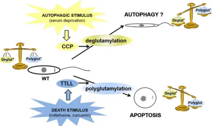
We illustrated this hypothesis in the model inFig 7where a death stimulus inLeishmania
would induce polyglutamylation, at the origin of apoptosis. On the contrary, an autophagic stimulus would induce overexpression of deglutamylases and therefore microtubule and/or other protein deglutamylation, inducing modifications mainly of the flagellum, at the origin of the autophagic phenotype and thus cell survival.
As a conclusion, through usingLeishmania, we highlighted a link between polyglutamylases
and cell death, suggesting the importance of the polyglutamylation/deglutamylation balance in the cell cycle. Imbalance would induce either apoptosis if polyglutamylation took precedence or autophagy if deglutamylation was prioritized. Even if the kinesins, microtubule-associated proteins or microtubule-severing factors interacting with microtubule modifications have to be identified in order to complete the proposed model, this work emphasized the role of PTM as essential regulators of protein function. This role has already been described, notably con-cerning microtubules, tubulin PTM having been linked to several pathologies: cilia-related dis-orders, neurodevelopmental and neurodegenerative disdis-orders, bleeding disdis-orders, cardiac diseases and cancer [41]. However, the importance of cytoskeleton modifications had not been emphasized in infectious diseases.
Supporting information
S1 Fig. Growth curves of WT cells and cells expressing the different TTLL-GFP. The cells expressing the recombinant TTLL had no growth defect in comparison to WT cells.
(TIF)
S2 Fig. Percentage of TUNEL-positive WT and polyglutamylase-overexpressing cells after the addition of 40μM of miltefosine for 24 hours. Means ± sd from three independent exper-iments. No significant difference was observed between the overexpressing and the WT cells. (TIF)
S3 Fig. Fluorescence microscopy showing, in differentL. major cells, localization of
CCP5A after fusion of the protein with GFP and localization of tubulin with an anti-α-tubulin antibody (bar = 5μm).
(TIF)
S4 Fig. Percentage of TUNEL-positive WT, CCP5A or CCP5B overexpressing cells after culture in autophagic conditions (PBS). Means± sd from minimum three independent experiments. Student t-test: ns: not significant,��: p<0.01.
(TIF)
Acknowledgments
We thank Carsten Janke from the Curie Institute (Paris-Sud 11 University) for the anti-α-tubulin antibody 12G10 and Patrick Bastien from the University of Montpellier for theL. major strain and the pTH6nGFPc and pTH6cGFPn expression vectors. We also thank
Lau-rence Berry from the University of Montpellier for very helpful discussions.
Author Contributions
Conceptualization: Magali Casanova.
Fig 7. Model: relationship between cellular deglutamylation/polyglutamylation balance, RCD and autophagy. In
the model based on the results obtained here, we suggest that a balance is established in WT cells between deglutamylation and polyglutamylation. An autophagic stimulus like serum deprivation induces overexpression of deglutamylases, inducing imbalance towards severe deglutamylation, responsible for the autophagic survival phenotype. On the contrary, a death stimulus such as miltefosine or curcumin induces severe polyglutamylation through activation of TTLL, inducing RCD.
Data curation: Louise Basmaciyan, Derrick R. Robinson, Magali Casanova. Formal analysis: Louise Basmaciyan, Derrick R. Robinson, Magali Casanova. Funding acquisition: Nadine Azas.
Investigation: Louise Basmaciyan, Magali Casanova. Methodology: Louise Basmaciyan, Magali Casanova. Project administration: Magali Casanova.
Supervision: Magali Casanova. Validation: Magali Casanova. Visualization: Magali Casanova.
Writing – original draft: Magali Casanova.
Writing – review & editing: Louise Basmaciyan, Derrick R. Robinson, Magali Casanova.
References
1. Sahasrabuddhe AA, Bajpai VK, Gupta CM. A novel form of actin in Leishmania: molecular characterisa-tion, subcellular localisation and association with subpellicular microtubules. Mol Biochem Parasitol. 2004; 134:105–14. PMID:14747148
2. Janke C, Bulinski JC. Post-translational regulation of the microtubule cytoskeleton: mechanisms and functions. Nat Rev Mol Cell Biol. 2011; 12:773–86.https://doi.org/10.1038/nrm3227PMID:22086369
3. Wloga D, Gaertig J. Post-translational modifications of microtubules. J Cell Sci. 2010; 123:3447–55.
https://doi.org/10.1242/jcs.063727PMID:20930140
4. Verhey KJ, Gaertig J. The tubulin code. Cell Cycle Georget Tex. 2007; 6:2152–60.
5. Janke C, Rogowski K, van Dijk J. Polyglutamylation: a fine-regulator of protein function? « Protein Modi-fications: beyond the usual suspects » review series. EMBO Rep. 2008; 9:636–41.https://doi.org/10. 1038/embor.2008.114PMID:18566597
6. Bonnet C, Boucher D, Lazereg S, Pedrotti B, Islam K, Denoulet P, et al. Differential binding regulation of microtubule-associated proteins MAP1A, MAP1B, and MAP2 by tubulin polyglutamylation. J Biol Chem. 2001; 276:12839–48.https://doi.org/10.1074/jbc.M011380200PMID:11278895
7. Boucher D, Larcher JC, Gros F, Denoulet P. Polyglutamylation of tubulin as a progressive regulator of in vitro interactions between the microtubule-associated protein Tau and tubulin. Biochemistry (Mosc). 1994; 33:12471–7.
8. Lacroix B, van Dijk J, Gold ND, Guizetti J, Aldrian-Herrada G, Rogowski K, et al. Tubulin polyglutamyla-tion stimulates spastin-mediated microtubule severing. J Cell Biol. 2010; 189:945–54.https://doi.org/ 10.1083/jcb.201001024PMID:20530212
9. Larcher JC, Boucher D, Lazereg S, Gros F, Denoulet P. Interaction of kinesin motor domains with alpha- and beta-tubulin subunits at a tau-independent binding site. Regulation by polyglutamylation. J Biol Chem. 1996; 271:22117–24. PMID:8703022
10. Janke C, Rogowski K, Wloga D, Regnard C, Kajava AV, Strub J-M, et al. Tubulin polyglutamylase enzymes are members of the TTL domain protein family. Science. 2005; 308:1758–62.https://doi.org/ 10.1126/science.1113010PMID:15890843
11. Kimura Y, Kurabe N, Ikegami K, Tsutsumi K, Konishi Y, Kaplan OI, et al. Identification of tubulin degluta-mylase among Caenorhabditis elegans and mammalian cytosolic carboxypeptidases (CCPs). J Biol Chem. 2010; 285:22936–41.https://doi.org/10.1074/jbc.C110.128280PMID:20519502
12. Rogowski K, van Dijk J, Magiera MM, Bosc C, Deloulme J-C, Bosson A, et al. A family of protein-deglu-tamylating enzymes associated with neurodegeneration. Cell. 2010; 143:564–78.https://doi.org/10. 1016/j.cell.2010.10.014PMID:21074048
13. van Dijk J, Rogowski K, Miro J, Lacroix B, Edde´ B, Janke C. A targeted multienzyme mechanism for selective microtubule polyglutamylation. Mol Cell. 2007; 26:437–48.https://doi.org/10.1016/j.molcel. 2007.04.012PMID:17499049
14. van Dijk J, Miro J, Strub J-M, Lacroix B, van Dorsselaer A, Edde B, et al. Polyglutamylation Is a Post-translational Modification with a Broad Range of Substrates. J Biol Chem. 2008; 283:3915–22.https:// doi.org/10.1074/jbc.M705813200PMID:18045879
15. Gull K. The cytoskeleton of trypanosomatid parasites. Annu Rev Microbiol. 1999; 53:629–55.https:// doi.org/10.1146/annurev.micro.53.1.629PMID:10547703
16. Robinson DR, Sherwin T, Ploubidou A, Byard EH, Gull K. Microtubule polarity and dynamics in the con-trol of organelle positioning, segregation, and cytokinesis in the trypanosome cell cycle. J Cell Biol. 1995; 128:1163–72. PMID:7896879
17. Portman N, Gull K. Proteomics and the Trypanosoma brucei cytoskeleton: advances and opportunities. Parasitology. 2012; 139:1168–77.https://doi.org/10.1017/S0031182012000443PMID:22475638
18. Casanova M, de Monbrison F, van Dijk J, Janke C, Pagès M, Bastien P. Characterisation of polygluta-mylases in trypanosomatids. Int J Parasitol. 2015; 45:121–32.https://doi.org/10.1016/j.ijpara.2014.09. 005PMID:25444861
19. Basmaciyan L, Berry L, Gros J, Azas N, Casanova M. Temporal analysis of the autophagic and apopto-tic phenotypes in Leishmania parasites. Microb Cell Graz Austria. 2018; 5:404–17.
20. Das M, Mukherjee SB, Shaha C. Hydrogen peroxide induces apoptosis-like death in Leishmania dono-vani promastigotes. J Cell Sci. 2001; 114:2461–9. PMID:11559754
21. Das R, Roy A, Dutta N, Majumder HK. Reactive oxygen species and imbalance of calcium homeostasis contributes to curcumin induced programmed cell death in Leishmania donovani. Apoptosis Int J Pro-gram Cell Death. 2008; 13:867–82.
22. Holzmuller P, Sereno D, Cavaleyra M, Mangot I, Daulouede S, Vincendeau P, et al. Nitric oxide-medi-ated proteasome-dependent oligonucleosomal DNA fragmentation in Leishmania amazonensis amasti-gotes. Infect Immun. 2002; 70:3727–35.https://doi.org/10.1128/IAI.70.7.3727-3735.2002PMID:
12065515
23. Lee N, Bertholet S, Debrabant A, Muller J, Duncan R, Nakhasi HL. Programmed cell death in the unicel-lular protozoan parasite Leishmania. Cell Death Differ. 2002; 9:53–64.https://doi.org/10.1038/sj.cdd. 4400952PMID:11803374
24. Mukherjee SB, Das M, Sudhandiran G, Shaha C. Increase in cytosolic Ca2+ levels through the activa-tion of non-selective caactiva-tion channels induced by oxidative stress causes mitochondrial depolarizaactiva-tion leading to apoptosis-like death in Leishmania donovani promastigotes. J Biol Chem. 2002; 277:24717– 27.https://doi.org/10.1074/jbc.M201961200PMID:11983701
25. Paris C, Loiseau PM, Bories C, Bre´ard J. Miltefosine induces apoptosis-like death in Leishmania dono-vani promastigotes. Antimicrob Agents Chemother. 2004; 48:852–9.https://doi.org/10.1128/AAC.48.3. 852-859.2004PMID:14982775
26. Kroemer G, Galluzzi L, Vandenabeele P, Abrams J, Alnemri ES, Baehrecke EH, et al. Classification of cell death: recommendations of the Nomenclature Committee on Cell Death 2009. Cell Death Differ. 2009; 16:3–11.https://doi.org/10.1038/cdd.2008.150PMID:18846107
27. Gannavaram S, Debrabant A. Programmed cell death in Leishmania: biochemical evidence and role in parasite infectivity. Front Cell Infect Microbiol. 2012; 2:95.https://doi.org/10.3389/fcimb.2012.00095
PMID:22919685
28. Shaha C. Apoptosis in Leishmania species & its relevance to disease pathogenesis. Indian J Med Res. 2006; 123:233–44. PMID:16778307
29. Proto WR, Coombs GH, Mottram JC. Cell death in parasitic protozoa: regulated or incidental? Nat Rev Microbiol. 2013; 11:58–66.https://doi.org/10.1038/nrmicro2929PMID:23202528
30. Genes CM, de Lucio H, Sa´nchez-Murcia PA, Gago F, Jime´ nez-Ruiz A. Pro-death activity of a BH3 domain in an aquaporin from the protozoan parasite Leishmania. Cell Death Dis. 2016; 7:e2318– e2318.https://doi.org/10.1038/cddis.2016.229PMID:27468694
31. Dubessay P, Blaineau C, Bastien P, Tasse L, Van Dijk J, Crobu L, et al. Cell cycle-dependent expres-sion regulation by the proteasome pathway and characterization of the nuclear targeting signal of a Leishmania major Kin-13 kinesin. Mol Microbiol. 2006; 59:1162–74. https://doi.org/10.1111/j.1365-2958.2005.05013.xPMID:16430691
32. Koide T, Nose M, Ogihara Y, Yabu Y, Ohta N. Leishmanicidal effect of curcumin in vitro. Biol Pharm Bull. 2002; 25:131–3. PMID:11824543
33. Rastrojo A, Carrasco-Ramiro F, Martı´n D, Crespillo A, Reguera RM, Aguado B, et al. The transcriptome of Leishmania major in the axenic promastigote stage: transcript annotation and relative expression lev-els by RNA-seq. BMC Genomics. 2013; 14:223.https://doi.org/10.1186/1471-2164-14-223PMID:
23557257
34. Wolff A, de Ne´chaud B, Chillet D, Mazarguil H, Desbruyères E, Audebert S, et al. Distribution of gluta-mylated alpha and beta-tubulin in mouse tissues using a specific monoclonal antibody, GT335. Eur J Cell Biol. 1992; 59:425–32. PMID:1493808
35. Rogowski K, Juge F, van Dijk J, Wloga D, Strub J-M, Levilliers N, et al. Evolutionary Divergence of Enzymatic Mechanisms for Posttranslational Polyglycylation. Cell. 2009; 137:1076–87.https://doi.org/ 10.1016/j.cell.2009.05.020PMID:19524510
36. Ambit A, Woods KL, Cull B, Coombs GH, Mottram JC. Morphological Events during the Cell Cycle of Leishmania major. Eukaryot Cell. 2011; 10:1429–38.https://doi.org/10.1128/EC.05118-11PMID:
21926331
37. Mariño G, Niso-Santano M, Baehrecke EH, Kroemer G. Self-consumption: the interplay of autophagy and apoptosis. Nat Rev Mol Cell Biol. 2014; 15:81–94.https://doi.org/10.1038/nrm3735PMID:
24401948
38. Kapoor P, Sahasrabuddhe AA, Kumar A, Mitra K, Siddiqi MI, Gupta CM. An Unconventional Form of Actin in Protozoan Hemoflagellate, Leishmania. J Biol Chem. 2008; 283:22760–73.https://doi.org/10. 1074/jbc.M800213200PMID:18539603
39. Nayak RC, Sahasrabuddhe AA, Bajpai VK, Gupta CM. A novel homologue of coronin colocalizes with actin in filament-like structures in Leishmania. Mol Biochem Parasitol. 2005; 143:152–64.https://doi. org/10.1016/j.molbiopara.2005.06.001PMID:16024104
40. Garcı´a-Salcedo JA, Pe´rez-Morga D, Gijo´n P, Dilbeck V, Pays E, Nolan DP. A differential role for actin during the life cycle of Trypanosoma brucei. EMBO J. 2004; 23:780–9.https://doi.org/10.1038/sj.emboj. 7600094PMID:14963487
41. Magiera MM, Singh P, Gadadhar S, Janke C. Tubulin Posttranslational Modifications and Emerging Links to Human Disease. Cell. 2018; 173:1323–7.https://doi.org/10.1016/j.cell.2018.05.018PMID:
29856952
42. O’Hagan R, Piasecki BP, Silva M, Phirke P, Nguyen KCQ, Hall DH, et al. The tubulin deglutamylase CCPP-1 regulates the function and stability of sensory cilia in C. elegans. Curr Biol CB. 2011; 21:1685– 94.https://doi.org/10.1016/j.cub.2011.08.049PMID:21982591
43. Pathak N, Austin-Tse CA, Liu Y, Vasilyev A, Drummond IA. Cytoplasmic carboxypeptidase 5 regulates tubulin glutamylation and zebrafish cilia formation and function. Mol Biol Cell. 2014; 25:1836–44.
https://doi.org/10.1091/mbc.E13-01-0033PMID:24743595
44. Lam HC, Cloonan SM, Bhashyam AR, Haspel JA, Singh A, Sathirapongsasuti JF, et al. Histone deace-tylase 6-mediated selective autophagy regulates COPD-associated cilia dysfunction. J Clin Invest. 2013; 123:5212–30.https://doi.org/10.1172/JCI69636PMID:24200693
45. Orhon I, Dupont N, Pampliega O, Cuervo AM, Codogno P. Autophagy and regulation of cilia function and assembly. Cell Death Differ. 2015; 22:389–97.https://doi.org/10.1038/cdd.2014.171PMID: