HAL Id: tel-03259625
https://tel.archives-ouvertes.fr/tel-03259625
Submitted on 14 Jun 2021HAL is a multi-disciplinary open access
archive for the deposit and dissemination of sci-entific research documents, whether they are pub-lished or not. The documents may come from teaching and research institutions in France or abroad, or from public or private research centers.
L’archive ouverte pluridisciplinaire HAL, est destinée au dépôt et à la diffusion de documents scientifiques de niveau recherche, publiés ou non, émanant des établissements d’enseignement et de recherche français ou étrangers, des laboratoires publics ou privés.
Unveiling the role of Rhizaria in the silicon cycle
Natalia Llopis Monferrer
To cite this version:
Natalia Llopis Monferrer. Unveiling the role of Rhizaria in the silicon cycle. Other. Université de Bretagne occidentale - Brest, 2020. English. �NNT : 2020BRES0041�. �tel-03259625�
L'UNIVERSITE
DE
BRETAGNE
OCCIDENTALE
ECOLE DOCTORALE N°598
Sciences de la Mer et du littoral Spécialité : Chimie Marine
Unveiling the role of Rhizaria in the silicon cycle
(Rôle des Rhizaria dans le cycle du silicium)
Thèse présentée et soutenue à PLouzané, le 18 septembre 2020
Unité de recherche : Laboratoire de Sciences de l’Environnement Marin
Par
Natalia LLOPIS MONFERRER
Rapporteurs avant soutenance :
Diana VARELA Professor, Université de Victoria, Canada
Giuseppe CORTESE Senior Scientist, GNS Science, Nouvelle Zélande Composition du Jury :
Président : Géraldine SARTHOU Directrice de recherche, CNRS, LEMAR, Brest, France Examinateurs : Diana VARELA Professor, Université de Victoria, Canada
Giuseppe CORTESE Senior Scientist, GNS Science, Nouvelle Zélande
Colleen DURKIN Research Faculty, Moss Landing Marine Laboratories, Etats Unis Tristan BIARD Maître de Conférences, Université du Littoral Côte d’Opale, France Dir. de thèse : Paul TREGUER Professeur des Universités, Université de Bretagne Occidentale, France Co-dir. de thèse : Aude LEYNAERT Directrice de recherche, CNRS, LEMAR, Brest, France
Co-dir. de thèse : Fabrice NOT Directeur de recherche, CNRS, Station Biologique de Roscoff, France
Invité(s)
iv
Unveiling the role of Rhizaria in
the Silicon cycle
v
vi
vii
ix
I would like to thank l’Université de Bretagne Occidentale and the LabexMer for funding these three years of PhD. I also thank Luis Tito de Morais, director of the Laboratoire des Sciences de l’environnement marin for hosting me in his laboratory.
I would like to extend my gratitude to the members of my committee, Diana Varela, Giuseppe Cortese, Colleen Durkin, Tristan Biard and Jill Sutton for accepting to evaluate my PhD thesis as well as the inclement timing of the presentation.
I would like to warmly thank all my supervisors, Paul, Aude et Fabrice. Merci beaucoup Aude de m’avoir fait confiance depuis notre premier Skype, quand je ne connaissais pas grand-chose sur les Rhizaria ni la silice. Merci d’avoir été là dès j’avais des questions, scientifiques et pas scientifiques. Merci pour ta bonne humeur sans limite, même quand j’oubliais les unités sur mes fichiers. Ça été un plaisir de partir à l’aventure avec toi et d’unifier les fabuleux mondes des Rhizaria et de la silice. Merci Paul de m’avoir ouvert les portes de l’univers de la silice et pour votre insatiable soif de connaissances, je n’aurais pas pu espérer meilleur mentor. Merci également d’avoir enrichi mon dictionnaire d’expressions françaises et brestoises. Fabrice, malgré l’encadrement à distance, je me suis toujours sentie soutenue. Ton esprit scientifique critique a vraiment marqué une différence dans mes travaux, bref, merci de m’avoir introduit au monde magnifique des Rhizaria. Merci à tous de m’avoir donné l’opportunité de voyager et découvrir des Rhizaria autour du globe. Je me rappellerai grâce à vous avec émotion de la traversée de l’équateur et des manchots de la mer de Ross.
Quería agradecer a Demetrio Boltovskoy toda su ayuda y paciencia y porque ha sido una gran fuente de inspiración. Merci Tristan pour ton aide et tes retours, grâce à toi, le monde de l’imagerie n’a (presque) plus de secrets pour moi. Mais je crois que je vais encore avoir besoin de ton aide pendant un petit moment.
Je voulais remercier le LEMAR et toutes les personnes que j’ai pu côtoyer quotidiennement pendant ces trois années. Je veux notamment remercier Morgane Gallinari, d’avoir égayé mes heures au laboratoire, qui auraient été bien moins sympas sans elle. Merci Manon pour l’aide avec les analyses et les petits footings du midi (il va falloir les reprendre). Merci beaucoup Jill pour tes conseils et ton soutien. Merci à Gene, Anne-So, Yves, Natalie, Elodie2, Elisabeth pour
x
l’aide avec la paperasse. Muchas gracias Bea, por ser tan linda y haberme enseñado la belleza del plancton.
Merci aux roskovites et l’équipe ECOMAP. Merci à Éric Thiébaut pour son aide en stats, Christian Jeanthon, Florence, Pris, Fabienne, Joost, Mathilde, Miguel, Morgane et plein d’autres !
I cannot forget to thank people with who I shared valuable time at sea. Many thanks to Chata, Hans, Andreia, Afonso, Cecilia, Carol, Wade, John, Giorgio, Igor, Glen, Karl, Mark, Pablo Escobar (yes, I can say I’ve been on a cruise with him!), Magali and so many other people I had the pleasure to sail with. Muchas gracias a Miguel, quien ha compartido todo, desde sus mejores fotos de radiolarios hasta las mejores pistas para diferenciar un spumellaria de una diatomea. Muchas gracias a Andrés por haber confiado en mí y por haberme ayudado a subirme, junto con mis cajas en el Tangaroa. Et bien sûr Briva, c’était un vrai plaisir de partager une belle campagne avec toi, sans oublier (même si ça peut arriver d’être tête en l’air), toutes les heures passées à sucer des radiolaires.
J’ai fait mes premiers pas d’océanographe en herbe à Villefranche-sur-Mer grâce à une très belle équipe. Merci Lars, Lionel, Fabien, Marc, Amanda, Baptiste, Corinne et Simon, qui m’ont aidé il y déjà quelques années et qui ont continué à m’aider durant ma thèse.
A huge thanks to my office mates, Hannah, Natasha, David, Marie, Debany, Manu, Wen-Hsuan and María to have shared all those wonderful times everywhere and always. Thanks for the patience, for correcting my English and listening to my scientific dramas, you are the best.
Je ne peux pas oublier mes amis doctorants. Chloé et Nico qui m’ont montré que coder peut être facile (pour eux), Jordon, qui m’a aidé à organiser mes campagnes et à renforcer mon psoas. Merci à tous les autres, Elyne, Pauline, Sarah, Julien, Justine2, Leslie, Gabin, Mariana,
Romina, José, Will, Aurélien, Anaïs, Kevin et un long etcétéra pour les bons moments de rigolade.
Muchas gracias a mis amigos, que a pesar de estar lejos, los siento siempre cerca. Palo y Lidia por seguirme hasta al polo norte si hace falta. Auro, por tus consejos, echo de menos las charlas después de la piscina. A Mau, Álvaro, Candel y todos los de la playa, por seguir siendo como sois, no cambiéis nunca. A todos los saguntinos, pero sobre todo a Paloma, por ser siempre la más luchadora, un verdadero ejemplo a seguir. No me puedo olvidar de mis
xi
Merci à Marc, pour toute la patience. Gracias por haber convertido el confinamiento y el final de la tesis en momentos inolvidables, el mejor compañero de viaje.
Por supuesto no me olvido de mi familia (la de Llopis y la de Monferrer). Si estoy aquí es gracias a vosotros, que siempre me habéis apoyado, desde el primer momento en que decidí embarcarme en esta aventura de oceanógrafa. Al tete, por aguantarme cada vez que le pido un dibujo o cualquier otra cosa que se le pueda ocurrir a mi cabecita. A Miguel, por haber conseguido sin saberlo, hacer de mi la tía más feliz del mundo.
Enfin, je voulais remercier toutes les personnes que je n’ai pas cité précédemment mais qui m’ont aidé de près ou de loin dans cette aventure.
xiii
Les radiolaires polycystines et les phaeodaires sont des organismes unicellulaires eucaryotes (i.e., protistes) hétérotrophes marins appartenant au super-groupe des Rhizaria. Ces protistes, qui présentent des tailles allant de quelques micromètres à quelques millimètres, sont présents à la surface et dans les profondeurs de l’ensemble des océans. De nombreux Rhizaria utilisent le silicium dissous dans l’eau de mer pour construire un squelette de silice. Les squelettes robustes des Rhizaria préservés dans les sédiments sont particulièrement précieux pour les micro-paléontologues pour la reconstitution des paléo-environnements. Bien qu’ils soient largement étudiés par les micro-paléontologues, l‘écologie et la contribution des Rhizaria au fonctionnement des écosystèmes marins actuels sont demeurés jusqu’à présent largement inexplorés.
La première partie de cette thèse est dédiée à la mesure des taux d'absorption du silicium par les Rhizaria à l'aide du radio-isotope 32Si et à la mesure de la composition élémentaire (C ; N ; Si)
d’organismes appartenant à plusieurs groupes taxinomiques. Les différents spécimens ont été collectés en 2017, 2018 et 2019, lors de trois campagnes océanographiques (MOOSE-GE 17, AMT28, TAN1901) en mer Méditerranée, océan Atlantique et mer de Ross. Nous avons observé une production de silice biogène par cellule très élevée chez les Rhizaria (jusqu'à 11.9 nmol de Si cellule-1j-1) en
comparaison avec les diatomées (0.001-21 pmol-Si cellule-1j-1). Avec des teneurs en silicium allant
jusqu'à 9 nmol-Si cellule-1, ces organismes apparaissent parmi les plus silicifiés des organismes
planctoniques. Une première évaluation de leur contribution au cycle biogéochimique de la silice dans l’océan mondial a été réalisée en combinant les résultats de nos expériences avec des données d’abondance publiées précédemment. Les Rhizaria pourraient contribuer de 4 à 22% à la production de silice biogène de l’ensemble des océans.
L’abondance, la biomasse et la diversité des Rhizaria ont été quantifiées dans le bassin nord-ouest de la Mer Méditerranée en combinant simultanément des techniques innovantes d’imagerie (FlowCAM, Zooscan et UVP) et des techniques metabarcode afin de couvrir la large gamme de taille de ces organismes. Cette approche a révélé que les petites cellules sont les plus nombreuses mais les grosses cellules constituent l’essentiel de la biomasse. L’ensemble représente jusqu’à 6% de la silice biogène des 500 premiers mètres de la colonne d’eau en Méditerranée.
Cette thèse fournit des données quantitatives uniques mettant en évidence le rôle fondamental des Rhizaria siliceux dans les océans contemporains. Elle souligne également la nécessité d'explorer davantage les profondeurs de l'océan pour affiner notre première estimation de la contribution des Rhizaria au cycle biogéochimique du Si à l'échelle locale et mondiale.
xiv
Abstract
Polycystine radiolaria and phaeodarians are unicellular eukaryotic (i.e., protist) heterotrophic marine organisms belonging to the Rhizaria supergroup. These protists, which range in size from a few micrometers to a few millimeters, are present from the surface to the bathypelagic waters of all oceans. Many Rhizaria use dissolved silicic acid in seawater to build a silica skeleton. The robust skeletons of Rhizaria preserved in sediments are particularly valuable for paleoceanographic reconstructions. Although widely studied by micro-paleontologists, the ecology and contribution of Rhizaria to the functioning of contemporary marine ecosystems has remained largely unexplored so far.
The first part of this thesis is dedicated to the measurement of silicic acid uptake rates by Rhizaria using the 32Si radioisotope and the elementary composition (C; N; Si) of organisms belonging to several
taxonomic groups. The different specimens were collected in 2017, 2018 and 2019 during three oceanographic expeditions (MOOSE-GE 17, AMT28, TAN1901) in the Mediterranean Sea, Atlantic Ocean and Ross Sea. We observed a very high production of biogenic silica per individual in Rhizaria (up to 11.9 nmol-Si cell-1d-1) compared to diatoms. With biogenic silica cellular contents up to 9
nmol-Si cell-1, Rhizaria appear among the most silicified of planktonic organisms. A first assessment of their
contribution to the biogeochemical cycling of silica in the world ocean has been made by combining the results of our experiments with previously published abundance data. Rhizaria could contribute between 4 and 22% to the biogenic silica production of the global ocean.
The abundance, biomass and diversity of Rhizaria have been quantified in the north-western basin of the Mediterranean Sea by simultaneously combining innovative imaging techniques (FlowCAM, Zooscan and UVP) and metabarcode techniques to cover the wide size range of these organisms. This approach has revealed that small cells are the most numerous but large cells constitute the bulk of the biomass. Together they account for up to 6% of the biogenic silica in the first 500 metres of the Mediterranean water column. This work provides unique quantitative data that highlight the impact of Rhizaria in the cycling of Si in contemporary oceans. This thesis also reveals the need to explore further the deep ocean to refine our first estimates of Rhizaria contribution to the Si biogeochemical cycle at local and global scales.
xvi
Contents
Introduction 1
1. Silica and the marine silicon cycle 3
2. Rhizaria, conspicuous organisms in the ocean 4
PhD Objectives 14
Chapter 1 – Estimating biogenic silica in the global ocean 15
Introduction 20
Material and Methods 22
Results 26
Discussion 29
Chapter 2 – Biogenic silica production of Rhizaria in contrasting environments 39
Chapter 2.1. – Rhizaria in the Southern Ocean 43
Introduction 44
Material and methods 45
Results 51
Discussion 59
Chapter 2.2 – Rhizaria in the Atlantic Ocean 67 Chapter 3 – Merging imaging technologies and metabarcoding to characterize the Rhizaria community 71
Introduction 76
Material and Methods 78
Results 82
Discussion 89
Discussion and perspectives 97
Bibliography 109
Annexes 119
AMT28 Cruise – Preliminary results 121
Oral and poster presentations 125
Oceanographic cruises 125
Others 126
3
1. Silica and the marine silicon cycle
Silicon (Si) is the second most abundant element in Earth’s crust. Silicon atoms bond with oxygen (O) atoms to create silicon dioxide (SiO2), also known as silica, which may be either
crystalline or amorphous. This element cycles through the marine environment (Figure 1), entering the ocean primarily from rivers. Other secondary sources include submarine groundwater, erosion of marine soils, hydrothermal vents and aeolian inputs. In the ocean, Si is essentially found as dissolved silicic acid (dSi), which is required for the growth of some phytoplankton groups like diatoms and other silicified planktons, such as radiolarians and phaeodarians, sponges, silicoflagellates and several species of choanoflagellates. These organisms, commonly called “silicifiers”, remove dSi from the ocean to build their cell tests. After they die, their test acts as ballast, thus causing themto sink toward the ocean floor. While sinking, most of the Si recycles throughout the water column, being again available as dSi for other silicifiers. The fraction of the cells that resists dissolution reaches the sea-floor, where they can either remain, forming a siliceous ooze, or dissolve and return to the upper layers of the ocean through upwelling processes.
| Introduction
4
Figure 1. Cycling of silica in the marine environment. Adapted from Tréguer and De La Rocha (2013).
The Si cycle is intimately connected to other biogeochemical cycles, like those of carbon and nitrogen. These cycles interact via marine primary production, which drives atmospheric CO2
sequestration to the deep ocean via the biological pump and, ultimately, exerts control over the Earth’s climate (e.g., Buitenhuis et al., 2006; Reynolds, 2001).
Among organisms that require Si to grow, diatoms have been the main focus so far. The production of bSi in the oceans by diatoms has been estimated to 240 Tmol Si yr-1 (Nelson et
al., 1995). Diatoms largely dominates all other biogeochemical fluxes in the Si cycle (Tréguer and De La Rocha, 2013). However, the contribution of other silicifiers, like sponges and Rhizaria, is poorly documented.
Since the late 20th century, silicified rhizarians have drawn attention for their role in the
marine Si cycle (Heath, 1974). Takahashi (1983) suggested that, in some oceanic areas, the daily flux of rhizarian biogenic silica (bSi) ranges around 20% to 30% of the overall bSi.
Recent studies combining genomic and in situ imaging data have evidenced that densities of siliceous Rhizaria, have so far been underestimated. Biard et al. (2016) reported that some Rhizaria taxa represent approximately 33% of the large zooplankton (>600 µm) in the upper water column. Guidi et al. (2016) pointed out Rhizaria’s significant involvement in the export of C to the deep ocean, with their abundances correlating with export C fluxes at 150-m depth in oligotrophic oceanic regions. These studies have raised awareness of the global significance of Rhizaria in the biological pump, as well as in the Si cycle.
2. Rhizaria, conspicuous organisms in the ocean
Oceanic Rhizaria are a very diverse microplankton group that have existed at least since the Cambrian era (~500 million years ago). This group of protists is a major lineage of eukaryotes, including Cercozoa and Retaria (Cavalier-Smith, 2002), with the latter grouping Radiolaria and Foraminifera (Figure 2).
5
Figure 2. Schematic of the eukaryotic tree based on a consensus of phylogenomic together with morphological
and cell biological information. SAR is the conglomerate of Stramenopiles, Alveolates, and Rhizaria, which together make an assemblage encompassing perhaps half of all eukaryote diversity. CRuMS is an amalgamation of several ‘orphan’ taxa: the Collodictyonids, Rigifilida, and Mantamonas. Adapted from Keeling and Burki (2019). Essentially unicellular though some are capable of forming colonies up to over 1 m in length, Retaria span a wide range of sizes, from tens to hundreds of micrometers (Boltovskoy et al., 2017).
Some species of Retaria have elaborate mineral skeletons of strontium sulfate (Acantharia), calcium carbonate (Foraminifera) and opaline silica (Nassellaria, Spumellaria and Phaeodaria). Radiolaria are divided into two major lineages: the Polycystinea (including the three orders: Nassellaria, Spumellaria and Collodaria) and the Spasmaria (including Acantharia and
| Introduction
6
Taxopodida; Krabberød et al., 2011). Phaeodaria, initially classified in Radiolaria, is now placed among the Cercozoa as revealed by molecular phylogeny (Polet, 2004).
The work performed during this thesis focuses on the silicifying Rhizaria (polycystine Radiolaria and Phaeodaria). The study of these planktonic protists has been essentially used for paleoceanographic reconstructions, based on the fossil record left by their silicified skeletons in oceanic sediments (Matsuzaki et al., 2014; Moore, 1978). Almost all polycystine species preserve well in the bottom sediments while most Phaeodaria skeletons dissolve more readily before reaching the sea-floor (Takahashi, 1983).
Despite their potential role as Si consumers and C exporters (e.g., Lampitt et al., 2009; Takahashi, 1983) in the marine environment, very little is known about their physiology, life cycle and ecology, particularly regarding silica. This is essentially due to the difficulty of culturing Rhizaria, as well as the delicate morphologies of living specimens which make them liable to collapse when using conventional sampling methods.
2.1. Cell structure
Polycystine radiolarian and Phaeodaria skeletal morphology can be very variable depending on the taxa considered (Figure 3). Generally, their ornamented skeletons surround a central and porous capsule from which pseudopodia (long and slender cytoplasmic projections) radiate.
Skeletons in protists are believed to play a role in essential functions. For instance, for the microalgae diatoms it has been proven that the skeleton provide mechanical protection for the cell against predators (Finkel and Kotrc, 2010; Hamm et al., 2003), as well as an effective pH buffer (Milligan, 2002). Less is known about the role of the Rhizaria’s skeletons, but it is likely to improve the uptake or storage of bio-essential elements (Suzuki and Not, 2015).
7
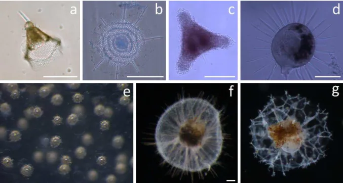
Figure 3. Representative examples of taxonomic groups analyzed in this thesis (scale bar when available 100 µm).
(a) Nassellarian of the family Plagiacanthidae, (b) Spumellarian of the superfamily Stylodictytoidea, (c) Spumellarian of the superfamily Spongodiscidae, (d) Phaeodarian of the family Challengeridae (Challengeron sp.), (e) Detail of central capsule of a colonie of Collodarian, family Sphaerozoidea, (f) Phaeodarian of the family Aulacanthidae (Aulacantha scolymanta), (g) Phaeodarian of the family Coelodendridae (Coelechinus sp.). Pictures N. Llopis Monferrer except picture (e), taken by A. Leynaert.
Polycystine Radiolaria. Most Nassellaria, Spumellaria and siliceous Collodaria possess
solid and dense skeletons of amorphous silica, so-called opal (SiO2) (Figure 4).
Nassellaria present a heteropolar skeleton with one or more sections aligned along an axis. From the narrower end, the cephalis, which is the first to be formed, several segments succeed: cephalis, thorax and abdomen (Boltovskoy et al., 2017). Skeletons vary from simple tripods to elaborate, helmet-shaped structure, often with spines or other ornamentations (Boltovskoy et al., 1990).
Most Spumellaria have radial or spherical symmetry with centrifugal shell-growth, but their skeleton can also be flat or elliptical (Suzuki and Not, 2015). The nucleus is located in the central capsule and it is surrounded by radially arranged lobes of cytoplasm, enclosed by a porous capsular wall.
Collodaria is the only taxon with colonial representatives, which can be composed of tens to thousand cells. Each colony consists of a spheroidal, hollow, gelatinous envelope containing numerous interconnected cells embedded within a gelatinous matrix (Anderson et al., 1987). Cells can be either naked (e.g., Collozoum sp.), or surrounded by a porous, spherical Si skeleton
| Introduction
8
(e.g., Collosphaera globularis) or provided with siliceous spines embedded in the cytoplasm (e.g., Sphaerozoum sp.).
Phaeodaria. Phaeodarians are often larger than polycystine individuals. Their cell size
ranges from several hundreds of micrometres up to several millimetres. Phaeodaria’s skeleton is also composed of amorphous silica but is more porous and less solid than polycystine’s skeletons (Nakamura et al., 2018). The porosity of their skeleton and the organic matter content in their test is presumably responsible for the poor preservation of these organisms in the fossil record (Takahashi et al., 1983). As for Radiolaria, their geometry is complex and varies among families. This group is mainly defined by a central capsule containing the phaeodium, a mass of partially digested food, generally darkly coloured that emanates from the astropyle, which resembles an oral aperture (Figure 4). Phaeodarians possess a double-walled central capsule and the skeletal network can be surrounded by filopods.
9
Figure 4. Scheme of the skeletal elements of the shell of a typical Nassellaria, Spumellaria and Phaeodaria
(Adapted from Boltovskoy et al., 2017). The right-hand scheme refers to the soft parts compared to the left-hand one, which refers to the skeleton alone.
2.2. General biology and ecology
Place in the trophic network. Despite the ubiquity of these organisms in the oceans,
fundamental information about their feeding behaviour is poor. These protists are mainly heterotrophic, as they can capture preys through adhesion to their pseudopodia.
Polycystine consume a wide variety of prey, from bacteria and algae up to small invertebrates (Gowing and Coale, 1989). Beside the heterotrophic behaviour, many polycystine inhabiting surface waters, exhibit symbiotic microalgae providing nutrients to the host (Decelle et al., 2015). Phaeodarians are omnivorous, they can feed on other plankton or on organic suspended matter in the water column (Gowing, 1986). Unlike many polycystine, no Phaeodaria have been reported harbouring microalgal symbiont so far.
Very little is known about the predatory pressure on Rhizaria, although there are evidences of other plankton such as Foraminifera, salps and small crustaceans preying on Rhizaria (Gowing 1989; Swanberg, 1979).
Reproduction. Rhizaria are difficult to keep alive in cultures and our knowledge about
their life cycle is incomplete (Suzuki and Not, 2015).
For several species of Nassellaria, Spumellaria and Collodaria it has been observed that after organisms have turned whitish, they release small bi-flagellated cells generally called swarmers (Suzuki and Not, 2015).
| Introduction
10
For Collodaria, binary fission within the central capsule has been reported as well as swarmer production (Anderson and Gupta, 1998; Biard et al., 2015).
Phaeodaria species have also been observed to reproduce by cell division and swarmer production (Hughes et al., 1989), but the entire life cycle has never been replicated in the laboratory.
Data about the longevity of Rhizaria is limited. Based on laboratory observations, it seems that Rhizaria can live from several weeks to several months, likely depending on the taxa, before reproducing (Boltovskoy et al., 2017).
2.3. Silicification
The general process of silicification has been essentially studied for diatoms. This complex process involves the transport of Si (mediated by Si transporters or by diffusion) across the cell membrane and then through the cytoplasm to the site of polymerisation within the silica deposition vesicle (SDV) (Martin-Jezequel et al., 2000; Thamatrakoln and Hildebrand, 2008). Although the presence of Si transporters that enable the uptake of dSi from the environment have recently been reported in rhizarians (Marron et al., 2016), the physiological and morphological factors regulating Rhizaria skeleton morphogenesis are poorly documented.
One of the first statements about the Rhizaria skeletal secretion was made by Haeckel in the 19th century:
“It may indeed be assumed that these skeletons arise directly by a chemical metamorphosis (silicification, acanthinosis, etc.) of the pseudopodia and protoplasmic network; and this view seems especially justified in the case of the Astroid skeleton of the Acantharia, the Spongoid skeleton of the Spumellaria, the Plectoid skeleton of the Nassellaria, the Cannoid skeleton of the Phaeodaria, and several other types. On closer investigation, however, it appears yet more probable that the skeleton does not arise by direct chemical metamorphosis of the protoplasm, but by secretion from it; for when the dissolved skeletal material (silica, acanthin) passes from the fluid into the solid state, it does not appear as imbedded in the plasma, but as deposited from it However, it must be borne in mind that a hard line of demarcation can scarcely, if at all, be drawn between these two processes.” [Haeckel, 1887, p.CXXXIV]
11 Anderson (1981, 1994) suggested the presence of SDV in polycystine radiolarians, similar to that found in diatoms. Recent studies have used a fluorescence compound, the PDMPO ((2-(4-pyridyl)-5-[(4-(2-dimethylaminoethylaminocarbamoyl) methoxy)-phenyl] oxazole) to elucidate the silicification process in Rhizaria. This compound binds with Si under acidic conditions, emitting a green fluorescence under ultraviolet light, that allows the imaging of newly deposited Si. These studies have also suggested the deposition of Si in an acidic compartment, likely a SDV (Ogane et al., 2010, 2009).
So far, three silicification processes have been reported for Polycystine radiolarians (Anderson et al., 1987; Boltovskoy et al., 1990) (i) Rim growth: which is found in porous shells. The Si deposition occurs on the rims/edges of the pores which become smaller in diameter during maturation. (ii) Bridge growth, which consists of the production of rod like elements that grow from one node of the cell to another resulting in a more complex skeleton (Anderson, 1983). (iii) Intermittent growth: polycystines intermittently assimilate siliceous matter within pseudopodia. This Si is quickly transferred to the cytokalimma —which is a cytoplasmic sheath— where it is deposited on the skeleton (Ogane et al., 2014).
For Collodaria and Phaeodaria, the silicification processes are less known; further application of the PDMPO method could solve this problem.
The porosity of the skeletons depends on the species considered. This is an important feature with respect to the Si biogeochemical cycle as it affects the sinking rate, the dissolution rate of the cell’s skeleton in the water column as well as in sediments after reaching the sea-floor (Takahashi, 1983).
2.4. Distribution and biogeography
Rhizaria are exclusively marine organisms, with the exception of the Nassellaria species
Lophophaera rioplatensis, whose presence has been reported in low salinity waters of Río de
la Plata in Argentina (Boltovskoy, 2003). In contrast to the diatoms, which are photosynthetic and reside in the upper layers of the ocean, Rhizaria live throughout the water column, from the surface waters to the deep sea (Suzuki and Not, 2015).
Latitudinal distribution and biogeography. Although Rhizaria can be found in every
| Introduction
12
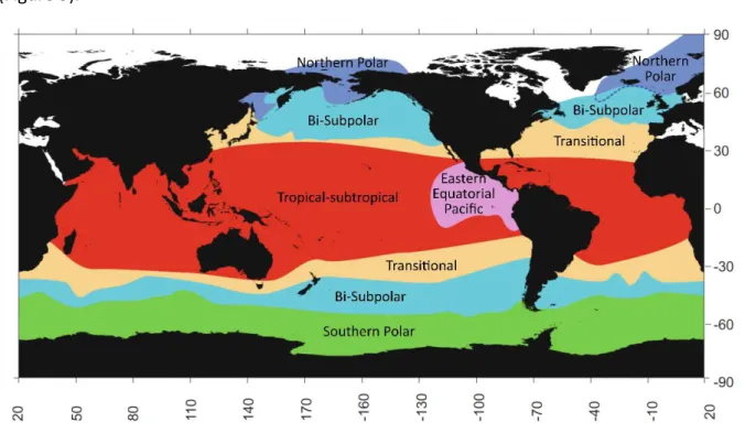
coastal regions with narrow continental shelf and abrupt depth changes, influenced by oceanic waters and upwelling systems, such as Villefranche-sur-mer at the Côte d’Azur (France), off the coast of California (USA) and in Norwegian fjords. Boltovskoy and Correa (2016) recently defined six major geographic provinces based on different polycystine assemblages collected using plankton net, sediment trap and surface sediment samples (Figure 5).
Figure 5. Major biogeographic provinces based on the distribution of polycystine radiolarians from plankton,
sediment trap and surface sediment samples. (Adapted from Boltovskoy and Correa (2016)).
Some of the major controlling factors of Rhizaria distribution seem to be water temperature and salinity. Numerous studies in laboratory conditions have demonstrated the broad tolerance of these organisms to changes in several parameters such as salinity, temperature and silica concentration (Anderson et al., 1989). Polycystine abundances peak at the equator (Boltovskoy et al., 2010). Nutrients and primary productivity seem also to affect distribution patterns of polycystine. Phaeodaria distribution is not clearly associated with temperature (Boltovskoy et al., 2017).
Vertical distribution. So far, the distribution of Rhizaria in the ocean has mostly been
derived from sediment data. In the last few decades, the development of new imaging and metabarcoding technologies has successfully revealed fine-scale distribution patterns of Rhizaria communities (Biard and Ohman, 2020). A substantial fraction of Polycystine
13 radiolarians harbour symbionts and therefore occur in the euphotic layer, especially in oligotrophic tropical and subtropical waters. In these waters polycystines are usually concentrated in the upper 50–100 m (Boltovskoy et al. 2010). In polar waters, however, peak abundances seem to be associated with deeper and warmer layers, at around 200–400 m (Boltovskoy and Alder 1992; Nimmergut and Abelmann 2002; Petrushevskaya 1967).
Regarding phaeodarians vertical distribution, it seems that they prefer to inhabit deep-waters, peaking in both abundances and diversity below 100 m (Boltovskoy et al., 2017). A vertical niche zonation was observed by Biard and Ohman (2020), with specific rhizarian taxa associated to each defined water layer. Phaeodarians preferentially inhabiting deeper waters may be due to their omnivorous nature, as these organisms feed on particulate organic matter which settles from the upper layers of the ocean.
Rhizaria abundances. In surface sediment samples, hundreds to thousands radiolarian
skeletons can be found in one gram of sediment (Boltovskoy et al., 1993). Phaeodarians, as mentioned above, are not well preserved in the sediment. In the water column, Rhizaria abundances rarely reach 5 cells per liter (Caron 1990), with lower values for phaeodarians (Gowing, 1989). Some studies reveal that densities of the Rhizaria group can be higher than expected in specific regions of the ocean in certain seasons. In the Japan Sea and the North Pacific Ocean high abundances for Phaeodaria and Nassellaria groups were reported, 208 and 234 cells m-3, respectively (Ishitani and Takahashi, 2007). Considering their occasional high
biomass and the fact that their skeleton is made of Si, Rhizaria can play an essential role in regional ecosystems and have an important impact in the global marine Si cycle.
| PhD objectives
14
PhD Objectives
Very little is known regarding the biology and ecology of Rhizaria. The general objective of this thesis is to better understand the role of silicifying Rhizaria in oceanic ecosystems and in the Si biogeochemical cycle. We developed a multidisciplinary approach merging marine ecology, ecophysiology, biogeochemistry and imaging (including laboratory tools and in situ instruments). We applied techniques previously used for diatoms in order to measure Rhizaria dSi consumption rates in situ in contrasted ecosystems’ settings.
The specific objectives of this thesis are:
1. To determine the elementary composition and the silicic acid uptake rates of a variety of taxonomic groups of siliceous Rhizaria. From these results and a compilation from the literature of rhizarians abundance data, we will estimate the potential role of Rhizaria in the Si cycle of the global Ocean.
2. To determine the relative importance in terms of biomass of siliceous Rhizaria planktonic community compared to diatoms, in contrasted oceanic regions (Mediterranean Sea, the Ross Sea and the Atlantic Ocean).
3. To obtain a holistic view of the Rhizaria community (i.e., considering the entire size spectra), and study more into detail, the relative abundance and biomasses of different taxonomic groups. To address this, optical and imaging tools (FlowCam, Zooscan and Underwater Vision Profiler-UVP) and metabarcoding methods are combined.
Chapter 1
– Estimating biogenic silica in the
global ocean
17
Context of the work
The first chapter of this thesis is dedicated to the determination of the elementary composition (biogenic silica, carbon and nitrogen) as well as the quantification of the rates of silicic acid uptake of living Rhizaria.
To carry out this objective, I participated in the 2017 Mediterranean Ocean Observing System for the Environment – Grande Echelle (MOOSE-GE) oceanographic cruise. During this cruise I collected Rhizaria specimens in the northwestern Mediterranean Sea and I conducted experiments using radioactive labeled silicon to measure the organisms’ biogenic silica production rates. We also analysed the elementary composition of several taxonomic groups. The results of these experiments were combined with previously published data on the abundance of polycystines and phaeodarians in the global ocean to generate the first estimates of the contribution of Rhizaria to the world’s biogenic silica production.
These findings challenge the view that diatoms have total control over oceanic silicon cycling in the modern ocean, a process that is coupled with other biogeochemical cycles, such as the carbon and nitrogen cycles.
19
Estimating Biogenic Silica Production of Rhizaria in the Global Ocean
Natalia Llopis Monferrer1,2, Demetrio Boltovskoy3, Paul Tréguer1, Miguel Méndez Sandin2,
Fabrice Not2, Aude Leynaert1
1Univ Brest, CNRS, IRD, Ifremer, LEMAR, F-29280 Plouzane, France
2Sorbonne University, CNRS, UMR7144, Ecology of Marine Plankton Team, Station Biologique de Roscoff, Roscoff,
France
3University of Buenos Aires-CONICET · Institute of Ecology, Genetics and Evolution of Buenos Aires, Argentina
Published in Global Biogeochemical Cycles - February 2020 https://doi.org/10.1029/2019GB006286
Abstract
Siliceous polycystines and phaeodarians are open-ocean planktonic protists found throughout the water column and characterized by complex siliceous skeletons that are formed, at least partly, through the uptake of silicic acid. These protists contribute to the marine organic carbon (C) and biogenic silica (bSi) pools but little is known about their contribution to the silica (Si) biogeochemical cycle. Here we report the first measurements of the Si uptake rate of polycystine and phaeodarian cells from samples collected in the Mediterranean Sea using the 32Si based method. The elementary composition (bSi, particulate organic carbon and
nitrogen) of these organisms was also measured. Combining our results with published data on the distribution and abundance of Polycystina and Phaeodaria in the global ocean, we conclude that these organisms could contribute from 0.2 to 2.2 mmol Si m-2 of the marine
standing stock of bSi and from 2 to 58 Tmol Si yr-1 (1 to 19%) of the global oceanic biogenic
silica production. The implications for the global marine Si cycle are discussed.
Silica production of Rhizaria | 1
20
1. Introduction
Rhizarians are eukaryotic, mostly heterotrophic single‐celled organisms, ranging in size from tens to hundreds of micrometers, although some are capable of forming gelatinous colonies up to over 1 m in length (Boltovskoy et al., 2017; Suzuki & Not, 2015). These protists are globally distributed, dwelling chiefly in the open ocean, from the surface down to bathypelagic depths. Their distribution and abundance are controlled by environmental factors, such as temperature, salinity, productivity, and nutrient availability (Boltovskoy, 2017a, 2017b; Boltovskoy et al., 2017; Boltovskoy & Correa, 2016). Some rhizarian taxa produce mineral skeletons of strontium sulfate (e.g., subclass Acantharia), calcium carbonate (e.g., order Foraminifera), and opaline silica (e.g., orders Spumellaria and Nassellaria and superorder Phaeodaria).
Silicifying organisms are a critical component of the global oceanic Si cycle. Diatoms, silicoflagellates, sponges, and siliceous rhizarians are all capable of using the silicic acid available in seawater to build elaborated skeletons that are believed to improve essential functions, such as mechanical protection for the cell (Hamm et al., 2003), an armor against predators (Finkel & Kotrc, 2010), an effective pH buffer (Milligan, 2002), or an improvement for the uptake or storage of bioessential elements (Suzuki & Not, 2015). Other studies have suggested that the frustule could confer diatoms an advantage due to its peculiar optical properties (Leynaert et al., 2018). Diatoms are considered the world's largest contributors to the Si cycle, dominating both the standing stock of water column biogenic silica (bSi) and its production rate (Ragueneau et al., 2000, 2006; Tréguer & De La Rocha, 2013). A number of studies ranging from sediment traps to environmental molecular surveys have emphasized the importance of rhizarians in biogeochemical cycles and export of C and bSi to the deep ocean (Biard et al., 2018; Guidi et al., 2016; Gutierrez‐Rodriguez et al., 2019; Lampitt et al., 2009). Moreover, recent studies combining genomic and in situ imaging approaches have shown that the contribution of large Rhizaria to the biomass of zooplankton has been largely underestimated (Biard et al., 2016), with their abundance correlating with carbon export fluxes at 150‐m depth in oligotrophic oceanic regions (Guidi et al., 2016). Globally, in terms of numbers, some rhizarian taxa can represent approximately 33% of large zooplankton (>600 μm) in the upper water column and up to 5% of the overall oceanic biota carbon reservoir (Biard et al., 2016; Stukel et al., 2018). These new findings suggest an unsuspected role of
21 these organisms in the biological carbon pump, as well as in the Si cycle, especially in oligotrophic areas where diatoms are poorly represented while siliceous rhizarians may be dominant in the tropical sediments (Dutkiewicz et al., 2015; Lisitzin, 1974).
The major taxa of silicifying rhizarians are represented by the Phaeodaria (Cercozoa, Thecofilosea) and by Spumellaria, Nassellaria, and Collodaria (Retaria, Radiolaria, Polycystinea) (Adl et al., 2018). The two groups differ in the robustness of their skeletal structures. While most polycystines possess solid and dense skeletons (Takahashi, 1983), phaeodarian skeletons are porous (Nakamura et al., 2018). The ballast produced by this skeleton causes them to sink toward the ocean floor where they can be incorporated into the sediment if their bSi has not been affected by the dissolution and recycling into Si(OH)4.
Polycystine bSi is more resistant to dissolution and often remains well preserved in the sediment, whereas that of Phaeodaria is rarely found in bottom deposits (Takahashi, 1983). Among Polycystina, Spumellaria and Nassellaria have been widely used for paleoceanographic reconstructions (Matsuzaki et al., 2014; Moore, 1978). However, essentially due to the difficulty of culturing planktonic rhizarians, our knowledge of their ecology, physiology, and biogeochemistry is very limited, especially with regard to the processes associated with Si sources, uses, stocks, and fluxes. Marron et al. (2016) reported the presence of Si transporters in rhizarians, which enable the uptake of Si (mainly in the form of silicic acid) from the environment.
In this study, we analyze the Si, carbon (C), and nitrogen (N) content of isolated rhizarian cells collected during two oceanographic cruises in the Mediterranean Sea (Mediterranean Ocean Observing System on Environment Grande Echelle, MOOSE‐GE) and from the Atlantic Ocean (Atlantic Meridional Transect, AMT28) cruise. We also measured their bSi production rates using the 32Si isotope, an approach successfully applied until now only to estimate diatom bSi production rates. We first discuss the relationship between rhizarians elemental composition and their cell size, and combining these measurements with available data on rhizarian abundances worldwide, we assess their potential contribution to the global marine Si cycle.
Silica production of Rhizaria | 1
22
2. Material and Methods
2.1. Sampling
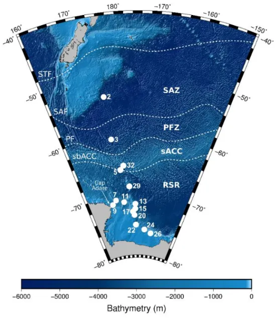
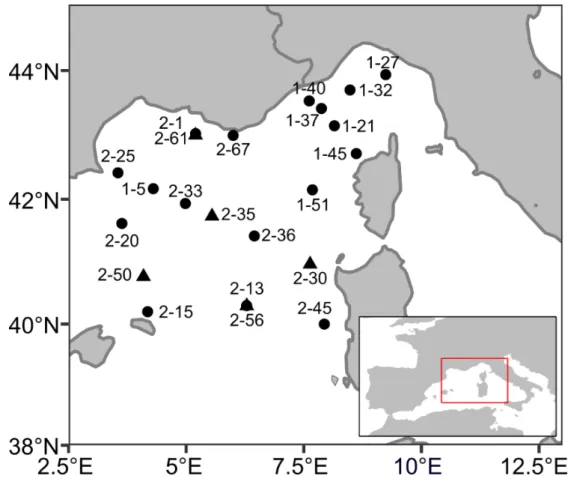
Samples were collected at 22 sampling sites in the western Mediterranean basin (Figure 1) during the MOOSE-GE expedition in September 2017 and June 2018 aboard the R/V Atalante and at one site during the AMT28 in October 2018 aboard the RRS James Clark Ross.
Figure 1. Sampling sites (Leg-Station) of the MOOSE-GE cruises, ● sites sampled in 2017 and ▲ sites sampled in
2018. Net was deployed between 0 and 500 m. The samples of the AMT cruise were collected at 3.69°S 24.98°W at depths between 0 and 200m.
Plankton samples were obtained at discrete depth-intervals, using vertical tows. Upon recovery, samples were immediately diluted in 0.2 µm filtered seawater and observed on-board under a stereomicroscope or an inverted microscope. Rhizarians were handpicked using a Pasteur pipette and transferred to 20 mL glass vials filled with filtered seawater. Cells were sorted according to targeted taxonomic groups, namely Nassellaria, Spumellaria and Collodaria, chiefly represented by Pterocorythidae, Hexastyloidea and Sphaerozoidae
23 respectively. Among the Phaeodaria, because of their high abundances, we differentiated the genera Aulacantha, primarily represented by Aulacantha scolymantha, and Challengeria. In each glass vial, we stored from 1 to 50 individuals, depending on cell size and abundance of the target organisms in the sample. Individual pictures were taken prior the experiments to obtain morphometric measurements using the ImageJ software. For each specimen, we measured length, width, and area, and subsequently calculated the biovolume associated using the most similar simple geometric shape (e.g., sphere, ellipsoid, cone). Due to logistic problems, for some of the stations, no pictures were available for biovolume estimates. However, samples from all the stations were analysed using the Flowcam (preserved with lugol’s solution) and the Zooscan (preserved with formaldehyde) immediately after the cruise. We also compared cell size from living and preserved individuals and we did not observed differences in cells dimensions between them. Therefore, we used the vignettes obtained with these imaging technologies to complete our set of measurements. For Collodaria (mostly colonial radiolarians comprising hundreds to thousands of siliceous shells or spicules embedded in a gelatinous matrix), the entire colony was measured, but the results are reported per individual. Capsules were counted and measured using ImageJ software. Seawater samples for the incubations were collected at each sampling location and at different depths using Niskin bottles.
2.2. Elementary composition
We analysed the bSi (in nmol Si cell-1) and the particulate organic carbon and nitrogen
(POC/PON in µmol cell-1) in 35 and 24 samples respectively (Table 1). In total, 1339 cells were
isolated and analysed.
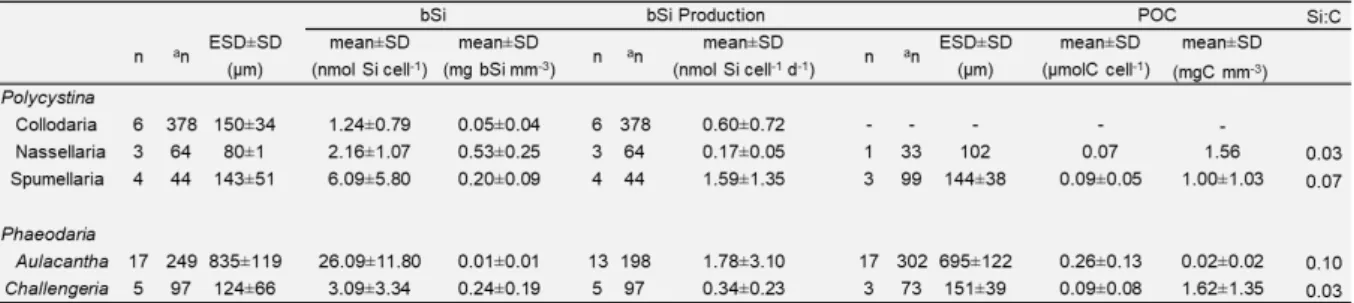
Table 1. Biogenic Silica Stocks, Production Rates and POC content of the rhizarians analyzed. Note: ESD denotes
equivalent spherical diameter, n denotes the number of samples analyzed and an the number of specimens
Silica production of Rhizaria | 1
24
The entire content of each vial with isolated cells in filtered seawater was filtered onto polycarbonate membrane (Nuclepore 47mm, 0.6 µm pore size) for bSi determination and onto 25 mm GF/F precombusted filters (at 450 °C during 4 h) for POC and PON analysis. Nuclepore membranes were 4-folded in a petri dish, dried and stored at ambient temperature until later analysis in the laboratory. GF/F filters were folded in a petri dish and stored at -20 °C until analysis.
The bSi of Polycystina and Phaeodaria was quantified by colorimetric determination of the orthosilicic acid after leaching. A single digestion in hydrofluoric acid (HF) was performed, since the samples only contained isolated rhizarians, i.e., there was no possible interference with lithogenic Si as there might be when filtering raw seawater (see Ragueneau et al., 2005). We added 0.2 ml of HF 2.5N to the polymethylpentene (PMP) tubes containing the filters. The filter was then compressed until submerged in HF and air bubbles were removed. The tube was tightly covered with a cap and kept under a fume hood, at room temperature for 48 h to allow for digestion of the bSi. We then added 9.8 ml of saturated H3BO3 solution. The
standards used for calibration were prepared with the same matrix as for the samples (HF/H3BO3), before analysis by colorimetric methods on a Technicon Auto-Analyzer II (Aminot
and Kérouel, 2007; Brzezinski and Nelson, 1989).
Concentrations of POC and PON were measured with a mass spectrometer (Delta plus, ThermoFisher Scientfic) coupled to a C/N analyser (Flash EA, ThermoFisher Scientifc). Standard deviations (SD) were 0.009 µM and 0.004 µM for POC and PON respectively. In order to avoid false positives, the detection limit was set at the control level plus ten times the standard deviation. Although POC and PON analyses were performed simultaneously using an elemental analyser, N was often close to the detection limit, in which case, the values were discarded, thus yielding more results for C than for N. The POC/PON measurements of Collodaria were rejected for the same reason.
2.3. Assessment of bSi production rates (𝝆𝑷)
For the measurements of bSi production rates (𝜌𝑃), we used the radioisotope of silicon (32Si)
(Leynaert et al., 1996; Tréguer et al., 1991). Immediately after isolation, glass vials containing between 1 and 50 cells of the same taxonomic group were incubated on deck with 800 becquerel (Bq) of high specific activity 32Si, for 24 h in a flowing-seawater incubator to
25 means of neutral screen to 50% of the incident light. The 32Si additions increased silicic acid
concentrations in the incubation bottles by less than 10 nM, a negligible value compared to the dissolved silica (DSi) concentration in seawater. A split of the seawater sample used for the incubation was stored for subsequent analyses of silicic acid concentration using the automated method of Aminot & Kérouel (2007).
After incubation, samples were filtered by gentle (<150 mmHg) vacuum filtration onto 47mm diameter, 0.6 µm pore-size polycarbonate membrane filters (Nuclepore), and rinsed twice with filtered seawater to wash away non-particulate 32Si. Each filter was then placed in a clean
20 ml polypropylene liquid scintillation vial and the vial was capped loosely to allow the sample to dry at room temperature for 48 h. The vials were then capped tightly and returned to the laboratory for counting.
The activity of 32Si in the samples from the incubation experiments was determined using the
Cerenkov counting method (Leynaert, 1993) three months after the samples were filtered, allowing 32Si and its daughter isotope 32P to return to secular equilibrium. Although this
method is less sensitive than some others (Brzezinski and Phillips, 1997), it was chosen because it allows using the materials for further bSi analyses. Twenty-four hours before assessing the activity on the filter, HF (2.0 ml of 2.5N) was added to each sample to dissolve all bSi. Samples were assayed using a Wallac Model 1414 scintillation counter. Because 32Si
does not produce Cerenkov emissions, the procedure allowed quantifying the amount of 32P
only. However, as 32Si and 32P are in secular equilibrium the activities of the two isotopes are
equal, and the 32Si activity can be deduced from that of 32P (Leynaert, 1993). Triplicate 40 min
counts were performed on each sample. Counting precision (95% Confidence Interval) was < ± 1%, except for a few very low-activity samples yielding < 250 CPM (counts per minute), for which counting precision was ± 2-5%. Counts yielding less than three times the background (8 CPM) were discarded. Collodarians without siliceous spicules or shells (e.g. Collozoum spp.) were incubated in parallel to obtain a production blank, which yielded production rates close to the detection limit, i.e., below 0.02 nmol Si cell-1 day-1 in all cases.
2.4. Extrapolation of siliceous rhizarians to the global ocean: bSi stocks and production rates
We performed an estimate of rhizarian abundances based on the compilation of worldwide data of Boltovskoy et al. (2010), supplemented with more recent studies (see Supplementary material). Our database contained 1191 data points of Polycystina and Phaeodaria densities
Silica production of Rhizaria | 1
26
(cells m-3) in plankton samples collated from 22 publications. Most of the studies used
plankton nets to collect rhizarians, including vertical and horizontal tows. Other studies sieved Niskin bottle samples to quantify abundances of these organisms (See details in Supplementary material).
These data were averaged over two bins: tropical-subtropical (40°N to 40°S), and colder waters (>40°N or S), each in turn was subdivided into two depth layers: 0-200 m, and below 200 m depth (Table 2). For the Phaeodaria, where the literature information is scarcer, all species were pooled in a single group (Phaeodaria). For the Polycystina, densities of Spumellaria and Nassellaria were estimated separately (and also used separately in subsequent calculations).
Table 2. Mean abundances in cells m-2 and [cells m-2] of Polycystina and Phaeodaria, as reported in 22
publications based on plankton materials. Note: values include all cells recorded (i.e., living and dead shells).
To convert abundance values reported in literature into bSistocks (nmol Si m-2) and production
rates (𝜌𝑃 µmol Si m-2 day-1), we used the estimates obtained in our cruises. For each taxonomic
group, we established ranges based on the minimum and maximum values for both bSi stock and 𝜌𝑃 (Table 1). To obtain global estimates, we considered an area of 2.68E14 m2 (40°N to
40°S) for the warm waters and an area of 9.3E13 m2 (>40°N or S) for the cold waters.
3. Results
3.1. Elemental composition: bSi, POC and PON
bSi content
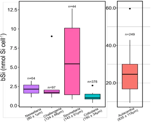
Overall, bSi per cell varied over one order of magnitude, from 1.24 ± 0.79 nmol Si cell-1 (mean
27 content differed significantly between Aulacantha (Kruskal-Wallis, P < 0.05) and the other groups (Figure 2).
Figure 1. bSi content and size of the taxa studied. n denotes the number of specimens (or individual cells, for the
Collodaria n*) analysed. Colours correspond to the taxonomic groups identified in Figure 3.
The largest cells (i.e., genus Aulacantha) have the highest Si content. In order for our data to be comparable with the units reported in the literature, Si concentrations were converted from nmol Si cell-1 to µg Si cell-1 assuming a molecular weight of 67 g mol-1 for hydrated
amorphous silica (Mortlock and Froelich, 1989).
We evaluated the log-log relationship between the Si content and cell size of the rhizarians surveyed. Based on the Si contents of 3 samples of Nassellaria (64 individuals), 4 samples of Spumellaria (65 individuals), 6 samples of Collodaria (378 individuals), 5 samples of
Challengeria (97 individuals) and 17 samples of Aulacantha (249 individuals), Si content (𝑄𝑏𝑆𝑖)
was significantly associated with the cell’s equivalent spherical diameter (ESD) (R2 = 0.8, F(1,
33)=129, P < 0.001) according to the following equation:
𝑙𝑜𝑔10(𝑄𝑏𝑆𝑖) = [−3.61 ± 0.29] + [1.30 ± 0.11] ∙ 𝑙𝑜𝑔10(𝐸𝑆𝐷) (1)
Where 𝑄𝑏𝑆𝑖 is in µg Si cell-1 ± standard error (SE) and ESD in µm ± SE.
Silica production of Rhizaria | 1
28
The POC and PON content of the rhizarians analysed differed significantly between taxa (ANOVA, P<0.001; excluding the Nassellaria, for which no data were available). Nitrogen measurements were obtained for 24 samples including a total of 507 siliceous rhizarians cells. Overall, PON concentrations ranged between 0.001 and 0.02 µmol N cell-1.
In these 24 samples, the POC content (𝑄𝐶) varied between 0.09 ± 0.05 and 0.26 ± 0.13 µmol C cell-1. The highest 𝑄
𝐶 was found for Aulacantha cells. However, the relationship between 𝑄𝐶
and ESD was not significant. The average C/N ratio was 12.
3.2. Rhizarian bSi production rates
We successfully measured, for the first time, the silicic acid uptake (𝜌𝑃) rates of 31 rhizarian samples (781 individuals). Rates of bSi production ranged from 0.17 ± 0.05 nmol Si cell-1 day-1
for Nassellaria to 1.78 ± 3.10 nmol Si cell-1 day-1 for Aulacantha (Table 1). Production rates
seem to be related to cell size.
In order to estimate specific uptake rate (𝑉𝑃, in day-1), 𝜌
𝑃 were normalized to the
concentration of bSi (𝜌𝑃/bSi) (Tables 1 and 3). The largest cells (Aulacantha) present the lower 𝑉𝑃, while Nassellaria, the smallest cells found in this study, had the highest 𝑉𝑃.
Table 3. Specific uptake rates for Polycystina, Phaeodaria and diatoms. Note: values for diatoms are from Claquin
et al. (2006) and references therein.
3.3. Worldwide Si standing stocks and production rates (𝝆𝑷)
According to the literature data collated, densities of Polycystina and Phaeodaria peak in the upper layers where their abundances (i.e., cells L-1) are ~1.5-8.5 times higher than below 200
29 throughout large depth intervals often yield larger standing stocks below 200 m than in the 0-200 m layer, especially at low latitudes (Table 2). Our estimates suggest that polycystine and phaeodarian bSi standing stocks in the water column range from 0.3 to 2.2 mmol of bSi m-2
(Table 4). For 𝜌𝑃, we obtained a tentative range of 5 to 58 Tmol yr-1 (Table 4). The 𝜌
𝑃 are of
more consequence in temperate waters than in cold waters, which reflects differences in abundances. Production rates throughout the water column are similar in the two oceanic zones considered.
4. Discussion
4.1. Filling the gaps in size-Si relationship. From small to giant protists.
Our work on the assessment of the elemental composition of siliceous rhizarians included several small-sized Polycystina (Collodaria, Nassellaria and Spumellaria), as well as the larger Phaeodaria, encompassing a wider size spectrum than that covered by previous studies. Takahashi (1981) was the first to report the Si content, weight, length, width, projected area, and biovolume of polycystines and phaeodarians. However, his studies were based on dead cells recovered from sediment traps. Using these data and specimens collected off the coast of California, Biard et al. (2018) established an allometric relationship that shows that the Si content of phaeodarians is closely associated with cell length and cell biovolume. These relationships were chiefly based on large specimens (0.6 to >10 mm, average: 2 mm), and have not hitherto been validated for the smaller rhizarians.
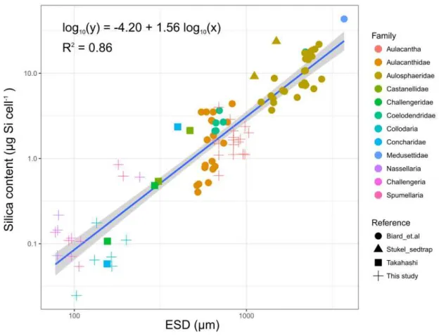
In order to investigate whether their Si content is associated with biovolume and cell size (expressed as ESD) throughout a wider size-range, we combined our measurements and Si content values with the log-log linear relationship shown by Biard et al. (2018). The result of this exercise showed that over a range of 103 to 3920 µm, rhizarian Si content is significantly associated with biovolume (R2 = 0.86, F(1, 93)=593, P<0.001):
𝑙𝑜𝑔10(𝑄𝑏𝑆𝑖) = [−4.05 ± 0.18] + [0.52 ± 0.02] ∙ 𝑙𝑜𝑔10(𝑏𝑖𝑜𝑣𝑜𝑙𝑢𝑚𝑒) (2)
Where 𝑄𝑏𝑆𝑖 is in µg Si cell-1 ± SE and biovolume in µm3 ± SE.
Si content is also correlated with the ESD (Figure 3; R2 = 0.86, F(1, 93)=593, P<0.001):
Silica production of Rhizaria | 1
30
Where 𝑄𝑏𝑆𝑖 is in µg Si cell-1± SE and ESD in µm ± SE. Biard’s length data were converted to ESD
for this analysis.
The slope of equation 3 is similar to those of Biard et al. (2018) for ESD (ANCOVA, P=0.41), confirming its validity for a wide spectrum of rhizarians sizes.
Figure 3. Relationship between the Si content of Polycystina and Phaeodaria and their ESD (µm) across all the
specimens assessed. Regression (blue line) and 95% confidence interval (gray shading). Combined data from Biard et al. (2018) and this study.
Silica content has previously been shown to be related to cell size in other siliceous organisms, such as diatoms. Conley et al. (1989) observed that the Si content of diatoms varies over five orders of magnitude depending on cell size and found a significant log-log linear relationship between Si content and biovolume. However, the relationship for diatoms gives much lower cellular bSi concentrations (100 times lower on average) than for rhizarians of comparable size. Thus, among the siliceous planktonic organisms in the ocean, rhizarians appear to be the most silicified.
Although the largest cells contain more Si, they are not necessarily the densest according to our conversion from Si biomass to density. Nassellarians, which are the smallest cells in our
31 study, had 530 µg bSi mm-3, while Aulacantha, the largest cells analysed (measurements based
on the solid skeleton), barely reached 10 µg bSi mm-3. As reported recently, the structure of
Polycystina and Phaeodaria differs considerably. Polycystine skeletons are solid, whereas phaeodarian skeletons are porous and in some species, including Aulacantha they are composed of loose spines protruding from the cell (Nakamura et al., 2018) (Figure 4). Sedimentation rates in the water column are likely to be affected by differences in density of these structures, as observed by Baines et al. (2010) for diatoms.
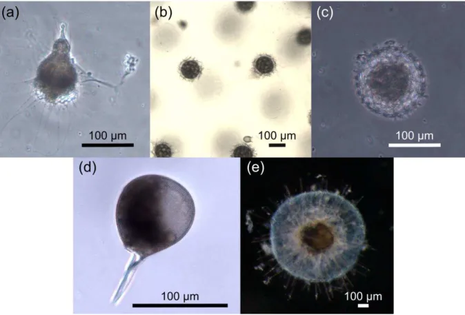
Figure 4. Images of the most abundant morphotypes surveyed in this work, including Polycystina (a-c), and
Phaeodaria (d-e). (a) Nassellaria - Pterocorythidae (b) Collodaria-Sphaerozoidae (c) Spumellaria - Hexastyloidea (d) Challengeridae - Challengeria xiphodon (e) Aulacanthidae - Aulacantha scolymantha.
As opposed to Si, we were not able to find any relationship between POC and PON and cell size, therefore an average was calculated per taxon (Table 1). Data on rhizarian 𝑄𝐶 are scarce. To our knowledge, the first 𝑄𝐶 published are those of Michaels et al. (1995) where they reported extremely variable 𝑄𝐶 values for solitary collodarians, ranging from 0.009 to 0.28 mg C mm-3. Stukel et al. (2018) estimated a 𝑄
𝐶 for Aulosphaeridae of 0.011 mg C mm-3 based on
a downward revision of Biard et al. (2016) measurements. Our data, are high when compared to those in these two studies. Polycystina and Phaeodaria Si:C molar ratios (0.03 to 0.1) are
Silica production of Rhizaria | 1
32
lower than those of diatoms (0.13 ± 0.04), according to data reported by Brzezinski (1985) for twenty-seven diatom species. On the other hand, our mean C/N ratio (~12) is higher than that reported by Michaels et al., (1995) for colonial collodarians (8.2), and much higher than the Redfield ratio (6.6). Higher molar ratios for the colonial radiolarians could be due to the presence of mucopolysaccharide material surrounding the colonies (Michaels et al., 1995).
4.2. The silicic acid uptake rates of rhizarians (𝝆𝑷)
In this study, 𝜌𝑃 were measured using the radioisotope 32Si, quantifying, for the first time the
ability of these protists to consume dissolved Si from seawater. Our results show consumption rates ranging from 0.17 nmol Si cell-1 day-1 (for Collodaria) to up to 1.78 nmol Si cell-1 day-1
(Aulacantha).
Comparison of these results with the 𝜌𝑃 of other plankton, in particular diatoms, is complicated by the fact that the rates measured for diatoms in the field are expressed in terms of volume (i.e. per liter), rather than “per cell”. For diatoms, 𝜌𝑃 per cell were only assessed in culture experiments in laboratory controlled conditions (Del Amo and Brzezinski, 1999; Riedel and Nelson, 1985) yielding values around 0.001-21 pmol Si cell-1 day-1. In contrast, the
consumption rates of silicic acid uptake by the rhizarians assessed in this work ranged from 200 to 2000 pmol Si cell-1 day-1, which is around 100 times higher relative to diatoms, and
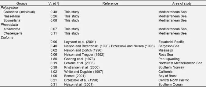
consistent with what was observed for the Si content, also 100 times higher in rhizarians. To gain insight into the role of rhizarians in the Si cycle we assessed the 𝑉𝑃 of these organisms by normalizing the silicic acid uptake rates to the bSi concentration (Table 3). Our 𝑉𝑃 ranged from 0.07 to 0.48 day-1, for Aulacantha and Collodaria, respectively. Despite differences in the
methods used, these values are generally, in line with results previously found for “larger rhizarians”, whose average 𝑉𝑃 was ~0.1-0.2 day-1 (Stukel et al., 2018). Our values also agree
with those found for diatoms worldwide (0.06-1.80 d-1) for a large temperature range (Table
4).
Based on these results, we evaluated the turnover rates of these protists (𝑡 = 𝑙𝑛2 𝑉⁄ ) 𝑃 assuming that the rhizarians analysed in this study reproduce by binary cell division, which might not always be the case, as sexual reproduction may also occur (Boltovskoy et al., 2017). Turnover rates range between 3 and 10 days (for Aulacantha and Spumellaria, respectively), which are comparatively low when compared to phytoplankton which usually span from several hours to a few days ((Flynn et al., 2018; Krause et al., 2017).
33
Table 4. Estimated values of bSi stock and production as derived from our experimental data using the minimum
and the maximum values for each group and the information detailed in Table 2. Note: we estimated for each toceanic area a value of production in Tmol Si yr-1.
4.3. Potential impact of siliceous rhizarians on the Si cycle of the World Ocean
Since the 1970s, silicified rhizarians (formerly collectively designated as Radiolaria) have drawn attention for their role in the marine Si cycle (Heath, 1974). Takahashi (1983) suggested that, in some oceanic areas, the daily flux of rhizarian bSi ranges around 20% to 30% of the overall bSi. Biard et al. (2016, 2018) found that the biomass of large Rhizaria, the so‐called “giant protists”, could constitute a substantial fraction of the >600‐μm plankton biomass representing more than a third of the bSi standing stocks in oligotrophic and high nutrient‐low chlorophyll regions of the ocean. These data conflict with the fact that in most global oceanic silicon budget estimates, only diatoms (Nelson et al., 1995; Tréguer et al., 1995) and more recently sponges (Maldonado et al., 2019; Tréguer & De La Rocha, 2013), are taken into account. This standpoint, however is gradually changing. For example, Tréguer and De La Rocha (2013) suggested that up to 23% of the bSi standing stocks in the upper 120 m of the
![Table 2. Mean abundances in cells m-2 and [cells m-2] of Polycystina and Phaeodaria, as reported in 22 publications based on plankton materials](https://thumb-eu.123doks.com/thumbv2/123doknet/13789242.440295/45.892.105.792.514.745/table-abundances-polycystina-phaeodaria-reported-publications-plankton-materials.webp)