• Aucun résultat trouvé

CT dose reduction in children

N/A
N/A
Protected

Academic year: 2021

Partager "CT dose reduction in children"

Copied!
2
0
0

Texte intégral

(1)

Eur Radiol (2005) 15: 2383–2384

DOI 10.1007/s00330-005-2906-7

E R R ATU M

Peter Vock

Published online: 10 September 2005

# Springer-Verlag 2005

CT dose reduction in children

Eur Radiol (2005) s00330-005-2856-0

Unfortunately, there were errors in Tables

3

and

5. The

corrected tables are given here.

The online version of the original article can be found at http://dx.doi.org/10.1007/s00330-005-2856-0

P. Vock (*)

Department of Radiology, University Hospital Inselspital, 3010 Bern, Switzerland e-mail: [email protected] Tel.: +41-31-6322435 Fax: +41-31-6324874

(2)

2384

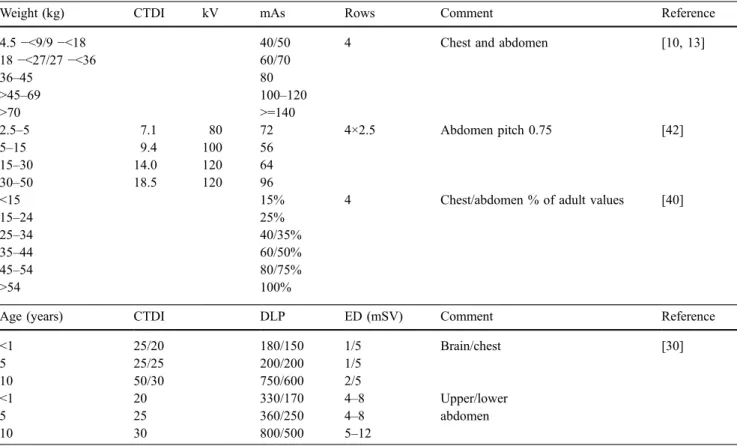
Table 3 Suggested paediatric CT protocols.CTDI CT dose index, DLP dose-length product, ED effective dose

Weight (kg) CTDI kV mAs Rows Comment Reference

4.5−<9/9 −<18 40/50 4 Chest and abdomen [10, 13]

18−<27/27 −<36 60/70 36–45 80 >45–69 100–120 >70 >=140 2.5–5 7.1 80 72 4×2.5 Abdomen pitch 0.75 [42] 5–15 9.4 100 56 15–30 14.0 120 64 30–50 18.5 120 96

<15 15% 4 Chest/abdomen % of adult values [40]

15–24 25%

25–34 40/35%

35–44 60/50%

45–54 80/75%

>54 100%

Age (years) CTDI DLP ED (mSV) Comment Reference

<1 25/20 180/150 1/5 Brain/chest [30] 5 25/25 200/200 1/5 10 50/30 750/600 2/5 <1 20 330/170 4–8 Upper/lower 5 25 360/250 4–8 abdomen 10 30 800/500 5–12

Table 5 Effective dose estimated from dose-length product (DLP)a

[8, 38]

Age Head Neck Chest Abdomen/pelvis

0 y 0.011/0.027 0.017/- 0.039/0.034 0.049/0.040 1 y 0.007/0.008 0.012/- 0.026/0.021 0.030/0.024 5 y 0.004/0.004 0.011/- 0.018/0.014 0.020/0.016 10 y 0.003/0.003 0.008/- 0.013/0.011 0.0150/0.014 15 y -/0.015 -/0.009 Adult 0.002/0.003 0.006/- 0.014/0.009 0.015/-a

Figure

Table 3 Suggested paediatric CT protocols. CTDI CT dose index, DLP dose-length product, ED effective dose

Références

Documents relatifs

Even though current advances in CT technology and dose reduction strategies are fully exploited worldwide in the clinic, some advocate the view that CT is still not a low- dose

To assess the proposed method we created different low-dose imaging scenarios by reducing the number of projections, and tested them on several bone samples.. The proposed

Qualitative and quantitative results show that the Gamma regularization based reconstruction can perform better in both edge-preserving and noise suppression when compared with

(a), the original FBP B70f reconstructed LDCT image; (b), the reference FBP B30f reconstructed SDCT image; (c), the general DL processed LDCT image; (d), the LDCT

Even if the trial has not been designed specifically for imaging, these SRCT data sets on patients are the first ever obtained on humans with a synchrotron source and clearly show

The 16-row multi-detector com- puted tomography (MDCT) scanners with 16 rows (S16) and 64-row MDCT scanners with 64 rows (S64) show no visible difference, whe- reas the values for

Stolzmann P, Scheffel H, Schertler T et al (2008) Radiation dose estimates in dual-source computed tomography coronary angiography. Schertler T, Scheffel H, Frauenfelder T et al

Bei der Lungenemboliediagnostik kann in Einzelfällen (Z. schwangere Patientinnen, sehr junge Patienten) die Scanlänge deutlich reduziert werden, in- dem der Lungenapex und