Peter Vock
Received: 24 June 2005 Accepted: 1 July 2005
Published online: 10 August 2005 # Springer-Verlag 2005
CT dose reduction in children
Abstract World wide, the number of CT studies in children and the radia-tion exposure by CT increases. The same energy dose has a greater bio-logical impact in children than in adults, and scan parameters have to be adapted to the smaller diameter of the juvenile body. Based on seven rules, a practical approach to paediatric CT is shown: Justification and patient prep-aration are important steps before scanning, and they differ from the preparation of adult patients. The subsequent choice of scan parameters aims at obtaining the minimal signal-to-noise ratio and volume coverage needed in a specific medical situation; exposure can be divided in two aspects: the CT dose index determin-ing energy deposition per rotation and the dose-length product (DLP)
deter-mining the volume dose. DLP closely parallels the effective dose, the best parameter of the biological impact. Modern scanners offer dose modula-tion to locally minimise exposure while maintaining image quality. Be-yond the selection of the physical parameters, the dose can be kept low by scanning the minimal length of the body and by avoiding any non-qualified repeated scanning of parts of the body. Following these rules, pae-diatric CT examinations of good quality can be obtained at a reasonable cost of radiation exposure.
Keywords Computed tomography (CT), children . Computed
tomography (CT), radiation exposure . Radiations . Injurious effects . Neoplastic
Introduction
Around 15 years ago, the industry and even radiologists expected CT to be a slowly dying methodology, leaving the field step-by-step to MRI, maybe with some residual use for emergency patients and for technically less developed regions of the world. This potential development would also have eliminated any concern about radiation exposure by this technique. Although over several decades the hypothesis may still be valued, the years since 1990 have shown an unexpected revival of CT with a major growth rather than a decline [3] and a parallel increase of radiation exposure in Western populations [26]. While this applies to the adult population, it is even truer in children, for several reasons. Unfortunately, neither referring physicians nor
radiologists seem to be aware of the significance of radiation exposure by CT examinations [25].
In this review, we will attempt to inform of the development of CT scans, to show reasons for the specific situation of children and paediatric CT and to make suggestions on how to best use CT in children.
Medical success as the main reason for a rising concern about radiation exposure
The introductions of spiral CT in 1989 [24] and of multi-row-detector CT shortly before the end of the last century have turned out to be the key factors responsible for an unexpected success in the clinical use of CT. Both enhance P. Vock (*)
Department of Radiology, University Hospital Inselspital, 3010 Bern, Switzerland e-mail: [email protected] Tel.: +41-31-6322435 Fax: +41-31-6324874
the potential of either imaging faster, of imaging a larger volume, or of getting the same volume scanned at an even better geometrical resolution (Table 1). All three of these factors that are often used in combination have contributed to an increase in the medical use of CT: faster scanning allows for examinations in many situations where motion artefacts had previously contraindicated its use, such as in dyspnoeic patients, in patients with severe pain excluding a long procedure, in young children, and in all medical situations requiring anaesthesia with intubation. The very chance of having CT without anaesthesia under minimal sedation as compared with a long-lasting MR examination under general anaesthesia opened the CT scanner for many children. In childhood neoplasia, staging of the neck and the entire trunk is often required, e.g. in case of malignant lymphoma. The availability of a test to survey such a large volume within a few minutes was especially welcome in paediatrics. Even more important for paediatric problems was the availability of reduced slice collimation for a better geometrical resolution in the Z-axis on new scanners without a compromise in volume coverage. Children not only have smaller organs than adults but also usually have less fat acting as a contrast layer between the organs of similar density. An optimised isotropic resolution is a pre-requisite for recognising organs in different planes. It may be medically important to observe contrast enhancement at different time delays: multi-phase imaging to study both the pre-contrast situation and arterial or venous vascular enhancement, parenchymal enhancement and urinary ex-cretion requires fast scanners. Nowadays, there is hardly any technical limit of tube heating, of generator power, or of computer capacity to multi-phase imaging [16]; radia-tion exposure, however, increases linearly with the number of phases scanned. Furthermore, 3D post-processing based on isotropic imaging has become an important tool for better understanding of CT findings by radiologists and, above all, clinicians. This also has eliminated one of the major disadvantages of CT as compared to MRI: the merely axial scan plane.
In summary, new technical developments have greatly enhanced the medical utility of CT, both by opening new indications and by improving the information on traditional ones. Clinicians seem to rely progressively on imaging. This has increased the number of CT examinations per-formed in adults and children. The larger the volume covered and the number of phases in protocols, the more radiation exposure per examination will increase, despite the reduc-tion offered by several technical advances of new scanners.
With more people getting CT scans, the population ex-posure grows even more than individual exex-posure per examination. For instance, the United States of America has seen a 600% increase of CT scans from the mid-1980s to the mid-1990s, and scans in children have increased from 4% to over 11% of all CT examinations, with one third of these being done in the first decade and one sixth already within the first five years of life [3,26,41].
Today, impressed by the powerful technology, most clinicians and many radiologists are not really aware of the amount of radiation delivered in a very short time by CT scanning [25]. There is hardly any doubt that CT has become the largest source of medical radiation exposure, contributing 40–67% to the medical radiation exposure of the population with around 5–10% of all imaging ex-aminations [26,36,41]; a growth to more than 10% of all studies would even increase the percentage in radiation exposure.
Biological impact of low-level radiation
Deterministic effects, i.e. those observed predictably above a certain threshold, are not seen below 100 mSv of local dose. Low-level radiation is mostly defined as ionising radiation in the range of up to 100 mSv of effective dose where only stochastic effects of ionising radiation are expected. Stochastic effects are those expected without any threshold and consisting mainly of radiation-induced cancers and genetic effects. Single CT examinations usu-ally provide for less than 1 mSv to more than 27 mSv [27, 41] although in children, this upper threshold may well be exceeded, e.g. when adult protocols are used.
Scientific facts about low-level radiation mostly are based on the epidemiologic evaluation of nearly 60 years of follow-up of the survivors of the atomic bombs in Japan [32]. Some data come from medical exposure in radiology, occupational exposure, molecular experiments, cellular ex-periments, animal experiments and mathematical calcula-tion of risk models. The data show a statistically significant increase of the risk of fatal cancer starting at the range of 50– 100 mSv, possibly already at 10–50 mSv [3,5]; 100 mSv are expected to cause a lifetime risk of 0.5% for fatal cancer. A single, full-body CT scan (from C3 to the pubic sym-physis) in a 45-year-old would cause an effective dose of about 12 mSv and an estimated lifetime cancer risk of 0.08% (95% confidence interval of 0.025–0.26%); should this be repeated for yearly screening, the same individual
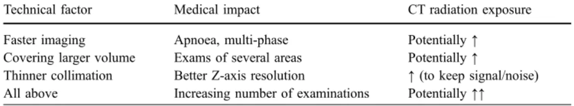
Table 1 Technical CT improve-ments, medical impact, and ra-diation exposure
Technical factor Medical impact CT radiation exposure
Faster imaging Apnoea, multi-phase Potentially↑
Covering larger volume Exams of several areas Potentially↑
Thinner collimation Better Z-axis resolution ↑ (to keep signal/noise) All above Increasing number of examinations Potentially↑↑
would run a 1.9% risk up to the age of 75 years [6]. Im-portant uncertainties in quantifying the risk estimate re-main; one important reason for this is the fact that below 100 mSv, the shape of the dose-response curve is still de-bated; a majority of researchers favour the linear extrapo-lation model without any threshold whereas other experts claim evidence for either increased or decreased effects at this lower end of the dose scale.
Immediate effects of ionising radiation cause cell dam-age by ionisation, excitation of molecules and formation of radicals; these are followed by changes at the chemical and molecular level, including genetic mutations, and may af-fect cells, tissues, organs and the entire organism. With follow-up CT examinations below 100 mSv, the likelihood of stochastic effects increases proportional to the total dose.
Biological need for CT dose reduction in children
Two factors require specific consideration of the biological effects of ionising radiation in children: their increased radio sensitivity and their life expectation at the moment of radiation exposure (Table2). In fact, this means that at the same effective dose [23], biological effects and lifetime risk will be higher for a child than for an adult. First, organs and tissues are more susceptible to radiation during childhood. Tissues are also distributed differently within the body in childhood. For instance, a CT examination of the lower extremities will hit nearly exclusively fatty bone marrow in an adult patient and thus cause a low effective dose whereas in a child, a significant proportion of the red marrow may be exposed, which is a reason for concern. In the trunk, since children have thinner layers of visceral fat, the natural contrast available usually in adulthood is reduced in childhood, similar to cachexy. At cellular and sub-cellular levels, proliferation during the growth period is one obvious factor for increased susceptibility.
Since most malignant tumours become manifest only years to decades after exposure, adult patients may have died of natural death before induced cancers have devel-oped; children, because of their longer life expectancy, have a higher chance of being alive at the time the tumour manifests clinically. Based on the 1990 recommendations of the International Commission on Radiation Protection
[23], Brenner estimates the lifetime risk for radiation-induced fatal cancer caused by one abdominal CT exam to be 0.18/0.15/0.12% at the ages of 1, 5, and 15 years, respectively [3]; the corresponding rates would be 0.07/ 0.05/0.02% for head scans. In babies the risk may be up to 10-fold that of the adult, and it is higher for girls than for boys. The lifetime risk of 100 mSv of effective dose, 5% for the whole population, reaches 15% below the age of 10 years but falls to less than 3% beyond 50 years of age [3,4]. The thyroid and the breast gland are just two organs characterised by a higher radio sensitivity during child-hood. Following an organ dose of 100 mGy to the breast, a girl of 15 years of age is likely to have breast cancer in 0.3%, and even 10 mGy received before 35 years of age will increase the spontaneous breast cancer rate by 14% [3,12,19].
Physical need for CT dose reduction in children
Until recently, it has not been unusual that radiologists simply applied adult protocols for children (Table2, [31]). The absolute energy imparted was smaller in children, according to the smaller volume of tissues exposed. For an abdominal study, only 72 mJ were deposited instead of 235 mJ in an adult [45]; however, the effective dose was significantly higher than in an adult, e.g. 6.1 mSv instead of 3.9 mSv, and similarly for a study of the head, it was 3.7 mSv instead of 1.0 mSv [22, 45]. Although not at all ideal to imitate the elliptic shape and the variable com-position of the body, cylindrical CT dose index (CTDI) phantoms are an important help in estimating and under-standing this phenomenon: 32 and 16 cm phantoms of polymethylmethacrylate are usually scanned to represent the adult trunk and head, respectively, whereas 16 and 6 cm phantoms may be a substitute for a child. Nickoloff [28, 29], using a constant protocol (kV, mAs), showed the relation of central to peripheral phantom dose to increase from around 50% at 32 cm to around 75% at 16 cm and to around 100% at 6 cm. Not only will many more photons reach the centre, but a higher percentage will also exit the smaller phantom. This explains that noise decreased by a factor of 6 at 140 kVp, of 8 at 120 kVp, and of even 13 at 80 kVp when the authors switched from the 32 cm to the 6 cm phantom [28, 29]. In contrast to conventional analogue radiography, digital techniques such as CT do not automatically show over-exposure by a poor black image; lower noise even improves image quality, which raises the need for optimising rather than maximising image quality.
In consequence, the standardised measurement of energy deposition per scanner rotation, the CTDI (mGy/100 mAs), shows an important increase of 33–100% from 32 cm to 16 cm and of 31–38% from 16 cm to 6 cm [28,29]. CTDI is the best parameter for estimating radiation exposure in the axial plane by one scanner rotation, and this easily Table 2 Reasons for a greater risk in paediatric as compared with
adult CT
Reason Explanation
Higher biological sensitivity at same effective dose
More proliferating tissues, different distribution Longer life expectancy Late manifestation of
radiation-induced cancer Use of standard adult CT protocols
independent of age and size
Smaller diameter of children requires reduced dose
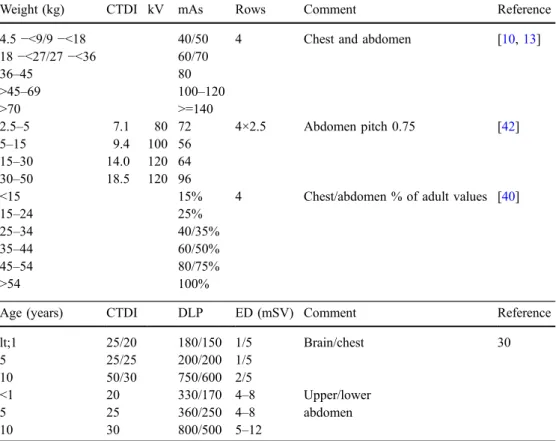
explains the higher effective dose in children undergoing the same protocol as adults. Several authors have shown that to reach the same photon flow at the detector, the tube output (mAs) can be lowered significantly in children. At 120 kVp, Huda reduced it by a total factor of 76 from 1,300 mAs for 120 kg of body weight to 200 mAs for 70 kg and to 17 mAs for 10 kg [21]. Boone [2] reached a constant contrast-to-noise ratio (CNR) for abdominal protocols when he decreased the current from 100% at 28 cm (adult phantom) to 56% at 25 cm, 20% at 20 cm, and 5% at 15 cm, respectively (different paediatric phantoms); the corre-sponding energy dose was lowered from 100% to 64%, 29% and 9%. Based on these physical facts, several authors have suggested weight-adapted paediatric CT protocols (Table3, [9,13,14,42]) aiming at a constant noise level. Alternative to CTDI measurements, measurement of the dose to humanoid phantoms during standard protocols or mathematical calculation of local doses using the Monte Carlo principles can give more realistic effective dose values [27,36]; unfortunately, this approach is not useful in practical scanning of patients.
Aside from the tube output, photon energy (kV) needed to penetrate the body of a child is lower than for the adult body. While in obese adults 140 kVp and in average adults 120 kVp are appropriate, 100 kVp and sometimes 80 kVp may be more adequate for children. Lowering the energy of the photon spectrum will shift the physical interaction of ionising radiation slightly from Compton scatter towards
photon absorption, enhancing the contrast between tissues of different mean atomic weight. In children, this will decrease the effective dose and at the same time increase the low contrast between tissues due to the lack of visceral fat; the lower mean energy of photons may furthermore improve the effect of iodinated contrast agents. However, there are limits to lowering the voltage of the tube: beam hardening artefacts will arise [9], and in practical protocols, 80 kVp are mainly suggested for babies up to 5 kg [42].
Another physical aspect to be considered is slice thickness. While the small dimension of a child asks for thin slices to improve geometric resolution, using identical exposure with thinner slices will automatically increase noise. Keeping the noise level constant requires a more than proportional increase of mAs and, thus, radiation ex-posure. Early multi-row-detector scanners with four detec-tor rows are less dose-efficient than single-row detecdetec-tors and need inappropriately high dose levels for thin slices. With 8–64 detector rows, this phenomenon is less im-portant; however, thinner multi-row detectors will physi-cally increase the dead space between rows in the Z-axis and inherently lower dose efficiency slightly as compared with single-row detectors. In practical paediatric scanning, this clearly asks for a compromise between dose and Z-axis resolution.
Finally, the choice of the minimal dose needed and maximal noise level allowed cannot be made without consideration of the body region studied and the medical
Table 3 Suggested paediatric CT protocols.
CTDI CT dose index, DLP dose-length product,ED effective dose
Weight (kg) CTDI kV mAs Rows Comment Reference
4.5−<9/9 −<18 40/50 4 Chest and abdomen [10,13] 18−<27/27 −<36 60/70 36–45 80 >45–69 100–120 >70 >=140 2.5–5 7.1 80 72 4×2.5 Abdomen pitch 0.75 [42] 5–15 9.4 100 56 15–30 14.0 120 64 30–50 18.5 120 96
<15 15% 4 Chest/abdomen % of adult values [40]
15–24 25%
25–34 40/35%
35–44 60/50%
45–54 80/75%
>54 100%
Age (years) CTDI DLP ED (mSV) Comment Reference
lt;1 25/20 180/150 1/5 Brain/chest 30 5 25/25 200/200 1/5 10 50/30 750/600 2/5 <1 20 330/170 4–8 Upper/lower 5 25 360/250 4–8 abdomen 10 30 800/500 5–12
information needed. In general, high-contrast tissues, such as the lung and the skeleton, tolerate much higher noise levels and still are adequately represented with low-dose protocols. Visceral organs, such as the liver, kidneys and pancreas, often show only minimal density differences between pathological lesions and normal parenchyma; they therefore require higher dose to maintain a diagnostic CNR. Similarly, defining the contours of bones for 3D surgical planning or following-up the size and number of metastatic pulmonary nodules may be done at dose levels of only a fraction of those needed for questions, such as the soft tissue extension of osteomyelitis or mediastinal in-filtration of lung cancer.
Seven rules for an optimised dose reduction in paediatric CT scanning
Justify rigourously
What may be good for the 70-year-old patient of inter-nal medicine may not be an appropriate solution for a pae-diatric patient. In either situation, imaging should not be used to merely demonstrate the morphology and to make a diagnosis; rather, to be justified, it is essential that the result of any examination will be needed to decide on patient management. Hopefully, it will even contribute to the suc-cess of treatment, whether this will be reconstitution of health, prolongation of life or improvement of quality of life; and finally, this should be achieved at a reasonable cost. Beyond the general medical justification, justification of the use of ionising radiation means weighing the ad-vantages of the test versus its disadad-vantages, and both may differ considerably between children and adults. The same effective dose will have more weight in childhood (Table4; [10,13,14,16,43]).
Before any imaging examination with X-rays is con-sidered, alternatives must be evaluated. Obviously in child-hood, ultrasound is the first-line imaging test since the slim body usually favours the access even to deep organs. In experienced hands, it can provide a lot of essential infor-mation, thus avoiding CT. When ultrasound and radiogra-phy are unlikely to answer—or have not answered—the specific medical question, the choice is often between CT and MRI [35]. In this situation, severity of suspected dis-ease, study duration, radiation exposure, side effects of contrast agents and anaesthesia, volume of interest and specific information required have to be evaluated. While there is no general answer, a problem concentrated in one organ or one limited region of the body requiring detailed information of soft tissues, nervous system, cardiovascular system or bone marrow is often best approached by MRI. A large volume of the body, time and anaesthetic re-strictions under emergency conditions such as multiple trauma, the need for information about cortical bone and
calcification favour CT. Malignant disease with a poor prognosis will decrease the weight of radiation exposure; however, with an increasing chance of curative treatment, the added risk of many follow-up studies under and after treatment must be considered.
Finally, follow-up CT scans are often performed too early when, according to the known biology of the disease, one cannot yet expect any effect. Justification has to be as rigourous as for the first examination, and alternatives may be adequate to observe known manifestations of disease.
This rule cannot be followed without profound knowl-edge and experience, which is the reason why education of referring clinicians and radiologists is essential [15,39]
Prepare the patient
In adulthood, patient preparation for CT usually means obtaining informed consent, checking renal function and, for the gastrointestinal tract, instructing the patient about oral bowel contrast application. In childhood, preparation is much more complex but an essential key for success. While older children want to be considered as individuals, in young children, the interaction is not just between the patient, the referring physician and radiology but also with the parents. They often have a better approach to the child and are essential in convincing the child about the need of the study, informing about the procedure and its possible discomfort but also in staying with the child during the examination, calming by hand contact or conversation. Specially trained staff experienced in addressing children and in making them feel comfortable is extremely helpful; similarly, an environment without machines may meet the child’s perception of the world and trigger trust. All actions that can avoid pain and anxiety and, thus, motion artefacts or movement of the child out of the region of interest during scanning should be considered well in advance; this will improve the quality of the examination and sometimes even avoid repeat scans with additional radiation exposure. Individually, it may include information, medication or fixation to allow for painless positioning, insertion of an i.v. line at a remote location well before the examination, sedation, anaesthetic supervision or even general anaes-thesia. For intravenous sedation, propofol is the preferred drug. While general anaesthesia is nowadays tolerated well by the young, retarded or handicapped children, it is more and more possible to avoid it thanks to the fast multi-row detector scanners. For studies of the trunk, it is useful to exercise the cooperation of the child in the scanner without radiation; while it is often wise to accept superficial respiration and not to instruct respiratory cooperation at ages 0–4 years, at ages 5–7 years, apnoea can mostly be achieved, and later on, inspiratory apnoea is suggested. However, these age limits are individually different and really require a test before the scan. The very same patient
may cooperate in a calm atmosphere and, next time, be anxious and uncooperative, maybe just because there is another accompanying person.
Local superficial protective devices have been suggested to protect sensitive organs, such as the lens, the thyroid gland, the breast gland and the gonads, from direct or Table 4 The seven rules for an optimised CT dose reduction in children.CTDI CT dose index, DLP dose-length product, US ultrasound, MRI magnetic resonance imaging, HRCT high-resolution computed tomography
1 Justify CT examination rigourously
- respect age-specific pathology and its prognosis - respect individual paediatric questions
- consider potential contribution of the scan to the management and outcome - respect cost and radiation exposure
- replace CT by examination without (US, MRI) or with lower radiation exposure - delay follow-up examination unless therapeutic decision based on scan is needed now 2 Prepare the patient
- informed consent (parents)
- check renal function and verify hydration - place intravenous line well in advance
- decrease anxiety; calm patient (information, accompanying person) - mark bowel (for abdominal scan)
- avoid pain (immobilisation, positioning, medication) - sedate, anaesthetise
- prepare monitoring, such as oximetry - exercise cooperation without radiation
- put protective device where indicated (lens, thyroid, breast, gonads) 3 Accept noise as long as the scan is diagnostic
- realise that in digital X-ray imaging, noise reduction requires higher exposure - reduce mAs (and possibly kV)
- reconstruct additional thick noise-reduced slices without increase of exposure 4 Optimise scan parameters within the axial plane
- increase tube filtration (if available)
- use maximal slice thickness appropriate for specific diagnosis - decrease kVp for thin objects
- use shortest rotation time available (only few exceptions in children) - decrease baseline mA (CTDI) according to body diameter and composition - use XY-plane dose modulation to minimise CTDI
5 Optimise scan parameters for volume coverage
- use representative volume sample when entire volume is not needed (by sequential scans with gaps) to reduce DLP - use spiral scan with pitch >1 (e.g. 1.5) to reduce DLP
- use thicker collimation with overlapping reconstruction when thin slices are not needed - use Z-axis dose modulation to decrease DLP
- in the near future, use noise-defined automatic exposure control 6 Scan minimal length
- be restrictive in defining upper-most and lower-most limits to keep DLP low - use localising projection scan extending just minimally beyond scan limits 7 Minimise repeated scanning of identical area
- avoid major overlap when scanning adjacent areas with different protocols - avoid non-enhanced scans unless specifically justified (e.g. for densitometry)
- optimise the protocol to obtain all the information requested during one scan (e.g. contiguous 5 mm images and 1 mm HRCT images every 10 mm); minimise number of scans in multi-phase scanning to decrease DLP
- in case of multi-phase scanning, use shorter scan length for additional scans - use lower CTDI for non-enhanced or repeat scans unless high quality is needed - use minimal number of additional sequential functional scans to keep DLP low - minimise length of scans and fluoroscopy time in interventional applications
scattered radiation. Protection from direct radiation using lead- or bismuth-containing devices is generally possible for the localising scout view; however, it will cause ar-tefacts when used in plane for CT scanning. Several authors suggest its use for the breast, eye, testes and thyroid gland [12,19,33]. For the testes, a good reason is the fact that the localiser will often cover a slightly larger Z-axis range and include the testes in abdominal CT examinations whereas the rotational scan will not need to include the male gonads [33]. Unfortunately, there is no good solution for the ovaries: their deep location does not allow superficial excentric protection without severe artefacts. Absorbing radiation scattered during CT scanning of an area close to a sensitive organ is the second approach. It has been shown to be extremely useful for the testes in abdominal scanning (77– 95% reduction, [17, 18, 33]) and still effective for the thyroid and breast glands in head scanning (23–76% reduction, [1,7,17]).
Accept noise as long as the scan is diagnostic
A medical doctor as well as a radiologist basically wants the best for the patient. Since images at higher dose look nicer than those obtained at lower dose, any doctor, equalling nice and good in the mind, tends to prefer the beautiful, higher-dose protocol. This mechanism has in-fluenced doctor-to-radiologist referral practice over many years. Nowadays, both the radiologist and the referring clinician have to realise that image quality cannot be the only criterion when biological facts tell us that ionising radiation may indeed induce cancer at a dose very close to the dose of one CT scan (in around 1‰ of small children). Unfortunately, it is not easy to balance an actual medical need with a rare statistical (stochastic) risk evident only within decades. Since we cannot easily quantify the risk, we should at least try to diminish it. Bringing the dose down to 50% mostly will not affect the diagnosis although the images will be slightly inhomogeneous. Often —de-pending on the organ and the medical question—a greater dose reduction will be tolerable. It is the radiologist’s important task to go to the limits, i.e. to accept as much noise as the specific medical task allows [34]. The practical ways of simultaneously achieving dose reduc-tion and controlling the noise level will be discussed under scanning rules 4 and 5. The acceptable noise level can be defined by guidelines on quality criteria for specif-ic medspecif-ical imaging tasks as initiated by the European Commission [11].
There is another way of reducing the dose and still maintaining the signal-to-noise ratio (SNR) by post-pro-cessing: With modern scanners, while one usually does not want to loose Z-axis resolution by prospectively scanning thicker slices, one can easily acquire noisy thin slices of 0.5–1.5 mm but simultaneously calculate thicker images of
2–6 mm, used primarily for interpretation. The thicker images have a good SNR; the thin images still are used to look at critical details and to get 2D reformation and 3D analysis.
Optimise scan parameters within the axial plane
Different scanners have different geometry, tube filtration, and slightly differing efficiencies of the detectors and the data acquisition system, factors that usually cannot be influenced by the radiologist or technician. It is likely that the market competition will minimise these differences within the next few years. It is also probable that additional filtration will be available for thin patients, decreasing the range of photon energies and therefore reducing the proportion of low-energy photons absorbed almost com-pletely in the body, similar to the current experience in radiography and fluoroscopy. On the other side, we are free to choose the kVp, rotation time and mA. The kVp value needed goes with the diameter of the patient, and paediatric protocols provided by the manufacturer may suggest the appropriate kVp, mostly following the arguments dis-cussed under the physical aspects above (Table 3). The shortest rotation time is mostly appropriate in paediatric CT; since with small objects the capacities of the tube and the acquisition system are not critical, this serves to minimise motion artefacts. Exceptions requiring slower rotation are the same as in adult patients but should be used restrictively. Defining the tube current (mA) needed is clearly the most critical and difficult choice. Again, the general physical rules discussed above apply, and scan-ner-specific suggestions for different regions and ages have been proposed (Table3). In practical work, it may be im-portant to realise that for every reduction of the patient diameter by 3.5 cm, there is roughly 50% less absorption, and the current can be reduced accordingly in children. Unfortunately, no standards of acceptable noise with a specific reconstruction algorithm needed in different med-ical indications have yet been described. Definition of the desired noise level will facilitate scan protocol selection in the near future thanks to interactive dose modulation mechanisms that are currently used in their first generation; since these options for automatic dose reduction are mostly effective in spiral volumetric scanning, they will be dis-cussed under rule 5.
CTDIw, the CTDI weighted for central and peripheral
locations, is the entity reflecting the selection of parameters during one rotation, such as is used in sequential axial scanning, but is also one of the most important parameters in spiral scanning. It is most helpful for comparing the relative exposure by different protocols. However, it is clearly based on a round phantom and neither respects the diameter, the shape nor the composition of the individual patient.
Optimise scan parameters for volume coverage
The way we scan the volume to be studied is the single-most important factor in CT radiation exposure. The term used to characterise volume exposure is the dose-length product (DLP), a parameter directly derived from the product of the CTDIwand the length of the scan. DLP has
the same restrictions as CTDIw of being a physical
parameter not adapted to the individual patient body. But DLP and CTDIw have the important advantage of being
measurable and thus offered by the scanner at the end of a study or even earlier for prospective planning. Since the literature gives factors to translate DLP values into effective dose (Table5, [8,38]), DLP as the only practical risk parameter must be checked regularly by both the radiologist and the technician. Historically, with sequential CT, contiguous slices were usually measured, giving a more or less homogeneous dose distribution that we define as 100%. If one wanted to improve Z-axis resolution, one had to use some overlap; an overlap of 20% (e.g. slice 5 mm, distance between slices 4 mm) increased exposure to 120%. On the other side, for high-resolution computed tomography (HRCT) in diffuse interstitial disease of the lung, scanning a sample of 10% of the organ (1 mm slice, distance between slices 10 mm), as often considered adequate, reduces exposure to 10%.
The introduction of spiral CT scanning with a single row of detectors avoided overlapping scanning, leaving expo-sure at 100% in the example cited, even when images were reconstructed at smaller distances of 1–4 mm; of course, this was only true with identical parameters and when table movement during one rotation was exactly the value of the slice collimation; this basic condition was defined as a pitch of 1 and, in consequence, a movement of twice the collimation was called a pitch of 2. For this type of scanner, it was therefore attractive to increase the pitch in order to reduce radiation exposure [10], with the only restriction that high pitch values caused a major thickening of the resulting slice above the collimation. Although not important for long Z-axis volume scans, spiral scanning means a small additional exposure outside the defined volume during the first and the last rotation of the gantry
since data are incomplete and have to be discarded partially.
Current multi-row detector scanners have increased the options for protocols enormously but also share a disadvantage in performing the HRCT protocol of the lung and other applications where partial sampling of a volume would be medically adequate. They may have to scan two or four slices instead of the single one needed, and collimation at the detector may cause a loss of signal. Aside from this restriction, however, they are mostly used in the spiral mode and have enhanced the speed and resolution of CT scanning, avoiding the problem of tube heating and offering real isotropic data for 3D analysis. The new scan geometry needs more complex image calculation to correct for the diverging beam of the outer detectors, but the operator does not have to take care of this modification. Also, the pitch factor has become less important since the increased speed offers other ways to cover a large volume and still control exposure; similarly, combining the in-formation of different detector rows for the reconstruction of one image has overcome the problem of slice thickening, as seen with early spiral scanners and higher pitch factors. The increased power of modern scanners has mostly eliminated hardware restrictions of older generations and made it easy to define protocols with a high radiation exposure, reaching the range of complex angiographic or fluoroscopic studies. This has increased the pressure of using any solution available to reduce radiation exposure. Current CT scanners offer one or several of the following options:
XY-plane dose modulation: This option was intro-duced to overcome the physical problem that the human body is neither round nor of homogeneous density. To achieve the same SNR, less radiation is required in the direction of the smaller diameter (anteroposterior at the level of the shoulders, Y-axis) than in the direction of the larger diameter (left to right at the same level, X-axis), and this difference is exaggerated by the presence of more bony mass in the X-axis. Modulation of the tube current according to the angle of the tube position around the patient is the logical solution; it is achieved either by estimating the global absorption at all Z-axis positions from an anteroposterior and a lateral localising projectional view or by using the information obtained during one rotation to interactively adapt the tube current for the same angle during the next rotation [40]. XY-plane dose modulation reduces the nominal mAs by around 20–40%, depending on the body region, and it is generally appropriate to use it. Specific new applica-tions of XY-dose modulation are appropriate for the heart and, maybe, the breast gland. This means pro-spectively ECG-triggered lower mA during the phases of the heart that are not used for reconstruction and higher mA during important phases, such as the mid-Table 5 Effective dose estimated from dose-length
product (DLP)a[8,38]
Age Head Neck Chest Abdomen/pelvis
0 y 0.011/0.027 0.017/- 0.039/0.034 0.049/0.040 1 y 0.007/0.008 0.012/- 0.026/0.021 0.030/0.024 5 y 0.004/0.004 0.011/- 0.018/0.014 0.020/0.016 10 y 0.003/0.003 0.008/- 0.013/0.011 0.0150/0.014 15 y -/0.015 -/0.009 Adult 0.002/0.003 0.006/- 0.014/0.009
0.015/-aNumbers give normalised effective dose per dose-length product
to-late diastoly. A similar approach might be used to decrease the radiation exposure of the breast gland in chest CT of young women by decreasing mA when the tube is located in front and—for compensation—by increasing mA when the tube is in the back of the patient.
Z-axis dose modulation: As for the axial plane, phys-ically in the longitudinal axis of the body (Z-axis), the radiation needed for an adequate SNR will vary with the diameter and density of the patient. For example, in cervicothoracic scanning, the cervical area and the lower chest require much less dose for a given image quality than the thoracic inlet and shoulder area. Sim-ilarly, until recently, one had to interrupt scanning at a level between physically different adjacent body areas; e.g. to use a lower radiation exposure for the upper than the lower abdomen, one had to stop the upper scan at the pelvic rim and to start another scan with modified parameters for the pelvis, often with a significant technical delay. Modern scanners allow for adapting the tube output during one single scan in this and other clinical applications. The option of Z-axis-dependent dose modulation is steered again either from the lo-calising view or interactively; it is clearly welcome to reduce radiation exposure and should be used generally. It must be mentioned that dose modulation is only one important step towards the final goal of noise-defined automatic exposure control and that the solutions im-plemented in current scanners may have rules for adapta-tion not easily understood by the user; one therefore has to be careful not to run into dose augmentation, e.g. by starting the scan at a level with low dose requirement at a nominal mAs value selected for the thickest scan level to be covered. Software tools will simplify the choice in the near future, e.g. will offer a selection of images with different noise.
Control of noise in the image is one approach whereas observation of the DLP per examination is another practical approach: Since in CT examinations the DLP is a good representative of effective dose to a specific area of the body, diagnostic reference levels (DRL) indicating an upper DLP not to be exceeded in typical clinical tasks are the practical solution [37, 44]. DRLs correspond to the third quartile (75% lower values obtained from a popula-tion with the same examinapopula-tion). They do not represent an absolute barrier; however, they should be defined for specific body areas according to the weight and the medical task. Since the DLP is available immediately during the study, each radiologist can prospectively plan the DLP to stay within the specific DRL or, exceptionally and with an appropriate justification, to exceed it for a concrete reason.
Scan minimal length
This rule applies both for the scout view and the rotational scan since there is really no value in going beyond the tissue volume where pathology is suspected. It has to be followed on at two levels: The referring physician and the radiologist have to find a compromise about the minimal body areas to be investigated; the radiologist and the technician have to fine-tune the upper and lower end of the examination [10]. In a lung scan, there is no reason to include the entire thoracic inlet with the thyroid gland as well as the upper half of the abdomen with multiple radiosensitive organs. In a pelvic scan of a boy, there is hardly ever a medical reason to include the testes. In-dependent of the organs included, any increase in scan length will proportionally increase energy deposition and the biological effects of ionising radiation. While other rules are the primary responsibility of the radiologist, the technician and her/his experience are most critical for this rule. In routine scanning, it is simply not justified to extend the length beyond the minimum required. For example, a chest scan has to cover the lowest part of the costophrenic sulcus and, in neoplastic disease, the adrenal glands; any inclusion of more abdominal structures will induce non-justified radiation exposure to sensitive organs.
For two reasons, the rule should be used less strictly for the localising than for the sectional scan. First, radiation exposure, although often neglected in dose estimation, is small during a localising projectional view, usually con-tributing a very low percentage to the global exposure. Second, the localiser has to include the starting and ending levels of the spiral scan and is a prerequisite for properly limiting the scan length to the minimum needed in the specific medical situation.
Avoid non-justified multiple scans of the same area Numerous opportunities exist with the current powerful scanners to scan the same volume of the body twice or even several times. Since there is no longer a technical re-striction, multi-phase studies can be performed without tube heating or data overflow.
Perhaps the most frequent neglection of this rule happens when two adjacent body areas are scanned with different protocols and a large overlap. The obvious example for this may be cervicothoracic scanning in malignant lymphoma; while the head and neck scan is planned on a lateral localiser, the scan of the trunk is planned on an anteroposterior localiser, and large overlaps at the thoracic inlet often cause multiple scanning of sensitive organs, such as the thyroid gland.
A number of medical reasons may require different types of repeat scans of the same area:
– native and contrast-enhanced scan after intravenous bolus injection,
– correct timing of scans, using a test bolus or repetitive scanning of one plane at low dose for bolus triggering of the proper diagnostic scan,
– dynamic enhancement studies, including arterial, pa-renchymal, venous and/or excretion phases of organs, such as the kidney or liver,
– functional lung scans to detect air trapping in inspira-tion and expirainspira-tion,
– supine and prone scans for demonstrating positional gravitational effects,
– CT-guided intervention, with or without fluoroscopy, – screening with thick slices and subsequent detailed
analysis with thin slices.
Some but by far not all of these technical possibilities are justified in medical problem solving, and it is probably the most difficult task of the resident in radiology to think of all these potential options but not to overuse them in view of radiation exposure. It is quite clear that double scanning means twice the radiation exposure as long as the same parameters are used, and even more scans will increase exposure proportionally. Aside from medical experience, a few general guidelines may help to appropriately select the number of scans. First of all, and again, the individual situation of the patient must be checked: Will any of the repeat scans help this patient? Will it influence the man-agement or even the outcome? Is it cost-efficient when
radiation exposure is added to the financial cost? Second, repeat scans can often be limited to a smaller volume or performed at lower dose that will not hide the additional information expected. Third, fixed standard scan timing can often replace individual triggering or test bolus unless cardiovascular disease is present and timing is very critical. Fourth, while CT fluoroscopy is a very helpful tool in case of difficult access, other biopsies or drainages can often be done under CT image control or even under ultrasound guidance. Fifth, in the lung, one single scan can usually be used to obtain all the information needed: using thin de-tector rows of around 1 mm will allow the calculation of both thin HRCT sections at any Z-axis level and thick 5 mm scans, as needed for tumour search or mediastinal analysis; for reformations and 3D post-processing, contin-uous and overlapping images can be prepared from the same raw data.
In conclusion, CT is characterised by a significantly higher radiation exposure than radiography. Based on its excellent diagnostic potential in a range of medical situations and on the increased biologic impact of radiation exposure in children, paediatric CT examinations should follow a strict justification and parameter as well as range selection. The seven rules will hopefully help the radiol-ogist to apply the “as low as reasonably achievable” (ALARA) principle when scanning children.
Acknowledgements The author thanks Rainer Wolf, M.D., for reviewing the manuscript, and Barbara Le Blanc for typing the manuscript.
References
1. Beaconsfield T, Nicholson R, Thornton A, Al-Kutoubi A (1998) Would thyroid and breast shielding be beneficial in CT of the head? Eur Radiol 8:664–667 2. Boone JM, Geraghty EM, Seibert JA,
Wootton-Gorges SL (2003) Dose re-duction in pediatric CT: a rational approach. Radiology 228:352–360 3. Brenner DJ, Elliston CD, Hall EJ,
Berdon WE (2001) Estimated risks of radiation-induced fatal cancer from pe-diatric CT. Am J Roentgenol 176: 289–296
4. Brenner DJ (2002) Estimating cancer risks from pediatric CT: going from the qualitative to the quantitative. Pediatr Radiol 32:228–231
5. Brenner DJ, Doll R, Goodhead DT, Hall EJ, Land CE, Little JB, Lubin JH, Preston DL, Preston RJ, Puskin JS, Ron E, Sachs RK, Samet JM, Setlow RB, Zaider M (2003) Cancer risks attrib-utable to low doses of ionizing radia-tion: assessing what we really know. Proc Natl Acad Sci U S A 100:13761– 13766
6. Brenner DJ, Elliston CD (2004) Esti-mated radiation risk potentially asso-ciated with full-body CT screening. Radiology 232:735–738
7. Brnic Z, Vekic B, Hebrang A, Anic P (2003) Efficacy of breast shielding during CT of the head. Eur Radiol 13:2436–2440
8. Chapple CL, Willis S, Frame J (2002) Effective dose in paediatric computed tomography. Phys Med Biol 47: 107–115
9. Cody DD, Moxley DM, Krugh KT, O’Daniel JC, Wagner LK, Eftekhari F (2004) Strategies for formulating ap-propriate MDCT techniques when im-aging the chest, abdomen, and pelvis in pediatric patients. Am J Roentgenol 182:849–859
10. Donnelly LF, Emery KH, Brody AS, Laor T, Gylys-Morin VM, Anton GA, Thomas SR, Frush DP (2001) Mini-mizing radiation dose for pediatric body applications of single-detector helical CT: strategies at a large chil-dren’s hospital. Am J Roentgenol 176:303–306
11. European Commission (2000) Euro-pean guidelines on quality criteria for computed tomography, EUR 16262EN. Office for Official Publications of the European Communities, Luxembourg
12. Fricke BL, Donnelly LF, Frush DP, Yoshizumi T, Varchena V, Poe SA, Lucaya J (2003) In-plane bismuth breast shields for pediatric CT: effects on radiation dose and image quality using experimental and clinical data. Am J Roentgenol 180:407–411 13. Frush DP, Soden B, Frush KS, Lowry
C (2002) Improved pediatric multi-detector CT using a size-based color-coded format. Am J Roentgenol 178:721–726
14. Frush DP (2002) Pediatric CT: practical approach to diminish the radiation dose. Pediatr Radiol 32:714–717 15. Frush DP, Donnelly LF, Rosen NS
(2003) Computed tomography and ra-diation risks: what pediatric health care providers should know. Pediatrics 112:951–957
16. Golding SJ, Shrimpton PC (2002) Commentary. Radiation dose in CT: are we meeting the challenge? Br J Radiol 75:1–4
17. Hidajat N, Schroder RJ, Vogl T, Schedel H, Felix R (1996) The efficacy of lead shielding in patient dosage reduction in computed tomography (German). Rofo 165:462–465
18. Hohl C, Mahnken AH, Klotz E, Das M, Stargardt A, Mühlenbruch G, Schmidt T, Günther RW, Wildberger JE (2005) Radiation dose reduction to the male gonads during MDCT: the effectiveness of a lead shield. Am J Roentgenol 184:128–130
19. Hopper KD, King SH, Lobell ME, TenHave TR, Weyver JS (1997) Rhe breast: in-plane x-ray protection during diagnostic thoracic CT-shielding with bismuth radioprotective garments. Radiology 205:853–858
20. Huda W, Gkanatsios NA (1997) Ef-fective dose and energy imparted in diagnostic radiology. Med Phys 24:1311–1318
21. Huda W, Scalzetti EM, Levin G (2000) Technique factors and image quality as functions of patient weight at abdomi-nal CT. Radiology 217:430–435 22. Huda W (2002) Effective dose to adult
and pediatric patients. Pediatr Radiol 32:272–279
23. International Commission on Radio-logical Protection (1990) 1990 recom-mendations of the international commission on radiological protection. ICRP publication 60. Pergamon Press, Oxford
24. Kalender WA, SeisslerW, Klotz E, Vock P (1990) Spiral volumetric CT with single breath-hold technique, continuous transport, and continuous scanner rotation. Radiology 176: 181–183
25. Lee CI, Haims AH, Monico EP, Brink JA, Forman HP (2004) Diagnostic CT scans: assessment of patient, physician, and radiologist awareness of radiation dose and possible risks. Radiology 231:393–398
26. Mettler FA, Wiest PW, Locken JA, Kelsey CA (2000) CT scanning: pat-terns of use and dose. J Radiol Prot 20:353–359
27. Mini RL, Vock P, Mury R, Schneeberger PP (1995) Radiation exposure of pa-tients who undergo CT of the trunk. Radiology 195:557–562
28. Nickoloff EL, Alderson PO (2001) Radiation exposures to patients from CT. Reality, public perception, and policy. Am J Roentgenol 177:285–287 29. Nickoloff E (2002) Current adult and
pediatric CT doses. Pediatr Radiol 32:250–260
30. Pages J, Buls N, Osteaux M (2003) CT doses in children: a multicentre study. Br J Radiol 76:803–811
31. Paterson A, Frush DP, Donnelly LF (2001) Helical CT of the body: are settings adjusted for pediatric patients? Am J Roentgenol 176:297–301 32. Pierce DA, Preston DL (2000)
Radia-tion-related cancer risks at low doses among atomic bomb survivors. Radiat Res 154:178–186
33. Price R, Halson P, Sampson M (1999) Dose reduction during CT scanning in an anthropomorphic phantom by the use of a male gonad shield. Br J Radiol 72:489–494
34. Ravenel JG, Scalzetti EM, Huda W, Garrisi W (2001) Radiation exposure and image quality in chest CT exam-inations. Am J Roentgenol 177: 279–284
35. Semelka R (2005) Radiation risk from CT scans: a call for patient-focused imaging. Medscape Radiol 6(01) 1/26/2005
36. Shrimpton PC, Edyvean S (1998) CT scanner dosimetry. Br J Radiol 71:1–3 37. Shrimpton PC, Wall BF (2000)
Refer-ence doses for paediatric computed tomography. Radiat Prot Dosim 90:249–252
38. Shrimpton PC, Hillier MC, Lewis MA, Dunn M (2005) Doses from computed tomography (CT) examinations in the UK-2003 review. UK Health protection agency, NRPB-W67
39. Slovis TL (2003) Children, computed tomography radiation dose, and the as low as reasonably achievable (ALARA) concept. Pediatrics 112: 971–972
40. Suess C, Chen X (2002) Dose optimi-zation in pediatric CT: current technol-ogy and future innovations. Pediatr Radiol 32:729–734
41. United Nations Scientific Committee on the Effects of Atomic Radiation (2000) UNSCEAR 2000 report to the general assembly, vol I: sources. United Nations, New York
42. Verdun FR, Lepori D, Monnin P, Valley JF, Schnyder P, Gudinchet F (2004) Management of patient dose and image noise in routine pediatric CT abdominal examinations. Eur Radiol 14:835–841 43. Vock P (2002) CT radiation exposure in
children: consequences of the Ameri-can discussion for Europe (German). Radiologe 42:697–702
44. Wall BF (2001) Diagnostic reference levels-the way forward. Commentary. Br J Radiol 74:785–788
45. Ware DE, Huda W, Mergo PJ, Litwiller AL (1999) Radiation effective doses to patients undergoing abdominal CT ex-aminations. 210:645–650