Original article
Received: 31 July 2013 / Accepted: 14 October 2013 / Published online: 31 January 2014 © The Author(s) 2013. This article is published with open access at Springerlink.com
Dose Reduction in Standard Head CT: First Results from a New
Scanner Using Iterative Reconstruction and a New Detector Type
in Comparison with Two Previous Generations of Multi-slice CT
C. Ozdoba · J. Slotboom · G. Schroth · S. Ulzheimer · R. Kottke · H. Watzal · C. Weisstanner
Signal-to-noise and contrast-to-noise ratios were best in S64; these differences also reached statistical significance. Image analysis, however, showed “non-inferiority” of scan-ner E regarding image quality.
Conclusions The first experience with the new scanner
shows that new dose reduction techniques allow for up to 40 % dose reduction while still maintaining image quality at a diagnostically usable level.
Keywords Tomography/X-ray computed/scanners ·
Dosage/radiation · Sv radiation dose equivalent · Image reconstruction · Neuroimaging
Abbreviations
CNR Contrast-to-noise ratio
CTDIvol Weighted volume computed tomography dose index
DLP Dose length product E CT scanner Somatom Edge
MDCT Multi-detector computed tomography
PACS Picture Archiving and Communication System S16, S64 16-row and 64-row MDCT scanners with 16
and 64 rows, respectively SNR Signal-to-noise ratio
Introduction
According to the annual reports published by the German Federal Office for Radiation Protection, computed tomogra-phy (CT) accounted for 6.1% of all radiologic procedures, but 51.9 % of the collective effective dose in 2003; by 2008, these percentages had risen to 8 and 60 %, respectively [1, 2]. As the number of installed CT scanners in
industria-Abstract
Purpose Computed tomography (CT) accounts for more
than half of the total radiation exposure from medical pro-cedures, which makes dose reduction in CT an effective means of reducing radiation exposure. We analysed the dose reduction that can be achieved with a new CT scanner [Somatom Edge (E)] that incorporates new developments in hardware (detector) and software (iterative reconstruction).
Methods We compared weighted volume CT dose index
(CTDIvol) and dose length product (DLP) values of 25
con-secutive patients studied with non-enhanced standard brain CT with the new scanner and with two previous models each, a 64-slice 64-row multi-detector CT (MDCT) ner with 64 rows (S64) and a 16-slice 16-row MDCT scan-ner with 16 rows (S16). We analysed signal-to-noise and contrast-to-noise ratios in images from the three scanners and performed a quality rating by three neuroradiologists to analyse whether dose reduction techniques still yield suf-ficient diagnostic quality.
Results CTDIVol of scanner E was 41.5 and 36.4 % less than the values of scanners S16 and S64, respectively; the DLP values were 40 and 38.3 % less. All differences were statisti-cally significant (p < 0.0001).
C. Weisstanner, MD () · C. Ozdoba, PD · J. Slotboom, PhD · Prof. G. Schroth · R. Kottke, MD · H. Watzal, MD
University Institute of Diagnostic and Interventional Neuroradiology, University of Bern/Inselspital, Freiburgstrasse 4,
3010 Bern, Switzerland
e-mail: christian.weisstanner@insel.ch S. Ulzheimer, PhD
Computed Tomography, Siemens Healthcare, Forchheim, Germany
lised countries has steadily increased in the past 2 decades [3], a further rise in both percentages may be expected.
The ever-increasing number of CT studies makes dose reduction one of the most important tasks for manufacturers and operators of CT scanners. This issue can be addressed by developments both in the scanner hardware and software, especially with advanced techniques for image reconstruc-tion that allow to acquire images with reduced dose levels without loss in image quality.
At the authors’ institution, the first machine from series production of a new CT scanner (Somatom Definition Edge; Siemens Healthcare, Erlangen, Germany) was installed in March 2012. This system is equipped with a new type of detector and routinely uses iterative reconstruction for image calculation. The detector has a highly integrated design, thereby reducing noise within the detector’s circuits, which allows for improved signal yield and reduced dose. Iterative reconstruction allows further reducing the applied dose compared with conventional filtered back projection. The fundamentals of iterative reconstruction have long been known and applied in other fields such as nuclear medicine [4]. Since 2009, CT manufacturers started introducing itera-tive reconstruction algorithms commercially on their clini-cal scanners. On the Somatom Definition Edge, an iterative reconstruction approach called Sinogram Affirmed Iterative Reconstruction is implemented.
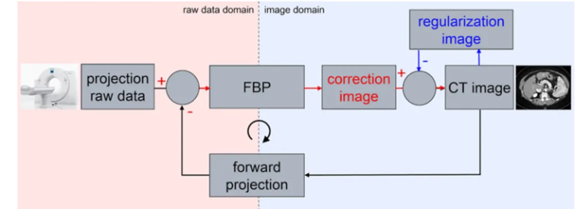
In an iterative loop, noise is removed from the image by modelling image noise and subtracting the resulting regula-risation image from the original data (Fig. 1). In contrast to simple image filters that can also reduce noise but lead to image blurring, iterative reconstruction maintains the high contrast resolution of the image and can remove artefacts from the image. A CT scan is simulated on the existing image data as if a real CT scan would be performed on the reconstructed image data in a step called ‘forward-projec-tion’. The result is simulated projection raw data that can then be compared with the actually measured data from the scanner. The differences between the simulated and mea-sured projection raw data can be used to reconstruct an
improved image; this can be repeated multiple times until the desired image quality level is reached.
The manufacturer claims that more than 30 % dose reduc-tion is possible depending on the applicareduc-tion and the clinical task. In this study, we tested this statement by comparing the dose levels that we achieved in routine imaging with those from the systems previously used for non-enhanced stan-dard brain CT. In addition, we analysed signal-to-noise ratio (SNR) and contrast-to-noise ratio (CNR) to assess whether image quality can be maintained with markedly reduced dose.
Material and Methods
We compared CT studies in routine head imaging in the first 25 patients studied with the new system with the values from the last 25 patients examined in the Edge’s predeces-sor (Somatom Sensation 64, Siemens Healthcare) and the respective values from a 16-slice multi-detector computed tomography (MDCT) system used in our institution’s emer-gency room (Somatom Sensation 16, Siemens Healthcare). This unselected cohort with numerous different diagnoses and a random age- and sex-mix was chosen to reflect a ‘real life’ situation.
We used only non-enhanced CT studies of the head in consecutive patients. Weighted volume computed tomogra-phy dose index (CTDIvol) and dose length product (DLP)
values were taken from the protocols that the scanners automatically send to the hospital’s Picture Archiving and Communication System (PACS). The standard examination protocols used in the three scanners are shown in Table 1. CT scanner Somatom Edge (E) was operated in helical mode, whereas incremental acquisition was used in 16-row MDCT scanner with 16 rows (S16) and 64-row MDCT scanner with 64 rows (S64), as these older machines do not allow helical acquisition with a tilted gantry.
As we measured attenuation values (Hounsfield units) in the frontal cortex and white matter, we excluded cases that might present Hounsfield values altered due to parenchyma Fig. 1 Schematic depiction of
the principle of Sinogram Affir-med Iterative Reconstruction, the iterative reconstruction approach implemented on the Somatom Definition Edge. Image quality is improved by iterative loops both in image space and raw data space to reduce image noise and artefacts, respectively
The non-parametric Mann–Whitney test (two-tailed, 95 % confidence interval) was used to test for significant differen-ces between these values, as these data did not show Gaus-sian distribution.
Image Quality Analysis
Three board-certified radiologists with a neuroradiological experience of at least 2 years assessed image quality in all 75 CT studies. The raters were blinded to the type of scanner used; studies were presented in a random manner. Image quality was evaluated in the supratentorial parenchyma, the basal ganglia (especially with delineation of the insular ribbon sign) and the infratentorial parenchyma. Image qua-lity readings were performed on PACS workstations with reporting-standard monitors; the raters were free to subjec-tively adjust window/level settings. Readers were free not to evaluate images that were degraded due to artefacts. Image quality was graded from 1 to 5:
1. not diagnostic, unusable,
2. reduced quality, diagnostic unsure, better repeat examination,
3. medium quality, still diagnostic, 4. slightly reduced, allows safe diagnostic, 5. good quality, diagnostically perfectly usable. compression or oedema, i.e. patients with
space-occupy-ing lesions, hydrocephalus and intracranial or intracerebral haemorrhage.
The study was approved by the institutional review board. All statistical analyses were performed with GraphPad Prism version 6.01 (GraphPad Software, La Jolla, CA).
Weighted Volume Computed Tomography Dose Index and Dose Length Product Values
In the first step, values were tested for normal (Gaussian) distribution with the D’Agostino–Pearson omnibus normal-ity test. Unpaired t-tests with a confidence interval of 95 % were performed for each possible pair of the three scanners to test for significant differences.
Signal-to-Noise and Contrast-to-Noise Ratios
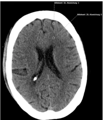
The ‘statistics/circle’ function of our hospital’s PACS (Sec-tra Imtec AB, Stockholm, Sweden; IDS 5, Release 11.4.1) was used to determine Hounsfield values in regions of inte-rest in the frontal cortex and adjacent white matter. The same-size region of interest was used in all patients; measu-red mean values and standard deviation (SD) were used for subsequent analysis. A typical example of the regions used is shown in Fig. 2.
The mean/SD quotient was calculated to determine the SNR. CNR was calculated according to the formula by Mul-lins et al. [5]. where GM Grey matter WM White matter SD Standard deviation mean GM mean WM SD GM SD WM −
(
)
⋅(
)
+(
⋅)
(
2 2 0 5)
.Table 1 Technical parameters of the examination protocols that were
used on the three scanners for standard brain studies Siemens Soma-tom Sensation 16 (S16) Siemens Soma-tom Sensation 64 (S64) Siemens Soma-tom Definition Edge (E) Gantry angulation Cantho-meatal Cantho-meatal Cantho-meatal
mAs 220 380 230 Kiloelectron volt (keV) 120 120 100 Field of view (mm) 220 220 220 Slice thickness/ table feed (mm) 4.5/4.5 4.8/4.8 3.0/3.0 Reconstruction kernel H41s H41s J45s
Fig. 2 Typical region-of-interest measurements in the frontal
cor-tex and white matter (diameter of region of interest: 5 mm).
S64 had statistically significantly lower CTDIvol than S16
(p < 0.0001); the absolute difference was 8 %. E was bet-ween 36 and 41 % lower in both parameters than the two older scanners; these differences were highly significant as well (p < 0.0001 in both instances).
Statistical comparison of the DLP values showed no significant difference between S16 and S64, whereas the The ratings were statistically compared in the same way as
the CTDIvol and DLP values.
Results
Demographic data of the patients are given in Table 2. Weighted Volume Computed Tomography Dose Index and Dose Length Product Values
The D’Agostino–Pearson test showed normal distribution for five of the six values; the only exception was CTDIvol
in S16. We still performed t-tests on all values taking into account that the results involving CTDIvol in S16 may be
imprecise. The CTDIvol and DLP values and the results of the statistical comparison are listed in Table 3 and shown in Figs. 3 and 4.
Table 2 Demographic characteristics of the patients studied in the
three scanners Siemens Soma-tom Sensation 16 (S16) Siemens Soma-tom Sensation 64 (S64) Siemens Soma-tom Definition Edge (E) Sex (male/female) 13/12 16/9 14/11
Age, mean (years) 49.6 59.0 60.3
Age, median
(years) 48.2 60.8 64.8
Age range
(min–max; years) 17.3–89.5 19.5–90.3 20.7–90.0
Table 3 CTDIvol and DLP values measured in the three scanners. Sta-tistical analysis shows a marked dose reduction in scanner E compared with the other models
Siemens Soma-tom Sensation 16 (S16) Siemens Soma-tom Sensation 64 (S64) Siemens Soma-tom Definition Edge (E) CTDI (mGy) Mean 59.63 54.86 34.88 Median 58.49 54.94 34.72 Standard deviation (SD) 2.80 2.93 4.79 DLP (mGycm) Mean 975.16 952.82 574.12 Median 933.00 906.49 559.00 SD 82.62 79.78 82.33 Statistical
comparison t-test CTDI Difference %
S16/S64 p < 0.0001 8.00 S16/E p < 0.0001 41.51 S64/E p < 0.0001 36.42 t-test DLP S16/S64 p = 0.3357 2.84 S16/E p < 0.0001 40.06 S64/E p < 0.0001 38.33
Fig. 3 Weighted volume computed tomography dose index (CTDIvol)
values in the three scanners. The box indicates the range (minimum– maximum), and the horizontal bar in the box denotes the mean value. Scanner ‘CT scanner Somatom Edge’s’ (E’s) values are markedly re-duced in comparison with the two older scanners
Fig. 4 Dose length product (DLP) values in the three scanners. The
box indicates the range (minimum–maximum), and the horizontal bar
in the box denotes the mean value. The 16-row multi-detector com-puted tomography (MDCT) scanners with 16 rows (S16) and 64-row MDCT scanners with 64 rows (S64) show no visible difference, whe-reas the values for CT scanner Somatom Edge are markedly lower
machines with two exceptions: the most experienced rater’s (more than 25 years in neuroradiology) evaluation yielded a statistically significant (p < 0.0001) advantage in scanner E in comparison with S16 for supratentorial parenchyma and basal ganglia. In some instances (Tables 5 and 6), statistical difference, with advantages for the older models, was mis-sed very closely, but scanner E never lost a direct compari-son significantly.
Discussion
The use of a new detector technology and the exclusive use of iterative reconstruction in the standard head clinical scanning protocol on the new scanner resulted in a marked reduction of radiation exposure to the patient; the difference of approximately 40 % that was established in clinical rou-tine is higher than previous manufacturer’s estimates.
CTDIvol and DLP are suitable parameters for such a study,
as they are objective technical parameters for the applied dose. In a recent overview, Huda and Mettler [6] came to the conclusion that these values can be used clinically, as they can be easily obtained from the scanner’s patient protocol; values obtained from E were significantly lower than those
from both S16 and S64 (p < 0.0001 in both instances). Signal-to-Noise and Contrast-to-Noise Ratios
The majority of the values did not pass the test for normal distribution. Therefore, data were compared with the non-parametric Mann–Whitney test. The results are listed in Table 4.
The SNR and CNR of the Somatom Edge are lower than those measured in the two older scanners; these values rea-ched statistical significance (p < 0.0001) only in comparison with the 64-row scanner. The S64 performed best in this comparison (SNR in white matter was 12.69 compared with 8.7 for the S16 and 7.41 for the E; CNR was 3.71 compared with 1.59 for the S16 and 2.19 for the E).
Image Quality Evaluation
The mean ratings are listed in Table 5, and the statistical comparisons are found in Table 6. An intra-rater analysis for the different scanners and anatomical regions showed no statistically significant differences between the three Table 4 Signal-to-noise ratio in cortex and white matter and
contrast-to-noise ratios in the three scanners. S64 shows significantly better va-lues than S16 and E (discussion)
Siemens Soma-tom Sensation 16 (S16) Siemens Soma-tom Sensation 64 (S64) Siemens Soma-tom Definition Edge (E) SNR (cortex) Mean 12.12 16.19 10.12 Standard devia-tion (SD) 3.43 7.88 2.00 SNR (white matter) Mean 8.70 12.69 7.41 SD 1.97 5.55 1.84
Contrast-to-noise (cortex/white matter)
Mean 1.59 3.71 2.19 SD 0.42 1.52 0.61 Statistical comparison p-value SNR (cortex) S16/S64 0.0230 S16/E 0.0434 S64/E < 0.0001 SNR (white matter) S16/ S64 < 0.0001 S16/ E 0.0213 S64/ E < 0.0001 CNR S16/S64 < 0.0001 S16/E < 0.0007 S64/E < 0.0001
CNR contrast to noise ratio, SNR signal to noise ratio
Table 5 Subjective image quality evaluation by the three raters.
Alt-hough the dose in E was significantly lower than in S16 and S64, the readings mostly did not reveal significant differences (exception and details in the text)
Rater 1 Rater 2 Rater 3
S16 Supratentorial 4.68 3.67 3.44 Basal ganglia 4.40 4.04 3.16 Infratentorial 3.76 3.61 3.24 S64 Supratentorial 3.80 3.78 3.72 Basal ganglia 3.92 3.74 3.68 Infratentorial 3.56 3.78 3.68 E Supratentorial 4.08 4.44 4.36 Basal ganglia 3.92 4.52 4.08 Infratentorial 3.72 3.92 4.16
Table 6 Inter-rater variability of subjective image quality ratings
Rater 1
(p-values) Rater 2 (p-values) Rater 3 (p-values) Supratentorial S16/S64 < 0.0001 0.7610 0.2321 Supratentorial S16/E 0.0004 0.0012 < 0.0001
Supratentorial S64/E 0.1056 0.0096 0.0048 Basal ganglia S16/S64 0.0216 0.2435 0.0137 Basal ganglia S16/E 0.0305 0.0369 < 0.0001
Basal ganglia S64/E > 0.9999 0.0007 0.0961 Infratentorial S16/S64 0.4877 0.5694 0.0808 Infratentorial S16/E 0.8052 0.3757 0.0002 Infratentorial S64/E 0.5754 0.8374 0.0574 Statistically significant differences (p < 0.0001) are given in italics
Further studies will have to be conducted to show whet-her more refined image reconstruction protocols will be able to improve image quality more markedly than just ‘non-in-ferior’; first results are promising [11]. For the time being, it can be stated that iterative reconstruction in combination with a high-yield detector is a very promising achievement in current CT technology with regard to dose reduction. Conflict of Interest The authors declare that there are no actual or
potential conflicts of interest in relation to this article.
Open Access This article is distributed under the terms of the
Creati-ve Commons Attribution Noncommercial License which permits any noncommercial use, distribution, and reproduction in any medium, provided the original author(s) and the source are credited.
References
1. Umweltradioaktivität und Strahlenbelastung. Jahresbericht 2005. Bundesministerium für Umwelt, Naturschutz und Reaktorsicher-heit (ed.). http://nbn-resolving.de/urn:nbn:de:0221–20100331990. Accessed 26 July 2013.
2. Umweltradioaktivität und Strahlenbelastung. Jahresbericht 2009. Bundesministerium für Umwelt, Naturschutz und Reaktorsicher-heit (ed.). http://nbn-resolving.de/urn:nbn:de:0221–201103025410. Accessed 26 July 2013.
3. OECD. Health at a Glance 2011: OECD Indicators, OECD Pu-blishing. (2011) http://dx.doi.org/10.1787/health_glance-2011-en (2011). Accessed 26 July 2013.
4. Li J, Jaszczak RJ, Greer KL, Coleman RE, et al. Implementation of an accelerated iterative algorithm for cone-beam SPECT. Phys Med Biol. 1994;39:643–53.
5. Mullins ME, Lev MH, Bove P, et al. Comparison of Image Quali-ty Between Conventional and Low-Dose Nonenhanced Head CT. AJNR Am J Neuroradiol. 2004;25:533–8.
6. Huda W, Mettler FA. Volume CT dose index and dose-length product displayed during CT: what good are they? Radiology. 2011;258:236–42.
7. Bundesamt für Gesundheit, Abteilung Strahlenschutz. Merkblatt R-06–06: Diagnostische Referenzwerte in der Computertomogra-phie. Eidgenössisches Departement des Innern EDI, April 1, 2010. http://www.bag.admin.ch/themen/strahlung/10463/10958/index. html?lang=de. Accessed 26 July 2013.
8. McCollough C, Branham T, Herlihy V, et al. Diagnostic reference levels from the ACR CT accreditation program. J Am Coll Radiol. 2011;8:795–803.
9. ACR Practice Guideline for Diagnostic Reference Levels in Me-dical X-Ray Imaging. Revised 2008 (Resolution 3). Available at: http://www.acr.org/~/media/ACR/Documents/PGTS/guidelines/ Reference_Levels.pdf. Accessed 26 July 2013.
10. Korn A, Fenchel M, Bender B, et al. Iterative reconstruction in head CT: image quality of routine and low-dose protocols in com-parison with standard filtered back-projection. AJNR Am J Neuro-radiol. 2012;33:218–24.
11. Buhk JH, Laqmani A, von Schultzendorff HC, et al. Intraindivi-dual evaluation of the influence of iterative reconstruction and fil-ter kernel on subjective and objective image quality in computed tomography of the brain. Fortschr Röntgenstr. 2013;185:741–8.
DLP allows estimating the patient’s effective dose. For the purpose of this study, these parameters therefore provide a good estimate and allow comparing dose levels from vari-ous scanners.
The increasing awareness for dose reduction has led to the introduction of ‘Diagnostic Reference Values’ by nume-rous organisations, which have partly been implemented in local legislation. The Swiss Federal Office of Public Health has defined this value for a standard brain CT study as a CTDIvol of 65 mGy and a DLP of 1,000 mGycm. The ‘tar-get values’ were set at 45 mGy and 600 mGycm, respec-tively [7]. In the United States, the applied dose has been markedly reduced since the introduction of the American College of Radiology’s (ACR) CT accreditation program [8]; currently, the ACR’s reference value for a head CT is a CTDIvol of 75 mGy [9].
With a CTDIvol of less than 35 mGy for an adult head CT,
the new scanner easily underruns these recommendations. Although responsible CT operators will welcome the new technology that allows for such marked decrease of patient dose, they will not accept a system where these redu-ced doses mean a reduction in image quality. Our results show that, although the dose is markedly reduced, the new scanner’s image quality fulfils the criteria of what is called ‘non-inferiority’ in clinical drug studies with regard to dia-gnostic usability despite reduced SNR and CNR. Although the subjective readers’ evaluations differed in some aspects, the older machines were never rated better than the new machine with statistical significance. We used a subjective readers’ rating instead of phantom studies for the same rea-son why we studied an unselected patient population: we wanted a ‘real-life’ test scenario that seemed more appro-priate to evaluate a new system in practical use (when eva-luating SNR, it should be noted that the protocol on the new scanner uses 3-mm sections compared with 4.5/4.8 mm in the older models; this change was introduced to improve diagnostic accuracy in routine imaging in cases of, e.g. trauma, subarachnoid haemorrhage or suspected cerebral ischaemia). A similar study with only one reader and a 30 % dose reduction has shown that image quality was not com-promised [10]; in this study, a CTDIVol of 42.3 mGy and a
mean DLP of 733 mGycm were achieved. Our data suggest that keeping up image quality is possible with even further reduction.
In conclusion, we may fairly assume from recent developments that the trend to an ever-increasing number of CT installations and CT studies will probably continue. These first data show that the use of state-of-the-art tech-nology provides a way to keep up diagnostic quality while reducing the general population’s radiation exposure.


