JSPP © 1990
Characteristics of Spinach Chloroplast Envelope, Thylakoid and Stroma
Polypeptides as Revealed by Triton X-114 Phase Partition
Paul-Andre Siegenthaler and Nicole Dumont
Laboratoire de Physiologie vigitale, Universite de Neuchatel, 20, rue de Chantemerle, CH-2000 Neuchatel, Switzerland
Comparison of the SDS-PAGE profiles of the spinach chloroplast stroma, thylakoid and envelope membranes shows that several polypeptides have the same electrophoretic mobility. To simplify these somewhat complex electrophoretic profiles and to verify whether the polypeptides having similar electrophoretic mobility are identical, we used Triton X-114 phase partition to ob-tain a separation of the polypeptides according to their relative hydrophobicity. The stroma poly-peptides partitioned essentially in the aqueous phase. About half of the thylakoid and envelope membrane polypeptides were exclusively recovered in either one of the two phases. Therefore, the phase partitioning of membrane polypeptides proved to be useful, as the organic phase con-tained true intrinsic polypeptides, while the aqueous phase was composed of peripheral ones and stroma components. Particularly interesting was the release of the RubisCO large subunit known to copurify with the envelope membranes. Additional experimental approaches were used (immunology, proteosynthesis in organello) to further characterize proteins which had ap-parent ambiguous phase partitioning. Here, we show that Triton X-l 14 is an excellent tool to un-mask polypeptides having identical electrophoretic mobility but different behaviour towards this detergent; its use leads to a clarification of the polypeptide SDS-PAGE profiles of chloroplast membranes.
Key words: Electrophoretic separation — Envelope membranes — Hydrophobicity — Immunol-ogy — Spinach chloroplast polypeptides — Triton X-114.
Chloroplasts from higher plants are made up of six The electrophoretic analysis of the inner and outer different compartments (the outer and inner envelope mem- envelope membranes in one-dimension shows that each branes, the intermembrane space, the stroma, the thyla- membrane has its own set of polypeptides which differ koid membranes and the thylakoid lumen), each of which from those of the thylakoid and the stroma (Joyard et al. having its own functions and a distinct set of polypeptides. 1982, Werner-Washburne et al. 1983). However, the two The envelope serves as a boundary between the cytosol and envelope membranes are linked together by contact sites as the chloroplast and is the site of many enzymatic activities shown by freeze-fracture electron microscopy (Cline et al. on top of its role as a selective barrier for the movement of 1985) and freeze-fracture and freeze-substitution combined molecules in and out of the chloroplast (Douce et al. with ultrathin sectioning (Cremers et al. 1988). Upon 1984). The envelope, the polypeptides of which represent separation of the two membranes in hypertonic medium only 1-2% of the total chloroplast proteins, is composed of followed by their purification, these contact sites give rise two membranes with different densities and permeabilities to stroma-containing vesicles migrating with the inner mem-(Douce et al. 1984). brane during centrifugation. The characterization of the two envelope membranes is thus complicated by the pres-Abbreviations: Mops, (3-[N-morpholino]propanesulfonic ence of stroma. Furthermore, even though the envelope acid); PMSF, phenylmethane sulfonyl fluoride; PAGE, polyacryl- membrane purification methods have been greatly improv-amide gel electrophoresis; IEF, isoelectric focusing; pi, isoelectric ed, it is not possible to obtain pure fractions and the elec-point; TX-114, Triton X-l 14 (alkylphenylpolyethyleneglycol); TX- trophoretic profiles may not be totally representative of the 100, Triton X-100; T, thylakoid; S, stroma; IM, inner membrane; subchloroplast fractions studied.
OM, outer membrane; RubisCO, ribulose 1,5-bisphosphate carb- Moreover, they are difficult to analyze as different poly-oxylase/oxygenase; Mr, apparent molecular weight. peptides may display the same electrophoretic mobility and
1102 P.-A. Siegenthaler and N. Dumont
migrate as a unique Coomassie blue band.
In an attempt to go deeper into the characterization of the two envelope membrane polypeptides and to assess and identify the source of any cross-contamination that may oc-cur during the purification of the two membranes, we have used TX-114 temperature-induced phase partition as described by Bordier (1981). TX-114 is the only detergent of the TX-100 series which forms clear micellar solutions in water at low temperature (4°C) and separates above 20°C into two phases, respectively enriched and depleted in detergent. The TX-114 phase partition has been used suc-cessfully by Bricker and Sherman (1982) on maize thyla-koid membranes and, more recently, by Kjellbom et al. (1989) on spinach leaf plasma membrane. Here, we have applied the TX-114 phase partition method to chloroplast envelope membranes with the specific aim of clarifying their complex electrophoretic profiles. For comparison, we have included stroma and thylakoid membrane elec-trophoretic separations in order to better detect possible contamination by these fractions. This technique gives rise to a preliminary separation of the proteins according to their hydrophobic/hydrophilic character while keeping them in a membrane-like environment, thus preserving their native form. This treatment, followed by electropho-resis, enables to separate several polypeptides of similar Mr but of different behaviour towards TX-114 phase partition and helps to identify polypeptides of different origin. However, due to ambiguous phase partitioning and to poly-peptide comigration that may still exist, it was imperative to include other criteria based on results from 2-D electro-phoresis, immunology and proteosynthesis in organello, to differentiate between those polypeptides that could have been otherwise mistaken for others. The present study ex-tends the results already presented in 1988 (Dumont et al.) and 1989 (Dumont and Siegenthaler).
Materials and Methods
Isolation of purified, intact chloroplasts—Chloro-plasts were obtained from 800 g of spinach leaves bought at the local market. The deribbed leaves were blended in a four-liter Waring blender for a total of 10 to 15 sec in a chilled grinding medium (250 ml/100 g of leaves) contain-ing 350 mM sorbitol, 25 mM Mops, 2 mM EDTA-Na2 and 2mM isoascorbate adjusted to pH7.6 with KOH. After filtration on 6 layers of muslin and one layer of cheesecloth, the resulting filtrate was centrifuged at 2,100xg (Sorvall GSA rotor) for 1 min. Each pellet was resuspended in
10 ml of grinding medium and layered on top of a 40% Per-coll solution containing the same ingredients as the grin-ding medium. After centrifugation (2,100 x g in a Sorvall HB-4 rotor for 3 min), the pellets of purified chloroplasts were resuspended in the grinding medium and centrifuged at 2,000 x g (Sorvall SS-34 rotor) for 3 min to wash out the
residual Percoll.
Purification of envelope membranes—The fractiona-tion of the chloroplasts into soluble and membrane pro-teins was achieved mainly according to Keegstra and You-sif (1986). The purified chloroplasts were resuspended in hypertonic medium and the chlorophyll adjusted at 2.5 mg per ml (Bruinsma 1961). After a 15 min incubation on ice, the chloroplasts were ruptured with a Dounce homogenizer and diluted with 2 vol of TE buffer (10 mM Tricine-NaOH, pH7.6, 2mM EDTA-Na2). The thylakoids were sedimented by a three 10 min steps differential centrifu-gation at 4,500, 13,500 and 23,500xg in a Sorvall HB-4 rotor using slow acceleration. This centrifugation pro-cedure led to a substantial increase in the envelope mem-brane yield. The yellow supernatant was layered on top of a discontinuous sucrose gradient consisting of 8 ml of each of 1 M, 0.65 M and 0.4 M sucrose solutions buffered with TE + MgCl2 (5 mM) in the presence of 1 mM PMSF and cen-trifuged for 6h at 100,000 x g in a Beckman SW-27 rotor. The stroma was recovered at the top of the gradient. The outer and inner envelope membrane fractions, found at the sucrose interfaces, were collected, diluted with TE + PMSF and sedimented at 100,000xg for 60min in a SW-27 rotor. After resuspension in TE+PMSF, the protein con-tent was measured (Bradford 1976).
Separation of stroma, thylakoid and envelope mem-brane proteins into hydrophobic and hydrophilic fractions —The proteins of the stroma, the thylakoid and the inner and outer envelope membranes were separated according to their hydrophobicity and hydrophilicity using the prop-erties of Triton X-l 14 as described by Bordier (1981). The original method was slightly modified: in order to avoid ex-cessive loss of proteins, no sucrose cushion was used be-tween the aqueous and the detergent phases; the detergent phase was extracted twice and its surface washed to eliminate contamination from the soluble proteins. The protein sample (100/ig) was adjusted to \% TX-114 with a solution containing 2% of precondensed TX-114, 10 mM Tris-HCl, pH7.4 and 150 mM NaCl and solubilized for
10 min at 0°C. To allow condensation, the sample was then incubated for 10 min at 30°C in a water bath before be-ing centrifuged for 3 min at 300 x g in a swbe-ingbe-ing bucket rotor (Universal Hettich 1200) at room temperature. From then on, the two phases were treated separately and the extraction was repeated once for the detergent phase and twice for the aqueous one. The surface of the detergent phase was washed with 500 (A of buffer. The pro-teins of the two phases of interest were precipitated over-night in ice cold 80% acetone at - 2 0 ° C . They were then centrifuged for 10 min at 10,500 x g in a Sorvall HB-4 rotor before being prepared for isoelectric focusing or SDS-poly-acrylamide gel electrophoresis (SDS-PAGE).
Electrophoresis and autoradiography—The precipi-tated proteins were denatured in boiling water for 3 min in
2% SDS, 0.005% bromophenol blue (w/v), 6.25% glycerol
and 5% mercaptoethanol (v/v) in 0 . 0 6 2 5 M Tris-HCl,
pH6.8. The samples were electrophoresed essentially ac-cording to Laemmli (1970) for 5 to 6 h on slab gels contain-ing either 12% acrylamide throughout or a linear 10 to 18% (w/v) acrylamide gradient accompanied by a 5 to 15% (w/v) sucrose gradient. The acrylamide to N,N"-methylenebisacrylamide ratio was 30 :0.8. The gels were fixed in a destaining solution containing 25% denatured ethanol and 8% acetic acid. They were either stained in 0.25% Coomassie brilliant blue R-250 (w/v) in 50% methanol and 10% acetic acid (v/v) or as described by Neuhoff (1988). After destaining, they were dried under vacuum. Prior to drying, the bands of the 10 to 18% gra-dient acrylamide gel were scanned at 600 nm in a Zeiss-Disc ZK4 gel scanner and apparent mol wt estimated by calcu-lating the relative mobility of the polypeptides compared to the following calibration proteins: phosphorylase b (94kDa), bovine serum albumin (67kDa), ovalbumin (43 kDa), carbonic anhydrase (30 kDa), soybean trypsin in-hibitor (20.1 kDa) and a-lactalbumin (14.4 kDa). Auto-radiographs were prepared on Kodak X-Omat AR films.
Isoelectric focusing—The organic and aqueous sam-ples, obtained from an initial amount of 300 fig of protein, were solubilized as described by Dunbar (1987). The isoelectric focusing procedure and pH determination were essentially according to Siegenthaler and Nguyen (1983). The gels were 7 cm long and contained 2% (v/v) TX-100 in-stead of Nonidet-P40. At the end of the run, the gels were treated as described by Jackie (1979) before being submit-ted to the second dimension on a 12% SDS-gel.
Western blotting—After SDS-PAGE, the polypeptides were transferred to cellulose nitrate sheets using a semi-dry electroblotting apparatus (Sartorius). The electrophoretic buffer contained 48 mM Tris, 39 mM glycine, 1.3 mM SDS and 20% (v/v) methanol (pH at about 9) (Heegaard and Bjerrum 1986).
Immunological studies—The control and TX-114 or-ganic and aqueous phase fractions of the thylakoid, stroma and envelope polypeptides were challenged with several antibodies by the method of Towbin et al. (1979), except that 10 and 3% bovine serum albumin were replaced by 5 and 1% powdered milk and that no carrier serum was used. The secondary antibody directed against the first antiserum was labelled with alkaline phosphatase (KPL Laboratories) and used as described by Leary et al. (1983). Antibodies raised against the ribulose 1,5-bisphosphate carboxy-lase/oxygenase were kindly provided by Dr. A. Radunz.
Results and Discussion
Although the electrophoretic patterns of the four purified chloroplast fractions, stroma (S), thylakoids (T), in-ner (IM) and outer (OM) envelope membranes, are
differ-ent, there are some comigrating polypeptides which may be either polypeptides with identical Mr but different physico-chemical properties or the result of some cross-contamina-tion. Furthermore, the stroma can be expected to contain polypeptides of nucleic origin which accumulate there when their insertion into the thylakoid membranes is prevented (Cline et al. 1989). Polypeptides of chloro-plastic origin can also be expected to accumulate there.
To investigate these possibilities, we separated the hy-drophobic from the hydrophilic polypeptides in each of these fractions, using the Triton X-114 phase partition method.
For sake of clarity, it is necessary to remind (Bennett 1982) that membrane proteins have been classified as in-tegral or peripheral proteins. Inin-tegral membrane proteins can be defined as being "globular" or "fibrous". The "globular" proteins, such as a membrane-spanning pro-tein, have a significant portion of their mass within the bilayer, while the "fibrous" ones have most of their mass protuding from the membrane and a hydrophobic poly-peptide tail dipping into the membrane bilayer. Peripheral proteins, on the other hand, are membrane bound through non-covalent, probably mainly electrostatic interactions with the hydrophilic regions of integral proteins or with headgroup regions of the lipid bilayer. Once separated from the membrane, these proteins behave as soluble pro-teins and will therefore be recovered in the detergent-free phase.
From the definition given above, it is expected that "globular" proteins will partition in the detergent phase, while the "fibrous" ones, depending on the relative impor-tance of their hydrophobic tail, may have an ambiguous behaviour and be recovered in the two phases.
TX-114 phase partition: extraction conditions
In his original procedure, Bordier (1981) recommend-ed one extraction of the organic phase and three for the hy-drophilic one. However, preliminary results showed that two extractions of the detergent phase were necessary to achieve a complete removal of the hydrophilic poly-peptides.
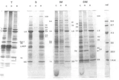
SDS-PAGE analysis of the four TX-114-treated fractions Introduction—Four fractions (T, S, IM and OM) were prepared and then submitted to TX-114 extraction. The electrophoretic patterns of the resulting aqueous and organ-ic phase polypeptides, along with their corresponding con-trols, are shown in Fig. 1. Although each phase contained its own set of polypeptides, there were still several similarities in the electrophoretic mobilities. Among the polypeptides having the same electrophoretic mobility, some had a similar behaviour towards TX-114 treatment and might therefore be identical, while others did not parti-tion in the same phase, thus indicating their different
1104 P.-A. Siegenthaler and N. Dumont nature. As no chlorophyll could be detected in the stroma
or the envelope membranes, contamination by thylakoid components could be ruled out. However, stroma materi-al, arising from inner membrane-derived vesicles, can be present in the envelope fractions (Cremers et al. 1988) as well as in the thylakoid one.
Membrane phase partition—The complexity of the electrophoretic patterns of the membrane polypeptides, especially those of the envelope membranes (IMc, OMc), can be partly overcome by separating these polypeptides ac-cording to their hydrophobic and hydrophilic properties. For instance, one can see (Fig. 1, IMo, OMa) that most of the inner membrane polypeptides were recovered in the or-ganic phase while those of the outer membrane partitioned preferentially in the aqueous phase. On close exam-ination, about half of the envelope membrane polypeptides were recovered in only one of the two phases. The parti-tioning of the polypeptides was therefore quite good whenever strongly hydrophobic or strongly hydrophilic polypeptides were concerned. Indeed, the inner mem-brane 94, 34 and 33 kDa, the outer memmem-brane 76, 66 and 22 kDa polypeptides, the thylakoid LHCP and Cyt. f (as lo-calized also by Bricker and Sherman 1982) as well as several thylakoid low Mr polypeptides were exclusively recovered in the organic phases (Fig. 1, IMo, OMo and To). In con-trast, the thylakoid 30 kDa and the outer membrane 109, 40, 24, 23 and 21 kDa polypeptides (Fig. 1, Ta and OMa) were only encountered in the aqueous phase. Consequent-ly, by discriminating between hydrophobic and hydrophilic polypeptides, TX-114 enhanced the SDS-PAGE resolution. Other polypeptides were common to the two phases and their characterization as integral ("globular" and
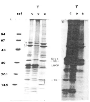
"fibrous") or peripheral is more difficult. As there are several answers to that situation, complementary methods have to be used. Here, we may be dealing with poly-peptides that were not totally removed from the membrane (Bordier 1981). These polypeptides could be "fibrous" pro-teins, the solubilization of which depends strongly on the nature of their hydrophobic tail and its interactions with the membrane. Such may be the case of the IM81 and 70 kDa polypeptides which were found simultaneously in both the organic and aqueous phases, as revealed by 2D electrophoresis (see arrows in Fig.2A and B). However, the a and P subunits of CF,, known as being hydrophilic polypeptides, failed to partition completely in the aqueous phase (see Figs. 1 and 3, To, a). As suggested by Bricker and Sherman (1982), a small amount of CF, may remain at-tached to CF0 or, as these polypeptides are very abundant, there could be a spilling over phenomenon. In other in-stances, we were dealing with two or more totally different polypeptides. Indeed, the thylakoid 15 kDa band (Fig. 1 and Fig. 3A, Tc, o, a) partitioned unequally between the two phases. Figure 3B shows that it was in fact resolved in-to a hydrophobic polypeptide synthesized in the chloro-plast (as attested by its 35S-labelling in the organic phase) and a hydrophilic one of cytoplasmic origin (no labelling in the aqueous phase).
Comigration may also occur in one of the phases. As shown in Fig. 2C, a unique Coomassie band (Control: aqueous phase 72 kDa) may contain two polypeptides hav-ing different pi (see arrows). In this case, they were not charge isomers but again polypeptides of different genomic origin as shown by proteosynthesis experiments (results not shown).
T S
c o s c o a
I I t
Fig. 1 Electrophoretic separation of thylakoid, stroma, inner and outer envelope membrane polypeptides. The TX-114 phases were extracted twice and the aqueous phases three times. The references (ref) and the separating gel containing a linear 10-18% acrylamide gradient are as described in Materials and Methods. Each control fraction (c) contained 30 fig of untreated polypeptides. The organic (o) and aqueous (a) fractions were obtained from an initial amount of 100/ig of proteins. T, thylakoid; S, stroma; IM, in-ner membrane; OM, outer membrane. Apparent mol wt values are indicated in kDa.
ref @ ^_ IEF — 0 IMO Stroma phase partition—All but five of the stroma - «•- _ polypeptides were recovered in the aqueous phase (Fig. 1
/81 j | Sa). The presence of Coomassie bands in the organic 9 4
" i f "**" /7 0 3 * phase (Fig. 1 So) was rather unexpected, but nevertheless, 67 -*P ""_-— ~* ~^~ TX-114 phase partitioning was highly reproducible. These ~ __ """* 3jt faint bands corresponded to polypeptides having Mr of 64, 4 3
w •-•••. 3 57, 54,44 and 41 kDa as determined by densitometric trac-'*" *# ings (results not shown). Since bands of equivalent Mr ^ could be seen in the organic phases of the membrane frac-30 - • & tions, envelope and/or thylakoid components might have r? contaminated the stroma. But, if contamination by mem-^ brane material occurred, one would have expected to find 20.1 • all or at least most of the membrane hydrophobic poly-peptides in the stroma organic phase (Fig. 1 So). Since it 1
I was obviously not the case, one can reasonably exclude any 14i
* * - ^jjl significant contamination of the stroma fraction by the —— envelope or thylakoid membranes. Furthermore, the stroma was unlikely to be contaminated by thylakoids since the stromal fraction did not contain any trace of chloro-r e f
© -~ IEF ^ 0 IMa phyll. T h e 63 kDa band was doubtful as it comigrated with an artifact sometimes present in our electrophoreses. _, _ ^ The presence of these hydrophobic proteins is best explain-6 7 • - / * -*J~ • S*^t ==** ec* by assuming that we are dealing with transit poly-peptides of chloroplastic (S57, 54 kDa) or nuclear (S44, 4 3
^ %s*.- " 41 kDa) origin, as suggested by proteosynthesis ex-periments (results not shown) and by Cline et al. (1989). Indeed, it is expected that these polypeptides, the fate of 3 0
* ^ ^ ^ ^ which is to be eventually integrated in a membrane, contain several hydrophobic domains which would favour an or-~ - ganic phase-partitioning.
* Peripheral membrane proteins or soluble
contami-': * . ' nants?—Another interesting feature of the polypeptide
1 4 4
^ -" *"f separation in the presence of TX-114 was the unexpectedly great number of envelope membrane polypeptides having a ^ hydrophilic character (Fig. 1 IMa, OMa). These may be peripheral (loosely membrane-bound) polypeptides or stroma components. The latter was indeed the case with
ref © — IEF — 0 Sa the large and small subunits of the RubisCO which are
known to copurify with the envelope membranes (Pineau et al. 1979, Joyard et al. 1982). Such was also the case 94 <«> i -^£ Z 4 with the inner membrane 72 kDa (Fig. 2B: pi 5.8) which, 67 m PI58 ~ - 5^L ~ 7 2 when present, reacted with the antibody against the stroma72 i 72 kDa raised in our laboratory (data not shown). On the 4 3
* ^ t o ^ * * - i _ *W o t n e r hand, OM 35, 23 and 21, found exclusively in the
I
membrane aqueous phase, did not have any counterpart in the stroma (Fig. 1 Sa). This strongly suggests that these 3 0** £ , outer membrane polypeptides are true peripheral
com-20.1 # ' ^ „ tr
% • • •• ' ^ ^ * Fig. 2 2-Dimension electrophoretic separation of the organic (A) .- _ _ _ -•»' •>„ ( and the aqueous (B) phases of the inner membrane and the aqueous 1 4 4
^ ""-^ • f c s s phase of the stroma (C). The IEF was as described in Materials - •• and Methods and the separating gel of the second dimension
1106 P.-A. Siegenthaler and N. Dumont ref c o a 94 67 43 30 20.1 14.4
Fig. 3 Electrophoretic separation of labelled thylakoid poly-peptides after TX-114 treatment (A) and its autoradiography (B). Experimental conditions and symbols as in Fig. 1. For proteosynthesis in organello, intact chloroplasts were resuspended (1 mg chl/ml) in an incubation medium containing 100 mM KC1, 2 mM MgCl2 and 66 mM Tricine-KOH at pH 7.6. The light-driven
incorporation of 35S-methionine (150/*Ci/mg chl; specific activ-ity > 1,000 Ci/mmol; SJ 1515, Amersham) was performed in a water bath at 20°C for 60min.
ponents of the envelope. It is thus clear that the preliminary separation obtained through the use of the TX-114 phase partition succeeds in eliminating all the stroma trapped in the inner or outer envelope membrane vesicles. The organic phase polypeptides are thus exclusively of mem-brane origin, while those found in the aqueous phase may still have a dual origin, namely membrane and stroma.
Proteins revealed by TX-114 phase partition—It is noteworthy that the use of this technique allowed the visualization of polypeptides which have close Mr and are normally partly overlapping. This was most obvious near the large subunit (LS) of the RubisCO. Indeed, a 54 kDa polypeptide was found in the organic phases of the stroma, of the inner and outer envelope membranes at the level of the RubisCO (Fig. 1 So, IMo, OMo). The RubisCO, in its
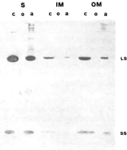
18 S holoenzyme form, is known to remain bound to the envelope membranes during their purification (Joyard et al. 1982) and has been, so far, impossible to eliminate (Pineau et al. 1979, Werner-Washburn et al. 1983). To assess the nature of this 54 kDa organic band, an antibody raised against the RubisCO was tested on the stroma, inner and outer envelope membrane TX-114-treated fractions (S, IM and OM) and their respective controls. The results are
shown in Fig. 4. A positive reaction was found only in the controls and the aqueous phases of the three fractions. No reaction could be detected on any of the organic phases. Furthermore, a polyclonal antibody was raised in our laboratory against the stromal organic phase 54 kDa poly-peptide. When tested, it did not react with LS in any of the aqueous phases but reacted with all three organic phases (see Fig. 5). The rather weak reaction of the con-trols can be explained by the presence of the LS which rep-resents most of the 30 fig of total protein content. There is, therefore, a fair probability that the hydrophobic 54 kDa polypeptide, unmasked in the three fractions by TX-114 treatment, is not the large subunit. It is noteworthy that the RubisCO, which is not released by sonication and other drastic treatments (Joyard et al. 1982, Werner-Washburne et al. 1983), can be eliminated from the envelope mem-branes by TX-114 phase partition. Moreover, the RubisCO small subunit band in the IM and OM fractions was split between the organic and aqueous phases. Fig. 4 shows that the polypeptide with hydrophobic behaviour was not the small subunit (SS) as there was no immuno-logical reaction with the RubisCO antibody.
Solubilization versus phase partition—The complex mixture of membrane proteins can also be fractionated by solubilizing them in chloroform/methanol (2 : 1 v/v) or in 0.1 N NaOH as described by Joyard et al. (1982). On the basis of their results, these authors concluded that 55 to 60% of the total envelope polypeptides are integral mem-brane proteins, most of them being characterized by a high Mr. A hydrophobic character was assigned to half of these integral proteins by virtue of their chloroform/ methanol solubility. The other 40 to 45% of the envelope polypeptides were released from the membrane by NaOH treatment and were thus considered as peripheral proteins. The above two solubilization methods may lead, through delipidation or limited saponification of lipids, to a total or partial destruction of the membrane. Furthermore, the quaternary and tertiary conformations of the proteins are probably not preserved, leading to the exposure of hydro-philic and hydrophobic domains which may be erroneously determinant towards the final solubilization and the charac-terization as intrinsic or extrinsic. This drawback does not happen with TX-114 phase partition, which has been shown by the works of Sanchez-Ferrer (1989a, b, 1990) and Soil and Bennett (1988) to keep the proteins in their native state, thus allowing the isolation of functional enzymes. The resulting protein separations obtained by TX-114 solu-bilization and phase partitioning or by solusolu-bilization in chloroform/methanol or NaOH are in fact based on very different properties and cannot be compared. Indeed, the polypeptides E37 and E24 described by Joyard et al. (1982), corresponding respectively to our IM34 and OM22 (Fig. 1, IMo, OMo) were not solubilized in chloro-form/methanol but partitioned in TX-114 as integral
poly-S c o a IM C O i OM c o a
S I M O M
c o a c o a c o a
ssFig. 5 Antibodies against the hydrophobic 54 kDa were tested on a Western blot as described in Fig. 4. Only the relevant part is shown. The antigen was purified on SDS gel electrophoresis. The desired gel bands were pooled, crushed in liquid nitrogen and dissolved in the complete Freund's adjuvant diluted 1 : 1 with distilled water. Three injections were made intradermically at 2 to 3 weeks intervals in a white New Zealand rabbit. Symbols are as in Fig. 1.
Fig. 4 Western blot of stroma, inner and outer envelope
mem-brane polypeptides after treatment with TX-114 and separation on a 12% acrylamide gel slab. Antibodies against the RubisCO (LS, SS) were tested. Symbols are as in Fig. 1.
peptides. However, the two results taken together could very well indicate that E37 and E24 are "fibrous" proteins according to the definition given above. However, it is in-teresting that the E14 (corresponding to our IM14 and OM14) which was not totally extracted by chloro-form/methanol (Joyard et al. 1982), was also found split between the aqueous and organic fractions of the inner and outer membranes (Fig. 1 IMa, o and OMa, o). As we have shown, theses organic bands were not related to the RubisCO small subunit as they did not react with the RubisCO antibody (Fig. 4). It is also likely that the faint band (at the E54 level), found by Joyard et al. (1982) in the chloroform/methanol-soluble fractions from stroma and envelope, is identical to the one found in our stroma and membrane organic fractions, and which was shown to be different from the RubisCO LS (Figs. 1, 4 and 5).
Conclusions—To decrease the complexity of the elec-trophoretic patterns of the envelope membrane poly-peptides, it is thus advisable to fractionate the total set of proteins prior to electrophoresis. The TX-114 phase parti-tion method, which is highly reproducible and easy to han-dle, has been shown here to be a valuable tool for chloro-plast envelope membranes. It provides a physico-chemical environment suitable for membrane protein purification and allows the separation of integral proteins from peripheral and soluble ones.
Furthermore, the organic phases of the inner and outer envelope membranes contain "globular" proteins probably involved in chloroplast transport and communica-tion. Moreover, the two RubisCO subunits, which are
always present in the three membraneous fractions, cannot be dislodged from the two envelope membranes unless TX-114 phase partition is used. However they are easily wash-ed out of the thylakoid. This suggests (as also proposwash-ed by Joyard et al. 1982) that the RubisCO could be specifically associated with the chloroplast envelope.
Therefore, the ability of TX-114 to separate mem-brane proteins according to their relative hydrophobicity while preserving their native form is a potentially very powerful method towards the characterization and purification of envelope chloroplast polypeptides.
The authors thank Miss Cecile Seuret and M. Daniel Leemann for their able technical assistance and Dr. A. Radunz for his generous gift of RubisCO antibody. This work is part of a doctoral program which is being carried out by N. D. in the Laboratoire de Physiologie v^getale, University de Neuchatel.
References
Bennett, J. P. (1982) Solubilisation of membrane-bound en-zymes and analysis of membrane protein concentration. In Techniques in Lipid and Membrane Biochemistry, Part 1, B408. pp. 1-22. Elsevier/North-Holland Scientific Publishers Ltd, County Clare, Ireland.
Bordier, C. (1981) Phase separation of integral membrane pro-teins in Triton X-l 14 solution. J. Biol. Chem. 256: 1604-1607. Bradford, M. (1976) A rapid and sensitive method for the quan-titation of microgram quantities of protein utilizing the princi-ple of protein-dye binding. Anal. Biochem. 72: 248-254. Bricker, T. M. and Sherman, L. A. (1982) Triton X-l 14
phase-fractionation of maize thylakoid membranes in the investiga-tion of thylakoid protein topology. FEBS Lett. 149: 197-202. Bruinsma, J. (1961) A comment on the spectrophotometric deter-mination of chlorophyll. Biochim. Biophys. Acta 52: 576-578.
1108 P.-A. Siegenthaler and N. Dumont
Cline, K., Keegstra, K. and Staehelin, L. A. (1985) Freeze-frac-ture electron microscopic analysis of ultrarapidly frozen envelope membranes on intact chloroplasts and after purifica-tion. Protoplasma 125: 111-123.
Cline, K., Fulsom, D. R. and Viitanen, P. V. (1989) An imported thylakoid protein accumulates in the stroma when insertion into thylakoids is inhibited. J. Biol. Chem. 264: 14225-14232. Cremers, F. F. M., Voorhout, W. F., van der Krift, T. P.,
Leunissen-Bijvelt, J. J. M. and Verkleij, A. J. (1988) Visualiza-tion of contact sites between outer and inner envelope mem-branes in isolated chloroplasts. Biochim. Biophys. Ada 933: 334-340.
Douce, R., Block, M.A., Dome, A.-J. and Joyard, J. (1984) The plastid envelope membranes: their structure, composition, and role in chloroplast biogenesis. Subcelt. Bioch. 10: 1-86. Dumont, N., Bovet, L. and Siegenthaler, P. A. (1988)
Separa-tion of stromal and envelope membrane proteins of spinach chloroplasts into hydrophilic and lipophilic fractions.
Ex-perientia 44: 44.
Dumont, N. and Siegenthaler, P. A. (1989) Separation of stromal, thylakoid, inner and outer envelope membrane pro-teins of spinach chloroplasts into hydrophilic and hydrophobic fractions. In Current Research in Photosynthesis. Pro-ceedings of the Vlllth International Conference on Photosyn-thesis, vol 3: 14.849. Edited by Baltscheffsky. Kluwer Academic Publishers.
Dunbar, B. S. (1987) Sample preparation and solubilization for one-dimensional polyacrylamide gel electrophoresis and isoelec-tric focusing. In Two-Dimensional Electrophoresis and Immu-nological Techniques, p. 217. Plenum Press, New York. Heegaard, N. H. H. and Bjerrum, O. J. (1986) In Handbook of
Immunoblotting of Proteins. Edited by Bjerrum, O. J. and Heegaard, H. H. CRC Press, Boca Raton, Florida.
Jackie, H. (1979) Visualization of proteins after isoelectric focus-ing durfocus-ing two-dimensional gel electrophoresis. Anal.
Bio-chem. 98: 81-84.
Joyard, J., Grossman, A., Bartlett, S. G., Douce, R. and Chua, N.-H. (1982) Characterization of envelope membrane poly-peptides from spinach chloroplasts. J. Biol. Chem. 257: 1095-1101.
Keegstra, K. and Yousif, A. E. (1986) Isolation and character-ization of chloroplast envelope membranes. Methods
En-zymol. 118: 316-325.
Kjellbom, P., Larsson, C , Rochester, C. P. and Andersson, B. (1989) Integral and peripheral proteins of the spinach leaf plas-ma membrane. Plant Physiol. Biochem. 27: 169-174. Laemmli, U. K. (1970) Cleavage of structural proteins during the
assembly of the head of bacteriophage T4. Nature 227, no 5259: 680-685.
Leary, J. J., Brigati, D. J. and Ward, D. C. (1983) Rapid and sen-sitive colorimetric method for visualizing biotin-labeled DNA probes hybridized to DNA or RNA immobilized on nitrocel-lulose: Bio-blots. Proc. Nail. Acad. Sci. USA 80: 4045-4049. Neuhoff, V., Arold, N., Taube, D. and Ehrhardt, W. (1988) Im-proved staining of proteins in polyacrylamide gels including isoelectric focusing gels with clear background at nanogram sen-sitivity using Coomassie brilliant blue G-250 and R-250.
Elec-trophoresis 9: 255-262.
Pineau, B., Ledoigt, G., Maillefer, C. and Lefort-Tran, M. (1979) Presence de sous-unites de la RubPcase dans les envelop-pes des chloroplastes d'epinard. Plant Sci. Lett. 25: 331-343. Sanchez-Ferrer, A., Villalba, J. and Garcia-Carmona, F. (1989a) Triton X-114 as a tool for purifying spinach polyphenol ox-idase. Phytochemistry 28: 1321-1325.
Sanchez-Ferrer, A., Bru, R. and Garcia-Carmona, F. (1989b) Novel procedure for extraction of a latent grape pol-yphenoloxidase using temperature-induced phase separation in Triton X-114. Plant Physiol. 91: 1481-1487.
Sanchez-Ferrer, A., Bru, R. and Garcia-Carmona, F. (1990) Par-tial purification of a thylakoid-bound enzyme using tempera-ture-induced phase partitioning. Anal. Biochem. 184: 279-282.
Siegenthaler, P. A. and Nguyen, T. D. (1983) Proteins and poly-peptides of envelope membranes from spinach chloroplasts.
Biochim. Biophys. Ada 722: 226-233.
Soil, J. and Bennett, J. (1988) Localization of a 64-kDa phospho-protein in the lumen between the outer and inner envelopes of pea chloroplasts. Eur. J. Biochem. 175: 301-307.
Towbin, H., Staehelin, T. and Gordon, J. (1979) Electrophoretic transfer of proteins from polyacrylamide gels to nitrocellulose sheets: Procedure and some applications. Proc. Natl. Acad.
Sci. USA 76: 4350-4354.
Werner-Washburn, M., Cline, K. and Keegstra, K. (1983) Analy-sis of pea chloroplast inner and outer envelope membrane pro-teins by two-dimensional gel electrophoresis and their com-parison with stromal proteins. Plant Physiol. 73: 569-575.