HAL Id: hal-01886410
https://hal.archives-ouvertes.fr/hal-01886410
Submitted on 22 May 2019
HAL is a multi-disciplinary open access
archive for the deposit and dissemination of sci-entific research documents, whether they are pub-lished or not. The documents may come from teaching and research institutions in France or abroad, or from public or private research centers.
L’archive ouverte pluridisciplinaire HAL, est destinée au dépôt et à la diffusion de documents scientifiques de niveau recherche, publiés ou non, émanant des établissements d’enseignement et de recherche français ou étrangers, des laboratoires publics ou privés.
Procedure of Recombinant Alzheimers Disease
Methionine-Modified Amyloid-beta Peptides
Marie Hoarau, Yannick Malbert, Romain Irague, Christelle Hureau, Peter
Faller, Emmanuel Gras, Isabelle André, Magali Remaud Simeon
To cite this version:
Marie Hoarau, Yannick Malbert, Romain Irague, Christelle Hureau, Peter Faller, et al.. A Robust and Efficient Production and Purification Procedure of Recombinant Alzheimers Disease Methionine-Modified Amyloid-beta Peptides. PLoS ONE, Public Library of Science, 2016, 11 (8), pp.e0161209. �10.1371/journal.pone.0161209�. �hal-01886410�
A Robust and Efficient Production and
Purification Procedure of Recombinant
Alzheimers Disease Methionine-Modified
Amyloid-β Peptides
Marie Hoarau1,2, Yannick Malbert1☯, Romain Irague1☯¤a, Christelle Hureau2‡, Peter Faller2¤b‡, Emmanuel Gras2‡, Isabelle André1‡, Magali Remaud-Siméon1‡*
1 Laboratoire d’Ingénierie des Systèmes Biologiques et Procédés, Université de Toulouse, CNRS, INRA, INSA, Toulouse, France, 2 Laboratoire de Chimie de Coordination, CNRS, Université de Toulouse, INPT, Toulouse, France
☯ These authors contributed equally to this work. ‡ These authors also contributed equally to this work.
¤a Current address: Unité Fonctionnalité et Ingénierie des Protéines, CNRS, Université de Nantes, Nantes, France
¤b Current address: Institut de Chimie, UMR 7177, Université de Strasbourg, Strasbourg, France *remaud@insa-toulouse.fr
Abstract
An improved production and purification method for Alzheimer’s disease related methio-nine-modified amyloid-β 1–40 and 1–42 peptides is proposed, taking advantage of the for-mation of inclusion body inEscherichia coli. A Thioflavin-S assay was set-up to evaluate inclusion body formation during growth and optimize culture conditions for amyloid-β pep-tides production. A simple and fast purification protocol including first the isolation of the inclusion bodies and second, two cycles of high pH denaturation/ neutralization combined with an ultrafiltration step on 30-kDa cut-off membrane was established. Special attention was paid to purity monitoring based on a rational combination of UV spectrophotometry and SDS-PAGE analyses at the various stages of the process. It revealed that this chromatogra-phy-free protocol affords good yield of high quality peptides in term of purity. The resulting peptides were fully characterized and are appropriate models for highly reproduciblein vitro aggregation studies.
Introduction
With a worldwide incidence of millions people, Alzheimer’s disease is considered as a major health issue, and much research effort is currently being devoted to better understand the causes and mechanisms of the disease. Diverse therapeutic approaches are being explored, including hormonotherapy [1], immunotherapy [2], genetics [3] or pharmaceutics [4].
Although their implication in the disease remains unclear, the presence of amyloid plaques and oligomers in the synaptic cleft is widely considered as one of the hallmarks of Alzheimer’s
a11111
OPEN ACCESS
Citation: Hoarau M, Malbert Y, Irague R, Hureau C, Faller P, Gras E, et al. (2016) A Robust and Efficient Production and Purification Procedure of Recombinant Alzheimers Disease Methionine-Modified Amyloid-β Peptides. PLoS ONE 11(8): e0161209. doi:10.1371/journal.pone.0161209 Editor: Koichi M Iijima, National Center for Geriatrics and Gerontology, JAPAN
Received: May 12, 2016 Accepted: August 1, 2016 Published: August 17, 2016
Copyright: © 2016 Hoarau et al. This is an open access article distributed under the terms of the
Creative Commons Attribution License, which permits unrestricted use, distribution, and reproduction in any medium, provided the original author and source are credited.
Data Availability Statement: All relevant data are within the paper and its Supporting Information files. Funding: This work was funded within the frame of APR-PRES 2013, project EVOLCAT.
Competing Interests: The authors have declared that no competing interests exist.
disease, and were found to be highly deleterious for neuron plasticity [5]. These aggregates are mainly constituted of amyloid-β (Aβ) peptides, namely Aβ1–40and Aβ1–42, resulting from the abnormal proteolytic cleavage of the amyloid precursor protein. Understanding the mecha-nism of formation and the behavior of these aggregates could pave the way to new therapeutic approaches against Alzheimer’s disease, promoting multiple investigations on Aβ peptide aggregation process [6].
Forin vitro experimentation, disposing of Aβ samples displaying reproducible behaviours is a key issue. Indeed, aggregation was shown to be highly sensitive to many factors, such as tem-perature, concentration, agitation, but also storage or sample preparation [7,8]. But most importantly, the presence of both contaminants and pre-aggregated forms of the peptide were shown to strongly alter the course of aggregation, either inhibiting it or yielding no fibrillary material. In this regard, the easy access to pure and homogeneous Aβ samples is still challeng-ing and essential forin vitro experimentation.
Most investigations are usually performed with chemically synthesized peptides. Although convenient and easily accessible, solid phase peptide synthesis can be complex in terms of puri-fication. Indeed, the presence of peptides with altered sequence caused by non-quantitative synthetic steps or of racemized peptides is observed, which can hardly be removed, even by up-to-date purification methods. Traces of salts or metal ions were also reported [9]. Finally, pres-ence of pre-formed aggregates due to storage conditions is frequent. All of this induce signifi-cant variations of aggregation properties from batch to batch. This has been recently
exemplified in a study, which showed that the recombinant peptide is more aggregation-prone and more neurotoxic than its synthetic analogue, demonstrating a deleterious effect of syn-thetic procedures on peptide properties [10]. This has encouraged the exploration of various biological routes to access recombinant Aβ, getting rid of such issues.
Aβ peptide recombinant production was attempted in yeast [11] or by combining recombi-nant and synthetic procedures [12] but, to date, the most common expression system remains Escherichia coli. Most often, peptides are appended to a fusion protein that enhance their solu-bility and ease their isolation and purification using affinity chromatography. However, such strategies raised several issues. First of all, despite the increased solubility of the fusion proteins, a significant amount of the peptide frequently remains insoluble, thus requiring an extra dena-turation step to achieve full recovery [13,14]. Moreover, a cleavage site has to be installed, its position being critical; indeed if not carefully chosen, upon cleavage amino-acids will be intro-duced at the terminal end of the peptide, potentially modifying its aggregation and metal-bind-ing properties [15,16]. The same drawback is also observed with smaller affinity tags [17,18]. This problem has been solved through the use of proteolytic enzymes whose cleavage sites enable the formation of native Aβ (Factor Xa, Enterokinase. . .) [19,20]. Still such methods are time-consuming and require many purification steps, including long affinity chromatography purification procedures that induce in a loss of peptide. A representative example was described by Zhang and co-workers, showing that only 24% of Glutathione S-transferase-tagged peptide is recovered in the soluble fraction after cell lysis. This yield is weakly affected by affinity chromatography (21%), but drops to 5.1% after thrombin cleavage. To overcome this drawback, it was proposed to express Aβ in its native form in E. coli [21,22]. Notably, Aβ
forms insoluble inclusion bodies (IBs) when expressed inE. coli in their native form. This was exploited by Walsh and co-workers to isolate pure recombinant methionine-modified Aβ pep-tides MAβ1–40and MAβ1–42in five purification steps [21].
We describe here an alternative and straightforward procedure of recombinant MAβ1–40 and MAβ1–42production and purification. First, expression conditions were optimized using Thioflavin-S (Th-S), a known fluorescent probe forin vivo imaging of IBs. We subsequently established a rapid purification process based on inclusion body treatment with NaOH followed
by an ultrafiltration step that has proven remarkably efficient to remove protein and nucleic acid contaminants. Finally, a detailed monitoring of the purification process was conducted, showing that only a combination of characterization techniques can ensure sample reliability.
Materials and Methods
All chemicals were purchased from Sigma-Aldrich (St. Louis, USA). pET28a plasmid was pur-chased from Novagen (Darmstadt, Germany). NcoI, XhoI and BamHI restriction enzymes Antarctic phosphatase and T4 DNA Ligase were purchased from New England Biolabs (Ips-wich, USA) and used with the provided buffer according to supplier’s recommendations. pET28a_His-Thromb-Aβ1–42plasmid was synthesized by Genecust (Dudelange, Luxembourg). E. coli TOP10 cells and E. coli BL21 (DE3) cells were obtained from Invitrogen (Carlsbad, USA). QIAprep spin MiniPrep kit was purchased from Qiagen (Hilden, Germany). Sequence analyses of the constructions were performed by GATC Biotech (Constance, Germany).
Vectors construction and cloning procedure
The commercial pET28a plasmid was digested by NcoI and XhoI restriction enzymes, dephos-phorylated using Antarctic phosphatase, and gel purified on an agarose 0.8% Tris acetate EDTA (TAE) gel. The pET28a_His-Thromb-Aβ1–42plasmid was used as template for PCR amplification of the Aβ1–42coding sequence using pR_pET28_Ab42: AGCGGTGGCAGCAG CCAACTCAGCT and pF_pET28_Ab42: GACCTACCCATGGACGCTGAATTTCGCCAC GACTCCGGCTAT as primers. The PCR product was digested by NcoI and XhoI, gel purified on an agarose 1.2% TAE gel and ligated in the NcoI/XhoI digested pET28a yielding the desired pET28a_MAβ1–42plasmid. The construction was used to transform chimiocompetentE.coli TOP10 cells that were grown on solid Luria-Bertani (LB) medium (10 g/L tryptone, 10 g/L NaCl, 5 g/L yeast extract) containing 50 mg/L kanamycin (1X). One colony was used to inocu-late 5 mL LB kanamycin 1X at 37°C. Cells were grown overnight, harvested before plasmid extraction using a MiniPrep kit.
The pET28a_MAβ1–40was obtained from pET28a_MAβ1–42using a similar procedure except that the sequence coding for Aβ1–40was PCR-amplified with pR_pet28_Ab40: AATGG ATCCTAATTAAACAACGCCGCCAACCATCAGACCGATG and pF_pet28_Ab40: AATT GTGAGCGGATAACAATTCCCCTCTAG as primers. The PCR-product was digested using NcoI and BamHI, and ligated into NcoI/BamHI digested and dephosphorylated pET28a.
Expression of MAβ
1–40and MAβ
1–42and inclusion body isolation
Chemically competentE. coli BL21 (DE3) cells transformed with pET28a_MAβ1–40or pET28a_MAβ1–42plasmids were used for peptide production. One single colony was used to inoculate 10 mL LB kanamycin 1X medium. The cells were grown for 16 h. 100 mL of LB medium containing kanamycin 1X were inoculated to an OD600of 0.05 with an overnight pre-culture performed from one single clone in 10 mL LB-Kana medium. Cells were grown at 37°C under agitation to an OD600of 0.8. Expression was induced with 0.5 mM Isopropyl β-D-1-thio-galactopyranoside (IPTG) during for 4 additional hours of culture at 37°C. Cells were harvested by centrifugation (8,000 g, 10 min) and re-suspended to an OD600of 80 in lysis buffer (50 mM Tris-HCl, 100 mM NaCl, pH 8) containing 1% Tergitol-type NP40 detergent. Cells were then lysed by 30s sonication on ice and the lysate was centrifuged (12,000 g, 10 min). The insoluble fraction was re-suspended in 1 mL lysis buffer, containing phenylmethanesulfonyl fluoride (15 mM) and lysozyme (300μg/mL), and incubated for 1 h at room temperature. IBs were har-vested by centrifugation (12,000 g, 10 min), washed with Triton X100 0.5% and twice with phosphate saline buffer (PBS) 1X pH 8. IBs were finally resuspended in PBS buffer and
submitted to 10 cycles of 10 s sonication followed by centrifugation to remove nucleic acid con-taminants contained in the supernatant and that could be stacked on their surface, according to a previously reported procedure [23].
Peptide purification
IBs were denatured by 2 h incubation in 500μL of NaOH 50 mM under smooth shaking at room temperature. The resulting mixture was centrifuged (12,000 g, 5 min), and the superna-tant neutralized by addition of 500 mM HCl. The formation of a white precipitate was
observed. The mixture was centrifuged once again, and the supernatant was collected. To opti-mize recovery, this denaturation/neutralisation cycle was applied twice. Supernatants were passed through a 30-kDa Amicon-Ultra centrifugal device, to remove high molecular weight contaminant proteins, and then concentrated on a 3-kDa Amicon-Ultra centrifugal device.
SDS-PAGE analysis
15μL protein samples were mixed with 5 μL Laemli Sample buffer (Bio-Rad) containing 10% β-mercaptoethanol, and denatured at 95°C for 10 min. Samples were then loaded on a Mini-protean TGX Stain-Free Any Kd Precast Gel (Bio-Rad) and electrophoresis was run for 30 min at 150 V in Tris/Glycine/SDS buffer (Bio-Rad). Gel was then stained using PageBlue Protein Staining solution (Bio-Rad).
Thioflavin-S staining
Samples of Aβ peptide producing cells or purified IBs are centrifuged (12,000 g, 10 min) and resuspended in 200μL Thioflavin-S 125 μM in PBS 1X pH 8. After 15 min incubation, cells or IBs were harvested by centrifugation, washed with 200μL PBS, re-suspended in PBS, and sub-mitted to fluorescence experiments.
Fluorescence microscopy
50μL samples of Th-S stained cells or IBs were dispensed on a microscope glass slide and imaged using a Leica microscope with a 63x lens with a DAPI filter (λexc= 340–380 nm, dichroic = 400,λem= 425 nm).
Fluorescence spectrophotometry
1 mL samples of Th-S stained cells or IBs were placed in a quartz cuvette and their emission spectra were recorded using a Jasco fluorimeter (λexc= 375 nm). Right after each measurement, OD600was measured, to normalize the signal.
1
H NMR
Pure samples of peptides in water were diluted to 100μM in phosphate buffer 50 mM pH 7 in D2O.1H NMR spectra were recorded on a 500 MHz NMR (Bruker) at 25°C using solvent as reference. As a control, synthetic Aβ1–40was dissolved in NaOD and diluted to 100μM under the same conditions.
LC-MS
Peptide samples were analysed by LC-MS on an Agilent 1200 HPLC fitted with a Macherey Nagel Nucleodur 300–5 C4 ec 250 mm x 2 mm 5 μm at 30°C coupled with a Thermo Fisher Scientific LCQ Fleet mass spectrometer fitted with an H-ESI II Probe.
Samples were eluted at 0.33 mL/min using a gradient of acetonitrile going from 5% to 90% in aqueous formic acid 0.1% in 12 min. Multiply-charged peptides were detected by mass spec-trometry (ESI +).
Thioflavin-T aggregation assay
To ensure of the homogeneity of the starting sample, monomeric peptides were isolated by ste-ric exclusion chromatography (SEC). An Aktä Purifier system equipped with a Superdex 75 10/300 column was equilibrated with 2 column volumes of 15 mM NaOH. Peptide samples in NaOH 50 mM were then eluted using NaOH 15 mM at 1 mL/min, with UV monitoring at 220 and 293 nm. Monomeric MAβ peptides display retention times around 9 min. Fractions con-taining peptides were collected, centrifuged, and their concentration was assessed by UV-Vis at pH 12 (λ = 293 nm, ε = 2400 L.mol-1.cm-1).
In a 384-wells microplate (Greiner) were added phosphate buffer (50 mM), Thioflavin-T (10μM) and freshly SEC-purified peptide (20 μM or 50 μM). Fluorescence intensity was recorded every 5 min for a total duration of 4 days using a ClarioStar plate reader (BMG Lab-tech) set at 37°C. Fluorescence parameters were set as follows:λexc= 440 nm,λem= 490 nm, gain = 650, shaking at 200 rpm for 12 s before each measurement. Aggregation curves were fit-ted using KaleidaGraph software.
Results and Discussion
Optimization of MAβ
1–40and MAβ
1–42expression
The genes encoding MAβ1–40and MAβ1–42were first cloned in pET28. They were then expressed by growingE. coli BL21 (DE3) transformed with the recombinant plasmids on LB broth supplemented with kanamycin. To determine optimal conditions for peptide expression, we envisioned a procedure based on Th-S monitoring.
Indeed, Th-S is usually used for staining amyloid plaques but was also recently shown to stain bacterial IBs formed by MAβ peptides, both in vivo and in vitro. Hence, Th-S was used to detect protein aggregation in bacteria [22], or to screenin vivo aggregation inhibitors [24]. In order to use Th-S staining as a tool for the optimization of peptide expression, the correlation between IB content and the Th-S fluorescence level had to be established. For this purpose, cul-tures expressing MAβ1–42were stopped at different induction times. The cells were sampled, incubated with Th-S, and their emission spectra were recorded. As shown inFig 1A, the fluo-rescence level increased during the first 4 hours of induction and then reached a plateau. The corresponding cultures were lysed and peptide was purified and analysed by SDS-PAGE. A good correlation was observed between the amount of purified peptide and the fluorescence level (Fig 1B). Similar results were obtained with MAβ1–40, showing that Th-S staining is very useful to estimate MAβ IB content in cells. Based on these results, a 4 h induction was consid-ered as adapted and applied throughout the rest of the study.
Th-S monitoring was then used to screen for the optimal concentration of IPTG inducer. No significant variations of IB production was observed with IPTG concentration comprised between 0.5 and 1.5 mM (Fig 1C). Contrariwise, increased levels of protein contaminants were observed when using 1 or 1.5 mM IPTG, which could be due to the overexpression ofE. coli chaperones [25]. Induction at 0.5 mM IPTG was thus found optimal. Finally, the optimal tem-perature of cell growth was investigated. As shown inFig 1D, the best temperature allowing the formation of a maximal amount of IBs was 37°C.
This preliminary work allowed us to establish the optimal conditions of MAβ peptide recombinant production as corresponding to recombinant cell cultures carried out at 37°C and
induced for 4 h with 0.5 mM IPTG. Under these conditions, both visible and fluorescence microscopy confirmed the presence of IBs located at the poles of the cells (Fig 1E).
Purification of MAβ
1–40and MAβ
1–42As Aβ peptides were produced as IBs, the first step of the purification procedure consisted in isolating and purifying IBs. The main challenge of this approach was to succeed in removing proteins and nucleic acids that could be trapped in the IBs or stacked on their surface.
First, the previously reported“soni-removal” method, which consists in multiple sonication and centrifugation cycles, was performed to roughly eliminate nucleic acids and proteins stacked at the IB surface [23]. At this stage, Th-S fluorescence visualization of isolated IBs con-firmed that their average size remained unchanged, showing no deterioration due to
sonication.
The protocol shown inFig 2Awas then applied. Aβ peptides are soluble at high pH, since their isoelectric point is about 5–6. The IBs were thus first incubated with 50 mM sodium hydroxide, to solubilize MAβ. Upon pH neutralization, a large amount of proteins is removed by precipitation, while MAβ peptides remain in the supernatant (Fig 2B, lanes S1 and P2). The high pH denaturation/neutralization cycle was applied twice to maximize MAβ recovery (Fig 2B, lane S2). Further completion of contaminant elimination was carried out by ultrafiltration of the solubilized peptides followed by concentration, respectively on a 30-kDa and a 3-kDa molecular weight cut-off membranes. As shown on the electrophoresis gel (Fig 2B, lane F), the
Fig 1. MAβ1–42expression monitoring using Thioflavin-S. A. Variations of normalized fluorescence of Th-S-treatedE. coli cells expressing MAβ1–42with culture induction time. B. SDS-PAGE of inclusion bodies formed duringE. coli cultures expressing MAβ1– 42; lane 1: 10–250 kDa protein ladder, lane 2 to 5: samples taken at 0, 2, 4, and 6 hours of growth, respectively. C. Normalized fluorescence intensity of Th-S-treatedE. coli cells expressing MAβ1–42as a function of IPTG concentration used for induction. D. Normalized fluorescence intensity of Th-S-treatedE. coli cells expressing MAβ1–42during 4h at various temperatures. E. Fluorescence microscopy ofE. coli cells expressing MAβ1–42as IBs after 4 h culture.
ultrafiltration step allowed getting one single band around 5 kDa, the presence of any protein contaminant being excluded.
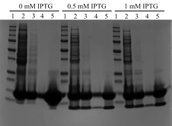
In parallel, the MAβ1–42IBs were also solubilized using 8M urea and then passed through a DEAE Sepharose ion-exchange resin to remove NA traces. As seen inFig 3, this protocol did not allow the elimination of a 13 kDa protein contaminant, regardless of IPTG concentration used for induction. Furthermore, this protein could not be eliminated by ultrafiltration indicat-ing that the urea treatment did not allow breakindicat-ing the interaction probably existindicat-ing between the 13 kDa protein and the peptide (Fig 3lanes 4 and 5). This clearly showed the limitation of the denaturation with urea and emphasizes the interest of the NaOH treatment with to elimi-nate proteins and nucleic acids.
Fig 2. Purification monitoring of MAβ. A. Simplified scheme of the purification procedure. Circled letters correspond to samples collected for either UV/Vis measurements or gel analysis. Supernatant S1 and S2, and pellet P1 and P2 correspond to the supernatant and the precipitate obtained for the two cycles of denaturation/neutralisation. B. SDS-PAGE at different steps of purification, compared with 10–250 kDa protein marker (lane M). C. UV-Vis spectra at pH 12 after IBs denaturation (D), after denaturation/neutralisation cycles (S1+S2), after 30-kDa ultrafiltration (F), after concentration (C), and after size exclusion chromatography (SEC).
At this point, one important issue was the monitoring of the sample purity at the various stages of the purification procedure. The Bradford protein assay was eliminated because it does not give any information on sample purity and only allows the determination of a global pro-tein content. Combination of UV-Vis spectrophotometry with gel electrophoresis was clearly preferred. Indeed, SDS-PAGE permits to visualize the presence of protein contaminants while UV-Vis analyses inform on the predominant presence of either nucleic acids (λ = 260 nm) or specific amino acids, namely tryptophan (λ = 280 nm), tyrosine/tyrosinate (λ = 274 nm/λ = 293 nm), phenylalanine (λ = 257 nm) [26]. Both methods were thus combined to assess the purity level of the peptide samples depending on the purification steps as shown inFig 2C. After denaturation (Fig 2C, spectrum D), a lot of species are evidenced. The supernatant har-vested after neutralization (Fig 2C, spectrum S1+S2), still contain proteins but is already devoid of nucleic acids. After ultrafiltration, the sample was too diluted to give an informative UV spectrum (Fig 2C, spectrum F). Finally, the concentration step yielded a pure sample showing a UV signature with the typical UV band atλ = 293 nm corresponding to the tyrosinate form (Fig 2C, spectrum C). This typical profile is conserved after size exclusion chromatography, which was performed to isolate only monomeric peptide before aggregation assays (Fig 2C, spectrum SEC). Overall, these analyses confirmed the high level of purity of the peptide preparation.
Fig 3. Purification protocol of MAβ1–42IBs dissolved in 8M urea. Lane 1: 10–250 kDa protein marker, lane 2: IBs after urea denaturation, lane 3: after passing through DEAE resin, lane 4: after 30-kDa ultrafiltration, lane 5: after 3-kDa concentration. doi:10.1371/journal.pone.0161209.g003
As the sample was devoid of any protein contaminants, the pH-dependent absorption of Tyr was used to estimate the peptide yield. Absorption spectra of the peptides were thus recorded at pH 12 (showing the absorption band of tyrosinate at 293 nm) and at pH 2 (display-ing no tyrosinate band). The precise quantification of the tyrosine content of the sample was performed by subtracting spectra. This enabled the estimation of the peptide yield as being around 4 mg/L of culture, which is in the same range as those previously reported, comprised between 3 and 7 mg/L [13,14,19,27,28].
Characterization of MA
β
1–40and MA
β
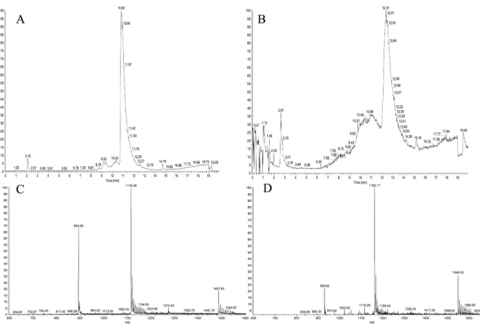
1–42To further characterize the peptides, LC-MS and NMR analyses were conducted right after the concentration step on both peptides. MAβ1–40displays a clean chromatographic trace with a major peak at 10.8 min. The mass spectrum associated to this peak is in good agreement with the expected masses (Fig 4A and 4C). MAβ1–42chromatogram is less resolved, the main peak being shouldered (Fig 4B and 4D). The masses detected for both the shoulder and the peak are consistent with the calculated masses of MAβ1–42, indicating that oligomeric forms of MAβ1–42 could be present. This is consistent with the high propensity of Aβ1–42to aggregate.
1H NMR spectra of both peptides were then recorded, and compared with commercially available synthetic Aβ1–40(Fig 5). They display the typical signals usually observed for Aβ1–40 peptide. In the aromatic region, signals corresponding to the Hδ and Hε of aromatic residues are well defined. As such, Phe 19 and 20 (ca. 7.2 ppm and ca. 7.3 ppm) and Tyr 10 (ca. 7.95 ppm andca. 6.8 ppm) can be distinguished. Slight shifts are observed for His 6, 13 and 14
Fig 4. LC-MS analysis of MAβ1–40and MAβ1–42. A. LC trace of MAβ1–40monitored by mass detection. B. LC trace of MAβ1–42monitored by mass detection. C. MS of MAβ1–40displaying [M+5H]5+, [M+4H]4+, and [M+3H]3+peaks (expected values: 892.65, 1115.56, 1487.07). D. MS of MAβ1–42displaying [M+5H]5+, [M+4H]4+, and [M+3H]3+peaks (expected values: 929.47, 1162.17, 1548.44).
(ca. 7.8 ppm and ca. 7.1 ppm), which can be attributed to pH variations. The absence of signal ca. 7.5 ppm confirms the absence of Trp residue in the peptides. Expectedly, signals corre-sponding to Hα (4.5–3.8 ppm) and Hβ (3.2–2.6 ppm and 2.3–1.5 ppm) are more difficult to attribute in details. In the aliphatic region, signals corresponding to Lys 16 and 28 (1.4 ppm) and to Val and Leu (1.0–0.7 ppm) are observed. Finally, the signal at 3.9 ppm reveals the pres-ence of a methionine residue for the recombinant peptides as expected.
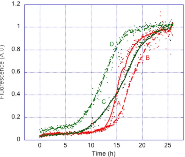
Because mostin vitro studies on Aβ peptides consist in studying their aggregative properties under various conditions, it was essential to ensure that the auto-assembling properties of the peptides are conserved after this purification process. Prior to this, it was important to remove traces of oligomeric species, to ensure a homogeneous aggregation process. The two peptides were thus submitted to SEC. To assess the apparent molecular weight of targeted peptides, a calibration curve was established using other unstructured peptides. It confirmed that the apparent molecular weight of the two peptides was about 4 kDa (S1 Fig). Fractions correspond-ing to monomeric peptides were then submitted to Th-T fibrillization assay. In 26 h, both pep-tides afford a characteristic sigmoidal aggregation curve, with aggregation half-times around 15 h (Fig 6). The MAβ peptides obtained through our purification procedure thus conserved their auto-assembling properties. The same results were obtained with peptide batches from different cultures, showing the high robustness of the purification procedure in terms of repro-ducibility. This purification protocol could thus be applied for routine lab-scale MAβ produc-tion. This also confirms that the presence of aN-ter Met residue does not affect the aggregation properties of MAβ peptides as previously reported [16]. Removing Met residue could be benefi-cial for specific studies such as metal coordination, but does not seem mandatory for classical aggregation assays.
Conclusion
We have here reported a rapid, straightforward and highly reproducible recombinant produc-tion and purificaproduc-tion of MAβ1–40and MAβ1–42. First, we showed that Th-S staining was reliable to monitor IB production during cell growth. Purification monitoring provided evidence that only the combination of UV-Vis spectrophotometry and gel electrophoresis analyses could properly allow the assessment of the peptide purity along the purification process.
Fig 5.1H NMR of MAβ
1–40and MAβ1–42compared with Aβ1–40. Samples of MAβ in water are diluted to 100 μM in 100 mM phosphate buffer pH 7 in D2O. Synthetic Aβ1–40is dissolved in NaOD and diluted to 100μM under the same conditions. Signals at 4.75 ppm correspond to H2O, and signals at 3.25 ppm and 1.25 ppm correspond to traces of ethanol.
In addition, we established that pH treatment of IBs is an efficient way to get rid of most nucleic acid and protein contaminants, contrasting with commonly used chaotropic agents. Additional purification steps consisting in simple ultrafiltrations, providing clean peptide prep-arations, as revealed by UV-Vis spectrophotometry, SDS-PAGE, LC-MS and1H NMR. With this protocol, up to 4 mg of peptides are produced within 2 days. In addition, the preparations are suitable for aggregation assays, which were highly reproducible from batch to batch, accounting for a robust purification procedure. Supplementary investigation might now be engaged to remove theN-ter methionine residue, in order to obtain the native Aβ, as already proposed by Walsh and co-workers and further improve the peptide yield [21].
Supporting Information
S1 Fig. SEC calibration curve with unstructured peptides.The different peptides were dis-solved in NaOH 50 mM to 10 mg/mL and injected on a Superdex 75 10/300 GL. Samples were eluted with NaOH 15 mM at 1 mL/min monitoring at 220 nm and 293 nm. The arrows indi-cate the elution volume found for synthetic Aβ1–40and the two recombinant MAβ1–40and MAβ1–42peptides.
(TIFF)
Fig 6. Normalized aggregation curves of MAβ1–40and MAβ1–42. Two different batches of MAβ1–40(A and B) and MAβ1–42(C and D) (20μM) aggregate in the presence of Th-T (10 μM) in phosphate buffer (50 mM, pH 7). Assay was run at 37°C with gentle shaking. Each experiment was run in triplicates.
Acknowledgments
Christian Bijani is acknowledged for NMR experiments. ICEO platform (GenoToul) is acknowledged for granting access to the Aktä Purifier system.
Author Contributions
Conceptualization:CH PF EG IA MRS. Funding acquisition:EG CH IA MRS. Investigation:MH. Methodology:MH YM RI MRS. Project administration:EG MRS. Supervision:EG MRS. Validation:YM RI MRS. Visualization:MH.Writing - original draft:MH.
Writing - review & editing:MH YM RI CH PF EG IA MRS.
References
1. Henderson VW. Alzheimer’s disease: review of hormone therapy trials and implications for treatment and prevention after menopause. J Steroid Biochem Mol Biol. 2014; 142: 99–106. doi:10.1016/j.jsbmb. 2013.05.010PMID:23727128
2. Delrieu J, Ousset PJ, Voisin T, Vellas B. Amyloid beta peptide immunotherapy in Alzheimer disease. Rev Neurol (Paris). 2014; 170: 739–748. doi:10.1016/j.neurol.2014.10.003
3. Tong LM, Fong H, Huang Y. Stem cell therapy for Alzheimer’s disease and related disorders: current status and future perspectives. Exp Mol Med. 2015; 47: e151. doi:10.1038/emm.2014.124PMID:
25766620
4. Schneider LS, Mangialasche F, Andreasen N, Feldman H, Giacobini E, Jones R, et al. Clinical trials and late-stage drug development for Alzheimer’s disease: an appraisal from 1984 to 2014. J Intern Med. 2014; 275: 251–283. doi:10.1111/joim.12191PMID:24605808
5. Chang Y-J, Chen Y-R. The coexistence of an equal amount of Alzheimer’s amyloid-β 40 and 42 forms structurally stable and toxic oligomers through a distinct pathway. FEBS J. 2014; 281: 2674–2687. doi:
10.1111/febs.12813PMID:24720730
6. Hamley IW. The Amyloid Beta Peptide: A Chemist’s Perspective. Role in Alzheimer’s and Fibrillization. Chem Rev. 2012; 112: 5147–5192. doi:10.1021/cr3000994PMID:22813427
7. Fu Z, Aucoin D, Davis J, Van Nostrand WE, Smith SO. Mechanism of Nucleated Conformational Con-version of Aβ42. Biochemistry (Mosc). 2015; 54: 4197–4207. doi:10.1021/acs.biochem.5b00467
8. Soto C, Castaño EM, Asok Kumar R, Beavis RC, Frangione B. Fibrillogenesis of synthetic amyloid-β peptides is dependent on their initial secondary structure. Neurosci Lett. 1995; 200: 105–108. doi:10. 1016/0304-3940(95)12089-MPMID:8614555
9. Zagorski MG, Yang J, Shao H, Ma K, Zeng H, Hong A. [13] Methodological and chemical factors affect-ing amyloidβ peptide amyloidogenicity. Methods in Enzymology. Elsevier; 1999. pp. 189–204. Avail-able:http://linkinghub.elsevier.com/retrieve/pii/S0076687999090151
10. Finder VH, Vodopivec I, Nitsch RM, Glockshuber R. The Recombinant Amyloid-β Peptide Aβ1–42 Aggregates Faster and Is More Neurotoxic than Synthetic Aβ1–42. J Mol Biol. 2010; 396: 9–18. doi:10. 1016/j.jmb.2009.12.016PMID:20026079
11. Shen M, Wang Q, Mu X, Xu H, Yan W. Expression, purification and characterization of recombinant humanβ-amyloid 1–42 in Pichia pastoris. Protein Expr Purif. 2009; 63: 84–88. doi:10.1016/j.pep.2008. 09.015PMID:18950715
12. Bockhorn JJ, Lazar KL, Gasser AJ, Luther LM, Qahwash IM, Chopra N, et al. Novel semisynthetic method for generating full lengthβ-amyloid peptides. Biopolymers. 2010; 94: 511–520. doi:10.1002/ bip.21391PMID:20593467
13. Lee EK, Hwang JH, Shin DY, Kim DI, Yoo YJ. Production of recombinant amyloid-β peptide 42 as an ubiquitin extension. Protein Expr Purif. 2005; 40: 183–189. doi:10.1016/j.pep.2004.12.014PMID:
15721787
14. Garai K, Crick SL, Mustafi SM, Frieden C. Expression and purification of amyloid-β peptides from Escherichia coli. Protein Expr Purif. 2009; 66: 107–112. doi:10.1016/j.pep.2009.02.009PMID:
19233290
15. Alies B, LaPenna G, Sayen S, Guillon E, Hureau C, Faller P. Insights into the Mechanisms of Amyloid Formation of ZnII-Ab11-28: pH-Dependent Zinc Coordination and Overall Charge as Key Parameters for Kinetics and the Structure of ZnII-Ab11-28 Aggregates. Inorg Chem. 2012; 51: 7897–7902. doi:10. 1021/ic300972jPMID:22765389
16. Szczepankiewicz O, Linse B, Meisl G, Thulin E, Frohm B, Sala Frigerio C, et al. N-Terminal Extensions Retard Aβ42 Fibril Formation but Allow Cross-Seeding and Coaggregation with Aβ42. J Am Chem Soc. 2015; 137: 14673–14685. doi:10.1021/jacs.5b07849PMID:26535489
17. Liao Y-H, Chen Y-R. A novel method for expression and purification of authentic amyloid-β with and without 15N labels. Protein Expr Purif. 2015; 113: 63–71. doi:10.1016/j.pep.2015.05.002PMID:
25969353
18. Wiesehan K, Funke SA, Fries M, Willbold D. Purification of recombinantly expressed and cytotoxic human amyloid-beta peptide 1–42. J Chromatogr B. 2007; 856: 229–233. doi:10.1016/j.jchromb.2007. 06.003
19. Sharma SC, Armand T, Ball KA, Chen A, Pelton JG, Wemmer DE, et al. A facile method for expression and purification of 15N isotope-labeled human Alzheimer’s β-amyloid peptides from E. coli for NMR-based structural analysis. Protein Expr Purif. 2015; 116: 82–89. doi:10.1016/j.pep.2015.07.012PMID:
26231074
20. Chhetri G, Pandey T, Chinta R, Kumar A, Tripathi T. An improved method for high-level soluble expres-sion and purification of recombinant amyloid-beta peptide for in vitro studies. Protein Expr Purif. 2015; 114: 71–76. doi:10.1016/j.pep.2015.05.015PMID:26118700
21. Walsh DM, Thulin E, Minogue AM, Gustavsson N, Pang E, Teplow DB, et al. A facile method for expres-sion and purification of the Alzheimer’s disease-associated amyloid β-peptide: Expression and purifica-tion of the amyloidβ-peptide. FEBS J. 2009; 276: 1266–1281. doi:10.1111/j.1742-4658.2008.06862.x
PMID:19175671
22. Espargaró A, Sabate R, Ventura S. Thioflavin-S staining coupled to flow cytometry. A screening tool to detect in vivo protein aggregation. Mol Biosyst. 2012; 8: 2839. doi:10.1039/c2mb25214gPMID:
22868714
23. Neerathilingam M, Mysore S, Gandham SHA. Soni-removal of nucleic acids from inclusion bodies. Bio-chem Biophys Res Commun. 2014; 448: 45–49. doi:10.1016/j.bbrc.2014.04.049PMID:24747565
24. Villar-Piqué A, Espargaró A, Sabaté R, de Groot NS, Ventura S. Using bacterial inclusion bodies to screen for amyloid aggregation inhibitors. Microb Cell Factories. 2012; 11: 55. doi: 10.1186/1475-2859-11-55
25. Allen SP, Polazzi JO, Gierse JK, Easton AM. Two novel heat shock genes encoding proteins produced in response to heterologous protein expression in Escherichia coli. J Bacteriol. 1992; 174: 6938–6947. PMID:1356969
26. Beaven GH, Holiday ER. Ultraviolet Absorption Spectra of Proteins and Amino Acids. In: Anson KB M. L. and JTE, editor. Advances in Protein Chemistry. Academic Press; 1952. pp. 319–386. Available:
http://www.sciencedirect.com/science/article/pii/S0065323308600224PMID:14933256
27. Long F, Cho W, Ishii Y. Expression and purification of 15N- and 13C-isotope labeled 40-residue human Alzheimer’s β-amyloid peptide for NMR-based structural analysis. Protein Expr Purif. 2011; 79: 16–24. doi:10.1016/j.pep.2011.05.012PMID:21640828
28. Kim E-K, Moon JC, Lee JM, Jeong MS, Oh C, Ahn S-M, et al. Large-scale production of soluble recom-binant amyloid-β peptide 1–42 using cold-inducible expression system. Protein Expr Purif. 2012; 86: 53–57. doi:10.1016/j.pep.2012.08.021PMID:22982229