Minimally invasive resection of thymomas with the da Vinci®
Surgical System
†
Didier Schneiter*, Sandra Tomaszek, Peter Kestenholz, Sven Hillinger, Isabelle Opitz, Ilhan Inci
and Walter Weder
Department of Surgery, Division of Thoracic Surgery, University Hospital Zurich, Zurich, Switzerland
* Corresponding author. Department of Surgery, Division of Thoracic Surgery, University Hospital Zurich, Rämistrasse 100, 8091 Zurich, Switzerland. Tel: +41-44-2558812; fax: +41-44-2558805; e-mail: [email protected] (D. Schneider).
Received 3 October 2011; received in revised form 18 March 2012; accepted 23 March 2012
Abstract
OBJECTIVES: The resection of thymic tumours requires completeness and may be technically challenging due to the anatomical proximity of the delicate mediastinal structures. An open approach by sternotomy is still recommended in all cases with locally extended disease. Video-assisted thoracoscopic surgery is feasible, but limited by the two-dimensional vision and the impaired mobility of the instruments. We evaluated the da Vinci® Surgical System for the resection of various mediastinal pathologies, particularly thymomas.
METHODS: Among 105 patients operated on by robotic assisted thoracoscopic surgery (RATS) for mediastinal tumours between 27 August 2004 and 12 July 2011, 20 patients with thymomas were studied prospectively. Of these, 10 males with a median age of 53 years, with a well-circumscribed thymic lesion on computed tomography (CT) and a diameter of <6 cm were resected by RATS alone, and selected ones (n = 3), with a diameter of >6 cm, underwent a hybrid procedure with a contralateral thoracotomy on the side of the main tumour extension. A regular follow-up with chest CT scans was performed every 6 months.
RESULTS: Thymoma resection was complete in all patients. Partial pericardial resection was needed infive and pulmonary resection in two patients. Eighty-five percent of patients had an R0 resection. Histological classifications included thymoma WHO type A (n = 3), AB (n = 8), B1–2 (n = 5) and B3 (n = 4). All B3 thymomas received adjuvant radiotherapy. No intraoperative complications occurred. The median hospitalization time was 5 days (range 2–14 days). There were no local, but two pleural, recurrences. After a median observa-tion time of 26 months, 19 patients (95%) are alive.
CONCLUSIONS: Well-circumscribed thymomas can be safely and completely resected with the da Vinci® Surgical System with excellent short- and mid-term outcomes. Selected tumours with large diameters may be resectable using a hybrid procedure combining RATS with a thoracotomy.
Keywords:Thymoma• da Vinci® Surgical System • Robotic-assisted thymectomy • Hybrid procedure
INTRODUCTION
Mediastinal pathologies are classified according to their anatomical localization. In the anterior mediastinum, they typically originate from the thymus, while in the middle compartment, lymphatic disorders and in the posterior compartment, neurogenic tumours are located. The surgical approach and the type of resection are often technically challenging due to the pathological variety, the degree of tumour spread as well as the anatomical proximity of large vessels or nerves. Therefore, open surgical techniques such as sternotomy or thoracotomy are recommended. This is consid-ered particularly important for thymomas since complete surgical resection is the key for successful treatment [1,2]. However, due to the technical improvements in video-assisted thoracoscopic
surgery (VATS), endoscopic access in the treatment of thymomas has become more and more popular in the last decade [3]. In the hands of skilled surgeons, the implementation of thoracoscopic procedures has shifted from diagnostic procedures to complex radical resections.
With the introduction of the da Vinci® Surgical System, a tool with unexpected prospects has become an additional option for endoscopic resections in thoracic surgery [4–6]. The outstanding visibility using a robotic-assisted system is due to its three-dimensional vision combined with the magnification of the op-eratingfield and the dexterity of the instruments which facilitates surgery with spectacular precision and security in contrast to VATS. Although the access through trocar ports is identical to the VATS procedure, the modality of surgical resection is different and comparable to what the surgeon is familiar with from the open surgical approach except for the lack of tactility. Hence the endoscopic surgeon is not limited by a two-dimensional view
†Presented at the 19th European Conference on General Thoracic Surgery,
Marseille, France, 5–8 June 2011.
© The Author 2012. Published by Oxford University Press on behalf of the European Association for Cardio-Thoracic Surgery. All rights reserved.
European Journal of Cardio-Thoracic Surgery 43 (2013) 288–292
ORIGINAL ARTICLE
and dissatisfying dissection of the anatomical structures due to limited manoeuvrability of the surgical instruments in a single di-mension. RATS has become an established approach in many centres for thymectomy in patients with myasthenia gravis (MG) [7]. However, only a few reports exist on its application in thymomas [4–8].
Here, we report our experience of a single academic institu-tion with minimally invasive resecinstitu-tions of thymomas with the da Vinci® Surgical System.
MATERIALS AND METHODS
Out of a cohort of the first 105 consecutive patients (47 men) who underwent the surgical resection of thoracic tumours with the da Vinci® Surgical System at the University Hospital Zurich between 27 August 2004 and 12 July 2011, 68 patients had a neoplasm in the anterior mediastinum (65%). In 58 of those, a thymic pathology was found (85.3%). For this study, we per-formed a sub-analysis of the 20 patients who underwent a robotic-assisted thymectomy for an underlying thymoma.
Resection with the da Vinci® Surgical System (three arms) was evaluated for all patients with a mediastinal pathology. Exclusion criteria were a lesion size of >6 cm, obvious signs of the invasion of vital organs, and expected unfeasibility of a single lung venti-lation. Later on, the approach was extended to a hybrid proced-ure combining RATS for the mediastinal dissection from one side and a thoracotomy for the main tumour resection from the opposite side in the selected patients with large (up to 8 cm), mainly paramediastinal tumours.
The preoperative workup of the patients included CT scan, lung function testing, ECG and basic blood tests in all patients. In addition, the specific parameters such as ACh-receptor, anti-MuSK and anti-Titin antibodies were measured in patients with MG before the operation. Clinical staging included medical history and physical examination, CT or optional positron emis-sion tomography scan, and a neurological workup in patients with MG. The intervention was performed preferentially from the left side except for tumours extending predominantly to the right side.
All resections were performed by three thoracic surgeons at our institution who were experienced in extended open as well as minimally invasive surgery including RATS. Individual patient consent was waived. The patient characteristics and perioperative outcome data were collected in a prospective database and a de-scriptive statistical analysis was performed retrospectively.
Medical charts were reviewed for demographic data, clinical presentation, diagnostic investigations, side of the approach, tumour invasion, histological diagnosis and staging, post-operative morbidity and mortality, follow-up and additional therapy. The follow-up data were collected through regular outpatient visits and individual phone calls if the follow-up was conducted in another centre.
Operative technique
All interventions were performed under general anaesthesia with single lung ventilation. The procedure was conducted with add-itional CO2insufflation (maximum pressure 6–8 mmHg) as it was found to facilitate the dissection along the anatomical layers, enlarge the operative field and clear the fume in front of the
camera. We use three of the four robotic arms of the new da Vinci®SiHDSurgical System and a 30° camera for thymectomies.
The positioning of the patients depended on the localization of the tumour and was carried out according to an earlier report [9,10]. In brief, all patients were positioned supine with the inci-sion side elevated by 30°. The arm of the patient was positioned parallel to the trunk allowing for free access to the mid-axillary line.
With increasing experience, we approached the surgical access from the right side (first 15 patients) to the left side for two main reasons: first, coming through the left thoracic cavity, the right phrenic nerve is protected by the superior vena cava in the delicate region of the right upper thymic horn (venous con flu-ence with the left brachiocephalic vein). Due to a more ventral localization, the left phrenic nerve is easier to identify and pre-serve. Secondly, accessory thymic tissue in the aortic-pulmonary window is accessible only from the left side.
The incision for the camera port is performed in the 5th inter-costal space in the anterior axillary line. The port for the left robotic arm is subsequently introduced under the direct vision in the 6th intercostal space in the mid-clavicular line and the right arm in the 3rd intercostal space in the anterior axillary line. In order to create more space in the thoracic cavity, we lift the thoracic cage up by 2–3 cm with the camera arm of the da Vinci® Surgical System once the camera arm is positioned.
For larger lesions (>6 cm) without invasiveness of vital struc-tures on CT scan, we developed the technique of a Hybrid pro-cedure in order to avoid a sternotomy or clamshell incision. This technique is typically applied in tumours arising from the midline of the mediastinum but with the main tumour mass being located paramediastinally. The mediastinal dissection of the tumour from the anonymous vein, the pericardium and the retrosternal region is done with the da Vinci® Surgical System on the side where the lesion is located centrallyfirst. Subsequently, a standard anterolateral thoracotomy is conducted from the contralateral and the tumour is freed by an open technique (with rib spreading). The advantage of this technique is that crit-ical structures in the upper mediastinum not controllable by an access like a thoracotomy, have already been dissected minimal-ly invasiveminimal-ly and hence the resection of the tumour can be com-pleted and the specimen removed through the thoracotomy.
RESULTS
Overall, 68 patients with lesions in the anterior mediastinum were resected by RATS; 58 of those were of thymic origin (85.3%) (Table 1). The current study population of thymomas (n = 20) included 10 females and 10 males with a median age of 53 years (range 21–77 years) and a median body mass index of 26.5 kg/m2 (range 19.0–37.0 kg/m2). MG was prevalent in 12 (60%) patients. The preoperative lung function test with forced expiratory volume in one second (FEV1), forced expiratory vital capacity (FVC) and diffusing capacity of the lung for carbon monoxide (TLCO) were available in 17 patients and are summar-ized in Table 2. All 20 procedures were performed as planned by RATS, three with a Hybrid approach.
There were six patients who needed an extended resection in-cluding the pericardium (n = 5) or a lung wedge resection (n = 1). Half of these were done entirely endoscopically and half with a Hybrid approach.
THOR
A
C
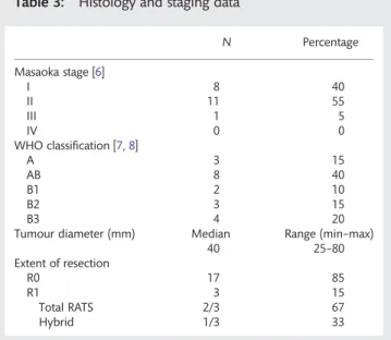
No emergency conversion to an open thoracotomy was ne-cessary. There were 15 patients who underwent a total thymec-tomy (80.0%). In five patients with small, well-circumscribed thymomas in the main body of the thymus in the absence of MG, a thymectomy without the complete resection of the upper horns was performed. A R0 resection was achieved in 85.0% of the patients and a R1 resection in three (15.0%) patients (two total RATS and one Hybrid approach). R1 resections were found in tumours either adjacent to the retrosternal pleura, pericar-dium, vena cava superior or phrenic nerves without macroscopic infiltration. Histology and staging data are summarized in Table3.
The median hospital length of stay of all 20 patients was 5 days (range 2–14 days), in the subgroup of RATS, 4 days (range 2–14 days), and in the hybrid group, 8 days (range 8–13 days). Only two patients suffered from postoperative complications (10.0%); one developed a sero-pneumothorax and one supra-ventricular arrhythmias. None of the patients needed re-intervention or had to be readmitted to the hospital within the first 90 days of dismissal. Adjuvant radiotherapy was con-ducted in all four patients with a B3 thymoma and in one patient with a B2 thymoma due to the R1 resection. The median follow-up time was 26 months (range 0.1–6.6 years), with one patient lost to the follow-up. Two patients (11.1%) had a recur-rence in the pleura after 1.3 and 4.9 years. One of those patients (5.0%) died because of the recurrence at 5.2 years after surgery.
DISCUSSION
Mediastinal pathologies are frequently asymptomatic and first noted on a routine chest radiograph or increasingly by CT. In any case, a chest CT scan with contrast medium should be con-ducted for further evaluation of the lesion as its specific
localization and appearance are highly suggestive of the various differential diagnoses. It is therefore essential for the planning of further diagnostic and therapeutic strategies. Primary tumours in the anterior mediastinum account for half of all mediastinal masses. The most common primary anterior mediastinal tumours are thymomas. Non-malignant lesions include thymic cysts, lymphangioma, intrathoracic goitre and ectopic parathy-roid adenoma [11]. Understanding of the pathology, clinical pres-entation and radiological prespres-entation of the main tumour types is mandatory in the safe and efficient work-up of a mediastinal mass. In spite of the improvement of imaging techniques, tissue is often needed for histological diagnosis and remains crucial for definitive treatment [12–14].
Nowadays in experienced centres, the majority of diagnostic but also many therapeutic procedures for thoracic malignancies are approached through a minimally invasive access. Shorter hospital stay, less pain, smaller and hence less noticeable scars as well as faster postoperative rehabilitation are sufficiently good reasons to promote endoscopic procedures. Furthermore, adju-vant treatment, if necessary, is better tolerated.
Irrespective of the approach, the complete surgical resection is of paramount importance in the treatment of thymomas and is considered as the most important prognostic factor. Any com-promise in achieving a radical resection due to an inadequate approach is undesirable. In more advanced tumour stages, par-ticularly after an R1 resection, adjuvant radiotherapy is commonly considered [15]. Due to the localization of thymic tumours in a narrow compartment with proximity to vital and vulnerable ana-tomical structures, traditionally an open approach is performed, via sternotomy or, in lesions located predominantly on one side, by thoracotomy or a hemi-clamshell incision. Thymic tumours have to be resected with the surrounding tissue in order to achieve the microscopic clear margins. Applying the da Vinci® Surgical System offers a clear benefit compared with VATS and in small well-circumscribed tumours, even with an open approach. This statement is scientifically difficult to prove, but reflects our own clinical experience shared by those who are skilled in the use of RATS. Due to the three-dimensional magnified view and the dexterity of the instruments, the surgeon is capable of performing
Table 3: Histology and staging data
N Percentage Masaoka stage [6] I 8 40 II 11 55 III 1 5 IV 0 0 WHO classification [7,8] A 3 15 AB 8 40 B1 2 10 B2 3 15 B3 4 20
Tumour diameter (mm) Median Range (min–max) 40 25–80 Extent of resection R0 17 85 R1 3 15 Total RATS 2/3 67 Hybrid 1/3 33
Table 1: Thymic pathologies resected with the da Vinci® Surgical System
N Percentage Anterior mediastinal lesions 68
Thymic disease 58/68 85.3 MG only 25 Thymoma without MG 8 Thymoma with MG 12 Thymic cyst 4 Thymic hyperplasia 9
Table 2: Preoperative pulmonary function testing
Median Range (min–max) FEV1 (l) 2.66 1.49–5.24 FEV1 (% predicted) 94 61–126 FVC (l) 3.2 1.78–6.23 FVC (% predicted) 96 55–123 TLCO (% predicted) 99 67–114
surgery with high precision and even extending the resection to the pericardium and the lung without any difficulties. In the majority of smaller thymomas, the complete resection can be achieved by robotic-assisted thymectomy in a safe and effective method. However, if the tumour exceeds a certain size or in fil-trates vital organs, conversion to sternotomy or thoracotomy is still the appropriate approach.
In recent years, thymectomy performed via robotic-assisted thoracoscopy (RATS) has gained importance and has been proven to achieve equivalent results to the VATS approach or open surgery in MG [7, 16]. However, little information about robotic resection for thymomas is found in the literature and no randomized study of any surgical approach is available [4–7,17–19].
Since the complete resection of thymomas is crucial, the surgeon must achieve R0 resection whenever technically feasible, and we followed this paradigm in all cases. The intrao-perative evaluation by the surgeon as to complete resection is essential because thefinal assessment by the pathologist is diffi-cult as he is not able to appreciate the intraoperative situation. This is commonly the case in tumours invading the pericardium. The thymoma is adherent to the pericardium, therefore, the pericardium is resected together with the thymoma. The micro-scopicfindings will still show tumour infiltration at the resection margin, yet there is absolutely no contact of the tumour with the underlying structure. Therefore, careful examination of the resected specimen by the pathologist in close collaboration with the surgeon is decisive, particularly when adjuvant radiotherapy is considered.
After more than 10 years of experience with thymectomy for MG without thymomas by VATS, we switched to the RATS approach in April 2004. Having experienced the technical advan-tages, we subsequently included smaller lesions up to 3 cm and over time also larger thymomas up to 6 cm in diameter.
In our series, 85% of patients underwent complete resection. Of the three patients with R1 resection, it was performed entirely endoscopically on two, and on one by the Hybrid procedure developed in our centre. The latter was performed in a woman suffering from severe, therapy-resistant MG with a large thymoma (WHO-Typ B3, Masaoka II) located paramediastinally to the left side who refused having a sternotomy. The mediastin-al part of the thymectomy including the dissection of the an-onymous vein was done from the right side and the main dissection and exstirpation of the large tumour of 8 cm, through an anterolateral thoracotomy from the left side. Eighteen months after extended surgical resection ( pericardium with re-construction and paramediastinal wedge-resection of the left upper lobe of the lung), the MG has improved and the CT scan shows no signs of local or distant recurrence. In conclusion, radical resection of well-circumscribed thymomas with the da Vinci® Surgical System is effective and safe and provides a short hospital stay and rapid recovery. Local control seems to be com-parable to an open approach based on this limited experience. Larger thymomas or lesions with invasion of the surrounding vessels are still preferentially resected by an open approach. In carefully selected patients, a hybrid may achieve equal radicality with the avoidance of a sternotomy, which is an advantage with respect to potentially associated morbidity and for cosmetic reasons, especially in female patients. Short- to mid-term out-comes are comparable with the data reported in the literature. Further and larger clinical studies with the longer follow-up data are required to substantiate our conclusion.
Conflict of interest: none declared.
REFERENCES
[1] Port JL, Ginsberg RJ. Surgery for thymoma. Chest Surg Clin N Am 2001; 11:421–37.
[2] Detterbeck FC, Parsons AM. Management of stage I and II thymoma. Thorac Surg Clin 2011;21:59–67.
[3] Odaka M, Akiba T, Yabe M, Hiramatsu M, Matsudaira H, Hirano Jet al. Unilateral thoracoscopic subtotal thymectomy for the treatment of stage I and II thymoma. Eur J Cardiothorac Surg 2010;37:824–6.
[4] Bodner J, Wykypiel H, Greiner A, Kirchmayr W, Freund MC, Margreiter R et al. Early experience with robot-assisted surgery for mediastinal masses. Ann Thorac Surg 2004;78:259–65.
[5] Savitt MA, Gao G, Furnary AP, Swanson J, Gately HL, Handy JR. Application of robotic-assisted techniques to the surgical evaluation and treatment of the anterior mediastinum. Ann Thorac Surg 2005;79:450–5. [6] Yoshino I, Hashizume M, Shimada M, Tomikawa M, Tomiyasu M,
Suemitsu R et al. Thoracoscopic thymomectomy with the da Vinci computer-enhanced surgical system. J Thorac Cardiovasc Surg 2001;122: 783–5.
[7] Rückert JC, Ismail M, Swierzy M, Sobel H, Rogalla P, Meisel A et al. Thoracoscopic thymectomy with the da Vinci robotic system for myas-thenia gravis. Ann N Y Acad Sci 2008;1132:329–35.
[8] Melfi F, Fanucchi O, Davini F, Viti A, Lucchi M, Ambrogi MC et al. Ten-year experience of mediastinal robotic surgery in a single referral centre. Eur J Cardiothorac Surg 2012;41:847–51.
[9] Rea F, Marulli G, Bortolotti L, Feltracco P, Zuin A, Sartori F. Experience with the "da Vinci" robotic system for thymectomy in patients with my-asthenia gravis: report of 33 cases. Ann Thorac Surg 2006;81:455–9. [10] Augustin F, Schmid T, Bodner J. The robotic approach for mediastinal
lesions. Int J Med Robot 2006;2:262–70.
[11] Priola AM, Priola SM, Cardinale L, Cataldi A, Fava C. The anterior medi-astinum: diseases. Radiol Med 2006;111:312–42.
[12] Masaoka A, Monden Y, Nakahara K, Tanioka T. Follow-up study of thym-omas with special reference to their clinical stages. Cancer 1981;48: 2485–92.
[13] Müller-Hermelink HK, Ströbel P, Zettl A, Marx A. Combined thymic epi-thelial tumours. In: Travis WD, Brambilla E, Müller-Hermelink HK, Harris CC (eds). Pathology and Genetics of Tumours of the Lung, Pleura, Thymus and Heart (WHO Classification of Tumours Series). Lyon, France: IARC Press, 2004, 196–8.
[14] Marx A, Ströbel P, Zetti A, Chan JKC, Müller-Hermelink HK, Harris NL et al. Thymomas. In: Travis WD, Brambilla E, Mueller-Hermelink HK, Harris CC (eds). Pathology and Genetics of Tumours of the Lung, Pleura, Thymus and Heart WHO (Classification of Tumours Series). Lyon, France: IARC Press, 2004, 152–71.
[15] Girard N, Mornex F, Van Houtte P, Cordier JF, van Schil P. Thymoma: a focus on current therapeutic management. J Thorac Oncol 2009;4: 119–26.
[16] Rückert JC, Ismail M, Swierzy M, Braumann C, Badakhshi H, Rogalla P et al. Minimally invasive thymus surgery. Chirurg 2008;79:20–5. [17] Cakar F, Werner P, Augustin F, Schmid T, Wolf-Magele A, Sieb Met al. A
comparison of outcomes after robotic open extended thymectomy for myasthenia gravis. Eur J Cardiothorac Surg 2007;31:501–4.
[18] Limmer KK, Kernstine KH. Minimally invasive and robotic-assisted thymus resection. Thorac Surg Clin 2011;21:69–83.
[19] Weksler B, Tavares J, Newhook TE, Greenleaf CE, Diehl JT. Robot-assisted thymectomy is superior to transsternal thymectomy. Surg Endosc 2012; 26:261–6.
APPENDIX. CONFERENCE DISCUSSION
Dr A. Duranceau (Montreal, QC, Canada): Can you tell us if you had any technical problem with access to the apices of the gland in the neck?
Dr Schneiter: We haven’t had problems with this access. We always come from the left side when we do thymectomies, and we have never had problems to go up to the neck. If we had not been sure that we had done the right operation, we would convert or we would do an additional incision in the jugulum.
THOR
A
C
Dr S. Bolukbas (Wiesbaden, Germany): My questions would be, is R1 resec-tion really a complete resecresec-tion? And second, we are all surgeons and inter-ested in demanding operations, and you perform a minimally invasive intrathoracic surgery but afterwards it ends up with a thoracotomy. What is the advantage for the patient?
Dr Schneiter: As to your first question, I didn’t say that it was a com-plete resection. I said it is a comcom-plete thymectomy. But in two patients it was not a complete resection because it was an R1 resection, you are right. And concerning the second question, there is a possibility to do either a hemiclamshell or a clamshell incision, or a sternotomy, and those patients were women, all of them, and they decided it might look better if they only had three small incisions that you don’t see very well on one side and a submammary incision on the other side. But you are right, it is always possible to do it in a more open method. But in this case we tried to combine this and it worked outfine. We could do it completely in all these cases.
Dr F. Detterbeck (New Haven, CT, USA): Two quick comments. I commend you for pushing the envelope in trying to define what we can do. I would exercise a word of caution, though. I think we have to be careful. We have good results with surgery with complete resection, and we should not be compromising that by trying to do a minimally invasive approach.
The other comment is within the ITMIG group we have spent a fair amount of time trying to define some principles about doing minimally inva-sive resection. Those are going to be published in the Journal of Thoracic Oncology within the next month, and I would urge people that are doing this
to try to adhere to some of those principles, because I think it will help us sort out a little bit better where thisfits and how it should be done.
Dr Schneiter: I completely agree. I think you should never do a minimally invasive resection if you are not doing it in the same way as you do it open. If this would be the case, you should convert, and this is what we did when we had problems.
Dr T. Lerut (Leuven, Belgium): Just in follow-up to what Dr Detterbeck said, I would caution to call 39-month follow-up long-term follow-up. That is medium follow-up in thymoma surgery.
Dr Schneiter: This is true. This is what we quoted.
Dr J. Kuzdzal (Krakow, Poland): I know that it is not the main topic of your presentation, but I would like to ask you to say a little bit more about the non-thymoma thymectomies for myasthenia. How could you define the extent of the thymectomy in these cases? Was it a simple thymectomy or an extended or maximal one and how far were you able to dissect the thymus and the fatty tissue in the upward direction, I mean towards the neck, towards the thyroid gland?
Dr Schneiter: We feel that we have a very good vision of the upper part of the thymus, and as I said before, we always come from the left side, so we have the possibility to go in the aortopulmonary window and go beneath the phrenic nerve, and we go to the base of the heart as well. So it was quite extended but not maximally extended as described by Jarezki. But we took out all the tissue we had from the left side, and we opened even the pleura on the other side and looked on the right side, and if there was more tissue we took it out as well.