ORIGINAL PAPER
Intestinal
Tritrichomonas foetus infection in cats
in Switzerland detected by in vitro cultivation and PCR
Caroline F. Frey&Marc Schild&Andrew Hemphill&Philipp Stünzi&Norbert Müller&Bruno Gottstein&
Iwan A. Burgener
Received: 7 August 2008 / Accepted: 23 October 2008 / Published online: 8 November 2008 # Springer-Verlag 2008
Abstract Tritrichomonas foetus, a parasite well known for its significance as venereally transmitted pathogen in cattle, has recently been identified as a cause of chronic large-bowel diarrhea in domestic cats in the US, UK, and, more recently, also in Norway. In a period of 3 months (October to December 2007), 45 cats of Switzerland suffering from chronic diarrhea were investigated for intestinal infections, including a search for trichomonads. A commercially available in vitro culture system was used to screen for infection, complemented with a PCR and subsequent amplicon sequencing to support speciation. The PCR is based upon amplification of a sequence derived from the internal transcribed spacer region 1 (ITS1) on the ribosomal RNA gene (rRNA) using primers designed to detect a broad range of genera and species belonging to the family of Trichomonadidae. The method was furthermore adapted to the uracil DNA glycosylase (UDG) system in order to prevent carry-over contamination and it included a recom-binant internal control to track for inhibitory reactions. Eleven out of the 45 cats were culture-positive, as revealed by microscopic identification of trichomonadid organisms. One of the isolates was subjected to scanning electron microscopy and findings revealed the presence of three flagella, thus placing the isolate into the gender Tritricho-monas sp. PCR and subsequent amplicon sequencing were
carried out with ten of the 11 isolates. A total homology with published T. foetus sequences was confirmed in all of the cases. T. foetus therefore appears to range among those organisms that can cause chronic diarrhea in cats in Switzerland.
Introduction
Tritrichomonas foetus is a significant veneral cause of infertility and abortion in naturally bred cattle in many countries. In Switzerland, T. foetus in cattle is a notifiable disease (Anonymous 1995). All Swiss bulls used for artificial insemination have to be strictly monitored for the absence of the parasite. The last case of T. foetus infection in cattle has been reported in 1997 (Anonymous 1997). Initially, in the USA, T. foetus was repeatedly identified in the feces of a large number of domestic cats with chronic diarrhea (Gookin et al. 1999, 2004), but recent reports showed that T. foetus infection is also fairly common in cats with diarrhea in the UK (Mardell and Sparkes2006; Gunn-Moore et al. 2007). More recently, a case study from Norway described a natural T. foetus infection of the feline uterus leading to pyometra, which is a common feature of T. foetus infection in cattle, but is a novel finding in cats (Dahlgren et al.2007). Experimental gastrointestinal infec-tion of cats with T. foetus isolated from a naturally infected cat resulted in the characteristic large-bowel diarrhea of the natural infection (Gookin et al. 2001). Cats affected by infection with T. foetus were mostly of young age, lived in multi-cat households and were predominantly pedigree cats (Gookin et al. 1999; Gunn-Moore et al. 2007). Most cats were presented with a history of chronic diarrhea and unsuc-cessful treatment against Giardia infection (Gookin et al. 1999). Usually, symptoms improved under treatment with DOI 10.1007/s00436-008-1255-2
C. F. Frey (*)
:
M. Schild:
A. Hemphill:
P. Stünzi:
N. Müller:
B. GottsteinInstitute of Parasitology, Vetsuisse Faculty, University of Bern, P.O. Box 8466, 3001 Bern, Switzerland
e-mail: [email protected] I. A. Burgener
Department of Clinical Veterinary Medicine, Vetsuisse Faculty, University of Bern,
metronidazole, but diarrhea relapsed after treatment, and had to be stopped. Metronidazole limited the infection temporar-ily, but was not able to clear the cat’s intestines from the parasite (Gookin et al.2006). The most efficacious cure for a trichomonad infection in cats was achieved with ronidazole (Gookin et al.2006). Besides T. foetus, other trichomonads have also been isolated from the cat’s intestine, such as Pentatrichomonas hominis (Levy et al.2003; Gookin et al. 2007), which is considered as a non-pathogenic organism.
The aims of the present study were to investigate whether trichomonads can be isolated from diarrheic cats in Switzerland and, if so, to address speciation of the detected organisms by molecular means.
Materials and methods Cats
Of the 45 cats assessed in this study, 26 were pedigree cats, 11 of unknown race and eight were domestic shorthair cats. Twenty-three cats were older than 1 year, 12 in their first year of life and for the remaining ten cats, the age was unknown. Sixteen cats were female, 23 were male and for six cats, the sex was unknown.
Protozoa
Different trichomonadid protozoa were obtained from the American Type Culture Collection (ATCC), Rockville, Md. T. foetus ATCC number 30924 was used for morphological and molecular comparative investigations.
Diagnostic in vitro cultivation
Trichomonads were cultivated in vitro by using the bovine InPouch™ TF system (Biomed diagnostics, San José, Ca.) as previously evaluated by Gookin et al. (2003). Thus, a freshly voided fecal sample was rapidly sent or directly brought to the Institute of Parasitology in Bern within 24 h after sampling. Specimens were kept at room temperature (i.e., neither refrigerated nor frozen) and no additives were added to the samples. In the laboratory, a small amount (size of a peppercorn, i.e., approximately 50 mg) of feces was immediately inoculated into the culture system. A positive control pouch was prepared and incubated in parallel for each sample, i.e., two drops of a trichomonad culture maintained in Diamond’s medium were added to a pouch. Initially, we used the T. foetus ATCC 30924 strain; later, a recovered cat isolate was used as positive control. The pouches were incubated in an upright position at 25°C for up to 11 days. Examination of cultures by light microscopy was carried out every 24 to 48 h. The microscopic revelation of motile trichomonads was
considered a positive finding. In this case, 200 µl of the culture were submitted to DNA extraction and subsequent PCR. In parallel, 200 µl of the positive culture were subcultured in 9 ml Diamond’s medium (Diamond 1957) supplemented with 11.1% heat-inactivated horse serum and antibiotics (50 U penicillin–streptomycin). Cultures were incubated at 37°C for 24 h and then at 25°C for up to 2 weeks until the next passage. Dense cultures of the first or second passage were transferred to freezing conservation medium (71% Diamond’s medium, 20% horse serum, 9% DMSO) and stored in liquid nitrogen.
Microscopy
Routine analysis of the culture systems was carried out by light microscopy using a 100× magnification. For better visualization of the parasites, they were treated with 4% formalin, mounted between a glass slide and coverslip and observed using differential interference contrast (DIC) microscopy at various magnifications.
For scanning electron microscopy, parasites from in vitro cultures were fixed in 2.5% glutaraldehyde in 100 mM sodium cacodylate buffer (pH 7.2) for 2 h at room temperature, followed by postfixation in 2% OsO4 in
100 mM sodium cacodylate buffer (pH 7.2) for 2 h at room temperature. Then, samples were washed in distilled water and dehydrated by sequential incubations in increasing concentrations of ethanol. The dehydrated specimens were finally immersed in hexamethyl disilazane, placed onto a glass coverslip, and air dried under a fume hood. They were then sputter-coated with gold and inspected on a JEOL 840 scanning electron microscope operating at 25 kV.
Purification of genomic DNA and PCR
Preparation of genomic DNA was performed with the DNeasy® Blood and Tissue Kit (Qiagen, Switzerland) using the protocol for cultured cells, and DNA was eluted in 200 µl elution buffer.
Primer design
Partial sequences of the 18S ribosomal RNA (rRNA) gene, the internal transcribed spacer region 1 (ITS1), and the 5.8S rRNA gene from five different trichomonad species available in GenBank were aligned using the Vector NTI Advance™ program (sequence analysis and data manage-ment software for PCs, Invitrogen). The alignmanage-ment included three T. foetus samples (AF466749, AF466750, AF466751), two Tetratrichomonas sp. (AF342740, AF342742), one Pentatrichomonas sp. (AF156964), one Trichomonas vaginalis (U86613), and one Trichomonas gallinae (U86614; data not shown). The alignment of ITS1
demonstrated diagnostically relevant polymorphism for trichomonad species differentiation. Conversely, this anal-ysis detected highly conserved sequence stretches within the 18S rRNA and the 5.8S rRNA gene adjacent to ITS1. These two conserved gene sequences flank ITS1 and thus allowed to design primers suitable for amplification of the entire diagnostic ITS1 region from the different trichomo-nad species. Accordingly, the PCR was performed with previously published (Grahn et al.2005) forward 18S primer (5′-GTAGGTGAACCTGCCGTTG-3′, MWG-Biotech Inc., Germany) and newly designed reverse 5.8S primer (5′-TTCAGTTCAGCGGGTCTTC-3′, MWG-Biotech Inc., Germany). Anticipated products for each trichomonad were T. foetus: 367 bp, Tetratrichomonas sp. 379 bp, Pentatricho-monas sp. 333 bp, T. vaginalis 363 bp, and T. gallinae 364 bp PCR conditions
PCR was performed in a 25 µl mixture containing 2.5 µl 10× PCR buffer (Perkin-Elmer, Rotkreuz, Switzerland), 0.2 mM each dATP, dGTP, and dCTP, 0.4 mM dUTP (Amersham Biosciences), 6.25 pmol each of the Trichomo-nas ssp.-specific primers (MWG-Biotech Inc., Germany), 2 units of AmpliTaq™ DNA polymerase (Perkin-Elmer) and 0.5 units of uracil DNA glycosylase (UDG; Roche, Switzerland). MgCl2 was supplemented to a final
concen-tration of 3.5 mM. UDG and dUTP were included in the reaction mixture to prevent carry-over contamination (Longo et al. 1990). The samples were incubated for 15 min at 20°C prior to the PCR reaction for UDG-mediated decontamination. This incubation was followed by a 3-min denaturation of the DNA and activation of the“Hot-Start” DNA at 95°C. Subsequent DNA amplification was done in 30 cycles (denaturation [94°C, 1 min], annealing [58°C, 1 min], and extension [72°C, 2 min]). After the last cycle, a final extension step of 5 min at 72°C was added.
Detection of putative PCR-inhibition
In order to avoid false-negative results potentially related to inhibition of our test by constituents of the diagnostic sample, a recombinant internal positive control was created by using an artificial DNA template molecule unrelated to the diagnostic target. This control DNA was developed with the composite primers P-Tri3-pBS-For/Rev, which contain sequences derived from pBluescript KS+ (Stratagene) flanked by the specific primer sequences. PCR with these composite primers and pBluescript KS+DNA as a template generated a 449-bp fragment including plasmid sequences (nt positions 1005 to 1417 of the plasmid) and the Trichomonas-primer sequences located at the very ends. This artificial fragment was then purified, and serial dilutions were used as templates for amplification by PCR. The limiting
amount of template DNA giving rise to an amplification product representing the equivalent of about ten template molecules was determined and was used as an inhibition control by adding it to a duplicate of each test sample. PCR product analysis
The specificity of the PCR reaction was confirmed by subsequent agarose gel electrophoresis (2%), which mon-itored the PCR product as a single DNA band; for T. foetus 367 bp, Tetratrichomonas sp. 379 bp, Pentatrichomonas sp. 333 bp, T. gallinae 364 bp, and T. vaginalis 363 bp. Amplification products from positive test samples were submitted to a commercial sequencing service (Microsynth, Balgach, Switzerland).
Results Cats
Of the 11 cats where a trichomonadid infection was revealed, nine animals were in their first year of life, one was 1.5 years old, and for one of the cats, the age remained unknown. Nine of the 11 positive cats were pedigree cats (three Norwegian Forest Cats, one Burma, one Abyssinian × Siam, one Abyssinian × Egyptian Mau, one Siamese, one Bengali, one Somali). Of two cats, the race remained unknown. Five of the positive cats were females and six were males. In vitro culture
Of 45 samples investigated for trichomonads, 11 were positive in the InPouch™ cultivation method after 24–72 h of incubation. Even though all cultures were incubated for 11 consecutive days at 25°C, none of the initially (24–72 h) negative cultures became positive at a later time point. Survival of the parasites was extremely variable: by subculturing the parasites in Diamond’s medium, only two isolates could be rescued as stable isolates. The other nine isolates either died already in the pouch system, or then they did not re-grow after transfer into Diamond’s medium. The two stable isolates, however, grew for weeks in the pouch system and subsequently for months after transfer into Diamond’s medium. From these two isolates, stabilates were generated and parasites could be recultivated after storage in liquid nitrogen.
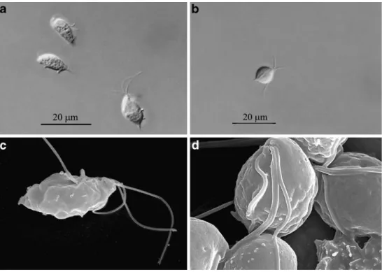
Light microscopy and scanning electron microscopy Light microscopy and scanning electron microscopy of T. foetus strain ATCC 30924 and of a stable cat isolate was performed. In both isolates, three flagella were visible. The
undulating membrane was clearly visible in the reference T. foetus strain. This reference strain also had the typical oval-lengthy shape (Fig.1, a and c). The cat isolate showed a rounded shape and the undulating membrane was hard to distinguish (Fig.1, b and d). The cat isolate seemed more agile in culture than the reference strain and more dividing parasites could be observed.
PCR analysis
An initial evaluation of the methodological sensitivity of the Tritrichomonas sp.-PCR was performed by using genomic DNA templates with different numbers of T. foetus parasites (ATCC strain 30924) each. This test revealed a high methodical sensitivity of the PCR, in that genomic DNA from ten T. foetus cells represented the
lowest limit of detection. Furthermore, the PCR was assessed with genomic DNAs prepared from different trichomonads including T. suis, T. gallinae, T. vaginalis, P. hominis, and Tetratrichomonas sp. and was able to detect all of them (Table 1). The PCR was assessed for the absence of nonspecific amplification reactions by testing genomic DNAs from negative InPouch™ and Diamond’s medium cultures (negative control DNA) (data not shown). Nonspecific amplification did not occur in PCRs with genomic DNA preparations representing a selection of parasitic and bacterial pathogens, and also some non-pathogenic control organisms (Table1).
PCR was carried out with ten of the 11 positive cultures. Unfortunately, one culture was wrongly discarded after the confirmation of motile trichomonads and could thus not be submitted to subsequent PCR. However, all ten assessed Fig. 1 Light micrographs
(×1,000, DIC) and scanning electron micrographs of repre-sentative specimen of the T. foetus isolate ATCC 30924 (a and c) and of tritrichomonads isolated from a cat (b and d). Note the three anterior flagellae and the undulating membrane present in both isolates and the rounded shape in the cat isolate
Table 1 Documentation of protozoal and bacterial isolates to assess specificity of diag-nostic PCR
Species Isolate PCR result Fragment size (bp)
Tritrichomonas foetus ATCC 30924 Positive 367
Tritrichomonas suis ATCC 30176 Positive 367
Trichomonas gallinae ATCC 30230 Positive 364
Trichomonas vaginalis ATCC 30240 Positive 363
Pentatrichomonas hominis ATCC 30098 Positive 333
Tetratrichomonas sp. AF342742 Positive 379
Giardia lamblia WBC6 Negative 0
Trichinella spiralis ISS3 Negative 0
Leishmania infantum ZH229/86 Negative 0
Toxoplasma gondii RH Negative 0
Neospora caninum NC1 Negative 0
isolates could be amplified by PCR and subsequent product-sequencing revealed total homology with T. foetus (GenBank accession No. AF466751) for all isolates. To our best knowledge, all published studies that included sequence data of isolates from naturally infected cats found 100% identity with the same T. foetus sequence (GenBank accession No. AF466751) (Gookin et al. 2002, 2003; Dahlgren et al.2007).
Discussion
In Switzerland, trichomonadid pathogens have not been diagnosed as a possible cause of diarrhea in cats so far. The reason for this might on one hand be the unfamiliarity with the organism in this context. On the other hand, routine coprological methods either destroy the fragile trichomo-nads (flotation-sedimentation method) or fix the parasites (SAF method), which leads to the loss of their characteristic movement and thus makes them hard to be recognized. The methods of choice for the detection of trichomonads in cat feces are (1) direct microscopical examination of saline-diluted feces, (2) cultivation methods, e.g., Diamond’s medium or InPouch™ TF culture system (Gookin et al. 2003), and (3) PCR analysis of the feces or of the cultures (Gookin et al.2002). The aims of our study were to assess whether trichomonads can be isolated from diarrheic cats in Switzerland and to determine to what species the tricho-monads belong to. This was of great interest to us because T. foetus, the species that has been isolated from cats by authors in the US, UK, and Norway (Gookin et al.2002, 2003; Gunn-Moore et al. 2007; Dahlgren et al.2007), is a notifiable disease in cattle in Switzerland and, for more than 10 years, no case of bovine infection had been reported. To answer the questions outlined above, we decided to screen clinical cases of cats suffering from chronic diarrhea with a commercially available culture system that has been accepted by the OIE for the surveillance of cattle (Anonymous2004), but which also has been evaluated for the diagnosis of intestinal trichomonadid infection in cats (Gookin et al.2003). Our study showed that about 25% of the diarrheic cats examined (11 out of 45 assessed cats) were positive in this pouch system. Since cultivation only detects parasites that survived until the sample was subjected to microscopical analysis, there is a possibility that some infected animals were misdiagnosed as being negative. However, the aim of this study was to assess whether trichomonads can be isolated from cats in Switzer-land and then to subject these isolates to PCR for molecular specification and not yet to perform an epidemiological investigation.
The PCR used for molecular speciation was designed to detect a broad range of trichomonads and yielded
amplifi-cation of an ITS-1 region which exhibits sufficient sequence heterogenicity to discriminate different trichomo-nad species. ITS-1 based assessment identified all our isolates from diarrheic cats as T. foetus (complete homology of the amplified sequence with reference strain sequence). Our molecular findings are also consistent with those revealed by scanning electron microscopy, which demon-strated the presence of three flagella in the assessed isolate rescued from an infected cat. Compared to the T. foetus reference strain 30924, the cat isolate did not show the typical elongated, slightly pear-shaped form but was rounded and full-bodied. Such changes in morphology have been described by various authors as signs of stress caused by variations in temperature or other physical influences or as effects of treatment with various drugs (Granger et al. 2000; da Silva et al. 2007; Carvalho and Gadelha 2007).
The results of the present study strongly indicated that T. foetus may be fairly common in diarrheic cats in Switzerland, and that with the given means (cultivation and morphology; PCR) we are presently unable to discriminate the parasites found in cats from T. foetus causing infertility and abortion in cattle. Furthermore, a recent study by Stockdale et al. (2007) has demonstrated that T. foetus isolated from a cat was able to cause endometritis and vaginitis upon experimental infection of heifers, although the endometrial damage caused by the cat isolate was less severe than that by T. foetus isolated from cattle in a parallel experiment. Conversely, T. foetus isolated from cattle could only successfully infect two out of five cats upon experimental infection (Stockdale et al.2008).
The mechanism of transmission between putatively different host species, as well as the route of transmission between feline hosts remains unknown so far. Thus, large-scale epidemiological studies accompanied by extended genetic comparisons between T. foetus strains from differ-ent hosts are necessary to address the potdiffer-ential hazard of this newly emerging parasite in cats to the Swiss cattle husbandry.
Acknowledgments We thank Dr. Gertrud Rosenberg, Caroline Müller, and Trang Nguyen for excellent technical assistance. Further-more, Dr. Beat Bigler of the laboratory Laupeneck in Bern is kindly acknowledged for providing feces from two cats with feline trichomonosis. The performed experiments comply with the current law of Switzerland.
References
Anonymous (1995) Tierseuchenverordnung. No 916.401 (www.bvet. admin.ch)
Anonymous (2004) Manual of diagnostic tests and vaccines for terrestrial animals, 5th ed. Chapter 2.3.6. (www.oie.int/fr/normes/ mmanual/a_00057.htm)
Carvalho KP, Gadelha AP (2007) Effects of three benzimidazoles on growth, general morphology and ultrastructure of Tritrichomonas foetus. FEMS Microbiol Lett 275:292–300
da Silva NS, Ribeiro C de M, Machado AH, Pacheco-Soares C (2007) Ultrastructural changes in Tritrichomonas foetus after treatments with AlPcS4 and photodynamic therapy. Vet Parasitol 146:175–181 Dahlgren SS, Gjerde B, Pettersen HY (2007) First record of natural Tritrichomonas foetus infection of the feline uterus. J Small Anim Pract 48:645–647
Diamond LS (1957) The establishment of various trichomonads of animals and man in axenic cultures. J Parasitol 43:488–490 Gookin JL, Breitschwerdt EB, Levy MG, Gager RB, Benrud JG
(1999) Diarrhea associated with trichomonosis in cats. J Am Vet Med Assoc 215:1450–1454
Gookin JL, Levy MG, Law JM, Papich MG, Poore MF, Breitschwerdt EB (2001) Experimental infection of cats with Tritrichomonas foetus. Am J Vet Res 62:1690–1697
Gookin JL, Birkenheuer AJ, Breitschwerdt EB, Levy MG (2002) Single-tube nested PCR for detection of Tritrichomonas foetus in feline feces. J Clin Microbiol 40:4126–4130
Gookin JL, Foster DM, Poore MF, Stebbins ME, Levy MG (2003) Use of a commercially available culture system for diagnosis of Tritrichomonas foetus infection in cats. J Am Vet Med Assoc 222:1376–1379
Gookin JL, Stebbins ME, Hunt E, Burlone K, Fulton M, Hochel R, Talaat M, Poore M, Levy MG (2004) Prevalence of and risk factors for feline Tritrichomonas foetus and giardia infection. J Clin Microbiol 42:2707–2710
Gookin JL, Copple CN, Papich MG, Poore MF, Stauffer SH, Birkenheuer AJ, Twedt DC, Levy MG (2006) Efficacy of
ronidazole for treatment of feline Tritrichomonas foetus infection. J Vet Intern Med 20:536–543
Gookin JL, Stauffer SH, Levy MG (2007) Identification of Penta-trichomonas hominis in feline fecal samples by polymerase chain reaction assay. Vet Parasitol 145:11–15
Grahn RA, BonDurant RH, van Hoosear KA, Walker RL, Lyons LA (2005) An improved molecular assay for Tritrichomonas foetus. Vet Parasitol 127:33–41
Granger BL, Warwood SJ, Benchimol M, De Souza W (2000) Transient invagination of flagella by Tritrichomonas foetus. Parasitol Res 86:699–709
Gunn-Moore DA, McCann TM, Reed N, Simpson KE, Tennant B (2007) Prevalence of Tritrichomonas foetus infection in cats with diarrhoea in the UK. J Feline Med Surg 9:214–218
Levy MG, Gookin JL, Poore M, Birkenheuer AJ, Dykstra MJ, Litaker RW (2003) Tritrichomonas foetus and not Pentatrichomonas hominis is the etiologic agent of feline trichomonal diarrhea. J Parasitol 89:99–104
Longo MC, Berninger MS, Hartley JL (1990) Use of uracil DNA glycosylase to control carry-over contamination in polymerase chain reactions. Gene 93:125–128
Mardell EJ, Sparkes AH (2006) Chronic diarrhoea associated with Tritrichomonas foetus infection in a British cat. Vet Rec 158:765–767
Stockdale H, Rodning S, Givens M, Carpenter D, Lenz S, Spencer J, Dykstra C, Lindsay D, Blagburn B (2007) Experimental infection of cattle with a feline isolate of Tritrichomonas foetus. J Parasitol 93:1429–1434
Stockdale HD, Dillon AR, Newton JC, Bird RC, Bondurant RH, Deinnocentes P, Barney S, Bulter J, Land T, Spencer JA, Lindsay DS, Blagburn BL (2008) Experimental infection of cats (Felis catus) with Tritrichomonas foetus isolated from cattle. Vet Parasitol 154:156–161