HAL Id: inserm-02530650
https://www.hal.inserm.fr/inserm-02530650
Submitted on 7 Apr 2020
HAL is a multi-disciplinary open access
archive for the deposit and dissemination of sci-entific research documents, whether they are pub-lished or not. The documents may come from teaching and research institutions in France or abroad, or from public or private research centers.
L’archive ouverte pluridisciplinaire HAL, est destinée au dépôt et à la diffusion de documents scientifiques de niveau recherche, publiés ou non, émanant des établissements d’enseignement et de recherche français ou étrangers, des laboratoires publics ou privés.
Advances in melanoma senescence and potential clinical
application
Sandy Giuliano, Mickaël Ohanna, Robert Ballotti, Corine Bertolotto
To cite this version:
Sandy Giuliano, Mickaël Ohanna, Robert Ballotti, Corine Bertolotto. Advances in melanoma senes-cence and potential clinical application. Pigment Cell & melanoma research, Blackwell Munksgaard, 2011, 24 (2), pp.295-308. �10.1111/j.1755-148X.2010.00820.x�. �inserm-02530650�
Advances in melanoma senescence and potential clinical application. Sandy Giuliano1,2, Mickaël Ohanna1,2, Robert Ballotti1,2,3, Corine Bertolotto1,2,3
1Inserm, U895, Equipe 1, Biologie et pathologies des mélanocytes: de la pigmentation cutanée au mélanome, C3M, Nice, F-06204, France
2Université of Nice Sophia-Antipolis, UFR Médecine, Nice, F-06107, France
3Centre Hospitalier Universitaire de Nice, Service de Dermatologie, Nice, F-06204, France E-mail: bertolot@unice.fr
Running head: Senescence in melanoma cells Key words: Melanoma, Senescence
Footnotes
This work was supported by INSERM, the Institut National du Cancer grant (INCA) R08009AP, the Ligue Nationale contre le Cancer, “Equipe labellisée”, the Association pour la Recherche sur le Cancer grant 4985 and the French Dermatology Society. M. Ohanna is a recipient from INCA.
Abstract
Normal cells possess a limited proliferative life span after which they enter a state of irreversible growth arrest called replicative senescence that acts as a potent barrier against transformation. Transformed cells have escaped the process of replicative senescence and could theoretically not re-enter senescence. However, recent observations have shown that transformed cells and particularly the melanoma cells can still undergo oncogene or stress-induced senescence. This senescence state is accompanied by many of the markers associated with replicative senescence such as flattened shape, increased acidic β-galactosidase activity (SA-β-gal), characteristic changes in gene expression and growth arrest. Interestingly, in some cancers, senescence induction following chemotherapy has been correlated to favorable patient outcome. Therefore, in this review, we wished to gather recent results describing senescence-like phenotype induction in melanoma cells and we discuss why senescence may also be exploited as a therapeutic strategy in melanoma.
1) Introduction
Normal cells possess a limited proliferative life span after which they enter a state of irreversible growth arrest. This process first observed by Hayflick and Moorhead and called replicative senescence is thought to result, in human cells, from telomere shortening as a consequence of cell division (Hayflick and Moorhead, 1961; Steinert et al., 2000). Replicative senescence can be identified by morphologic, biochemical and typical chromatin changes. Cells become enlarged, flattened and enriched in cytoplasmic vacuoles. They display senescence-associated β-galactosidase (SA-β-gal) reactivity at pH 5.5-6 which appears as a blue perinuclear staining (Kurz et al., 2000) and lack of BrdU incorporation. Senescent cells may show activation of two main signaling pathways p16INK4A/Rb and p14ARF/p53 (Ben-Porath and Weinberg, 2005) leading to the retinoblastoma protein (RB) held in an active hypophosphorylated state. They are characterized by senescence associated heterochromatic foci (SAHF) enriched in trimethylated histone H3 at lysine 9 (3MeK9H3) that recruit members of the heterochromatin-binding protein family (HP1) (Narita et al., 2006). Heterochromatic foci are responsible for epigenetic silencing at genes such as E2F target genes implicated in cell proliferation, mediating thereby the sustained growth arrest that characterizes senescence (Rastogi et al., 2006).
In addition, senescence-like phenotypes can also occur earlier, in the absence of telomere attrition, but in response to a variety of stimuli such as activation or inactivation of oncogene (or proteins that behave as oncogenes), oxidative stress, DNA damage, and drug-like inhibitors, and have been termed premature senescence, oncogene-induced senescence (OIS), oncogene-inactivation induced sennescence (OIIS), stress-and-aberrant signaling-induced senescence (STASIS) or stress-induced premature senescence (SIPS). Although unlike replicative senescence, premature senescence cannot be bypassed by over-expression of telomerase, it is nevertheless morphologically indistinguishable and the same markers accepted for replicative senescence can be employed for its detection.
While senescence is mainly associated with a G1 arrest, the spatio-temporal roles of the direct or associated-cell cycle regulators such as p16INK4A, p21CIP1, CDK4, p14ARF, p53 and PML for commitment and maintenance of the senescent state are still not fully understood, and at least some of them work in a context-, cell type and
species-dependent way (Kamijo et al., 1997; Sharpless et al., 2004). Although SA-β-Gal reactivity is the most widely-used marker of senescence, silencing of Glb1, that encodes the lysosomal beta-D-galactosidase gene, disrupts SA-β-Gal reactivity but does not impair cell entry into senescence (Lee et al., 2006), indicating that SA-β-Gal is a marker, but not a contributor to the senescence phenotype. In fact, SA-β-Gal reactivity reflects a change in lysosomal number or activity associated with the senescence phenotype (Kurz et al., 2000). Moreover, SAHF can be hardly detectable in some senescent situations (Vance et al., 2005), although this does not mean that extensive heterochromatinisation of proliferation-associated genes does not take place. Additionally, differentiated cells also undergo a G1 arrest that in most cases is irreversible but with a program of gene expression leading to a specialized cellular function. Taken together, only examining one marker will likely not reliably assign a cell as being senescent or not. On the other hand, the detection of several of these markers and a sustained cessation of proliferation will determine whether a cell is truly senescent.
Whatever replicative or premature, cellular senescence is characterized by a sustained growth arrest and it is commonly observed in pre-malignant lesions but rarely in the transformed state (Braig et al., 2005; Chen et al., 2005; Collado et al., 2005), supporting the notion that senescence acts as a potent tumor suppressor mechanism restricting cell proliferation.
2) Senescence in melanocytes
As observed in other cell types, melanocytes display progressive natural erosion of telomere at each round of cell division, and as a consequence of DNA damage resulting from too short telomeres finally undergo replicative senescence (Schwahn et al., 2005; Sviderskaya et al., 2002).
Replicative senescence of normal human melanocytes is in part p16-dependent as illustrated by the extended life-span of cultured p16-deficient relative to normal melanocytes (Gray-Schopfer et al., 2006; Sviderskaya et al., 2002). However, p16INK4A -deficient melanocytes are still able to respond to telomere attrition and to enter senescence suggesting that additional factors, such as p53, may impose a p16INK4A -independent checkpoint (Sviderskaya et al., 2003; Terzian et al., 2010). Melanocyte senescence is also associated with down-regulation of CDK2 and CDK4 kinase activities (Bandyopadhyay and Medrano, 2000), the dephosphorylation of the retinoblastoma
protein RB and subsequent repression of genes under the control of E2F1 (Sage et al., 2000). In addition to p16INK4A loss, some melanomas express p16-resistent CDKs (Zuo et al., 1996) or overexpress cyclin D1 (Sauter et al., 2002; Smalley et al., 2008), enabling cells to bypass Rb-induced senescence by a different mechanism.
Melanocytes can undergo premature senescence in response to oxidative stress (Leikam et al., 2008) and expression of various oncogenes such as BRAFV600E (Michaloglou et al., 2008), NRASQ61R (Zhuang et al., 2008) and HRASG12V (Denoyelle et al., 2006).
One of the best examples of in vivo senescence in melanocytes is nevi formation. Nevi, are benign melanocytic lesions, composed of senescent melanocytes and have been thought to occur from telomere attrition (Bastian, 2003). However, more recently, no apparent telomere loss was detected in nevi samples and together with the presence of BRAFV600E mutation in a large proportion of nevi samples, the hypothesis of premature senescence was suggested for these lesions (Gray-Schopfer et al., 2006; Michaloglou et al., 2008). However, as a single eroded telomere can trigger senescence (Abdallah et al., 2009), it cannot be excluded that this process is involved in nevi formation.
The serine/threonine kinase BRAF, which is mutated in approximately 50-60% of melanoma cells, is a downstream effector of NRAS in which mutations are also observed in about 15-20% of melanoma cells (Bennett, 2008). Forced expression of these oncogenic proteins in normal human melanocytes elicits OIS, that seems to be independent of telomere attrition (Michaloglou et al., 2005), but might result from replicational stress as described in other cell systems (Bartek et al., 2007; Di Micco et al., 2006; Mallette et al., 2007). Additionally, BRAFV600E and NRASQ61R-induced senescence in normal melanocytes proceeds independently of p16INK4A and p53 (Zhuang et al., 2008). Amplified or mutated H-RAS (HRASG12V) may also lead to OIS in a restricted subset of small melanocytic neoplasms, the Spitz nevi. Indeed, cells in which HRASG12V is over-expressed undergo senescence (Denoyelle et al., 2006). In that case, OIS is p16INK4A and p53-independent, but involves activation of the endoplasmic reticulum stress pathway. The activation of the MAPK/ERK pathway can also be achieved through hyperactivation of upstream-acting receptor tyrosine kinases such as the epidermal growth factor receptor (EGFR) orthologue Xiphophorus melanoma receptor kinase (Xmrk) (Wellbrock and Schartl, 1999). Accordingly, melanocytes harboring high expression level of Xmrk undergo senescence, through reactive oxygen species production and mitotic defect induction (Leikam et al., 2008). In contrast, low or moderate Xmrk expression level favors cell proliferation. This is reminiscent of previous data reporting gene dosage-dependent
senescence (Chen et al., 2005; Sarkisian et al., 2007). Support of these data also comes from observations that HRASG12V-driven melanoma is associated with transcriptional induction of EGFR ligand, providing an autocrine EGFR-stimulating loop (Bardeesy et al., 2005). Despite the oncogenic nature of these mutations, benign or spitz nevi typically remain in a growth-arrested state for decades and only rarely progress into melanoma, indicating that oncogene-induced melanocyte senescence acts as an effective cellular brake against melanoma development.
Mutation of senescence checkpoints allows continued proliferation and sustained DNA damage leading to genetic instability and cancer progression. One of the most important genetic and epigenetic changes in melanoma is associated with the CDKN2A locus encoding p16INK4A and p14ARF. Germline mutations affecting this locus have been linked to melanoma incidence (Goldstein et al., 2006; Kannengiesser et al., 2009). Intriguingly, while inactivation of the p53 pathway represents another common feature observed in human cancer, in melanoma, mutation or deletion of p53 is relatively rare (Hussein et al., 2003). However, the p53 signaling cascade is often disrupted ensuing the loss of its positive regulator ARF (Freedberg et al., 2008; Pomerantz et al., 1998; Zhang et al., 1998) or deregulation of some of its downstream target genes such as APAF1 (Soengas et al., 2001). Animal models have revealed the importance of p16INK4A and p53 in constraining cancer progression through senescence induction. Indeed, in zebrafish and mouse, BRAFV600E or oncogenic Ras (HRASG12V and NRASQ61R) expression promote benign melanocytic hyperplasias that resemble nevi while melanomas develop when these mutations are introduced in p16INK4A/p14ARF- or p53-deficient backgrounds (Ackermann et al., 2005; Bardeesy et al., 2001; Chin et al., 1997; Goel et al., 2009; Ha et al., 2007; Patton et al., 2005) or when p16INK4A expression is silenced via activation of β-catenin (Delmas et al., 2007). Noteworthy, melanoma can develop in oncogenic BRAFV600E mouse model, without p16INK4A inactivation, although it can not be ruled out that p16INK4A is non functional (Dhomen et al., 2009). Thus, results in vivo reveal the requirement of p16INK4A and p53 in restraining OIS. PTEN also restrains melanomagenesis (Dankort et al., 2009; Nogueira et al., 2010; You et al., 2002) and its inactivation, leading to activation of PI3K and of some of its downstream targets such as β-catenin, Ral or C-MYC, can be involved in melanocyte senescence suppression (Dankort et al., 2009; Mishra et al., 2010; Zhuang et al., 2008).
Therefore, oncogene activation in combination with genetic or epigenetic inactivation of gatekeepers of tumor suppression allows cells to bypass senescence checkpoints and to
favor cell transformation. In agreement, senescent cells can re-enter the cell cycle if DNA-damage checkpoints are inactivated (Di Micco et al., 2006). Contrastingly, tumor cells theoretically could not re-enter the senescence process. However, recent observations show that cellular senescence remains latently functional in tumors cells including melanoma cells (Giuliano et al., 2010; Khodadoust et al., 2009; Vance et al., 2005).
The process of replicative and premature senescence in melanocytes has been reviewed elsewhere (Bennett, 2003; Bennett and Medrano, 2002; Ha et al., 2008; Michaloglou et al., 2008) and will not be more detailed in this review. Contrastingly, there was a relative dearth of information regarding the ability of melanoma cells to enter a senescence program. Since advances have been made, this review will mostly focus on recent findings reporting induction of cellular senescence in melanoma cells and the potential therapeutic use of this process in the management of melanoma.
3) Senescence in melanoma cells
Cancers are the consequence of multiple epigenetic and genetic changes, therefore, the inactivation of only one factor, even critically required for tumor cell proliferation may not be sufficient to cause sustained tumor regression. Moreover, cancers display strong genomically instability that may readily overcome or compensate for the inactivation of a single oncogene. However, several reports demonstrate that the inactivation of a single oncogene, the restoration of one tumor suppressor or chemotherapeutic drugs may surprisingly induce cellular senescence. In this review, we will focus our attention on recent reports indicating that senescence remains latently functional in tumor cells from melanoma and can be reactivated upon gain or loss of function of tumor suppressor genes (or considered as tumor suppressors) or oncogenes (or behaving like oncogenes) respectively, that have been deregulated during the process of transformation. Hence, the inactivation of C-MYC, MITF, TBX2 or DEK can commit melanoma cells to senesce as well as forced expression of p16INK4A or SYK that are generally absent in melanoma cells. Since melanoma cells are highly resistant to apoptotic processes, these observations have important implications, rendering cellular senescence an attractive therapeutic strategy to limit melanoma cell proliferation.
MITF
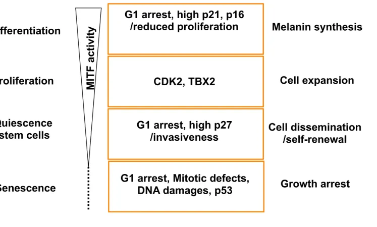
The microphthalmia-associated transcription factor, MITF, is a member of the basic helix-loop-helix-leucine zipper (bHLH-LZ) family. The MITF M-isoform, specifically expressed in melanocytes, plays a key role in melanocyte homeostasis and in the pathogenesis of melanoma where it has been described as a lineage-addiction oncogene. Indeed, MITF controls the transcription of genes involved in cell cycle progression, cell survival, migration and angiogenesis (Cheli et al., 2010) and point mutations, that affect its ability to regulate transcription, have been found in melanoma cells (Cronin et al., 2009). Additionally, MITF lies downstream from a number of oncogenic pathways, such as ERK pathway (Jane-Valbuena et al., 2010; Wellbrock et al., 2008; Zhu et al., 2009) and is amplified in some melanoma cases (Garraway et al., 2005). Consequently, targeting MITF is a potential new avenue for the management of melanoma. In this context, our group has shown that MITF acts as an anti-senescence factor and hence MITF-silencing by siRNA triggers senescence of melanoma cells (Giuliano et al., 2010). Nicely in line with our findings are the data from the group of E. Medrano, which shows an absence of MITF expression in senescent cultured melanocytes (Schwahn et al., 2005) and that of C. Goding showing, in a more physiological context, a reduced MITF expression in nevi compared to melanoma specimens (Carreira et al., 2006). However other studies showed that MITF expression is conserved in nevi, although an accurate quantification of the number of MITF positive and negative cells in nevi relative to melanoma as well as a correlation between the level of MITF expression and cellular senescence has not been investigated (King et al., 2001; Nazarian et al., 2010). MITF-suppression triggers senescence-like phenotypes in several human and mouse melanoma cell lines from different genetic backgrounds, thereby demonstrating that this process is not restricted to a specific species or to a unique melanoma cell line. Importantly, MITF-silencing also induces senescence in melanoma cells harboring BRAFV600E, the most frequently mutated oncogene in this disease. It should also be noted that MITF-silencing, induces SA-β-Gal reactivity in freshly isolated melanoma cells, although to a lesser extent than in cell lines. Culture-established cell lines may enter the senescence state more easily than primary tumors due to the genetic defects that the cells may accumulate during cell culture and expansion. The observation that MITF controls a senescence program in the melanocyte lineage adds a new step in the rheostat model (Figure 1) proposed by the group of C. Goding several years ago (Carreira et al., 2006).
Senescence triggered by MITF-invalidation is p16INK4A-independent but p53-dependent (Giuliano et al., 2010). The increase in p53 is the consequence of activation of the DNA-Damage Response cascade that was suspected to result from mitotic defects. In line with this view, ChIP-seq and RNA-seq analysis allowed the repertoire of direct MITF direct target genes to be identified, among which several are involved in mitotic progression and DNA damage/repair processes (Strub et al., 2010). Severe mitotic defects usually end into cell death through mitotic catastrophe or apoptosis. Impairment of apoptotic processes found in melanoma cells may tip the balance towards senescence instead of cell death in these cells.
Oxidative stress has also been shown to cause DNA damage and cellular senescence through the p53 pathway (Lewis et al., 2008). To prevent ROS accumulation, cells have developed detoxifying enzymes (superoxide dismutase, catalase, glutathione peroxidase) which transform ROS to H2O2 and also use redox-sensitive molecules such as apurinic/apyrimidic endonuclease1/redox factor-1 (APE1/Ref1) that decreases the ROS by inhibiting Rac-1-regulated NAD(P)H oxidase (Angkeow et al., 2002; Veal et al., 2007). In this context, MITF has been shown to control the expression of APE1/Ref1 (Liu et al., 2009) and down-regulation of APE1/Ref1 triggers cellular senescence of mesenchymal stem cells (Heo et al., 2009). As mentioned above, MITF-silencing promotes p53 up-regulation and p53 is a negative-regulator of APE1/Ref1 by interfering with Sp1 binding to the APE1/Ref1 promoter (Zaky et al., 2008). Thus, ROS production might account for MITF-silencing induced senescence. Accordingly, ROS levels are enhanced upon MITF suppression (C. Bertolotto, personal communication) but this increase is observed at a time where senescence is already established. Hence, ROS accumulation is unlikely to represent the initiator event, but it may act to reinforce the MITF-silencing-induced senescence program in melanoma cells. In agreement with these hypotheses, a DNA damage-ROS signaling cascade has been reported to play a critical role in the maintenance of the senescence state (d'Adda di Fagagna, 2008; Passos et al., 2010).
MYC
The proto-oncogene C-MYC, another transcription factor of the b-HLH-LZ family, acts as a heterodimer with MAX in various biological processes such as cell proliferation, growth, metabolism and angiogenesis (Pelengaris et al., 2002). Over-expression of C-MYC is associated with tumorigenesis in a wide range of cancers (Adhikary et al., 2005).
Conversely, its inactivation can reverse the process of transformation and induces tumor regression via apoptosis or differentiation. C-MYC-inactivation by RNA interference has been shown to trigger cellular senescence in diverse tumor types including melanoma cells (Biroccio et al., 2003; Guney et al., 2006; Wu et al., 2007; Zhuang et al., 2008). This process has been termed oncogene-inactivation induced senescence (OIIS) (Wu et al., 2007). C-MYC inactivation in these different cell types does not engage the same cell cycle regulators and checkpoint proteins, indicating that OIIS, caused by C-MYC suppression, operates in a context-dependent manner. Senescence growth arrest in osteosarcomas is p53-, p16INK4A- and Rb-dependent. It does not involve DNA-damage response, ERK or p38 MAPK pathways. Senescence mediated by C-MYC suppression in melanoma cells, is p53- and p16INK4A-independent but relies on the ERK or PI3K signaling pathway activation according to whether c-MYC inactivation operates in a BRAFV600E or NRASQ61R background (Zhuang et al., 2008). BRAFV600E and to a lesser extent NRASQ61R-induced melanocyte senescence is associated with C-MYC down-regulation and enforced C-MYC expression prevents this senescence program suggesting that MYC may function to overcome senescence. Furthermore, pharmacological inhibition of the ERK or PI3K pathways impairs some of the C-MYC-induced senescence phenotypes in melanoma cells harboring BRAFV600E or NRASQ61R respectively. It is unknown how C-MYC-silencing promotes the growth arrest in melanoma cells. In most cell lines used in the study of Nikiforov and co-workers, C-MYC suppression mainly triggered G1 cell cycle arrest, although G2/M arrest is observed for some cell lines. Noteworthy, G1 cell cycle arrest was associated with p53-dependent transcriptional repression of C-MYC in various mouse and human cell lines and mouse tissues (Ho et al., 2005). MITF-silencing, which increases p53 and promotes G1 arrest, does not affect the level of c-MYC, indicating that MITF-silencing induced senescence is not mediated through c-MYC down-regulation (personal communication).
Several cell cycle inhibitors, such as p21CIP1 or p27KIP1, are known to be transcriptionally repressed by C-MYC (Gartel and Shchors, 2003). In melanoma cells, p21CIP1 and p27KIP1 level increase following C-MYC suppression and may favor the cell cycle arrest. p21CIP1 and p27KIP1 are also the targets of the PI3K pathway (Liang and Slingerland, 2003) which positively controls C-MYC and which acts as an anti-senescence factor (Zhang and Yu, 2010). Finally, ROS production has also been shown to play an important role for the senescence program mediated by C-MYC inhibition (Biroccio et al., 2003).
TBX2
TBX2 is one of the T-box family of genes encoding key developmental transcription factors that govern cell fate decisions, and deregulation of some members of this family, namely the highly-related transcriptional repressors TBX2 and TBX3, has been observed in cancers (Papaioannou, 2001; Tada and Smith, 2001). TBX2 and TBX3 are overexpressed in melanoma cell lines (Hoek et al., 2004; Vance et al., 2005) and TBX2 overexpression in normal cells favors senescence bypass and tumor progression by repressing the expression of the cyclin-dependent kinase (CDK) inhibitor p21 and the tumor suppressor ARF (Brummelkamp et al., 2002; Jacobs et al., 2000; Prince et al., 2004; Yarosh et al., 2008). Conversely, TBX2-inhibition by forced expression of an inducible dominant-negative mutant, which maintains the ability to bind DNA but lacks the carboxyl-terminal repressor domain promotes senescence entry (Vance et al., 2005). The mechanism occurs in CDKN2a-deficient cells and involves de-repression of p21CIP1 expression, most likely through displacement of the TBX2-HDAC1 complex at the p21CIP1 promoter (Prince et al., 2004; Vance et al., 2005). Histone deacetylase 1 (HDAC1) belongs to the large HDAC family of enzymes, which play important roles in chromatin remodeling and signal regulation (Witt et al., 2009). Increased expression of HDAC1 in melanocytes undergoing senescence has been observed (Bandyopadhyay et al., 2002) and HDAC1 immunoreactivity is found in nevi (Bandyopadhyay et al., 2007), one of the best examples of in vivo senescence. Consistent with this, HDAC1 forced expression using a tet-on system commits melanoma cells to senescence (Bandyopadhyay et al., 2007). This process involves the cooperative activity of HDAC1 with other chromatin remodeling effectors, such as the SWI/SNF member Brahma (BRM1), which, by triggering a stable association of RB protein with chromatin, will allow a sustained DNA heterochromatization and the ultimately irreversible cell cycle arrest and senescence phenotype. These observations indicate that HDAC1 plays an essential role in cellular senescence. Thus, one might also hypothesize that TBX2-HDAC1 dissociation from DNA will not only engage p21CIP1 derepression but will favor HDAC1 release from this complex. In turn, free HDAC1 will lead to histone lysine deacetylation, an event required for histone H3 trimethylation at lysine 9 and associated with the senescent state (Willis-Martinez et al., 2010). Whether this process also implicates the activity of TBX3 or other T-box factors, that can potentially be inhibited by the dnTBX2, remains unknown, though Tbx3 can target the p21CIP1 promoter (Hoogaars et al., 2008) and has been described as an anti-senescence factor in other systems (Brummelkamp et al., 2002). The role of
TBX2 as an anti-senescence factor is strengthened by at least two other observations. First, TBX2 is up-regulated in a PI3K-dependent manner (Ismail and Bateman, 2009) a signaling pathway involved in senescence bypass and in tumor development. Second, TBX2 is a downstream target gene of MITF (Carreira et al., 2000), for which inactivation also triggers senescence (Giuliano et al., 2010).
DEK
DEK is a nuclear factor that acts as a regulator of chromatin architecture being involved in DNA replication, splice site recognition, and gene transcription (Alexiadis et al., 2000; Soares et al., 2006; Waldmann et al., 2004). DEK has been initially discovered as the target of a chromosomal translocation in acute myelogenous leukemia (von Lindern et al., 1992) and now has been found frequently up-regulated in several neoplasms (Carro et al., 2006; Wu et al., 2008). In melanoma cells, DEK level is higher than in normal melanocytes or benign nevi (Khodadoust et al., 2009). DEK expression is regulated either by genomic amplification, or by post-transcriptional and transcriptional modifications, and has been associated with an extended cellular lifespan (Wise-Draper et al., 2005). At the transcriptional level, DEK increase depends on pRB loss and E2F1 activity (Carro et al., 2006). Therefore, one can expect that DEK will be decreased in cells undergoing senescence as a consequence of RB activation and E2F1 repression. Additionally, DEK-DNA interactions can be modulated by a series of chromatin-associated factors such as histone deacetylases and methyltransferases (Riveiro-Falkenbach and Soengas, 2010) that play an active role in the control of cellular senescence.
In line with these observations, in papilloma-positive cervical cancer cells, down-regulation of the viral E7 oncogene, a classical RB inactivator, results in cellular senescence that is associated with DEK repression (Wise-Draper et al., 2005). Furthermore, long-term DEK suppression by shRNA triggers premature senescence of melanoma cells in absence of p16INK4A regulation and in p53 proficient or deficient cell lines (Khodadoust et al., 2009), indicating that both p16INK4A and p53 are not essential for DEK depletion induced senescence in melanoma cells. Noteworthy, short-term depletion of DEK increased response of melanoma cells to apoptotic drugs (Khodadoust et al., 2009). This is in agreement with the anti-apoptotic function of DEK previously reported in HeLa cells (Wise-Draper et al., 2006) and with the notion that senescence renders cells resistant to apoptosis, indicating that apoptotic stimuli may exert their effects before
senescence establishment (Ryu et al., 2006; Ryu et al., 2007; Yeo et al., 2000). The molecular mechanisms, by which DEK inhibition mediates senescence of melanoma cells, have yet to be determined. DEK can enhance resistance to DNA-damaging agents by mediating DNA repair (Gamble and Fisher, 2007; Kappes et al., 2008), suggesting a role for DEK in genomic integrity. The inhibition of DEK might elicit DNA damages that could end into apoptotic or senescence responses. It appears that DEK or MITF suppression elicits similar defects, suggesting that they may function in a same pathway, although, until now the link between DEK and MITF remains to identify.
b) Oncogene-Induced Senescence (OIS)
a)
BRAF/RAS
BRAF mutation mostly at codon 600, and NRAS mostly at codon 61, are mutated with high frequency in melanoma lesions (50% and 15-20% respectively), where they bring important proliferative and survival properties (Inamdar et al., 2010). Contrastingly, each of these mutations triggers senescence of normal melanocytes (Denoyelle et al., 2006; Michaloglou et al., 2008). It seems worth noting that although at high frequency, mutations of these two oncogenes are mutually exclusive in melanoma cells, suggesting that they functionally overlap or are incompatible with cell survival. To explain the mutually exclusive mutation profile, the group of Sensi has evaluated the effect of co-expressing BRAFV600E and NRASQ61R in same melanoma cells (Petti et al., 2006). They show that co-expression of both oncogenic proteins activates a p16INK4A- and p53-independent senescence program characterized by G0/G1 growth arrest and SA-β-Gal reactivity. This data may provide some explanation as to why BRAFV600E and NRASQ61R co-expression are rarely found in the same melanoma. The authors also find an increase in p21CIP1, while p53 level does not change when activated oncogenes are expressed both in the same cell, suggesting a p53-independent induction of p21CIP1 as previously reported (Lodygin et al., 2002). One mechanism by which ERK over-activation upon BRAF or NRAS mutation might cause senescence is through an increase in BRN2 that acts as a repressor of MITF (Goodall et al., 2008; Goodall et al., 2004). However, no change in MITF expression was observed in cells co-expressing BRAFV600E and NRASQ61R co-expression (Petti et al., 2006), although a change in MITF transcriptional activity can not be ruled out. This hypothesis has yet to be investigated. It is interesting to note that senescence entry is not the only mechanism leading to selection against
“double mutant” cells as BRAFV600E and NRASQ61R co-expressing cells display increased lysability by cytotoxic-T cells (Petti et al., 2006), showing that senescent melanoma cells may be actively eliminated by the immune system.
c) Restoration of tumor suppressor-induced senescence SYK
The non-receptor tyrosine kinase SYK (Spleen Tyrosine kinase) that was initially thought to be restricted to the hematopoietic system where it exerts essential functions in immunoreceptor signalling, natural killer cell-mediated cytotoxicity and maturation of lymphocytes, has been recently detected in non-hematopoietic tissues such as breast epithelium and melanocytes, where its physiological function remains to be clearly defined (Coopman et al., 2000; Hoeller et al., 2005; Mocsai et al., 2010). SYK is expressed in normal human mammary gland tissue and in melanocytes but is found downregulated in breast carcinoma cell lines and melanoma cells. The loss of SYK expression in these tumors is associated with a CpG island hypermethylation in the SYK promoter region (Bailet et al., 2009; Coopman et al., 2000; Wang et al., 2004). Reexpression of SYK in melanoma reduces their invasive properties and tumor formation in vivo (Hoeller et al., 2005; Muthusamy et al., 2006), indicating that SYK functions as a tumor suppressor. Recently, a senescence-like phenotype has been observed upon SYK reintroduction into melanoma cells (Bailet et al., 2009). SYK-repleted melanoma cells become flattened, enlarged, vacuolized and are positive for the SA-β-Gal staining. SYK-induced senescent cells also show an up-regulation of p53 and p21CIP1 and further additional p53 target genes are witnesses of senescence entry. A fraction of SYK has been found at the centrosomes, that control mitotic progression and consequently, sustained expression of a transfected SYK engendered abnormal cell division and non-apoptotic cell death resembling mitotic catastrophe (Zyss et al., 2005). Hence, SYK may act in a tissue-specific manner inducing cell death by mitotic catastrophe or senescence. The mechanisms by which SYK induces a senescence program remain undetermined. As mentioned above, genomic instability in highly apoptotic resistant melanoma cells may be one of these processes. Furthermore, SYK negatively controls phosphatidylinositol (PI) 3'-kinase activity (Mahabeleshwar and Kundu, 2003), that functions upstream of TBX2 or C-MYC (Ismail and Bateman, 2009; Zhu et al., 2008). Therefore, one can envision that SYK may act through deregulation of TBX2 or C-MYC to mediate its pro-senescence effects.
Indeed, qRT-PCR experiments show that C-MYC mRNA level decreases upon forced expression of SYK (Bailet et al., 2009), suggesting that C-MYC might play a role in SYK-induced senescence in melanoma cells.
p16INK4A
One of the most important genetic and epigenetic changes in melanoma is associated to the CDKN2A locus. Germline mutations affecting the INK4a/ARF locus have been linked to melanoma incidence (Goldstein et al., 2006). This locus encodes two tumor suppressor proteins namely p16INK4A and the p53 activator p14ARF. Melanocytes isolated from melanoma-prone individuals harboring biallelic p16INK4A inactivating mutations undergo delayed senescence (Jones et al., 2007; Sviderskaya et al., 2003) and can easily be immortalized by telomerase reverse transcriptase expression, strengthening the key role of p16INK4A as a potent barrier against neoplastic transformation. The reintroduction of wild type p16INK4A triggers growth arrest and cellular senescence phenotypes in INK4a/ARF-deficient murine melanocytes (Sviderskaya et al., 2002) and in human melanoma cell lines (Haferkamp et al., 2008). The senescence program mediated by p16INK4A in melanoma cells does not involve p53, p21CIP1, the endoplasmic reticulum stress or the DNA-damage response pathways, but does require RB. Furthermore, ectopic expression of CDK4 and its homologue CDK6, but not CDK2, overcomes p16INK4A-induced senescence. To further substantiate these observations, the establishment of senescence in melanoma cells failed when reintroducing mutants of p16INK4a that do not bind CDK4 (Haferkamp et al., 2008). In support of a role of p16INK4a in restraining tumorigenesis, CDK4 mutations, rendering this kinase insensitive to p16INK4a have also been identified in melanoma-prone families (Soufir et al., 1998; Zuo et al., 1996) and mice carrying the corresponding oncogenic CDK4 are highly susceptible to melanoma development (Sotillo et al., 2001).
d) Chemotherapy induced senescence
Standard chemotherapy regimens are now recognized to exert their therapeutic potential via forcing cancer cells to die and/or to enter a senescence-growth arrest phenotype. The observation that cancer cells have maintained the ability to senesce in vivo has opened a new window of opportunity for cytostatic drugs in chemotherapy, and more particularly
with the demonstration of senescence induction in melanoma by compounds that target specific pathways.
The DNA-alkylating agent temozolomide (TMZ), one of the first-line chemotherapeutic drugs for the treatment of metastatic melanoma, triggers G2/M arrest and senescence-like properties in some melanoma cells (Mhaidat et al., 2007). Cells adopt an enlarged and flattened morphology and display a SA-β-Gal reactivity. Blocking p21CIP1 induction by siRNA prevents melanoma cell growth arrest, in agreement with findings in glioblastoma cells where TMZ mediates senescence through p21CIP1 up-regulation and inhibition of CDK2 (Hirose et al., 2001). TMZ has also been reported to induce apoptosis (Naumann et al., 2009). Noteworthy, in patients with melanoma, the response rates to TMZ remain low and do not exceed 10-20%. p53 mutation in some cases or high level of O(6)-methylguanine-DNA methyltransferase (MGMT) might explain the resistance of melanoma cells to TMZ-induced senescence (Mhaidat et al., 2007), supporting the notion that the genetic make-up can determine outcome. Moreover, it is possible that not all the senescent competent cells will execute the arrest program due to heterogeneity in drug supply within the tumor. It will be interesting to determine the level of senescence or apoptosis in tumor from patients who showed an objective response to TMZ and to determine whether TMZ affects MITF level.
A subset of human melanoma cell lines with BRAF or NRAS activating mutations can also adopt senescence-like phenotypes in response to the diterpene ester PEP005, a novel anticancer agent (Cozzi et al., 2006). Treatment with PEP005 induces SA-β-Gal reactivity, RB activation, p21CIP1 up-regulation and induction of an irreversible G2-M cell cycle arrest. By contrast, MEK inhibitors prevent senescence commitment by PEP005. In this context, cDNA microarrays identified the HRAS-like suppressor 3 (HRASLS3) as part of a molecular signature for sensitivity or resistance to diterpene ester treatment (Cozzi et al., 2006). This is reminiscent of Nikiforov and coworkers’ observations demonstrating that C-MYC silencing-induced melanoma senescence involves the MAPK/ERK pathway (Zhuang et al., 2008), presumably with the ERK-pathway promoting hyperproliferation-related stress.
In conclusion, several independent reports demonstrate that melanoma cells retain the capacity to senesce. One cannot rule out that cultured cancer cell lines may be more sensitive to senescence commitment in vitro than that will be observed in vivo due to either the lack of the micro-environment effects or the genetic defects acquired during cell
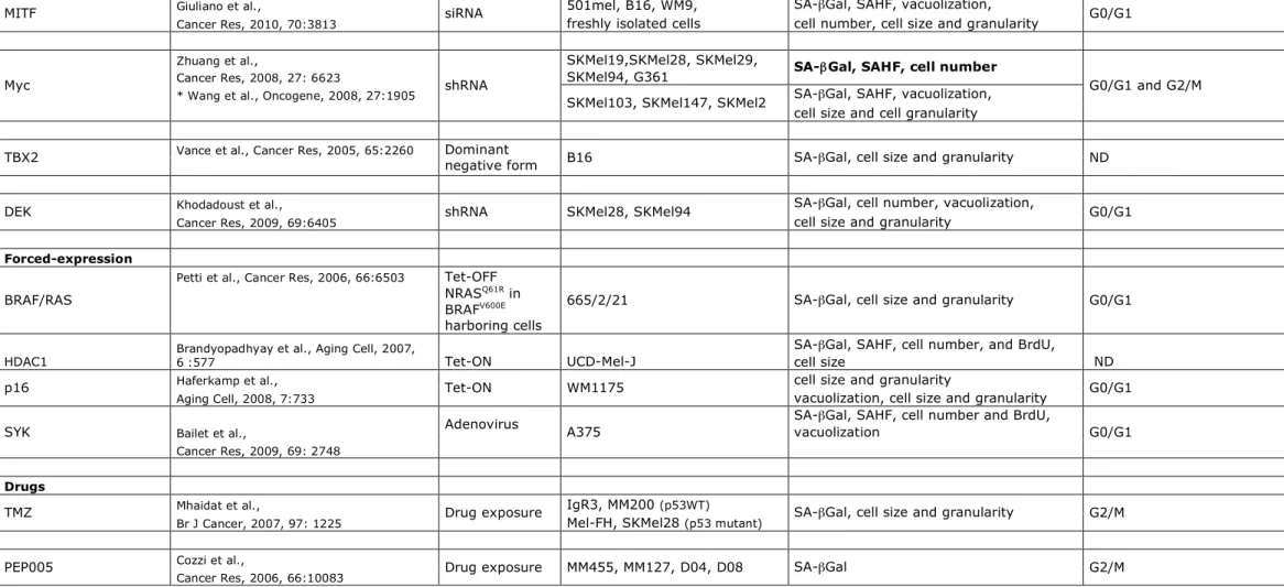
culture maintenance. Nevertheless, the observation by Wu et al. that MYC invalidation also induces senescence in transplanted liver tumors in vivo argues against this hypothesis (Wu et al., 2007). Moreover, in all these studies, the senescence arrest has been investigated over a one or two-week period. Whether this arrest is really equivalent to the irreversible growth arrest observed in truly senescent cells, like in nevi, remains to be fully determined. Furthermore, a combination of different markers has been used to determine the senescence phenotypes (Table 1), but the causal role and requirement of most of these biomarkers in senescence induction remains to be elucidated in greater details. The data, collected in this review, highlight the existence of a complex and overlapping regulatory network of senescence-inducing stimuli (Figure 2). Further, MYC, MITF, TBX2, DEK and SYK have been associated with the control of genomic stability (Davis et al., 2008; Felsher and Bishop, 1999; Giuliano et al., 2010; Zyss et al., 2005), suggesting additional levels of senescence control that are not mentioned in Figure 2. Thus, it remains to be clearly elucidated how the senescence programs, induced by modulation of these factors, are connected. Future insights gained from the molecular events of senescence in melanoma cells will likely lead to the identification of potential targets for pro-senescence drugs.
4) Potential side-effects of senescence
a) Senescence-associated secretory phenotype (SASP)
Senescence has been considered, for decades, as a potent barrier against tumor progression. However, recent observations have demonstrated that, although growth stopped, senescent cells display secretory capacity known as the Senescence-Associated Secretory Phenotype (SASP). The SASP comprises metalloprotease (MMP1, MMP3 or MMP10), pro-inflammatory chemokines (CXCL2, IL6, IL8 and TGF-β) and growth factors (GROα, HGF, VEGF) with auto- and paracrine effects (Coppe et al., 2010; Freund et al., 2010). Some of these secreted molecules may have autocrine effects that enforce senescence (Acosta et al., 2008; Kuilman et al., 2008), whereas SASP may also exert non-cell-autonomous effects favoring tumorigenesis in nearby non-senescent cells (Olumi et al., 1999). Thus, it will be important to identify the SASP’s full repertoire and to determine the pro- and anti-senescent factors. Whether senescent melanoma cells developed a SASP is still unknown.
b) Neosis
Another side-effect of senescence has been the observation that some tumoral cells engaged in a senescence program seem to be able to escape senescence by a process called neosis (Rajaraman et al., 2006). Neosis applies to polyploid senescent cells that can generate small mononucleated active cells, called Raju cells, by a mechanism of nuclear budding. It has been proposed that Raju cells initially expand through asymmetric division and next through classic mitosis. Given that Raju cells are produced by nuclear budding, the control of the mitotic spindle is absent, resulting in strong aneuploidy and genomic instability at the origin of highly heterogeneous tumoral cells. It is not known however, whether Raju cells emerge from fully senescent or pre-senescent cells and whether senescent melanoma cells are capable of neosis.
c) Reversibility of the senescence phenotype
It is worth noting that senescence reversibility has been observed in vitro in response to the inactivation of tumor-suppressors in senescent cells (Beausejour et al., 2003). However, whether in vivo senescent cells are able to acquire genetic or epigenetic defects in the absence of DNA synthesis to truly revert towards proliferative cells remains to be clearly demonstrated. In favor of this idea is the emergence of melanoma within or contiguous to nevi (Bevona et al., 2003; Gruber et al., 1989). Tumors with BRAF mutations were also significantly more likely to occur in association with a contiguous nevus (Poynter et al., 2006). Therefore, these observations are in support of a nevus-to-melanoma progression model where bypass of OIS of nevus cells due to oncogenic BRAF or NRAS favor progression towards melanoma.
5) Exploiting senescence induction as a therapeutic strategy against melanoma Apoptosis and senescence are cellular failsafe programs that counteract excessive mitogenic signaling observed in cancer cells. Chemotherapy-induced reduction in tumor load mainly functions through apoptotic cell death, orchestrated by intracellular caspases. However, the effectiveness of these therapies is compromised by mutations affecting specific genes, controlling and/or regulating apoptotic signaling. This is especially true for melanoma, which is known for its notorious resistance to apoptotic processes (Soengas
and Lowe, 2003). Therefore, it is desirable to identify novel anti-proliferative pathways, which could function in tandem with or in the absence of efficient apoptotic machinery. Senescence may be one of these processes, although no pro-senescence drugs have been described so far to provide an effective and durable inhibition of established melanomas.
First of all, cellular senescence can act as a potent tumor-suppressor mechanism firmly halting tumor progression, as illustrated for nevi that usually remain unchanged for decades. Furthermore, results from animal models show that senescent tumor cells can elicit an immune response in vivo leading to their clearance (Wu et al., 2007; Xue et al., 2007). Anti-natural killer-neutralizing antibody impairs natural killer cell function and prevents the elimination of senescent cells (Krizhanovsky et al., 2008), demonstrating the requirement of an efficient immune system to eradicate senescent cells. In agreement, senescent melanoma cells display an increased lysability relative to their non-senescent counterpart (Petti et al., 2006). Thus, in vivo, the negative effect of the SASP, produced by senescent cells and previously reported in this review, may be overcome by the presence of an effective immune system. Conversely, a defective immune system may favor SASP accumulation and may reveal detrimental effects of cellular senescence. These observations suggest nevertheless that, in vivo, chemotherapy-induced senescence may exert objective anti-tumor effects. Consistent with this, in humans, senescence-induction in human breast cancer and lung carcinoma following chemotherapy is correlated to favorable outcome (Schmitt et al., 2002; te Poele et al., 2002).
It is interesting to note that melanoma cells already have a powerful pro-senescence signal via activation of BRAF or NRAS that distinguishes the cancer cells from their normal counterparts. In support of this notion is the work from the group of Nikiforov showing that BRAFV600E or NRASQ61R dependent senescence exists in dormancy in advanced melanoma cells and could be reactivated by depletion of C-MYC (Zhuang et al., 2008). As such pro-senescence therapy already has a built-in specificity for cancer cells. Importantly, it is postulated that chemotherapeutic drugs trigger senescence when they are used at low doses and promote apoptosis when they are used at higher doses. From a clinical viewpoint, chemotherapeutic drug cytotoxicity is a major problem and therefore whether cure may be achieved with lower drug concentrations may represent a clinical advantage for the well-being and management of the patient.
Senescence is commonly observed in pre-malignant lesions but not in the malignant one, suggesting that the detection of senescence markers- acidic SA-β-Gal reactivity, the expression of cell cycle inhibitor (p14ARF, p16INK4A, p21CIP1 and p27KIP1 for the most studied), the tumor suppressor p53, the active form of RB and the formation of SAHF- may be used as prognostic indicator of the tumor stage (Collado et al., 2005; Lazzerini Denchi et al., 2005; Michaloglou et al., 2008). Induction of cellular senescence appears to be a promising therapeutic strategy but the molecular mechanisms of senescence, and in particular here of melanoma cell senescence, remain to be explored in greater details in order to determine whether pro-senescence drugs will truly have beneficial effects.
Acknowledgments
References
Abdallah, P., Luciano, P., Runge, K.W., Lisby, M., Geli, V., Gilson, E., and Teixeira, M.T. (2009). A two-step model for senescence triggered by a single critically short telomere. Nat Cell Biol 11, 988-993.
Ackermann, J., Frutschi, M., Kaloulis, K., McKee, T., Trumpp, A., and Beermann, F. (2005). Metastasizing melanoma formation caused by expression of activated N-RasQ61K on an INK4a-deficient background. Cancer Res 65, 4005-4011.
Acosta, J.C., O'Loghlen, A., Banito, A., Guijarro, M.V., Augert, A., Raguz, S., Fumagalli, M., Da Costa, M., Brown, C., Popov, N., et al. (2008). Chemokine signaling via the CXCR2 receptor reinforces senescence. Cell 133, 1006-1018.
Adhikary, S., Marinoni, F., Hock, A., Hulleman, E., Popov, N., Beier, R., Bernard, S., Quarto, M., Capra, M., Goettig, S., et al. (2005). The ubiquitin ligase HectH9 regulates transcriptional activation by Myc and is essential for tumor cell proliferation. Cell 123, 409-421.
Alexiadis, V., Waldmann, T., Andersen, J., Mann, M., Knippers, R., and Gruss, C. (2000). The protein encoded by the proto-oncogene DEK changes the topology of chromatin and reduces the efficiency of DNA replication in a chromatin-specific manner. Genes Dev 14, 1308-1312.
Angkeow, P., Deshpande, S.S., Qi, B., Liu, Y.X., Park, Y.C., Jeon, B.H., Ozaki, M., and Irani, K. (2002). Redox factor-1: an extra-nuclear role in the regulation of endothelial oxidative stress and apoptosis. Cell Death Differ 9, 717-725.
Bailet, O., Fenouille, N., Abbe, P., Robert, G., Rocchi, S., Gonthier, N., Denoyelle, C., Ticchioni, M., Ortonne, J.P., Ballotti, R., et al. (2009). Spleen tyrosine kinase functions as a tumor suppressor in melanoma cells by inducing senescence-like growth arrest. Cancer Res
69, 2748-2756.
Bandyopadhyay, D., Curry, J.L., Lin, Q., Richards, H.W., Chen, D., Hornsby, P.J., Timchenko, N.A., and Medrano, E.E. (2007). Dynamic assembly of chromatin complexes during cellular senescence: implications for the growth arrest of human melanocytic nevi. Aging Cell 6, 577-591.
Bandyopadhyay, D., and Medrano, E.E. (2000). Melanin accumulation accelerates melanocyte senescence by a mechanism involving p16INK4a/CDK4/pRB and E2F1. Ann N Y Acad Sci 908, 71-84.
Bandyopadhyay, D., Okan, N.A., Bales, E., Nascimento, L., Cole, P.A., and Medrano, E.E. (2002). Down-regulation of p300/CBP histone acetyltransferase activates a senescence checkpoint in human melanocytes. Cancer Res 62, 6231-6239.
Bardeesy, N., Bastian, B.C., Hezel, A., Pinkel, D., DePinho, R.A., and Chin, L. (2001). Dual inactivation of RB and p53 pathways in RAS-induced melanomas. Mol Cell Biol 21, 2144-2153.
Bardeesy, N., Kim, M., Xu, J., Kim, R.S., Shen, Q., Bosenberg, M.W., Wong, W.H., and Chin, L. (2005). Role of epidermal growth factor receptor signaling in RAS-driven melanoma. Mol Cell Biol 25, 4176-4188.
Bartek, J., Lukas, J., and Bartkova, J. (2007). DNA damage response as an anti-cancer barrier: damage threshold and the concept of 'conditional haploinsufficiency'. Cell Cycle 6, 2344-2347.
Bastian, B.C. (2003). Understanding the progression of melanocytic neoplasia using genomic analysis: from fields to cancer. Oncogene 22, 3081-3086.
Beausejour, C.M., Krtolica, A., Galimi, F., Narita, M., Lowe, S.W., Yaswen, P., and Campisi, J. (2003). Reversal of human cellular senescence: roles of the p53 and p16 pathways. Embo J 22, 4212-4222.
Ben-Porath, I., and Weinberg, R.A. (2005). The signals and pathways activating cellular senescence. Int J Biochem Cell Biol 37, 961-976.
Bennett, D.C. (2003). Human melanocyte senescence and melanoma susceptibility genes. Oncogene 22, 3063-3069.
Bennett, D.C. (2008). How to make a melanoma: what do we know of the primary clonal events? Pigment Cell Melanoma Res 21, 27-38.
Bennett, D.C., and Medrano, E.E. (2002). Molecular regulation of melanocyte senescence. Pigment Cell Res 15, 242-250.
Bevona, C., Goggins, W., Quinn, T., Fullerton, J., and Tsao, H. (2003). Cutaneous melanomas associated with nevi. Arch Dermatol 139, 1620-1624; discussion 1624.
Biroccio, A., Amodei, S., Antonelli, A., Benassi, B., and Zupi, G. (2003). Inhibition of c-Myc oncoprotein limits the growth of human melanoma cells by inducing cellular crisis. J Biol Chem 278, 35693-35701.
Braig, M., Lee, S., Loddenkemper, C., Rudolph, C., Peters, A.H., Schlegelberger, B., Stein, H., Dorken, B., Jenuwein, T., and Schmitt, C.A. (2005). Oncogene-induced senescence as an initial barrier in lymphoma development. Nature 436, 660-665.
Brummelkamp, T.R., Kortlever, R.M., Lingbeek, M., Trettel, F., MacDonald, M.E., van Lohuizen, M., and Bernards, R. (2002). TBX-3, the gene mutated in Ulnar-Mammary Syndrome, is a negative regulator of p19ARF and inhibits senescence. J Biol Chem 277, 6567-6572.
Carreira, S., Goodall, J., Denat, L., Rodriguez, M., Nuciforo, P., Hoek, K.S., Testori, A., Larue, L., and Goding, C.R. (2006). Mitf regulation of Dia1 controls melanoma proliferation and invasiveness. Genes Dev 20, 3426-3439.
Carreira, S., Liu, B., and Goding, C.R. (2000). The gene encoding the T-box factor Tbx2 is a target for the microphthalmia-associated transcription factor in melanocytes. J Biol Chem
275, 21920-21927.
Carro, M.S., Spiga, F.M., Quarto, M., Di Ninni, V., Volorio, S., Alcalay, M., and Muller, H. (2006). DEK Expression is controlled by E2F and deregulated in diverse tumor types. Cell Cycle 5, 1202-1207.
Cheli, Y., Ohanna, M., Ballotti, R., and Bertolotto, C. (2010). Fifteen-year quest for microphthalmia-associated transcription factor target genes. Pigment Cell Melanoma Res
23, 27-40.
Chen, Z., Trotman, L.C., Shaffer, D., Lin, H.K., Dotan, Z.A., Niki, M., Koutcher, J.A., Scher, H.I., Ludwig, T., Gerald, W., et al. (2005). Crucial role of p53-dependent cellular senescence in suppression of Pten-deficient tumorigenesis. Nature 436, 725-730.
Chin, L., Pomerantz, J., Polsky, D., Jacobson, M., Cohen, C., Cordon-Cardo, C., Horner, J.W., 2nd, and DePinho, R.A. (1997). Cooperative effects of INK4a and ras in melanoma susceptibility in vivo. Genes Dev 11, 2822-2834.
Collado, M., Gil, J., Efeyan, A., Guerra, C., Schuhmacher, A.J., Barradas, M., Benguria, A., Zaballos, A., Flores, J.M., Barbacid, M., et al. (2005). Tumour biology: senescence in premalignant tumours. Nature 436, 642.
Coopman, P.J., Do, M.T., Barth, M., Bowden, E.T., Hayes, A.J., Basyuk, E., Blancato, J.K., Vezza, P.R., McLeskey, S.W., Mangeat, P.H., et al. (2000). The Syk tyrosine kinase suppresses malignant growth of human breast cancer cells. Nature 406, 742-747.
Coppe, J.P., Desprez, P.Y., Krtolica, A., and Campisi, J. (2010). The senescence-associated secretory phenotype: the dark side of tumor suppression. Annu Rev Pathol 5, 99-118.
Cozzi, S.J., Parsons, P.G., Ogbourne, S.M., Pedley, J., and Boyle, G.M. (2006). Induction of senescence in diterpene ester-treated melanoma cells via protein kinase C-dependent hyperactivation of the mitogen-activated protein kinase pathway. Cancer Res 66, 10083-10091.
Cronin, J.C., Wunderlich, J., Loftus, S.K., Prickett, T.D., Wei, X., Ridd, K., Vemula, S., Burrell, A.S., Agrawal, N.S., Lin, J.C., et al. (2009). Frequent mutations in the MITF pathway in melanoma. Pigment Cell Melanoma Res 22, 435-444.
d'Adda di Fagagna, F. (2008). Living on a break: cellular senescence as a DNA-damage response. Nat Rev Cancer 8, 512-522.
Dankort, D., Curley, D.P., Cartlidge, R.A., Nelson, B., Karnezis, A.N., Damsky, W.E., Jr., You, M.J., DePinho, R.A., McMahon, M., and Bosenberg, M. (2009). Braf(V600E) cooperates with Pten loss to induce metastatic melanoma. Nat Genet 41, 544-552.
Davis, E., Teng, H., Bilican, B., Parker, M.I., Liu, B., Carriera, S., Goding, C.R., and Prince, S. (2008). Ectopic Tbx2 expression results in polyploidy and cisplatin resistance. Oncogene
27, 976-984.
Delmas, V., Beermann, F., Martinozzi, S., Carreira, S., Ackermann, J., Kumasaka, M., Denat, L., Goodall, J., Luciani, F., Viros, A., et al. (2007). Beta-catenin induces immortalization of melanocytes by suppressing p16INK4a expression and cooperates with N-Ras in melanoma development. Genes Dev 21, 2923-2935.
Denoyelle, C., Abou-Rjaily, G., Bezrookove, V., Verhaegen, M., Johnson, T.M., Fullen, D.R., Pointer, J.N., Gruber, S.B., Su, L.D., Nikiforov, M.A., et al. (2006). Anti-oncogenic role of the endoplasmic reticulum differentially activated by mutations in the MAPK pathway. Nat Cell Biol 8, 1053-1063.
Dhomen, N., Reis-Filho, J.S., da Rocha Dias, S., Hayward, R., Savage, K., Delmas, V., Larue, L., Pritchard, C., and Marais, R. (2009). Oncogenic Braf induces melanocyte senescence and melanoma in mice. Cancer Cell 15, 294-303.
Di Micco, R., Fumagalli, M., Cicalese, A., Piccinin, S., Gasparini, P., Luise, C., Schurra, C., Garre, M., Nuciforo, P.G., Bensimon, A., et al. (2006). Oncogene-induced senescence is a DNA damage response triggered by DNA hyper-replication. Nature 444, 638-642.
Felsher, D.W., and Bishop, J.M. (1999). Transient excess of MYC activity can elicit genomic instability and tumorigenesis. Proc Natl Acad Sci U S A 96, 3940-3944.
Freedberg, D.E., Rigas, S.H., Russak, J., Gai, W., Kaplow, M., Osman, I., Turner, F., Randerson-Moor, J.A., Houghton, A., Busam, K., et al. (2008). Frequent p16-independent inactivation of p14ARF in human melanoma. J Natl Cancer Inst 100, 784-795.
Freund, A., Orjalo, A.V., Desprez, P.Y., and Campisi, J. (2010). Inflammatory networks during cellular senescence: causes and consequences. Trends Mol Med 16, 238-246.
Gamble, M.J., and Fisher, R.P. (2007). SET and PARP1 remove DEK from chromatin to permit access by the transcription machinery. Nat Struct Mol Biol 14, 548-555.
Garraway, L.A., Widlund, H.R., Rubin, M.A., Getz, G., Berger, A.J., Ramaswamy, S., Beroukhim, R., Milner, D.A., Granter, S.R., Du, J., et al. (2005). Integrative genomic analyses identify MITF as a lineage survival oncogene amplified in malignant melanoma. Nature 436, 117-122.
Gartel, A.L., and Shchors, K. (2003). Mechanisms of c-myc-mediated transcriptional repression of growth arrest genes. Exp Cell Res 283, 17-21.
Giuliano, S., Cheli, Y., Ohanna, M., Bonet, C., Beuret, L., Bille, K., Loubat, A., Hofman, V., Hofman, P., Ponzio, G., et al. (2010). Microphthalmia-associated transcription factor controls the DNA damage response and a lineage-specific senescence program in melanomas. Cancer Res 70, 3813-3822.
Goel, V.K., Ibrahim, N., Jiang, G., Singhal, M., Fee, S., Flotte, T., Westmoreland, S., Haluska, F.S., Hinds, P.W., and Haluska, F.G. (2009). Melanocytic nevus-like hyperplasia and melanoma in transgenic BRAFV600E mice. Oncogene 28, 2289-2298.
Goldstein, A.M., Chan, M., Harland, M., Gillanders, E.M., Hayward, N.K., Avril, M.F., Azizi, E., Bianchi-Scarra, G., Bishop, D.T., Bressac-de Paillerets, B., et al. (2006). High-risk melanoma susceptibility genes and pancreatic cancer, neural system tumors, and uveal melanoma across GenoMEL. Cancer Res 66, 9818-9828.
Goodall, J., Carreira, S., Denat, L., Kobi, D., Davidson, I., Nuciforo, P., Sturm, R.A., Larue, L., and Goding, C.R. (2008). Brn-2 represses microphthalmia-associated transcription factor expression and marks a distinct subpopulation of microphthalmia-associated transcription factor-negative melanoma cells. Cancer Res 68, 7788-7794.
Goodall, J., Wellbrock, C., Dexter, T.J., Roberts, K., Marais, R., and Goding, C.R. (2004). The Brn-2 transcription factor links activated BRAF to melanoma proliferation. Mol Cell Biol
24, 2923-2931.
Gray-Schopfer, V.C., Cheong, S.C., Chong, H., Chow, J., Moss, T., Abdel-Malek, Z.A., Marais, R., Wynford-Thomas, D., and Bennett, D.C. (2006). Cellular senescence in naevi and immortalisation in melanoma: a role for p16? Br J Cancer 95, 496-505.
Gruber, S.B., Barnhill, R.L., Stenn, K.S., and Roush, G.C. (1989). Nevomelanocytic proliferations in association with cutaneous malignant melanoma: a multivariate analysis. J Am Acad Dermatol 21, 773-780.
Guney, I., Wu, S., and Sedivy, J.M. (2006). Reduced c-Myc signaling triggers telomere-independent senescence by regulating Bmi-1 and p16(INK4a). Proc Natl Acad Sci U S A
Ha, L., Ichikawa, T., Anver, M., Dickins, R., Lowe, S., Sharpless, N.E., Krimpenfort, P., Depinho, R.A., Bennett, D.C., Sviderskaya, E.V., et al. (2007). ARF functions as a melanoma tumor suppressor by inducing p53-independent senescence. Proc Natl Acad Sci U S A 104, 10968-10973.
Ha, L., Merlino, G., and Sviderskaya, E.V. (2008). Melanomagenesis: overcoming the barrier of melanocyte senescence. Cell Cycle 7, 1944-1948.
Haferkamp, S., Becker, T.M., Scurr, L.L., Kefford, R.F., and Rizos, H. (2008). p16INK4a-induced senescence is disabled by melanoma-associated mutations. Aging Cell 7, 733-745. Hayflick, L., and Moorhead, P.S. (1961). The serial cultivation of human diploid cell strains. Exp Cell Res 25, 585-621.
Heo, J.Y., Jing, K., Song, K.S., Seo, K.S., Park, J.H., Kim, J.S., Jung, Y.J., Hur, G.M., Jo, D.Y., Kweon, G.R., et al. (2009). Downregulation of APE1/Ref-1 is involved in the senescence of mesenchymal stem cells. Stem Cells 27, 1455-1462.
Hirose, Y., Berger, M.S., and Pieper, R.O. (2001). p53 effects both the duration of G2/M arrest and the fate of temozolomide-treated human glioblastoma cells. Cancer Res 61, 1957-1963.
Ho, J.S., Ma, W., Mao, D.Y., and Benchimol, S. (2005). p53-Dependent transcriptional repression of c-myc is required for G1 cell cycle arrest. Mol Cell Biol 25, 7423-7431.
Hoek, K., Rimm, D.L., Williams, K.R., Zhao, H., Ariyan, S., Lin, A., Kluger, H.M., Berger, A.J., Cheng, E., Trombetta, E.S., et al. (2004). Expression profiling reveals novel pathways in the transformation of melanocytes to melanomas. Cancer Res 64, 5270-5282.
Hoeller, C., Thallinger, C., Pratscher, B., Bister, M.D., Schicher, N., Loewe, R., Heere-Ress, E., Roka, F., Sexl, V., and Pehamberger, H. (2005). The non-receptor-associated tyrosine kinase Syk is a regulator of metastatic behavior in human melanoma cells. J Invest Dermatol
124, 1293-1299.
Hoogaars, W.M., Barnett, P., Rodriguez, M., Clout, D.E., Moorman, A.F., Goding, C.R., and Christoffels, V.M. (2008). TBX3 and its splice variant TBX3 + exon 2a are functionally similar. Pigment Cell Melanoma Res 21, 379-387.
Hussein, M.R., Haemel, A.K., and Wood, G.S. (2003). p53-related pathways and the molecular pathogenesis of melanoma. Eur J Cancer Prev 12, 93-100.
Inamdar, G.S., Madhunapantula, S.V., and Robertson, G.P. (2010). Targeting the MAPK pathway in melanoma: why some approaches succeed and other fail. Biochem Pharmacol
Ismail, A., and Bateman, A. (2009). Expression of TBX2 promotes anchorage-independent growth and survival in the p53-negative SW13 adrenocortical carcinoma. Cancer Lett 278, 230-240.
Jacobs, J.J., Keblusek, P., Robanus-Maandag, E., Kristel, P., Lingbeek, M., Nederlof, P.M., van Welsem, T., van de Vijver, M.J., Koh, E.Y., Daley, G.Q., et al. (2000). Senescence bypass screen identifies TBX2, which represses Cdkn2a (p19(ARF)) and is amplified in a subset of human breast cancers. Nat Genet 26, 291-299.
Jane-Valbuena, J., Widlund, H.R., Perner, S., Johnson, L.A., Dibner, A.C., Lin, W.M., Baker, A.C., Nazarian, R.M., Vijayendran, K.G., Sellers, W.R., et al. (2010). An oncogenic role for ETV1 in melanoma. Cancer Res 70, 2075-2084.
Jones, R., Ruas, M., Gregory, F., Moulin, S., Delia, D., Manoukian, S., Rowe, J., Brookes, S., and Peters, G. (2007). A CDKN2A mutation in familial melanoma that abrogates binding of p16INK4a to CDK4 but not CDK6. Cancer Res 67, 9134-9141.
Kamijo, T., Zindy, F., Roussel, M.F., Quelle, D.E., Downing, J.R., Ashmun, R.A., Grosveld, G., and Sherr, C.J. (1997). Tumor suppression at the mouse INK4a locus mediated by the alternative reading frame product p19ARF. Cell 91, 649-659.
Kannengiesser, C., Brookes, S., del Arroyo, A.G., Pham, D., Bombled, J., Barrois, M., Mauffret, O., Avril, M.F., Chompret, A., Lenoir, G.M., et al. (2009). Functional, structural, and genetic evaluation of 20 CDKN2A germ line mutations identified in melanoma-prone families or patients. Hum Mutat 30, 564-574.
Kappes, F., Fahrer, J., Khodadoust, M.S., Tabbert, A., Strasser, C., Mor-Vaknin, N., Moreno-Villanueva, M., Burkle, A., Markovitz, D.M., and Ferrando-May, E. (2008). DEK is a poly(ADP-ribose) acceptor in apoptosis and mediates resistance to genotoxic stress. Mol Cell Biol 28, 3245-3257.
Khodadoust, M.S., Verhaegen, M., Kappes, F., Riveiro-Falkenbach, E., Cigudosa, J.C., Kim, D.S., Chinnaiyan, A.M., Markovitz, D.M., and Soengas, M.S. (2009). Melanoma proliferation and chemoresistance controlled by the DEK oncogene. Cancer Res 69, 6405-6413.
King, R., Googe, P.B., Weilbaecher, K.N., Mihm, M.C., Jr., and Fisher, D.E. (2001). Microphthalmia transcription factor expression in cutaneous benign, malignant melanocytic, and nonmelanocytic tumors. Am J Surg Pathol 25, 51-57.
Krizhanovsky, V., Yon, M., Dickins, R.A., Hearn, S., Simon, J., Miething, C., Yee, H., Zender, L., and Lowe, S.W. (2008). Senescence of activated stellate cells limits liver fibrosis. Cell 134, 657-667.
Kuilman, T., Michaloglou, C., Vredeveld, L.C., Douma, S., van Doorn, R., Desmet, C.J., Aarden, L.A., Mooi, W.J., and Peeper, D.S. (2008). Oncogene-induced senescence relayed by an interleukin-dependent inflammatory network. Cell 133, 1019-1031.
Kurz, D.J., Decary, S., Hong, Y., and Erusalimsky, J.D. (2000). Senescence-associated (beta)-galactosidase reflects an increase in lysosomal mass during replicative ageing of human endothelial cells. J Cell Sci 113 ( Pt 20), 3613-3622.
Lazzerini Denchi, e., Attwool, c., Pasini, d., and Helin, k. (2005). Deregulated E2F activity induces hyperplasia and senescence-like features in the mouse pituitary gland. Mol Biol Cell
25, 2660-2672.
Lee, B.Y., Han, J.A., Im, J.S., Morrone, A., Johung, K., Goodwin, E.C., Kleijer, W.J., DiMaio, D., and Hwang, E.S. (2006). Senescence-associated galactosidase is lysosomal beta-galactosidase. Aging Cell 5, 187-195.
Leikam, C., Hufnagel, A., Schartl, M., and Meierjohann, S. (2008). Oncogene activation in melanocytes links reactive oxygen to multinucleated phenotype and senescence. Oncogene
27, 7070-7082.
Lewis, D.A., Yi, Q., Travers, J.B., and Spandau, D.F. (2008). UVB-induced senescence in human keratinocytes requires a functional insulin-like growth factor-1 receptor and p53. Mol Biol Cell 19, 1346-1353.
Liang, J., and Slingerland, J.M. (2003). Multiple roles of the PI3K/PKB (Akt) pathway in cell cycle progression. Cell Cycle 2, 339-345.
Liu, F., Fu, Y., and Meyskens, F.L., Jr. (2009). MiTF regulates cellular response to reactive oxygen species through transcriptional regulation of APE-1/Ref-1. J Invest Dermatol 129, 422-431.
Lodygin, D., Menssen, A., and Hermeking, H. (2002). Induction of the Cdk inhibitor p21 by LY83583 inhibits tumor cell proliferation in a p53-independent manner. J Clin Invest 110, 1717-1727.
Mahabeleshwar, G.H., and Kundu, G.C. (2003). Syk, a protein-tyrosine kinase, suppresses the cell motility and nuclear factor kappa B-mediated secretion of urokinase type plasminogen activator by inhibiting the phosphatidylinositol 3'-kinase activity in breast cancer cells. J Biol Chem 278, 6209-6221.
Mallette, F.A., Gaumont-Leclerc, M.F., and Ferbeyre, G. (2007). The DNA damage signaling pathway is a critical mediator of oncogene-induced senescence. Genes Dev 21, 43-48.