HAL Id: hal-02110345
https://hal.sorbonne-universite.fr/hal-02110345
Submitted on 25 Apr 2019
HAL is a multi-disciplinary open access archive for the deposit and dissemination of sci-entific research documents, whether they are pub-lished or not. The documents may come from teaching and research institutions in France or abroad, or from public or private research centers.
L’archive ouverte pluridisciplinaire HAL, est destinée au dépôt et à la diffusion de documents scientifiques de niveau recherche, publiés ou non, émanant des établissements d’enseignement et de recherche français ou étrangers, des laboratoires publics ou privés.
Spike burst-pause dynamics of Purkinje cells regulate
sensorimotor adaptation
Niceto Luque, Francisco Naveros, Richard Carrillo, Eduardo Ros, Angelo
Arleo
To cite this version:
Niceto Luque, Francisco Naveros, Richard Carrillo, Eduardo Ros, Angelo Arleo. Spike burst-pause dynamics of Purkinje cells regulate sensorimotor adaptation. PLoS Computational Biology, Public Library of Science, 2019, 15 (3), pp.e1006298. �10.1371/journal.pcbi.1006298�. �hal-02110345�
Spike burst-pause dynamics of Purkinje cells
regulate sensorimotor adaptation
Niceto R. Luque1
*, Francisco NaverosID2, Richard R. CarrilloID2, Eduardo Ros2‡,
Angelo ArleoID1‡*
1 Sorbonne Universite´ , INSERM, CNRS, Institut de la Vision, Paris, France, 2 Department of Computer Architecture and Technology, CITIC-University of Granada, Granada, Spain
‡ These authors are joint senior authors on this work. *[email protected](NRL);[email protected](AA)
Abstract
Cerebellar Purkinje cells mediate accurate eye movement coordination. However, it remains unclear how oculomotor adaptation depends on the interplay between the characteristic Purkinje cell response patterns, namely tonic, bursting, and spike pauses. Here, a spiking cerebellar model assesses the role of Purkinje cell firing patterns in vestibular ocular reflex (VOR) adaptation. The model captures the cerebellar microcircuit properties and it incorpo-rates spike-based synaptic plasticity at multiple cerebellar sites. A detailed Purkinje cell model reproduces the three spike-firing patterns that are shown to regulate the cerebellar output. Our results suggest that pauses following Purkinje complex spikes (bursts) encode transient disinhibition of target medial vestibular nuclei, critically gating the vestibular signals conveyed by mossy fibres. This gating mechanism accounts for early and coarse VOR acquisition, prior to the late reflex consolidation. In addition, properly timed and sized Pur-kinje cell bursts allow the ratio between long-term depression and potentiation (LTD/LTP) to be finely shaped at mossy fibre-medial vestibular nuclei synapses, which optimises VOR consolidation. Tonic Purkinje cell firing maintains the consolidated VOR through time. Importantly, pauses are crucial to facilitate VOR phase-reversal learning, by reshaping pre-viously learnt synaptic weight distributions. Altogether, these results predict that Purkinje spike burst-pause dynamics are instrumental to VOR learning and reversal adaptation.
Author summary
Cerebellar Purkinje cells regulate accurate eye movement coordination. However, it remains unclear how cerebellar-dependent oculomotor adaptation depends on the inter-play between Purkinje cell characteristic response patterns: tonic, high frequency bursting, and post-complex spike pauses. We explore the role of Purkinje spike burst-pause dynam-ics in VOR adaptation. A biophysical model of Purkinje cell is at the core of a spiking net-work model, which captures the cerebellar microcircuit properties and incorporates spike-based synaptic plasticity mechanisms at different cerebellar sites. We show that Pur-kinje spike burst-pause dynamics are critical for (1) gating the vestibular-motor response association during VOR acquisition; (2) mediating the LTD/LTP balance for VOR
a1111111111 a1111111111 a1111111111 a1111111111 a1111111111 OPEN ACCESS
Citation: Luque NR, Naveros F, Carrillo RR, Ros E,
Arleo A (2019) Spike burst-pause dynamics of Purkinje cells regulate sensorimotor adaptation. PLoS Comput Biol 15(3): e1006298.https://doi. org/10.1371/journal.pcbi.1006298
Editor: Francesco P. Battaglia, Radboud
Universiteit Nijmegen, NETHERLANDS
Received: June 12, 2018 Accepted: January 8, 2019 Published: March 12, 2019
Copyright:© 2019 Luque et al. This is an open access article distributed under the terms of the
Creative Commons Attribution License, which permits unrestricted use, distribution, and reproduction in any medium, provided the original author and source are credited.
Data Availability Statement: All relevant data are
within the paper and its Supporting Information files. All software-related files are available from the URL: www.ugr.es/~nluque/restringido/Burst-pause_Purkinje_dynamics_regulate_motor_ adaptation_NEURON_MODEL_COMPLETE.rar;
www.ugr.es/~nluque/restringido/CODE_Burst-pause_Purkinje_dynamics_regulate_motor_ adaptation_EDLUT.rar(user: REVIEWER, password: REVIEWER).
Funding: This work was supported by the
consolidation; (3) reshaping synaptic efficacy distributions for VOR phase-reversal adap-tation; (4) explaining the reversal VOR gain discontinuities during sleeping.
Introduction
The cerebellum controls fine motor coordination including online adjustments of eye
move-ments [1]. Within the cerebellar cortex, the inhibitory projections of Purkinje cells to medial
vestibular nuclei (MVN) mediate the acquisition of accurate oculomotor control [2,3]. Here,
we consider the role of cerebellar Purkinje cells in the adaptation of the vestibular ocular reflex (VOR), which generates rapid contralateral eye movements that maintain images in the fovea
during head rotations (Fig 1A). The VOR is crucial to preserve clear vision (e.g., whilst
read-ing) and maintain balance by stabilising gaze during head movements. The VOR is mediated by the three-neuron reflex arc comprised of connections from the vestibular organ via the
medial vestibular nuclei (MVN) to the eye motor neurons[3–5]. VOR control is purely
feed-forward [6] and it relies on several cerebellar-dependent adaptive mechanisms driven by
sen-sory errors (Fig 1C). Because of its dependence upon cerebellar adaptation, VOR has become
one of the most intensively used paradigms to assess cerebellar learning [6]. However, very few
studies have focused on the relation between the characteristics spike response patterns of Pur-kinje cells and VOR adaptation, which is the main focus of this study.
Purkinje cells provide the major output of the cerebellum through MVN. Purkinje cells
receive two main excitatory (glutamatergic) afferent currents (Fig 1B). The first excitatory input
originates from the parallel fibres (PFs), i.e. the axons of the granule cells (GCs). The second comes from the climbing fibres (CFs), i.e. the projections of the inferior olive (IO) cells. These excitatory inputs drive Purkinje cell simple or complex spike patterns, respectively [9,10]. Sim-ple spikes of Purkinje cells are elicited topically at high frequencies [11,12]. Complex spikes consist of a fast initial large-amplitude spike followed by a high-frequency burst [13]. This burst is made of several slower spikelets of smaller amplitude separated from one another by 2–3 ms
[13–15]. Complex spikes are caused by the activation of a single IO neuron that produces a
large electrical event in the soma of the post-synaptic Purkinje cell. This electrical event gener-ates calcium-mediated action potentials in the Purkinje cell dendrites that, in turn, shape the
complex spike. Simple spike activity is, in fact, mostly suppressed during complex spiking [15].
After each CF-evoked burst, a spike pause prevents Purkinje cells from firing for a period that
increases in the presence of extra dendritic spikes [16–18]. The CF-evoked spike burst-pause
sequences of Purkinje cell responses critically regulate the inhibitory (GABAergic) drive of MVN synapses, which determines the cerebellar output during sensorimotor adaptation. There-fore, understanding the dynamics of the characteristic Purkinje cell spike patterns is relevant to linking cerebellar cell properties to cerebellar-dependent behavioural adaptation. Recent studies have paved the road in gaining knowledge on the behavioural implication of Purkinje cell spike modes [3,15,19]. In particular, Herzfeld and colleagues suggested that the cerebellum predicts real-time motion of the eye through the organisation of Purkinje cells into clusters that share
similar CF projections from the IO [3]. The combined activity of bursting and silent Purkinje
cell populations can predict both the actual speed and direction of rapid accurate eye move-ments (saccades). However, these studies have not assessed the interplay between the different Purkinje cell spike patterns and the plasticity mechanisms at stake at MVN synapses in shaping sensorimotor adaptation. MVN neurons, in addition to receiving the inhibitory inputs from Purkinje cells, are also innervated by the excitatory afferents from the mossy fibres (MFs),
which convey vestibular signals about head movements (Fig 1B). This vestibular information
Burst-pause Purkinje dynamics regulate motor adaptation
SpikeControl 658479 (recipient NL), the Spanish Agencia Estatal de Investigacio´n and European Regional Development Fund (www.ciencia.gob.es/ portal/site/MICINN/aei), Project CEREBROT TIN2016-81041-R (recipient ER), and the French National Research Agency ( www.agence-nationale-recherche.fr) – Essilor International (www.essilor. com), Chair SilverSight ANR-14-CHIN-0001 (recipient AA). The funders had no role in study design, data collection and analysis, decision to publish, or preparation of the manuscript.
Competing interests: The authors have declared
also converges onto Purkinje cells through the mossy fibre-granule cell-parallel fibre pathway
(MF-GC-PF;Fig 1B). Therefore, the characteristic firing patterns of Purkinje cells are likely to
play a key role in driving the associative plasticity mechanisms operating at MF-MVN
excit-atory synapses [20–22] and at Purkinje cells-MVN inhibitory synapses [23–26]. The CF-evoked
spike burst-pause sequences of Purkinje cells depend indeed upon the activation of CFs, which are assumed to convey an ‘instructive’ signal encoding sensory error information [6,15,27]. Therefore, the properties of the CF-evoked spike burst-pause patterns (e.g., the relative duration of the bursts versus the pauses) reflect sensory error related information [15,19,28]. The activa-tion of CFs is critical for inducing different forms of plasticity at PF-Purkinje cell synapses and,
indirectly, at Purkinje cell-MVN synapses [29,30]. Importantly, plasticity at MF-MVN synapses
also seems to be dependent on Purkinje cell signals [31–33], generated through the MF-GC-PF
pathway and through CF activation. Some computational studies have proposed that plasticity mechanisms at MF-MVN and Purkinje cell-MVN synapses as key factors in determining cere-bellar adaptive gain control [31,32,34]. These models support the hypothesis of a two-state cer-ebellar adaptation process [35,36], with a fast adaptive phase mediated by the cerebellar cortex (involving plasticity at Purkinje cell synapses) and a slow adaptive process occurring in deeper
structures, involving plasticity at MVN synapses [33,35–39]. However, these computational
studies do not account for the interaction between the different spiking modes of Purkinje cells (in particular CF-evoked spike burst-pause dynamics) and the distributed plasticity
mecha-nisms underpinning cerebellar adaptive control [34].
The spiking cerebellar model presented here addresses these issues within a VOR
adapta-tion framework (Fig 1A and 1C). We simulate horizontal VOR (h-VOR) experiments with
mice undertaking sinusoidal (~1 Hz) whole body rotations in the dark [40]. The model
incor-porates the main anatomo-functional properties of the cerebellar microcircuit, with synaptic plasticity mechanisms at multiple cerebellar sites (Fig 1B; seeMaterials & Methods).
Results
Spike burst–pause properties of model Purkinje cell responses
The detailed Purkinje cell model reproduces the characteristic response patterns observed experimentally: tonic simple spiking (20–200 Hz), complex spiking (bursts with high-frequency
spikelet components up to 600 Hz), and post-complex spike pauses (Fig 2A). In the model, CF
discharges trigger transitions between the Purkinje cell Na+spike output, CF-evoked bursts,
and post-complex spike pauses. As evidenced in [41], inin-vitro slice preparations at normal
physiological conditions, 70% of Purkinje cells spontaneously express a trimodal oscillation: a
Na+tonic spike phase, a Ca-Na+bursting phase, and a hyperpolarised quiescent phase. On the
other hand, Purkinje cells also show spontaneous firing consisting of a tonic Na+spike output
Fig 1. Vestibular ocular reflex (VOR) and cerebellar control loop. (A) Horizontal VOR (h-VOR) protocols compare head rotational movements
(input) against the induced contralateral eye movements (output) via two measurements: the VOR gain, i.e. the ratio between eye and head speeds (Evand Hv, respectively); and the VOR phase, i.e. the temporal lag between eye and head velocity signals. (B) Schematic representation of the main
neural layers, cells, connections, and plasticity sites considered in the cerebellar model. Mossy fibres (MFs) convey the sensory signals from the vestibular organ and they provide the input to the cerebellar network. MFs project sensorimotor information onto granular cells (GCs) and medial vestibular nuclei (MVN). GCs, in turn, project onto Purkinje cells through parallel fibres (PFs). Purkinje cells also receive excitatory inputs from the climbing fibres (CFs). CFs deliver the error signals encoding instructive terms that drive motor control learning. Purkinje cells integrate CF and PF inputs, thus transmitting the difference between head and eye movements. Finally, MVN are inhibited by Purkinje cells and provide the main cerebellar output. The cerebellar model implements different spike timing dependent plasticity mechanisms at multiple sites: PF-Purkinje cell, MF-MVN, and Purkinje cell-MVN synapses. (C) Cerebellar feed-forward control system comparing a known reference (head velocity or input variable) to the actual output (eye velocity) to quantify an error signal, whose delay matches the sensory-motor pathway delay (~100 ms) [7]. The cerebellum compensates for the difference between actual eye (represented as an inverter logic gate in this scheme) and head velocity profiles. The head velocity consists of a 1 Hz sinusoidal function iteratively presented to the cerebellar model, mimicking the sinusoidal frequency of the head rotation in experimental protocols [8].
https://doi.org/10.1371/journal.pcbi.1006298.g001
Fig 2. Spike burst–pause properties of model Purkinje cell responses. (A) Simulated (left) and electrophysiological (right) recordings of Purkinje cell spike outputs in
response to CF spike excitatory postsynaptic potentials occurring at physiological frequencies (arrows) (data from [41]). CF discharges trigger transitions between Purkinje cell Na+spike output and CF-evoked bursts and pauses via complex spikes. Here, the Purkinje cell model was run on the EDLUT simulator (seeMethods). (B)
Simulated (left) and experimental (right) Purkinje cell tonic spike frequency during CF discharges aligned with spike-grams in A (data from [41]). N = 10 Purkinje cells were simulated to compute the tonic spike frequency. (C) Relation between pause duration and pre-complex spike (pre–CS) inter spike intervals (ISIs) when increasing the amplitude of the injected current: model data (red circles, n = 1000) vs. experimental data [44] (grey to black dots). Grey-to-black lines represent individual cells (n = 10). The blue dashed line is the linear regression curve fitting model data. The model captures the relation between spike pause duration and pre-complex spike ISI
without Ca- Na+bursts [41–43]. McKay et al. [41] report Purkinje cell recordings exhibiting a
tonic Na+phase sequence followed by CF-evoked bursts (via complex spikes) and the
subse-quent pause (Fig 2A). The frequency of Purkinje cell Na+spike output decreases with no
corre-lation with the intervals between CF discharges [41]. The model mimics this behaviour under
similar CF discharge conditions (Fig 2B). It also replicates the relation between spike pause
duration andpre-complex spike inter-spike interval (ISI) duration observed through
electrophysiological recordings [44] (Fig 2C; R2= 0.9879; p<0.0001). Only ISIs immediately fol-lowing complex spikes were considered for this analysis. This relation was measured by main-taining the CF stimulation constant whilst incrementally increasing the amplitude of the PF
input current. The probability distribution ofpost-complex spike ISIs is also consistent with
experimental data [44] (Fig 2D). The kurtosis (‘peakedness’) of the ISI distribution is 4.24,
which is in the range of kurtosis values measured after tetanisation of mouse Purkinje cells [44].
Modelpost-complex spike ISI values are skewed rightward (positive skewness value of 0.6463),
consistently with the asymmetric distribution shape observed experimentally [44]. Finally, the
duration of the model post-complex spike pauses is non-linearly related to burst duration (S1A
and S1B Fig), assuming that CF stimuli carrying large error-related signals (as during VOR adaption) elicit both somatic and extra dendritic Purkinje spikes [16–18].
Role of cerebellar Purkinje spike burst-pause dynamics in VOR adaptation
We assessed h-VOR adaptation by simulating a 1 Hz horizontal head rotation to be
compen-sated by contralateral eye movements (Fig 1A). First, we tested the role of Purkinje spike
burst-pause dynamics in the absence of cerebellar learning, i.e. by blocking synaptic plasticity across all model projections (i.e., MF-MVN, PF-Purkinje cell, Purkinje cell-MVN). Synaptic weights were initialised randomly and equally within each projection set. The CF input driving Purkinje cells was taken as to signal large retina slips, which generated sequences of complex spikes made of 4 to 6 burst spikelets [15] (Fig 3A, top). The elicited Purkinje spike burst-pause sequences shaped the temporal disinhibition of target MVN neurons, allowing the incoming
input from MFs to drive MVN responses (Fig 3A, middle). This facilitated a coarse baseline
eye motion (Fig 3A, bottom). Blocking complex spiking in the Purkinje cell model (through
the blockade of muscarinic voltage-dependent channels, seeMethods) prevented MF activity
from eliciting any baseline MVN compensatory output (Fig 3B). These results suggest that the
gating mechanism mediated by Purkinje spike burst-pause sequences, which encode transient disinhibition of MVN neurons, is useful for early and coarse VOR, prior to the adaptive con-solidation of the reflex through cerebellar learning.
We then activated the LTD/LTP plasticity mechanisms at MF-MVN, PF-Purkinje cell, and
Purkinje cell-MVN synapses (seeMaterials & Methods). During 10000 s, the model faced a 1
Hz horizontal head rotation, and cerebellar h-VOR learning took place to generate compensa-tory contralateral eye movements. A sensitivity analysis identified the critical LTD/LTP bal-ance at MF-MVN and PF-Purkinje cell synapses in order to achieve VOR adaptation (in terms of both gain and phase). This analysis predicts a very narrow range of values for which LTP slightly exceeding LTD at MF-MVN synapses ensures learning stability through time. By con-trast, PF-Purkinje cell synapses admitted a significantly broader range for the LTD/LTP ratio
(S2andS3Figs). The same parameter sensitivity analysis for the cerebellar model with no
duration observed electro physiologically [44]. (D) Distribution of ISI values following the complex spike (post-CS). The ISI duration is normalised to pre-CS ISI values. The Kurtosis for the distribution of post-CS ISI values is 4.24. The skewness is positive (0.6463), thus indicating an asymmetric post-CS ISI distribution. Kurtosis and skewness values were consistent with Purkinje cell data [44].
https://doi.org/10.1371/journal.pcbi.1006298.g002
bursting and pause dynamics shows a much wider range of values for the LTD/LTP balance at
both PF-Purkinje cell and MF-MVN synapses (S4 Fig).
A comparison of VOR adaptation accuracy in the presence vs. absence of CF-evoked Pur-kinje spike burst-pause dynamics shows that VOR gain plateaued three times faster in the
presence of Purkinje complex spikes (Fig 4A, left). Also, the VOR gain converged to [0.8–0.9],
which is consistent with experimental recordings in mice [40], monkeys [45], and humans
[46] (S5 Fig). Conversely, without Purkinje bursting-pause dynamics the VOR gain saturated
Fig 3. Purkinje post–complex spike pauses act as a gating mechanism for early coarse VOR in the absence of cerebellar adaptation. Only half of
h-VOR cycle is represented. Two equal cerebellar network configurations except for the Purkinje cell dynamics were compared under equal stimulation. (A) The first model accounts for CF-evoked Purkinje spike burst-pause dynamics. CF stimulation generates complex spikes and subsequent post–complex spike pauses. The latter allows MFs to drive directly the immediate activation of MVN, which facilitates an early but rough eye movement compensation for head velocity. (B) The second model only exhibits Purkinje tonic firing (i.e., complex spiking is blocked through the blockade of muscarinic voltage-dependent channels, seeMethods), which prevents MFs from eliciting any baseline MVN compensatory output. SeeS2
andS3Figs for a sensitivity analysis of parameters regulating the LTD/LTP balance at PF-Purkinje cell and MF-MVN synapses. See alsoS4 Figfor the same parameter sensitivity analysis in the absence of Purkinje spike burst-pause dynamics.
to a value >1 (i.e. over learning) at the end of the adaptation process. In terms of VOR phase, convergence to 180˚ (i.e., well synchronised counter-phase eye movements) was reached after
approximately 1000 s under both conditions (Fig 4A, right).
A more accurate VOR gain adaptation in the presence of Purkinje complex spiking
reflected a more selective synaptic modulation across learning (Fig 4B–4D). In particular,
Pur-kinje spike burst-pause dynamics facilitated a sparser weight distribution at MF-MVN
synap-ses (Fig 4B), which ultimately shaped VOR adaptation [21]. Indeed, Purkinje burst sizes,
which were assumed to reflect the sensed errors [15,19,28], regulated the inhibitory action of
Purkinje cells on MVN, and induced error-dependent LTD at MF-MVN synapses (see
Materi-als & Methods). On the other hand, post-complex spike pauses (disinhibiting MVN) induced error-dependent LTP at MF-MVN synapses (the larger the error, the larger the burst size, and the wider the post-complex spike pause in the presence of extradendritic Purkinje cell spikes,
S1 Fig. At the beginning of VOR adaptation, the error was larger, and so were the burst and
pause durations. Because the durations of pauses remained always larger than bursts (S1 Fig.
LTP dominated over LTD at MF-MVN synapses, increasing the learning rate. Therefore, the spike burst-pause dynamics enhanced the precision of cerebellar adaptation at MVN cells, by
(i) recruiting the strictly necessary MF-MVN projections (i.e., higher kurtosis value of the
syn-aptic weight distribution;Fig 4B),(ii) making a better use of the synaptic range of selected
pro-jections (larger standard deviations with lower overall gains;Fig 4C), and the rate by(iii)
varying synaptic weights selectively (lower averaged synaptic weight variations;Fig 4D).
Purkinje spike burst-pause dynamics facilitates VOR phase-reversal
learning
Phase-reversal VOR is induced when a visual stimulus is given simultaneously in phase to the
vestibular stimulation but at greater amplitude (10% more) [29]. This creates a mismatch
between visual and vestibular stimulation making retinal slips reverse direction[47]. Cerebellar
learning is deeply affected by VOR phase reversal since the synaptic weight distribution at both PF-Purkinje cell and MF-MVN synapses must be reversed. Here, we first simulated an h-VOR adaptation protocol (1 Hz) during 10000 s (as before). Then, h-h-VOR phase reversal took place during the next 12000 s. Finally, the normal h-VOR had to be restored during the last
12000 s (Fig 5). Our results suggest that the presence of Purkinje spike burst-pause dynamics is
instrumental to phase-reversal VOR gain adaptation (Figs5AandS7) allowing for fast VOR
learning reversibility consistently with experimental recordings [2] (Fig 5B). Conversely, the absence of Purkinje complex spiking led to impaired VOR phase-reversal learning with
signifi-cant interference (Fig 5A and 5B). The two models (i.e., with and without Purkinje complex
spiking) behaved similarly in terms of VOR phase adaptation during the same reversal learn-ing protocol (S6 Fig).
VOR phase-reversal learning demanded first the reduction of the VOR gain, which can be regarded as a ‘forgetting phase’ (Fig 5B, days 1&2). Then, a ‘synchronisation phase’ took place with a reverse adaptive action that gradually increased the VOR gain (Fig 5B, days 3&4).
Fig 4. Role of Purkinje spike burst-pause dynamics in VOR cerebellar adaptation. (A) VOR gain and phase adaptation with (purple curve) and without
(green curve) CF-evoked Purkinje spike burst-pause dynamics. VOR cerebellar adaptation starts with zero gain owing to the initial synaptic weights at PF and MVN afferents (Table 5). Purkinje spike burst-pause dynamics provides better VOR gain adaptation (in terms of both rate and precision) converging to gain values within [0.8–0.9] (S5 Fig), which are consistent with experimental data [40,45,46]. (B) Purkinje complex spiking allows a sparser weight distribution (with higher Kurtosis) to be learnt at MF-MVN synapses, with significantly lesser MF afferents needed for learning consolidation. (C) The model endowed with Purkinje complex spiking updates less MF afferents during learning consolidation but their synaptic range is fully exploited. (D) The averaged synaptic weight variations are more selective during the adaptive process in the presence of Purkinje spike burst-pause dynamics, yet the standard deviation remains equal.
Fig 5. Purkinje spike burst-pause dynamics facilitates VOR phase-reversal learning. (A) VOR gain adaptation with
(red curve) and without (green curve) Purkinje spike burst-pause dynamics during: VOR adaptation (first 10000 s), phase-reversal learning (subsequent 12000 s), and normal VOR restoration (remaining 12000 s). (B) Purkinje spike burst-pause dynamics provides fast learning reversibility, consistently with experimental recordings [2]. By contrast, phase-reversal VOR learning is impaired in the absence of Purkinje complex spiking. SeeS6 Figfor the time course of VOR phase-reversal learning.
https://doi.org/10.1371/journal.pcbi.1006298.g005
During the forgetting phase, LTD dominated over LTP at MF-MVN synapses (Purkinje burst sizes were maximal), thus erasing the memorised weight patterns. During the synchronisation phase, Purkinje post-complex spike pauses led to a dominant LTP at MF-MVN synapses, reversing the learnt configuration. The interplay between bursts and post-complex spike pauses allowed synaptic adaptation at MF-MVN projections to be highly selective, which resulted in a sparser weight distribution as compared to the case without Purkinje complex
spiking (Fig 6A). Therefore, VOR reverse learning required the adjustment of fewer MF-MVN
synapses, thus facilitating the eye counteraction of the head velocity movement (S8 Fig), and
the weight distribution was reshaped more efficiently with negligible interferences from the previously learnt patterns (Fig 6B and 6C).
LTP blockades (by dominant LTD) during REMs explain reversal VOR
gain discontinuities between training sessions
VOR phase-reversal learning can take place across several days [2] (Fig 5). Dark periods
in-between training sessions cause reversal VOR gain discontinuities (Fig 7). This phenomenon
has been assumed to result from the decaying of synaptic weights back to their initial values
during sleep [2]. However, the mechanisms underlying this decaying process remain
unknown. We explored possible cerebellar LTD/LTP balance modulation scenarios occurring during sleep as a consequence of changes in cerebellar activity. During rapid eye movement sleep (REMs), the mean firing activity of Purkinje cells shows increased tonic firing and
decreased bursting in both frequency and size [48]. The CF average activity during REMs
remains constant at a low frequency regime, showing a tendency in many IO neurons to
diminish their overall frequency [49]. The activation of MFs varies during REMs, unrelatedly
to any apparent behavioural changes, up to 60 MFs/s on average [49].
We modelled Purkinje cell, CF and MF activities during REMs. CFs were stochastically acti-vated at 1 Hz [48,49] following a Poisson distribution (S9 Fig). CF activations were also modu-lated to generate a large event in the Purkinje soma able to elicit bursts of 3 spikes on average
[48]. MFs were stochastically activated by mimicking their activity during REMs (with an
upper bound firing rate of 8–13 Hz). We tested three hypotheses, based on different levels of cerebellar activity during 6 REMs stages of 3000 s each (i.e., 18000 s of simulation) between days 1 and 2. In the first scenario, we considered high levels of MF activity (average firing rate 10 Hz), which led to a dominance of LTP at both PF-Purkinje cell and MF-MVN synapses dur-ing REMs. Consequently, the cerebellar model kept ‘forgettdur-ing’ the memory traces as durdur-ing
the reversal VOR learning of day 1 (Fig 7, blue curve). In the second scenario, we considered
an average MF activity of 2.5 Hz, which made the LTP driven by vestibular activity counterbal-ance the LTD driven by the CFs. Under this condition, the cerebellar model consolidated reversal VOR adaptation thus maintaining the synaptic weights at PF-Purkinje and MF-MVN
synapses (Fig 7, green curve). Finally, we considered a low level of MF activity (average 1 Hz),
which made LTD block the LTP action driven by the vestibular (MF) activity. Under this third scenario, the cerebellar model showed a consistent tendency for weights at PF-Purkinje and
MF-MVN synapses to decay back towards their initial values (Fig 7, red curve). Therefore, the
model predicts that LTP blockades during REMs stages might underlie the reversal VOR gain
discontinuities in-between training sessions, in agreement with experimental data [2] (Fig 7,
black curve).
Purkinje complex spike-pause dynamics under stationary VOR conditions
During transient VOR adaptation and phase reversal learning, retina slips were large causing vigorous CF discharges (up to 10 Hz) to encode the sensed errors. Consequently, Purkinje cell
complex spike-pauses were elicited at high frequency during adaptation (Fig 8). As the VOR error decreased, the frequency of CF-evoked Purkinje bursts decayed to ~1 Hz upon
comple-tion of adaptacomple-tion (Fig 8). Therefore, during post (and pre) VOR adaptation, model Purkinje
Fig 6. Evolution of synaptic weight distributions during VOR phase-reversal learning. (A) Only the sparser and more selective distribution of MF-MVN
synaptic weights resulting from the interplay between bursts and post-complex spike pauses facilitates an efficient reshaping of the learnt patterns (B), allowing phase-reversal learning to be achieved (C).
https://doi.org/10.1371/journal.pcbi.1006298.g006
Fig 7. LTP blockades (due to dominant LTD) during REMs explain reversal VOR gain discontinuities between training sessions. We
simulated 6 REMs stages (for a total of 18000 s of simulation) between day 1 and 2 of VOR phase-reversal learning. High levels of MF activity (10 Hz) leads to a dominance of LTP at both PF-Purkinje cell and MF-MVN synapses during REMs. Hence, during REMs the cerebellar model keeps ‘forgetting’ the memory traces as during day 1 (blue curve). A smaller MF activity (2.5 Hz) leads to a balance of LTP (driven by vestibular activity) and LTD (driven by the CFs). Thus, the model tends to maintain the synaptic weights learnt during day 1 (green curve). A very low MF activity (1 Hz) makes LTD to block LTP at PF-Purkinje and MF-MVN synapses. Under this third hypothesis, the synaptic weights tend to decay back towards their initial value (red curve) in accordance with experimental data [2] (black curve). SeeS9 Figfor the modelled probabilistic Poisson process underpinning CF activation.
https://doi.org/10.1371/journal.pcbi.1006298.g007
Fig 8. Purkinje complex spike-pause frequency and VOR gain error during adaptation and post/pre adaptation. The frequency of Purkinje complex spike-pauses
(red squares) diminishes through VOR adaptation from 8–9 Hz to 2–3 Hz under a sinusoidal vestibular stimulus of ~1 Hz. After VOR adaptation, a direct random stimulation of CFs at 7 Hz during 30 min as in [50] impairs the VOR reflex. The evolution of the VOR gain error (Mean Absolute Error; black curve) during adaptation, post-adaptation, and artificial random stimulation of CFs.
tonic Na+spike output dominated and Purkinje cells tended to fire steadily (similar to sponta-neous activity) with only rare complex spike-pause firing. Under stationary VOR conditions,
(i.e., during pre/post VOR adaptation) model CFs were stochastically activated at ~1 Hz (S9
Figshows the Poisson-based generative model for the IO firing). Such a CF baseline discharge
at ~1 Hz allowed non-supervised LTP to be counterbalanced at PF-Purkinje cell synapses (see
Materials & Methods), thus preserving pre/post cerebellar adaptation.
Luebke and Robinson [50] found that directly stimulating CFs at 7 Hz during 30 min after
3 days of VOR adaptation would impair the reflex. Model CFs discharged at frequencies larger than 1 Hz only to signal retina slips (i.e., during VOR adaptation). However, a direct (and error independent) high-frequency stochastic stimulation of CFs would lead to VOR
impairment. To illustrate this, we simulated a protocol similar to the one used by [50]. As
expected, the number of CF-evoked Purkinje burst-pauses increased as the CF frequency was
artificially incremented through a 7 Hz direct stimulation (Fig 8). Therefore, the VOR gain
error tended to increase indicating an impairment/blockade of the acquired reflex (Fig 8) and
a decrease in VOR gain even with similar CFs discharges observed during VOR adaptation.
Discussion
Marr and Albus theory [51,52] elicited a large body of research on the link between behavioural
adaptation and the cellular and network properties of the cerebellum. This extensive effort crys-tallised into a broad range of cerebellar models based on divergent premises. On the one hand, detailed models were grounded on cellular and synaptic properties observed experimentally
[53–57]. Most of these biophysical models did not aim at driving behavioural adaptation
explic-itly through network-level dynamics. On the other hand, numerous large-scale solutions were engineered to be computationally efficient for learning sensorimotor tasks, regardless of the
anatomo-functional constraints governing cellular and network cerebellar processes [58–61].
The approach presented here conjugates these two vantage points and focuses on the role of the multiple spiking patterns of Purkinje cells in cerebellar adaptation. It is well known that Pur-kinje cells can express fast tonic firing as well as a characteristic burst-pause spiking pattern in
response to excitatory parallel fibre (PF) and climbing fibre (CF) inputs [44]. Nevertheless, we
address here the still uncovered question of how these different spiking patterns regulate the inhibitory action of Purkinje cells onto target medial vestibular nuclei (MVN) and ultimately shape the adaptive behavioural control mediated by the cerebellum.
We model cerebellar-dependent adaptation of the rotational vestibulo-ocular reflex (VOR) (Fig 1A). For natural head rotation frequencies (0.5–5.0 Hz), the VOR gain (i.e., eye velocity divided by head velocity) and the VOR phase shift (i.e., the time lag between eye and velocity
profiles) are close to 1 and 180˚, respectively [8]. Thus, synchronised counter-phased eye and
head movements stabilise visual targets on the fovea, minimising retina slips and improving visual acuity [62]. Cerebellar learning, and particularly Purkinje cell response adaptation, is
necessary to mediate online changes in VOR gain control [63,64]. Thus, numerous VOR
models focused on the cerebellar mechanisms at stake during VOR adaptation.Functional
VOR models capture the input-to-output relationship by abstracting specific cerebellar
opera-tions involved during VOR adaptation. Some functional models are derived from the
biologi-cally inspired principle of feedback-error learning (FEL) [65,66], combined with
non-parametric statistical learning networks [67,68]. Other functional VOR models assume that
the cerebellum would operate like a bank of recurrent adaptive linear filters supervised by the
CF acting as an instructive signal [69,70]. Another set of functional VOR models use locally
weight projection regression (LWPR) algorithms [71] as nonlinear approximators of the
gran-ular and molecgran-ular cerebellar layers. The output of these LWPR functions is then used as input
to Purkinje cells (readout neurons) to control gaze stabilization [72].Cellular-level VOR models
capture the features of cerebellar neuronal topology and processing. Amongst these
approaches, analogue VOR models (i.e., assuming a neural rate code) can elegantly reproduce
behavioural experimental data [2,26,73,74]. Other cellular-level VOR models focus on
spatio-temporal spiking representations, by capturing STDP mechanisms as well as Purkinje spike burst-pause dynamics. However, the ability of spiking cerebellar models to cross link cellular, network, and behavioural VOR data remains partly addressed (despite attempts to model and interconnect certain sub-circuits as granular layer [57] or olivary nucleus [75–77]).
The approach presented here belongs to the cellular-level spiking VOR models, and tries to combine neuronal, network, and behavioural description levels. The proposed model mimics the main properties of the cerebellar microcircuit, and it embodies spike-based LTP/LTD plasticity mechanisms at multiple synaptic sites (Fig 1C). At the core of the spiking cerebellar network, a detailed single-compartment model of Purkinje cell reproduces the characteristic tonic, complex
spike, and post-complex spike pause patterns [78,79]. In order to focus on how CF-evoked spike
burst-pause dynamics of Purkinje cell responses can regulate the adaptive output of the cerebel-lum, we also use a simpler Purkinje neuron model that cannot express complex spike firing (i.e., it can only operate in tonic mode). The main finding of this study is that the CF-evoked spike burst-pause dynamics of the Purkinje cell is a key feature for supporting both early and consoli-dated VOR learning. The model predicts that properly timed and sized Purkinje spike burst-pause sequences are critical to: (1) gating the contingent association between vestibular inputs (about head rotational velocity) and MVN motor outputs (to determine counter-rotational eye movements), mediating an otherwise impaired VOR coarse acquisition; (2) allowing the LTD/ LTP balance at MF-MVN synapses to be accurately shaped for optimal VOR consolidation; (3) reshaping previously learnt synaptic efficacy distributions for VOR phase-reversal adaptation. Finally, the model predicts that the reversal VOR gain discontinuities observed after sleeping
peri-ods in-between training sessions [2] are due to LTD/LTP balance modulations (and in particular
LTP blockades) occurring during REM sleep as a consequence of changes in cerebellar activity. Our model captures the fact that similar CF discharges occur during both VOR gain increase and decrease adaptation [80,81]. The direction of retinal slips relative to the vestibular stimulus
(assumed to be encoded by CF signals [82]) induces either an increase or a decrease in VOR gain
[83]. Interestingly, the relation between CF activity and the induction of plasticity at Purkinje cell synapses is described as gating mechanism that varies under these two VOR adaptation
para-digms [81]. Furthermore, optogenetic CF stimulation in VOR gain-decrease paradigms suggest
that changes in Purkinje cell complex spike responses do not only depend upon CF activation
[81]. Our cerebellar model accounts for these observations by means of the mechanism that
bal-ances LTD/LTP plasticity at PF-Purkinje cell synapses. During VORgain–increase adaptation,
LTD predominantly blocks LTP at modelled PF-Purkinje cell synapses. This results in a synaptic efficacy decrease as a CF spike reaches the target Purkinje cell (error-related signal). In particular, a CF spike is more likely to depress a PF-Purkinje cell synapse if the PF has been active within
50–150 ms of the CF spike arrival [84–86]. Increasing LTD at PF-Purkinje cell synapses reduces
the inhibitory action of Purkinje cells on MVN activity, which in turn, increases the VOR gain.
During VORgain–decrease adaptation [29,80], LTP dominates at PF–Purkinje cell synapses,
despite the fact that CF inputs are similar to those occurring during gain-increase phases. A raise in synaptic efficacy at PF-Purkinje cell synapses increases the inhibition of MVN neurons, which in turn, reduces the VOR gain. LTP at modelled PF-Purkinje cell synapses is non-supervised and it strengthens a connection upon each PF spike arrival at the target Purkinje cell. This plasticity mechanism does not need to modulate the input provided by CFs (and then the CF-evoked spike burst-pause dynamics of Purkinje cells) to counter LTD and decrease the VOR gain, in accordance to in-vitro experiments [87–89].
The cerebellar model endowed with CF-evoked Purkinje cell spike burst-pause dynamics performs better, in terms of adaptation accuracy and consolidation rate, than the model with Purkinje cells expressing tonic firing only. CF-evoked spike burst-pause patterns appear par-ticularly useful in a disruptive task such as VOR phase-reversal adaptation. Nevertheless, our results indicate that complex spikes, post-complex spike pauses, and their relative modulation, are not essential for VOR control learning and adaptation. This is in agreement with recent experimental findings challenging the hypothesis that Purkinje cell complex spikes are neces-sarily required in cerebellar adaptation, and suggesting that their role in motor learning is
par-adigm dependent [90,91]. Overall, this work provides insights on how the signals provided by
the CFs may instruct, either directly or indirectly, plasticity at different cerebellar synaptic sites [6,15,92]. The results point towards a key role of CF-evoked Purkinje cell spike burst-pause dynamics in driving adaptation at downstream neural stages. This testable prediction may help to better understand the cellular-to-network principles underlying cerebellar-dependent sensorimotor adaptation.
Model assumptions & limitations
This work assumes a gradually modulated CF activity capable of providing an ‘instructive’
sig-nal to Purkinje cells [92]. Evidence exists showing that the presence of the CF signal enables
VOR acquisition even in the absence of PF-Purkinje LTD [93], whereas erasing the CF signal
impairs VOR adaptation [90]. Nonetheless, the information conveyed by CFs onto Purkinje
cells and its potential role in sensorimotor adaptation is under strong debate. The controversy about the nature of CF activity has been further roused by the fact that IO functional properties have so far not been univocally identified [63,91,94,95]. On the one hand, proponents of the Marr-Albus-Ito motor learning theory hypothesise that CFs carry a binary feedback-error
sig-nal computed by the IO [96]. Yet, recent studies have questioned the hypothesis of a binary CF
signal by demonstrating that the duration of Purkinje cell complex spikes (evoked by CF affer-ents) can accurately be adjusted based on information that a binary instructive signal could not support [15,16,77,97,98]. Our model embraces this second hypothesis. On the other
hand, despite the CF instructive-role hypothesis is widely accepted in cerebellar learning [6],
the overall assumption about IO-mediated feedback-error learning is contrasted by a body of
research proposing different roles for the IO rather than coding error [99]. These works focus
on the periodic nature of CF activity and they put the CF signalling in relation to the timing aspects of motion [99,100], and, in particular, to the onset of motion [101]. These counter
hypotheses may be classified under five categories:(i) CFs may act as a temporal information
encoder which operates independently of awareness [102–104]. Subjects were scanned (using event-related functional MRI) whilst observing changes in stimulus timing that were presented near each subject’s detection threshold such that subjects were aware of such changes in only approximately half the trials. The IO and multiple areas within the cerebellar cortex showed a robust response to time changes regardless of whether the subjects were aware of these changes.(ii) CFs may play a key role in associative somatosensory learning [105]. In this approach, CFs are thought to provide little or no information about self-produced motion and, therefore, they are not useful for correcting or improving motor performance. Yet, during classical conditioning, the IO may provide the cerebellum with a representation of the
uncon-ditioned stimulus for associative learning.(iii) CF may act as a periodic low-frequency
synchro-niser [106]. Under this framework, CFs are believed to convey many different types of
information, each of which is supposedly assigned to a different narrow time window. Because movement parameters (i.e., end-point error in a cerebellar target-reaching task) are different from trial to trial, it is further hypothesised that a group of CFs innervating a longitudinal
synchronous band shall be recruited to convey one particular form of information in each trial
at a particular timing. Rhythmicity, randomness, and synchrony could therefore coexist.(iv)
CFs may be responsible for motor timing and reset [107,108]. Some of the most characteristic
morphological features of the olivary neuropil, the glomeruli with their dendrodendritic gap junctions, seem to enable the synchronous activation of clusters of neurons that may act as a temporal clock for motricity. Conversely, subthreshold IO oscillation would allow for a
“clock” resetting of a group of neurons.(v) CFs may be functioning in both motor timing and
motor learning [109]. Although synchronous activation of clusters of IO neurons seem to favor
the timing hypothesis, it was hypothesised that the olivary micro circuitry (with its unique characteristics, such as the combined excitatory and inhibitory input to the olivary spine) might be able to support both the timing and learning hypotheses, but not the original Marr-Albus-Ito comparator hypothesis.
The cerebellar model presented here assumes a perfect transmission of CF bursts to target Purkinje cells, thus neglecting occasional spike transmission failures observed in vivo [16]. Thus, in the model, there exists a linear relationship between the number of CF stimuli and the length of Purkinje complex spikes. Another limitation is that no distinction between somatic and dendritic spikes is drawn because the Purkinje model consists of a single compartment. Therefore, a key assumption of the model is that CF stimuli elicit both somatic spikes and extra dendritic spikes. Under this assumption, the model predicts a non-linear relation between the length of CF-evoked bursts and the duration of post-complex spike pauses. Indeed, since we adopt a graded representation of the CF instructive signal [15,19,28], incremental errors are translated into incremental Purkinje dendritic stimulation intensities. Purkinje calcium-depen-dent potassium channels activated by Ca2+ influx provoke an after-hyperpolarisation that
inhibits the spike generation and modulates the lengthening of the pause [110]. Furthermore,
larger numbers of CF stimuli were observed to trigger extra Purkinje dendritic spikes, which
influenced Purkinje cell pauses [17,18]. Also, increasing the number of spikes within the CF
burst in the absence of additional dendritic calcium spikes was reported to lead to a decrease in
the length of the Purkinje post-complex spike pauses [16]. The model thus assumes a cerebellar
operation with a non-linear modulation of the lengths of Purkinje cell post-complex spike pauses due to extra dendritic spikes and large dendritic stimulations during VOR adaptation. Finally, the proposed model considers stationary physiological conditions in the generation of the Purkinje post-complex spike pause (indeed, temperature increase of the cerebellar tissue [111] or delivery of anaesthesia [112] can induce longer, >500 ms, Purkinje simple spike pauses, which ultimately may compromise the spike burst-pause dynamics).
A simplification of our model is that it does not account for the putative role of inhibitory interneurons in the supervised learning mechanism. Understanding the role of this inhibitory
network has stimulated numerous experiments and fuelled a debate [113,114]. It was observed
that genetic removal of GABAAreceptors of Purkinje cells does not significantly impair mice’s
gait and baseline VOR [115], although it does affect the adaptive cerebellar motor control
[116]. It was also proposed that this inhibition might regulate the excitatory drive on Purkinje
cells by granule cell activity [117]. In the rat cerebellar cortex, GABAergic molecular layer interneurons (which converge on Purkinje cells) are only a small fraction (about 2.3%) of
gran-ule excitatory neurons [118]. Overall, these observations point towards the fact that this
inhibi-tory network might not be at the core of the discriminability of input states, whereas it might sub-serve the processes of facilitating cerebellar learning and the correct operation of the
cere-bellar network [31,110,119,120]. The model suggests that CF-evoked Purkinje cell spike
burst-pause dynamics is critical to shape MF-MVN synapses, as to optimise the accuracy and consolidation rate of VOR adaptation. We show that burst and spike pause sequences facilitate sparser MF-MVN connections, which increases coding specificity during the adaptation
process. The results predict that the spike burst-pause dynamics should be central to retune MF-MVN synapses during VOR phase-reversal adaptation. First, it is shown that blocking complex spike responses (and post-complex spike pauses) in Purkinje cells impairs reverse VOR adaptation. More strikingly, the results indicate that Purkinje cell bursting and spike pauses ensure the reversibility of the adaptation process at MF-MVN synapses. Bursts selec-tively facilitate LTD at MF-MVN connections, which rapidly erases previously learnt memory traces at these synapses. Subsequently, post-complex spike pauses induce strong LTP at MF-MVN synapses, which allows the cerebellar output to become rapidly reverse-correlated to the sensed error. In addition, the memory consolidation of VOR adaptation during sleeping
[2,73,121] is also supported by the CF-evoked Purkinje cell spike burst-pause dynamics. CF
stochastically activations at a low frequency (0.9 Hz) during REMs stages maintain a base Pur-kinje bursting that ultimately facilitates LTP blockades at PF-PurPur-kinje cell and MF-MVN syn-apses, and it preserves the on-going learning process.
Materials and methods
VOR analysis and assessment
We simulated horizontal VOR (h-VOR) experiments with mice undertaking sinusoidal (~1
Hz) whole body rotations in the dark [40]. The periodic functions representing eye and head
velocities (Fig 1A) were analysed through a discrete time Fourier transform. The VOR gain
was calculated as the ratio between the first harmonic amplitudes of the forward Fourier eye– and head–velocity transforms:
VOR GAIN G ¼ A
eye velocity
1
Ahead velocity1
ð1Þ In order to assess the VOR shift phase, the cross-correlation of the eye and head velocity time series was computed:
xcorr ¼ ðx � yÞ½g� ¼defX
þ1
n¼ 1
x�ðnÞyðn þ gÞ ð2Þ
wherex�is the complex conjugate ofx, and g the lag (i.e., shift phase). The ideal eye and head
velocity lag is±0.5 after normalisation, with cross-correlation values ranged within [–1, 1],
which is equivalent to a phase shift interval of [–360˚ 360˚].
Simulation of VOR protocols
We simulated a rotational chair test, in which a subject (mouse, monkey, or human) is seated in a rotatory table. In this protocol, the velocity of rotation is controlled and the subject’s head is restrained, assuming that the movement of the table is equal to the subject’s head movement. During normal VOR adaptation, a visual target is provided in anti-phase with vestibular stim-ulation. The eyes must follow the visual target, thus minimising retinal slips. The vestibular stimulation and the eye output functions in our simulation were taken as:
Vestibular stimulation ¼ sinð2 � p � tÞ
Eye output function ¼ AE�sinð2 � p � t þ p � �EÞ
ð3Þ
where the ideal VOR experiment values correspond toAE¼1; �E¼0 (visual field fixed).
Dur-ing VOR phase-reversal learnDur-ing, the visual stimulus is given in-phase with the visual field but it turns twice the distance of the turntable, i.e.AE= -1.
Cerebellar spiking neural network model
The cerebellar circuit was modelled as a feed–forward loop capable of compensating head
movements by producing contralateral eye movements (Fig 1B). The connectivity and the
topology of the simulated cerebellar network involved five neural populations: mossy fibres (MFs), granule cells (GCs), medial vestibular nuclei (MVN), Purkinje cells, and inferior olive
(IO) cells [33,122–125]. During simulated 1 Hz head rotations, sensorimotor activity was
translated into MF activity patterns that encoded head velocity. MFs transmitted this informa-tion to both MVN and GCs. The latter generated a sparse representainforma-tion of head velocity sig-nals, which was sent to Purkinje cells through the PFs. Purkinje cells were also driven by the CFs, which conveyed the instructive signal encoding sensory error information (i.e., retina
slips due to the difference between actual and target eye movements, [82]). Finally, Purkinje
cells’ output inhibited MVN neurons, which closed the loop by shaping cerebellar-dependent VOR control. The CF-Purkinje cell-MVN subcircuit was divided in two symmetric micro-complexes for left and right h-VOR, respectively. The input-output function of the cerebellar network model was made adaptive through spike-timing dependent plasticity (STDP) at stake
at multiple sites (Fig 1C). These STDP mechanisms led to both long-term potentiation (LTP)
and long-term depression (LTD) of the ~50000 synapses of the cerebellar model see [126].
This spiking neural network model was implemented in EDLUT [86,127,128] an efficient
open source simulator mainly oriented to real time simulations.
Purkinje cell model
We used a detailed Purkinje cell model based on the experimental work by Middelton et al.
[78], and on the modelling work by Miyasho et al. [79]. The model consisted of a single
com-partment with five ionic currents:
dV dt ¼ gK�n 4 � ðV þ 95Þ gNa�m0½V� 3 �h � ðV 50Þ gCa�c 2 � ðV 125Þ gL� ðV þ 70Þ gM�M � ðV þ 95Þ ð4Þ
withgKdenoting a delayed rectifier potassium current,gNaa transient inactivating sodium
cur-rent,gCaa high-threshold non-inactivating calcium current,gLa leak current, andgMa
musca-rinic receptor suppressed potassium current (seeTable 1).
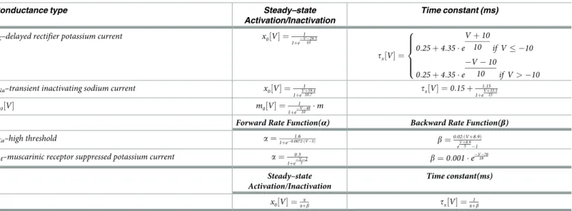
The dynamics of each gating variable evolved as follows:
x・¼x0½V� x
tx½V� ð5Þ
where x indicates the variables n, h, c, and M. The implemented equilibrium function is deter-mined by the termx0½V� and time constant tx½V� (Table 2).
The sodium activation variable was replaced and approximated by its equilibrium function
m0½V�. M-current presents a temporal evolution significantly slower than the rest of the five
Table 1. Ionic conductance densities.
Conductance type Soma (mho/cm2) gK–delayed rectifier potassium current 0.01
gNa–transient inactivating sodium current 0.125
gCa–high threshold 0.001
gM–muscarinic receptor 0.75
gL–leak current (anomalous rectifier) 0.02 https://doi.org/10.1371/journal.pcbi.1006298.t001
variables thus provoking a slow-fast system able to reproduce the characteristic Purkinje cell spiking modes (Fig 2).
The final voltage dynamics for the Purkinje [78,79]cell model was given by:
dV dt ¼ gK�n4� ðV þ 95Þ gNa�m0½V� 3 �h � ðV 50Þ gCa�c 2� ðV 125Þ g L� ðV þ 70Þ gM�M � ðV þ 95Þ þ Injected Current Membrane Area Membrane Capacitance ð6Þ
where the parametersMembrane Area and Membrane Capacitance are provided inTable 3,
andInjected Current is the sum of all contributions received through individual synapses (see
Eqs7–9below).
First, we validated the detailed Purkinje cell model (Eqs Eqs4–6) in the Neuron simulator.
Subsequently, we reduced the Purkinje cell model to make it compatible with an event-driven lookup table simulator (i.e, the EDLUT simulator) for fast spiking neural network simulations
[86,127]. In the reduced Purkinje cell model,IKandINacurrents were implemented through a
simple threshold process that triggers the generation of a triangular voltage function each time
the neuron fires [129]. This triangular voltage depolarisation drives the state of ion channels
similarly to the original voltage depolarisation during the spike generation. We inserted the
differential equation defined inEq 6within EDLUT. We used an in-house fixed-step
numeri-cal integration method compatible with GPUs (bi-fixed integrative method). Our numerinumeri-cal integration method provided similar accuracy than the variable step-size numerical integration
methods provided by NEURON with considerably less computational cost [128]. To make
NEURON simulation comparable with EDLUT as inS1 Fig, the stimulation of the Purkinje
cell was carried out by spike trains through an AMPA synapse (single decaying exponential
withτ = 1 ms, Eexc= 0 mV). We emulated the effect of the PF over the Purkinje cell through a
spike train of 55 Hz and a synaptic weight of gexc= 8μS. We used CF synaptic stimulations
through an AMPA synapse with weight of gexc= 80μS, (see asterisks inFig 2C). The spike
tim-ing traces of the Purkinje spike burst-pause dynamics under equal Purkinje stimulation were consistent in NEURON and EDLUT. Both the EDLUT and NEURON source codes are avail-able at the following URLs:
www.ugr.es/~nluque/restringido/Burst-pause_Purkinje_dynamics_regulate_motor_ adaptation_NEURON_MODEL_COMPLETE.rar
Table 2. Ionic conductance kinetic parameters.
Conductance type Steady–state
Activation/Inactivation
Time constant (ms)
gK–delayed rectifier potassium current x0½V� ¼ 1 1þeV 29:510 tx½V� ¼ 0:25 þ 4:35 � e V þ 10 10 if V � 10 0:25 þ 4:35 � e V 10 10 if V > 10 8 > > > < > > > :
gNa–transient inactivating sodium current x0½V� ¼ 1 1þeVþ59:410:7 tx½V� ¼ 0:15 þ 1:15 1þeVþ33:515 m0½V� m0½V� ¼ 1 1þeV 4810 �m
Forward Rate Function(α) Backward Rate Function(β)
gCa–high threshold a ¼1þe0:0072�ðV 5Þ1:6 b ¼0:02�ðVþ8:9Þ
eVþ8:95 1
gM–muscarinic receptor suppressed potassium current a ¼ 0:3
1þeV 25 b ¼0:001 � e V 70 18 Steady–state Activation/Inactivation Time constant(ms) x0½V� ¼ a aþb tx½V� ¼aþb1 https://doi.org/10.1371/journal.pcbi.1006298.t002
www.ugr.es/~nluque/restringido/CODE_Burst-pause_Purkinje_dynamics_regulate_ motor_adaptation_EDLUT.rar
User: REVIEWER, password: REVIEWER (for both).
Other cerebellar neuron models
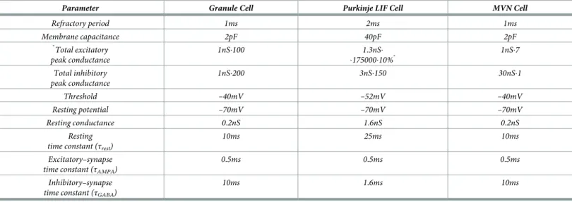
The other cerebellar neurons (granule cells, MVN cells, . . .) were simulated as leaky integrate– and–fire (LIF) neurons, with excitatory (AMPA) and inhibitory (GABA) chemical synapses:
Cm�
dVm c
dt ¼gAMPAðtÞ � ðEAMPA Vm cÞ þgGABAðtÞ � ðEGABA Vm cÞ þGrest� ðErest Vm cÞ ð7Þ
whereCmdenotes the membrane capacitance,EAMPAandEGABAare the reversal potential of
each synaptic conductance,Erestis the resting potential, andGrestindicates the conductance
responsible for the passive decay term towards the resting potential. ConductancesgAMPAand
gGABAintegrate all the contributions received by each receptor type (AMPA and GABA)
through individual synapses and they are defined as decaying exponential functions [86,130]:
gAMPAðtÞ ¼ 0 ; t � t0 gAMPAðt0Þ �e ðt t0Þ tAMPA ; t > t 0 ð8Þ 8 > < > : gGABAðtÞ ¼ 0 ; t � t0 gGABAðt0Þ �e ðt t0Þ tGABA ; t > t 0 ð9Þ 8 > < > :
witht representing the simulation time, t0being the time arrival of an input spike, andτAMPA
andτGABAdenoting the decaying time constant for AMPA and GABA receptors, respectively.
Note that we also used the LIF neuronal model (Eqs7–9) to simulate Purkinje cells that
could only express tonic spike firing (Fig 3B). These Purkinje cells with compromised
CF-evoked spike burst-pause dynamics provided a coarse phenomenological model of Kv3.3-defi-cient Purkinje neurons (as in Kcnc3 mutants, in which the absence of voltage-gated potassium channel Kv3.3 drastically reduces spikelet generation within complex spikes of cerebellar
Pur-kinje cells) [131]. Note, however, that completely suppressing CF-evoked spike burst-pause
dynamics would require more severe actions. A plausible way for obtaining
no-bursting-after-CF stimulus may consist in modulating GABABPurkinje cell receptors via Baclofen, the
spe-cific GABABagonist (as shown in [132]).Table 4summarises the parameters used for each cell
and synaptic receptor type.
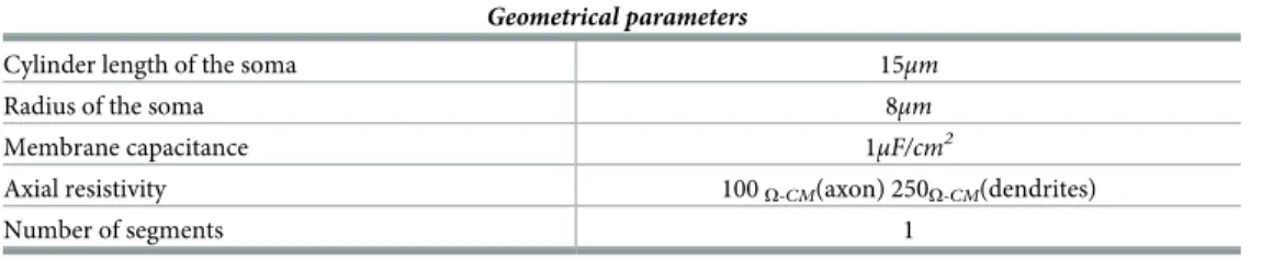
Table 3. Geometrical parameters.
Geometrical parameters
Cylinder length of the soma 15μm
Radius of the soma 8μm
Membrane capacitance 1μF/cm2
Axial resistivity 100O-CM(axon) 250O-CM(dendrites)
Number of segments 1
Cerebellar neural population models
Mossy fibres (MFs). N = 100 MFs were modelled as LIF neurons (Eqs7–9). Consistently
with the functional principles of VOR models of cerebellar control [2], the ensemble MF
activ-ity was generated following a sinusoidal shape (1 Hz with a step size of 0.002 ms) to encode
head movements [2,142,143]. The overall MF activity was based on non-overlapping and
equally sized neural subpopulations that allowed a constant firing rate of the ensemble MFs to be maintained over time. Importantly, two different times always corresponded to two differ-ent subgroups of active MFs ensuring the overall constant activity. (Network connectivity
parameters summarised inTable 5).
Granular cells (GCs). The granular layer included N = 2000 GCs and it was implemented
as a state generator [144–147], i.e. its inner dynamics produced time–evolving states even in
Table 4. Parameters of the LIF cell types.
Parameter Granule Cell Purkinje LIF Cell MVN Cell
Refractory period 1ms 2ms 1ms Membrane capacitance 2pF 40pF 2pF � Total excitatory peak conductance 1nS�100 1.3nS� �175000�10%� 1nS�7 Total inhibitory peak conductance 1nS�200 3nS�150 30nS�1 Threshold –40mV –52mV –40mV Resting potential –70mV –70mV –70mV Resting conductance 0.2nS 1.6nS 0.2nS Resting time constant (τrest)
10ms 25ms 10ms
Excitatory–synapse time constant (τAMPA)
0.5ms 0.5ms 0.5ms
Inhibitory–synapse time constant (τGABA)
10ms 1.6ms 10ms
Parameters obtained from the following papers:
Granule cell (GC)[133–137]. Only the rapidly decaying component of AMPA is modelled (τAMPA=0.5ms) the presence of slowly decaying components in some GC caused
by spill overs of glutamate was not taken into consideration [133](τAMPA=3ms)[138] Purkinje cell (PC) [137,139–141]. MVN data were extracted from unpublished
material from Prof. D’Angelo’s lab.
�Where 10% means the ratio of active connections PF–PC (out of the total 175000 PFs)
https://doi.org/10.1371/journal.pcbi.1006298.t004
Table 5. Summary of neurons and synapses.
Neurons Synaptic weights (nS)
Presynaptic cell number Postsynaptic cell Number of synapses Type Initial weight (Detailed/non Detailed PC) Weight range Mossy Fibres (100) Granular Cells 8000 AMPA 0.35/0.35�
Medial Vestibular Nuclei 200 AMPA 0.0/0.0 [0, 10] /[0, 10] Climbing Fibres (2) Purkinje Cells 20 AMPA 40/2.5
Granular Cells (1000) Purkinje Cells 40000 AMPA 3.4/3.75 [0, 3.75] / [0, 5.5] Purkinje cell (20) Medial Vestibular Nuclei 20 GABA 0.15/0.15 [0 10] / [0, 10] Medial Vestibular Nuclei (2)
�Parameter used for generating the Granular layer activity. Since this activity remained invariant during VOR adaptation, it was stored offline in a file and then loaded in computation time.
https://doi.org/10.1371/journal.pcbi.1006298.t005
the presence of a constant MF input [59]. The granular layer generated non-overlapped
spatio-temporal patterns that were repeatedly activated in the same sequence during each learning trial (1 Hz rotation for 1 s)). 500 different states encoded each second of the 1 Hz learning trial, each state consisting of four non-recursively activated GCs.
Climbing fibres (CFs). N = 2 CFs carried the instructive signal (from the IO) to the
popu-lation of Purkinje cells. The two CFs handled clockwise and counter–clockwise sensed errors.
CF responses followed a probabilistic Poisson process. Given the normalised error signalεðtÞ
and a random number ηðtÞ between 0 and 1, a CF fired a burst if εðtÞ > ηðtÞ, otherwise it
remained silent [84,120,148]. Thus, a single CF burst encoded time information regarding
the instantaneous error. Furthermore, the probabilistic spike sampling of the error ensured a proper representation of the whole error region over trials, whilst maintaining the CF activity
below 10 Hz per fibre (similar to electrophysiological data; [149]). The evolution of the sensed
error could be sampled accurately even at such a low frequency [148,150]. A graded
represen-tation of the instructive signal [15,19,28] enabled the correlation between the intensity of the sampled instantaneous error and the number of the spikes within the olivary burst:
Sspikes: ½0; 1� � R ! R ε!y¼SspikesðεÞ SspikesðεÞ ¼ 2 if 0:25 �ε � 0:50 3 if 0:50 �ε � 0:75 4 if 0:75 �ε � 0:85 5 if 0:85 �ε � 0:95 6 if 0:95 �ε � 1:0 ð10Þ 8 > > > > > > > > > > < > > > > > > > > > > :
The model assumed a perfect linearly correlated transmission of olivary bursts from CFs to the target Purkinje cells. Hence, the number of spikes in the Purkinje complex spikes linearly
depended on the number of stimuli in the CF burst [16]. CFs were simulated to transmit from
2 to 6 CF stimuli, delivered at inter-stimulus intervals of 2 ms [16], signalling the sensed error to be compensated. For the sake of computational efficiency, only 2 CFs were simulated
(instead of 20). In the cerebellum, each Purkinje cell is innervated by a single CF [151] coming
from the associated IO in the olivary system. However, no olivary system was considered here and, consequently, CFs sensing clockwise and counter–clockwise errors were equally activated (it would suffice 1 CF sensing clockwise and 1 CF sensing anti-clockwise errors).
Purkinje cells. N = 20 Purkinje cells were divided in two subpopulations of 10 neurons
each. Each subpopulation received the inputs from one CF encoding the difference between (either rightward or leftward) eye and head movements. Each Purkinje cell also received 2000
PF inputs. Since real Purkinje cells are innervated by about 150000 PFs [152], the weights of
the PF–Purkinje cells synapses of the model were scaled so as to obtain a biologically plausible amount of excitatory drive. Each of the two subgroups of 10 Purkinje cells targeted (through inhibitory projections) one MVN cell, responsible for either clockwise or counter-clockwise compensatory motor actions (ultimately driving the activity of agonist/antagonist ocular muscles).
Medial vestibular nuclei (MVN). The activity of N = 2 MVN cells produced the output of
the cerebellar model. The two MVN neurons handled clockwise and counter–clockwise motor correction, respectively. Each MVN neuron received excitatory projections from all MFs (which determined the baseline MVN activity), and inhibitory afferents from the