HAL Id: inserm-00482495
https://www.hal.inserm.fr/inserm-00482495
Submitted on 2 May 2011
HAL is a multi-disciplinary open access
archive for the deposit and dissemination of
sci-entific research documents, whether they are
pub-lished or not. The documents may come from
teaching and research institutions in France or
abroad, or from public or private research centers.
L’archive ouverte pluridisciplinaire HAL, est
destinée au dépôt et à la diffusion de documents
scientifiques de niveau recherche, publiés ou non,
émanant des établissements d’enseignement et de
recherche français ou étrangers, des laboratoires
publics ou privés.
A model of transient unilateral focal ischemia with
reperfusion in the P7 neonatal rat: morphological
changes indicative of apoptosis.
Sylvain Renolleau, Djamila Aggoun-Zouaoui, Yezekiel Ben-Ari, Christiane
Charriaut-Marlangue
To cite this version:
Sylvain Renolleau, Djamila Aggoun-Zouaoui, Yezekiel Ben-Ari, Christiane Charriaut-Marlangue. A
model of transient unilateral focal ischemia with reperfusion in the P7 neonatal rat:
morphologi-cal changes indicative of apoptosis.. Stroke, American Heart Association, 1998, 29 (7), pp.1454-60;
discussion 1461. �inserm-00482495�
Reperfusion in the P7 Neonatal Rat
Morphological Changes Indicative of Apoptosis
S. Renolleau, MD; D. Aggoun-Zouaoui, PhD; Y. Ben-Ari, PhD; C. Charriaut-Marlangue, PhD
Background and Purpose—The mechanisms leading to delayed cell death after hypoxic-ischemic injury in the developingbrain remain to be elucidated. The aim of this study was to develop a model of transient focal ischemia in the neonatal rat in an attempt to create a reperfusion phase since in the filament model of reversible middle cerebral artery occlusion, size limitations precluded performing this procedure before 14 to 18 days. We then analyze whether apoptosis or necrosis occurs in this model.
Methods—Seven-day-old Wistar rat pups (n596) underwent permanent left middle cerebral artery occlusion in association
with 1-hour occlusion of the left common carotid artery. Evolution of the brain infarction was studied from 24 hours to 3 months on cresyl violet–stained coronal sections. Infarct volume was determined with the use of the mitochondrial stain 2,3,5-triphenyltetrazolium chloride. Neuronal death was demonstrated by the silver staining method of Gallyas et al (1980). Chromatin condensation was shown by DNA fragmentation assessed with the use of terminal deoxynucleo-tidyl transferase–mediated dUTP-biotin nick end-labeling (TUNEL) assay in cryostat sections and electron microscopic analysis.
Results—Almost all of the animals who survived had reproducible cortical infarcts. The mean infarct volume was
3167 mm3
(mean6SD). The ipsilateral hemisphere showed a well-delineated lesion in the frontoparietal cortex at 3-month recovery. Argyrophilic (dying) neurons were observed a few hours after reperfusion and increased with time. Cells exhibiting DNA fragmentation were shown as early as 6 hours, increased up to and peaked at 24 to 96 hours, then progressively decreased and persisted for several days, suggesting an ongoing process. Electron microscopy analysis demonstrated high condensation and clumping of chromatin beneath nuclear membrane in shrunken neurons.
Conclusions—Our study demonstrates the feasibility of performing ischemia-reperfusion in 7-day-old rats that develop
progressive neuronal death with features characteristic of apoptosis. The reperfusion phase mimics events that occur during neonatal human hypoxic-ischemic encephalopathy at birth, since perinatal intensive care most often permits recirculation. (Stroke. 1998;29:1454-1461.)
Key Words: cell deathn chromatin n reperfusion injury n rats
D
espite recent advances in the understanding of neuronaldeath during cerebral ischemia in adult rodent models, only a few reports discuss neonatal ischemia. Reduction of oxygen supply during the perinatal period may affect central nervous system development and lead to neurological dys-function.1
The traditional model of neonatal hypoxia-ische-mia in a 7-day-old rat was that of a permanent unilateral carotid ligation followed by a hypoxic episode of several hours.2
This results in a lesion similar to that observed in the full-term infant who has undergone a hypoxic-ischemic episode such as perinatal asphyxia. To investigate the acute and long-term pathophysiology of neonatal stroke, particu-larly the phenomenon of reperfusion injury3,4
and its sequelae in the developing nervous system, new models of transient focal ischemia were recently developed in rats aged 14 to 185
See Editorial Comment, page 1461
and 106days. However, there is no model of ischemia with
reperfusion in 7-day-old rat pups, although reperfusion has been reported to be a deleterious event in young7and adult8
rats. Previous neuropathologic studies showed that at this stage of development the animal’s brain is histologically similar to that of a stillborn infant.3,9 In addition, it was
recently demonstrated that the rodent and primate models could be used for long-term neurological and behavioral outcome experiments, whereas the fetal sheep, newborn lamb, and piglet models are well suited for the study of acute and subacute metabolic and physiological end points.10
In adult cerebral ischemic models, two types of neuronal cell death have been described: apoptosis and necrosis. Since
Received December 29, 1997; final revision received March 6, 1998; accepted April 6, 1998. From the Universite´ Rene´ Descartes, Paris, France.
Presented in part at the 18th International Symposium on Cerebral Blood Flow and Metabolism, Baltimore, Md, June 15–19, 1997, and published in abstract form.
Correspondence to C. Charriaut-Marlangue, INSERM U29, 123 Boulevard de Port-Royal, 75014 Paris, France. E-mail cm@u29.cochin.inserm.fr © 1998 American Heart Association, Inc.
1993, an increased number of reports suggest that neuronal death after cerebral ischemia in rodents occurs through an apoptotic pathway (for review, see References 11 and 12). Strong evidence of apoptosis has been provided by combin-ing DNA gel electrophoresis, light and electron microscopy, in situ DNA nick end-labeling assessed by TUNEL staining, and apoptosis-associated gene expression.13,14
Necrosis was shown to occur in the core of the ischemic lesion, a zone in which the degree of injury was severe. In contrast, apoptosis was mainly detected in the periphery, termed the penum-bra.15,16 Internucleosomal DNA fragmentation in the cortex,
hippocampus, striatum, and thalamus was reported after unilateral occlusion and exposure to hypoxia in 7-day-old rats.17
These data demonstrated that cell death involves the action of the specific endonuclease that is accepted as the hallmark of apoptosis in other systems18and is not the result
of classic necrosis.
The objective of the present report was to develop a model of transient unilateral cerebral ischemia in 7-day-old rats. The combination of permanent left MCA electrocoagulation and transient left carotid occlusion induces neuronal death in the ipsilateral cortex. We then analyzed the temporal profile of cells undergoing apoptosis by the use of the TUNEL assay and electron microscopy to detect nuclear changes. Part of the present investigation has been reported in abstract form.19
Materials and Methods
Focal Ischemia Model
Experiments were performed in strict accordance with guidelines of the National Institutes of Health and the French Department of Agriculture (license No. 01352). Ipsilateral focal ischemia was induced in 7-day-old Wistar rats (weight, 17 to 23 g; n596) of both sexes. Pups were anesthetized with an intraperitoneal injection of chloral hydrate (300 mg/kg). After 15 minutes, rats were placed on
their backs, and a median incision was made in the neck to expose the left common carotid artery. Rats were placed on the right side, and an oblique dermal incision was made between the ear and eye. After excision of the temporal muscle, the cranial bone was removed from the frontal suture to a level below the zygomatic arch. The left MCA, exposed just after its apparition over the rhinal fissure, was permanently electrocoagulated at the inferior cerebral vein level before the MCA bifurcated into frontal and parietal branches (Figure 1). After this procedure, a clip was placed to occlude the left common carotid artery (Figure 1). The vascular clip was removed after 60 or 90 minutes. Carotid blood flow restoration was verified with the aid of a microscope. Both neck and cranial skin incisions were then closed. The duration of this procedure was 20 minutes. During the surgical procedure, body temperature was maintained at 37°C to 38°C by means of a heating pad. Rat pups were then placed in an incubator maintained at 37°C until they awoke, and then they were transferred to their mothers for the long-term survival period. This new model was compared with a model of permanent MCA occlusion alone and with a model of transient (1-hour) carotid artery occlusion alone. Sham-operated brains and control pups were used as controls.
Measurement of Infarct Volume
Neuropathologic evaluation of brain injury at 48 hours of recircula-tion (gliosis being detected at 72 hours) was accomplished with the use of the mitochondrial stain 2,3,5-triphenyltetrazolium chloride (n55), as previously reported.20,21In another set of animals (n56), pups were killed and brains were removed and frozen in isopentane (240°C). Cryostat coronal sections were stained with cresyl violet. On each section, cortical areas of infarction were measured with an image analyzer (IMSTAR). The volume of infarction was calculated by integrating the necrotic areas.
Tissue Preparation
Cell death studies were performed in a separate set of animals subjected to left MCA electrocoagulation and 1 hour of left common carotid artery occlusion. Rats were killed at various times after reperfusion (4 to 96 hours, 7 and 14 days, 1 and 3 months; n56 each). Rats were perfused through the ascending aorta under deep anesthesia (chloral hydrate, 300 mg/kg) with warm heparinized saline followed by PBS (0.12 mol/L, pH 7.4) containing 4% paraformaldehyde. Brains were then removed, kept for 2 hours in the same fixative solution, and placed in 0.1 mol/L PBS containing 10% sucrose for 2 days. Brains were rapidly frozen in isopentane (240°C) and subsequently stored at 270°C until used. Coronal cryostat sections (20mm thick) were collected on gelatin-coated slides.
For electron microscopy, rats at 24 hours of recirculation (n52) were anesthetized with chloral hydrate and perfused with 50 mL of saline followed by 150 mL 4% paraformaldehyde and 1% glutaral-dehyde in 0.1 mol/L PBS (pH 7.4). Brains were removed, kept
Figure 1. Ischemia-induced model in 7-day-old
Wistar rat pup. Left, MCA branching pattern with electrocoagulation site A (arrow). Right, Blood supply to the chest and the base of the brain; B is the position of the vascular clip to occlude the left common carotid artery. Selected Abbreviations and Acronyms
MCA5 middle cerebral artery PBS5 phosphate-buffered saline TdT5 deoxynucleotidyl transferase
TUNEL5 terminal deoxynucleotidyl transferase–mediated dUTP-biotin nick end-labeling
overnight in the same fixative, and cut with a vibratome (50mm thick). Selected areas were processed for electron microscopy by postfixation in 1% osmium tetroxide for 2 hours, washed in PBS, dehydrated in ethanol, and embedded in epoxy resin (Epon/Araldite). Polymerization was made at 60°C for 48 hours. Blocks were cut on a Reichert ultramicrotome in 1-mm-thick sections and stained with toluidine blue for light microscopy. Ultrathin sections (100 to 200 nm) were poststained with uranyl acetate and lead citrate and viewed with a Phillips EM 300 electron microscope.
Silver Staining Procedure
We used the method of Gallyas et al22 modified by Nadler and Evenson23to visualize both degenerating terminals and cell bodies of neurons and lysosomes that bind silver (ie, become argyrophilic). Briefly, the staining procedure included alkaline pretreatment, silver impregnation, development at pH between 5.5 and 6.3, washing in acetic acid, dehydration, and mounting in Permount.
In Situ Labeling of Fragmented DNA
Coronal cryostat sections were processed for TUNEL assay as previously reported.24Briefly, sections were incubated with TdT (0.2 U/mL; Gibco) and biotin-16-dUTP (20 mmol/L; Boehringer Mannhein), then visualized with streptavidin-biotin-peroxidase com-plex and diaminobenzidine. Cells exhibiting DNA fragmentation (TUNEL positive) were counted in the cortex by the use of a320 objective. Counting was performed by an investigator who was blinded to the experimental protocol. Data are presented as mean6SD per tissue section.
Results
Neither permanent MCA occlusion alone nor transient carotid occlusion alone in 7-day-old rat pups induced an ischemic lesion visible at 96 hours. A reproducible ischemic lesion was found when permanent left MCA electrocoagulation associ-ated with 1 hour of left carotid occlusion was performed (Figure 1). All pups showed an infarct, and fewer than 10% of animals died during the first 2 hours of reperfusion. In contrast, all pups subjected to permanent left MCA electro-coagulation associated with more than 1 hour of left carotid occlusion died during the procedure.
Histology
Examination of cresyl violet sections with recovery periods of less than 6 hours did not show detectable differences between the two cerebral hemispheres. At 18 to 48 hours of recovery, the ipsilateral hemisphere appeared slightly larger than the opposite hemisphere (18% edema). Infarct lesion (determined by pale cresyl violet and TTC staining) was located in the cerebral
cortex, and the mean infarct volume was 3167 mm3
(n510). Figure 2 depicts the six serial cross-sections, with the black shaded area indicating the mean area of infarction for that section at 48 hours of reperfusion. Slight damage in the head of
the caudate putamen was detected in 20% of animals (18 of a total of 96 pups; Figure 3A). During the next 2 weeks, the infarct evolved into a smooth-walled cavity that was more visible at posterior levels; the volume of the ipsilateral hemisphere was reduced compared with the contralateral side (Figure 3C). Three months after transient ischemia, the ipsilateral hemisphere was significantly reduced compared with the contralateral hemi-sphere and showed a well-delineated lesion in the frontoparietal cortex (Figure 3D). High magnification of cresyl violet staining demonstrated cells with pyknotic nuclei and chromatin clumping at 48 hours of reperfusion (Figure 3B).
Cell Death
With the selective Gallyas silver staining, silver-impregnated cell bodies were seen in the cortex at 6 hours after ischemia (Figure 4A and 4B), and they increased with time of recir-culation. TUNEL labeling was detected in a few scattered cells of the sham-operated or control rat pup brains, which corresponds to the programmed cell death that occurs during development. In contrast, TUNEL-positive nuclei appeared as early as 4 hours of reperfusion in the frontoparietal cortex, increased up to 24 hours (Figure 4C and 4D and Figure 5), and remained stable until 96 hours (Figure 5). A progressive decrease in the number of apoptotic cells was observed from 7 to 30 days (Figure 5). The stained nuclei showed the morphological criteria of apoptosis, ie, cytoplasmic shrinkage and cytoplasmic membrane convolutions, chromatin conden-sation below the nuclear membrane, followed by fragmenta-tion of the nucleus into rounded or oval bodies (apoptotic bodies, Figure 4E through 4G). Necrotic cells, detected by diffused nuclear and cytoplasmic staining,24
were not detected in normal and sham-operated rats or in the contralateral hemisphere of ischemic pups. A few necrotic cells were evident in the MCA site, probably due to a mechanical tissue lesion during the surgical procedure (Figure 4G).
Semithin plastic sections through the cortex of animals killed at 24 hours after reperfusion were examined. Neurons in the contralateral cortex appeared unaffected, showing clear cyto-plasm and nuclei (not shown). Ipsilaterally, dying neurons in the early and late stages of degeneration exhibited abnormal mor-phology (Figure 6A). At the ultrastructural level, neurons in the early stages of degeneration exhibited dark nuclei and cytoplasm with chromatin coalescence beneath the nuclear membrane. All organelles were well preserved (Figure 6C). In the late stage of degeneration, neurons showed a prominent cytoplasmic shrink-age with abnormal cytoplasmic organelles. A particular conden-sation and segregation of the chromatin was observed (Figure
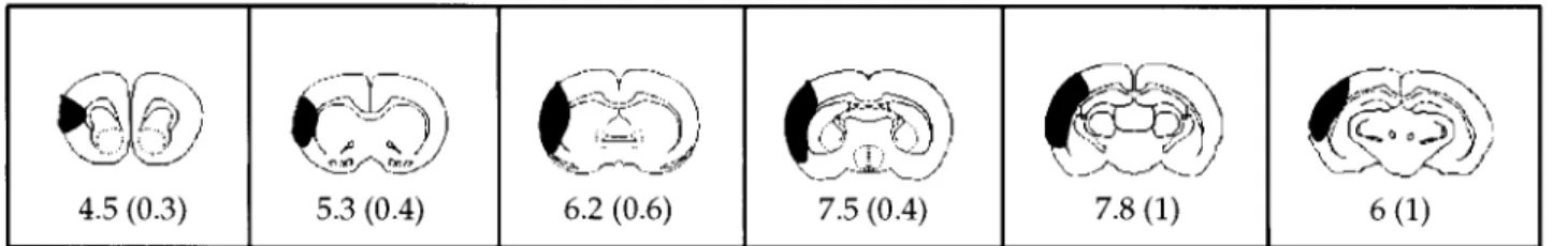
Figure 2. Representative cross-sections from the brain of ischemic rat pups (n56) are depicted from anterior (left) to posterior (right).
The darkened areas represent the mean percent area of infarction for that group at the particular cross-sectional level. The mean (6SD) percent area of infarction is given under each section.
6D). These ultrastructural results are consistent with apoptotic neuronal death.
Discussion
The major objectives of the present investigation were two-fold: (1) to induce ischemia by permanent and/or transient occlusion of artery(ies) in neonatal (P7) rats and (2) to determine whether immature ischemia-induced cell death markedly exhibited features of programmed cell death (apo-ptosis). The data presented here show that permanent left MCA occlusion associated with 1 hour of left carotid occlu-sion produced a cortical infarct in 7-day-old rats, and the majority of injured neurons demonstrated punctate condensed chromatin indicative of apoptosis.
No ischemic lesion after occlusion of the MCA alone was found in neonatal rats, as previously described in a model of 20-day-old rats.25
The numerous anastomoses between cere-bral arteries (anterior, middle, and posterior) in rat brain are so efficient that they protect MCA cerebral territory from ischemic injury.26
In contrast, the association of transient homolateral carotid artery and permanent MCA proximal occlusion probably created a situation of low cerebral blood flow in the ipsilateral hemisphere that was sufficient for anastomoses to no longer be efficient despite Willi’s polygon. However, anastomoses may allow a secondary recirculation phase after removal of the carotid artery microclip. This recirculation was difficult to prove without a study of cerebral
blood flow in different cerebral arteries and anastomoses. We were unable to determine these measurements because of the age of the rat pups. However, detection of polymorphonu-clear cells and macrophages in and around the ischemic lesion is good evidence of the blood-brain barrier opening and the occurrence of an inflammatory response, respectively.27
In the newborn, stroke models have been difficult to develop, and few studies have been published in which infarct volume or cerebral blood flow has been measured. Compared with the model of Rice et al,2
which associated permanent unilateral carotid occlusion and hypoxia (FIO28%) for 1 hour
in 7-day-old rat pups, our model has a reperfusion phase in the anatomoses through the carotid artery. Recently, two authors described new models of transient ischemia in young rats5,6
that could not be considered pure neonatal stroke models but rather juvenile stroke models. They performed MCA occlusion using an endovascular nylon filament. The filament was removed after 1 hour, allowing recirculation. However, the models of both Aschwal et al5and Mitsufuji et
al6 used rats older than 7 days (14 to 18 and 10 days old,
respectively), but many biochemical, physiological, and an-atomic changes occur in the rat pup between day 7 and days 14 to 18.28
In the model of Mitsufuji et al, the survival rate was very poor (27.8% of rat pups died during the occlusion period, and 38.5% of the surviving rats died within the first hour of reperfusion). In our conditions, almost all of the
Figure 3. Evolution of infarction after transient focal ischemia in the rat pup. Representative cresyl violet–stained coronal sections from
animals killed at 48 hours (A and B), 14 days (C), and 3 months (D) of reperfusion. A, Large ill-defined pale area is seen in the left cere-bral hemisphere. Arrows indicate border of the pale area. B, Enlarged panel of cresyl-violet–stained section in A at higher magnification (bar represents 10mm) showing pyknotic nuclei (arrowheads) and cytoplasm (small arrows). C, At 14-day recovery, a smooth-walled cavity (arrowhead), surrounded by astrocytosis, is seen as well as the reduced size of the ipsilateral hemisphere compared with the contralateral side. D, At 3-month recovery, a substantial cortical infarction is shown. No dilation of the lateral ventricle was observed.
animals survived and displayed a smaller infarct size than that obtained in 7-day-old Wistar rats after hypoxic-ischemic injury29or reversible MCA occlusion with the use of filament
in 14- to 18-day-old spontaneously hypertensive rats.5
Dam-age in pup brains was limited to the MCA distribution, similar to that seen in rat adult brains.30,31
The small variability of ischemic area, at the level of the head of the caudate putamen, that we found may be a direct consequence of the different anatomic variations of the MCA division arteries before or after the level of the inferior cerebral veins.32,33
In contrast to hypoxic-ischemic exposures in immature rat brain, we did not observe either prominent white matter injury2
or damage in
the different zones of the hippocampus.34 Furthermore, the
reduction in thickness and the loss of the frontoparietal cortex were complete at 3-month recovery without a compensatory dilation of the lateral ventricle, as previously reported.35
Towfighi et al35
reported time-dependent neuropathologic evolution after neonatal ischemia. Two recent studies, in which genomic DNA gel electrophoresis and in situ labeling of nuclear DNA fragmentation were used, demonstrated that neuronal death was indicative of apoptosis after
hypoxia-is-chemia.17,36 Permanent left MCA and 1-hour left carotid
occlusion in rat pups induced principally apoptosis, as as-sessed with the TUNEL assay and electron microscopic analysis. Morphological analysis of TUNEL-positive cells showed conspicuous chromatin condensation and apoptotic bodies. The characteristic features of apoptotic cell death are now well documented in several pathologies in the central nervous system (for review, see References 9 and 10). A karyorrhexic or apoptotic morphology with TUNEL-labeled punctate chromatin predominates in our neonatal transient focal ischemia model, as previously reported in rat pup37and
newborn piglet.38
Since this is not the case in the adult
Figure 4. Cell death in ipsilateral neonatal ischemic cortex during reperfusion. Photomicrographs show representative sections of
corti-cal tissue performed with the argyrophilic staining of dying cells according to Gallyas et al22(1980) and modified by Nadler and
Even-son23(1983) (A and B) and the TUNEL assay (C through G). A, Presence of numerous silver-stained lysosomes in healthy cells (gray
dots) of the contralateral hemisphere. B, Dark silver-stained degenerating neurons at 6 hours of recovery. C and D, TUNEL-positive nuclei in the cerebral cortex and MCA site, respectively, at 6 and 18 hours of recovery. E through G, High magnification of TUNEL-pos-itive nuclei. Note chromatin-dense masses and apoptotic body formation (arrowheads), a nucleus divided into two masses (arrow in F), and a necrotic nucleus (arrow in G). Bar represents 25mm (A and B), 50 mm (C and D), and 6 mm (E through G).
Figure 5. Number of TUNEL-positive cells in coronal sections at
the level of the anterior commissure after ischemia and various reperfusion times in rat pups (n54). A progressive increase in the number of apoptotic cells occurs with increasing reperfusion time from 4 to 24 hours followed by a steady state until 96 hours and a progressive decrease from 7 to 30 days of reperfu-sion. Numbers are mean6SD per tissue section.
ischemic rat in which apoptosis and necrosis were generally reported to occur,16,39,40 these data suggest that immature
neurons may be more prone to apoptotic death, while termi-nally differentiated neurons exhibit pyknosis or die by necro-sis. Furthermore, a prolonged presence of TUNEL-positive nuclei from 6 hours to 30 days after reperfusion suggests that cell damage in this model is a persistent and ongoing process, as previously reported after MCA occlusion in adult rats.15
Electron microscopic analysis demonstrated that neurons die through an apoptotic process (chromatin condensation and segregation), as previously reported after transient focal ischemia in adult rats.41,42
These apoptotic features are in agreement with recent data demonstrating that in our model the apoptosis-associated proteins p53 and Bax, which are not or are basically expressed, respectively, in control situations, are sequentially upregulated in neurons exhibiting DNA
fragmentation,43 suggesting that neuronal apoptosis is an
important event in the developing central nervous system. The presence of the same karryorrhexic morphology and the formation of apoptotic bodies in pontosubicular necrosis in the human neonate44 – 46
are noteworthy.
In conclusion, the two models of cerebrovascular injury in 7-day-old rat pups—the model of unilateral carotid ligation and 8% O2according to Rice et al
2
(1981) and our ischemia-reperfusion model— can be considered complementary since they examine two different types of cerebral insults (hypoxic-ischemic injury and stroke). The clinical relevance of devel-oping a model of neonatal stroke has become apparent over the past decade as neuroimaging studies have convincingly demonstrated that such lesions are more common than pre-viously recognized and account for serious neurological morbidity.47– 49
The main advantage of our model is the reperfusion phase in a P7 rat, which is truly neonatal and more relevant to distressed infants. This reperfusion mimics processes that occur during neonatal human hypoxic-ische-mic encephalopathy at birth, since perinatal intensive care most often permits recirculation. Furthermore, a well-defined infarct is created by occlusion of arteries rather than a hemispheric ischemic insult caused by ligation of one artery in combination with a severe hypoxic insult to the entire brain. Thus, our data demonstrating apoptotic neuronal death
Figure 6. Ultrathin sections showing neuronal degeneration in the ipsilateral cortex of ischemic rat pups at 24 hours of reperfusion. A,
Semithin section (1mm). Note the presence of normal neurons (clear cytoplasm and nucleus [n]) near dying neurons (in early and late stages of degeneration [arrowhead and arrow, respectively]). Magnification3400. B, Ultrastructure of normal neuron showing clear cytoplasm and nucleus (n). All organelles were present and preserved. Magnification33300. C, Ultrastructure of early stage of neuronal degeneration. Note cytoplasmic compaction, increased electron density, and nuclear chromatin condensation beneath the nuclear membrane (arrowheads). Magnification33300. D, Ultrastructure of late stage of apoptotic neuronal death showing a high compaction of cytoplasm and nucleus. Note marginated coalesced and segregated chromatin (arrow). Magnification,310 000.
may lead to further advances in therapeutic approaches for the preservation of neurons in neonatal stroke.
Acknowledgments
The authors are grateful to Drs M. Plotkine and A. Gelot for their helpful comments and to E. der Terrossian for critical reading of the manuscript.
References
1. Younkin D. Hypoxic-ischemic brain injury of the newborn: statement of the problem and overview. Br Pathol. 1992;2:209 –210.
2. Rice J, Vannucci R, Brierley J. The influence of immaturity on hypox-ia-ischemic brain damage in the rat. Ann Neurol. 1981;9:131–141. 3. Vannucci R. Experimental biology of cerebral hypoxia-ischemia: relation
to perinatal brain damage. Pediatr Res. 1990;27:317–326.
4. DelZoppo G, Schmid-Scho¨nbein G, Mori E. Polymorphonuclear leu-kocytes occlude capillaries following middle cerebral artery occlusion and reperfusion in baboons. Stroke. 1991;22:1276 –1283.
5. Ashwal S, Cole D, Osborne S, Osborne T, Pearce W. A new model of neonatal stroke: reversible middle cerebral artery occlusion in the rat pup.
Pediatr Neurol. 1995;12:191–196.
6. Mitsufuji N, Yoshioka H, Okano S, Nishiki T, Sawada T. A new model of transient cerebral ischemia in neonatal rats. J Cereb Blood Flow
Metab. 1996;16:237–243.
7. Fellman V, Raivio K. Reperfusion injury as the mechanism of brain damage after perinatal asphyxia. Pediatr Res. 1997;41:599 – 606 . 8. Traytsman R, Kirsch J, Koehler R. Oxygen radical mechanisms of brain
injury following ischemia and reperfusion. J Appl Physiol. 1991;71: 1185–1195.
9. Dobbing J, Sands J. Comparative aspects of the brain growth spurt. Early
Hum Dev. 1979;3:79 – 83.
10. Roohey T, Raju T, Moustogiannis A. Animal models for the study of perinatal hypoxic-ischemic encephalopathy: a critical analysis. Early
Hum Dev. 1997;47:115–146.
11. Charriaut-Marlangue C, Aggoun-Zouaoui D, Represa A, Ben-Ari Y. Apoptotic features in ischemia, epilepsy and gp120 toxicity. Trends
Neurosci. 1996;19:109 –114.
12. Choi D. Ischemia-induced neuronal apoptosis. Curr Opin Neurobiol. 1996;6:667– 672.
13. Koistinaho J, Ho¨kfelt T. Altered gene expression in brain ischemia.
Neuroreport. 1997;8:I-VIII.
14. MacManus J, Linnik M Gene expression induced by cerebral ischemia: an apoptotic perspective. J Cereb Blood Flow Metab. 1997;17:815– 832. 15. Li Y, Chopp M, Jiang N, Yao F, Zaloga C. Temporal profile of in situ
DNA fragmentation after transient middle cerebral artery occlusion in the rat. J Cereb Blood Flow Metab. 1995;15:389 –397.
16. Charriaut-Marlangue C, Margaill I, Represa A, Popovici T, Plotkine M, Ben-Ari Y. Apoptosis and necrosis following reversible focal ischemia: an in situ DNA fragmentation analysis. J Cereb Blood Flow Metab. 1996;16:186 –194.
17. Hill I, MacManus J, Rasquinha I, Thor U. DNA fragmentation indicative of apoptosis following unilateral cerebral hypoxia-ischemia in the neonatal rat. Brain Res. 1995;676:398 – 403.
18. Schwartzman R, Cidlowski J. Apoptosis: the biochemistry and molecular biology of programmed cell death. Endocr Rev. 1993;14:133–151. 19. Charriaut-Marlangue C, Renolleau S, Plotkine M, Ben-Ari Y. Neuronal
apoptosis in a new model of transient unilateral focal ischemia in rat pups. In: Program and abstracts of the 18th International Symposium on Cerebral Blood Flow and Metabolism; June 15–19, 1997; Baltimore, Md. 1997;17:S258.
20. Cole D, Drummond J, Ghazal E, Shapiro H. A reversible component of cerebral injury as identified by the histochemical stain 2,3,5-triphenyltet-razolium chloride (TTC). Acta Neuropathol (Berl). 1990;80:152–155. 21. Isayama K, Pitts L, Nishimura N. Evaluation of
2,3,5-triphenyltetrazoli-um chloride to delineate rat brain infarcts. Stroke. 1991;22:1394 –1398. 22. Gallyas F, Wolff J, Bo¨ttcher H, Zaborszky L. A reliable and sensitive
method to localize terminal degeneration and lysosomes in the central nervous system. Stain Technol. 1980;55:299 –306.
23. Nadler V, Evenson D. Use of excitatory amino acids to make axon-sparing lesions of hypothalamus. Methods Enzymol. 1983;103:393– 400. 24. Charriaut-Marlangue C, Ben-Ari Y. A cautionary note on the use of
TUNEL stain to describe apoptosis. Neuroreport. 1995;7:61– 64.
25. Coyle P. Middle cerebral artery in the young rat. Stroke. 1982;13: 855– 859.
26. Menzies S, Hoff J, Betz A. Middle cerebral artery occlusion in rats: a neurological and pathological evaluation of a reproducible model.
Neu-rosurgery. 1992;31:100 –107.
27. Coeroli L, Renolleau S, Arnaud S, Plotkine D, Cachin N, Plotkine M, Ben-Ari Y, Charriaut-Marlangue C. Nitric oxide formation and perivascular nitrotyrosine following focal ischemia in immature rats.
J Neurochem. In press.
28. McDonald J, Johnson M. Physiological and pathophysiological roles of excitatory amino acids during central nervous system development. Brain
Res Rev. 1990;15:41–70.
29. Saeed D, Goetzman B, Gospe S. Brain injury and protective effects of hypothermia using triphenyltetrazolium chloride in neonatal rat. Pediatr
Neurol. 1993;9:263–267.
30. Levine S. Anoxic-ischemic encephalopathy in rats. Am J Pathol. 1960; 36:1–17.
31. Du C, Hu R, Csernansky C, Hsu C, Choi D. Very delayed infarction after mild focal cerebral ischemia: a role for apoptosis. J Cereb Blood Flow
Metab. 1996;16:195–201.
32. Rubino G, Young W. Ischemic cortical lesions after permanent occlusion of individual middle cerebral artery branches in rats. Stroke. 1988;19:870 – 877. 33. Fox G, Gallacher D, Shevde S, Lotfus J, Swayne G. Anatomic variation of the middle cerebral artery in Sprague-Dawley rat. Stroke. 1993;24: 2087–2093.
34. Towfighi J, Yager J, Housman C, Vannuci R. Neuropathology of remote hypoxic-ischemic damage in the immature rat. Acta Neuropathol (Berl). 1991;81:578 –587.
35. Towfighi J, Zec N, Yager J, Housman C, Vannuci R. Temporal evolution of neuropathologic changes in immature rat model of cerebral hypoxia: a light microscopic analysis. Acta Neuropathol (Berl). 1995;90:375–386. 36. Ferrer I, Tortosa A, Macaya A, Sierra A, Moreno D, Munell F, Blanco R,
Squier W. Evidence of nuclear DNA fragmentation following hypoxia-ischemia in the infant rat brain, and transient forebrain hypoxia-ischemia in the adult gerbil. Br Pathol. 1994;4:115–122.
37. Sidhu R, Tuor U, Bigio MD. Nuclear condensation and fragmentation following cerebral hypoxia-ischemia occurs more frequently in immature than older rats. Neurosci Lett. 1997;223:129 –132.
38. Yue X, Mehmet H, Penrice J, Cooper C, Cady E, Wyatt J, Reynolds E, Edwards A, Squier M. Apoptosis and necrosis in the newborn piglet brain following transient cerebral hypoxia-ischaemia. Neuropathol Appl
Neu-robiol. 1997;23:16 –25.
39. Ferrer I, Martin F, Serrano T, Reiriz J, Perez-Navarro E, Alberch J, Macaya A, Planas A. Both apoptosis and necrosis occur following intrastriatal admin-istration of excitotoxins. Acta Neuropathol (Berl). 1995;90:504 –510. 40. Nitatori T, Sato N, Waguri S, Karasawa Y, Araki H, Shibanai K,
Komimani E, Uchiyama Y. Delayed neuronal death in the CA1 pyramidal cell layer of the gerbil hippocampus following transient ischemia is apoptosis. J Neurosci. 1995;15:1001–1011.
41. Li Y, Victor G, Jiang N, Zaloga C, Sabbah H, Chopp M. Ultrastructural and light microscopic evidence of apoptosis after middle cerebral artery occlusion in the rat. Am J Pathol. 1995;146:1045–1051.
42. Aggoun-Zouaoui D, Margaill I, Borrega F, Represa A, Ben-Ari Y, Charriaut-Marlangue C. Ultrastructural morphology of neuronal death following reversible focal ischemia in rat. Apoptosis. 1998;3:133–147. 43. Renolleau S, Benjelloun N, Ben-Ari Y, Charriaut-Marlangue C.
Regu-lation of apoptosis-associated proteins in cell death following transient focal ischemia in rat pup. Apoptosis. 1997;2:368 –376.
44. Sohma O, Mito T, Mizuguchi M, Takashima S. The prenatal age critical for the development of the pontosubicular necrosis. Acta Neuropathol
(Berl). 1995;90:7–10.
45. Bruck Y, Bruck W, Kretzschmar H, Lassmann H. Evidence for neuronal apoptosis in pontosubicular neuron necrosis. Neuropathol Appl
Neu-robiol. 1996;22:23–29.
46. Scott R, Hegyi L. Cell death in perinatal hypoxic-ischaemic brain injury.
Neuropathol Appl Neurobiol. 1997;23:307–314.
47. Friede R. Perinatal lesions of gray matter. In: Developmental
Neuropa-thology. Go¨ttingen, Germany: Springer-Verlag; 1989:82–97.
48. Rademakers R, Vanderknaap M, Verbeeten BJ, Barth P, Valk J. Central cortico-subcortical involvement: a distinct pattern of brain damage caused by perinatal and postnatal asphyxia in term infants. J Comput Assist
Tomogr. 1995;19:256 –263.
49. Volpe J, Pasternak J. Parasagittal cerebral injury in neonatal hypoxic-is-chemic encephalopathy: clinical and neurological features. Pediatrics. 1977;91:472– 476.
Editorial Comment
In this article, the authors demonstrate the development of a model of transient unilateral cerebral ischemia in 7-day-old rats. This model was produced through a combination of permanent left middle cerebral artery electrocoagulation with transient left carotid artery occlusion, a combination that induces neuronal death in the ipsilateral cortex. The authors then analyzed the temporal profile of cells undergoing apo-ptosis through use of the TUNEL assay and electron micros-copy to detect nuclear changes. This is an important model of global ischemia, and its main advantage is the reperfusion phase in a 7-day-old rat pup, which is truly neonatal and perhaps more relevant to distressed infants. But it must be remembered that this is not a complete reperfusion, because the middle cerebral artery is permanently occluded. This particular model can be compared with the unilateral carotid
ligation plus 8% O2, in accordance with the Rice and
Vannucci model, and is more a hypoxic-ischemic model than the model presented here, which is more a model of ische-mia-reperfusion model. Thus, these two models should be considered complementary, because they put forth two types of cerebral insult: the hypoxic-ischemic model versus the
ischemia-reperfusion model. In the present model, the authors characterize the apoptotic findings that occur. A karyorrhexic or apoptotic morphology with the TUNEL-labeled punctate chromatin predominates in this neonatal model of transient focal ischemia. Because this is not the case in the adult ischemic rat, in which apoptosis and necrosis both are reported to occur, these data suggest that immature neurons may be more prone to apoptotic death whereas terminally differentiated neurons exhibit pyknosis or die by necrosis. It is important to note that the same karyorrhexic morphology and formation of apoptotic bodies in pontosubicular necrosis in the human neonate is similar to that occurring in this model. Thus, the new aspect of this study is that it involves transient, unilateral focal ischemia with partial reperfusion in the 7-day-old neonatal rat. This is important and presents a new model of ischemia to compare with the hypoxic-ischemic model of Rice and Vannucci.
Richard J. Traystman, PhD, Guest Editor
Anesthesiology/Critical Care Medicine Johns Hopkins Medical Institutes Baltimore, Maryland