Chromatin packaging and morphology in ejaculated human
spermatozoa: evidence of hidden anomalies in normal
spermatozoa
Patrizia Grace Bianchi1, Gian Carlo Manicardi2, Francoise Urner1, Aldo Campana1 and Denny Sakkas13
1Clinic for Infertility and Gynaecological Endocrinology-WHO Collaborating Centre, Hdpital Cantonal Universitaire de
Geneve, 20 rue Alcide-Jentzer, 1211 Geneve 14, Switzerland, and 2Department of Animal Biology, University of Modena,
Modena, Italy
•'To whom correspondence should be addressed
This study aimed to investigate the association between anomalies in sperm chromatin packaging, morphology and fertilization in patients undergoing routine in-vrtro fertilization (IVF) or subzonal insemination (SUZI). Sperm chromatin packaging was assessed using chromomycin A3 (CMA3), a f luorochrome specific for guanine-cytosine rich sequences of DNA. One hundred to 150 sperm cells were assessed in 55 patients to compare sperm chromatin packaging and morphology to fertilization after IVF or SUZI. When the morphology and CMA3 fluorescence of individual spermatozoa was assessed, >75% of the macrocephalic sperm fluoresced in all patients. In contrast, a mean of 37% of the spermatozoa with normal morphology fluoresced in IVF patients compared with 58% of the normal spermatozoa in male factor patients treated by SUZI. SUZI patients displaying a high fluorescence (>70%) in their spermatozoa also had a significantly lower fertilization rate. Lower packaging quality in morphologically normal spermatozoa may represent a major limiting factor in the fertilizing ability of male factor patients. This study confirms that a high percentage of CMA3 positivity is present in certain forms of male factor infertility and that such a test may be used to distinguish separate populations in morphologically normal spermatozoa.
Key words: chromatin/chromomycin A^in-vitro fertilization/male infertility/spermatozoa
Introduction
Semen quality is conventionally determined according to the number, motility and morphology of spermatozoa in an ejacu-late (World Health Organization, 1989). In turn, it is generally accepted that an association exists between these semen parameters and fertilizing ability (Mahadevan and Trounson, 1984; Kiugeretal, 1986; Liu etal., 1988). Sperm morphology, in particular, seems to be the most significantly related para-meter to fertilization rates (Pousette et al, 1986; Oehninger
et al, 1988). With the advent of in-vitro fertilization (TVF)
and related techniques such as subzonal insemination (SUZI) and intracytoplasmic sperm injection (ICSI), it has become increasingly apparent that the number, motility and morphology of spermatozoa are not always indicative of a male's fertility status. Significantly different fertilization rates have been reported for patients with similar semen parameters, suggesting that a more sensitive test is needed to identify the inherent defects which render certain spermatozoa unable to fertilize (Jeyendran et al, 1989; Wolf et al, 1992).
A failure of the conventional semen parameters to predict fertilization indicates that hidden anomalies, lying at the sperm membrane level or at the chromatin level, should also be evaluated. A number of studies have shown that spermatozoa with abnormal nuclear chromatin organization are more fre-quent in infertile men than in fertile men (Evenson et al, 1980; Monaco and Rasch, 1982; Foresta et al, 1992). In these studies, specific dyes and fluorochromes were used to assess
the chromatin of mature mammalian spermatozoa and relate them to fertilization results. The accessibility of specific dyes and fluorochromes to DNA can give clues to the packaging of the chromatin, which occurs during spermiogenesis. How-ever, the targets of these fluorochromes or dyes vary and may not be adequate in identifying specific anomalies in chromatin structure. For example, Monaco and Rasch (1982) compared DNA fluorochromes specific for GC-rich sequences, mithramy-cin and chromomymithramy-cin A3 (CMA3), and for AT-rich sequences,
2'-[4-hydroxyphenyl]-5-[4-methyl-1 -piperazinyl]-2,5'-bi-1H-benzimidazole and 4-6, diamidino-2-phenylindole. They con-cluded that, although fluorescence was evident in spermatids and spermatocytes, a decline in staining with GC-specific dyes during maturation probably reflected changes in protein composition and DNA packaging ratios.
In our own studies (Bianchi et al, 1993), we have found that the amount of protamines present in mature spermatozoa represented at least one of the limiting factors controlling the accessibility of CMA3 to the DNA. In the same study we
postulated that the CMA3 fluorochrome could be used as a
tool in routine laboratory analysis for the rapid screening of certain conditions of subfertility and infertility in man, as it seemed to allow an indirect visualization of protamine-defi-cient, nicked and partially denatured DNA. In this study we investigate the use of CMA3 in relation to individual sperm
morphology. Furthermore, we assess the relationship between CMA3 fluorescence, in conjunction with morphology, and the
Table I. Distribution of the semen characteristics in treatmen in-vitro fertilization (IVF) and subzonal insemination (SUZI) (a) sperm concentration and motility and (b) morphology
Sperm concentration (X lO'/ml) 0-5 6-20 >20 Morphology (% normal forms) 0-20 21-40 >40 Sperm motility ( 0-20 IVF SUZI I 5 0 4 0 1 IVF SUZI" 4 18 17 8 10 2 %) 21-50 IVF i 0 6 SUZI 6 7 4 t cycles of according to >50 IVF 0 2 21 SUZI 2 0 1
Table II. The mean percentage fluorochrome stain according to
Sperm concentration (X lO'/ml) 0-5 6-20 >20 Motility (%) 0-20 21-40 >40
of spermatozoa positive to the CMA3
i sperm concentration and motility No. of cycles 14 13 34 11 22 28 •Values are means ± SD.
bP < 0.05 compared to 0-5 X 10*/ml sperm concentration
CP < 0.05 compared to 0-20% motility group.
%CMA3 fluorescence* 68.0 + 18.1 59.8 ± 23.0 47.7 ± 16.5b 71.5 ± 20.6 55.4 ± 2 1 . 8 48.0 ± 15.01 group.
1 No assessment in two cycles due to insufficient spermatozoa.
outcome of fertilization for patients being treated by routine IVF and SUZI.
Materials and methods
Patient characteristics and sperm preparation
Semen samples were all taken from 55 patients undergoing treatment for infertility at the Clinic for Infertility and Gynaecological Endo-crinology-WHO Collaborating Centre, University Hospital of Geneva, Geneva, Switzerland. Ejaculated human spermatozoa were collected and the greater part prepared for routine IVF or SUZI. The criteria for selection of patients performing either procedure was: for IVF, an insemination droplet of at least 100 000 motile spermatozoa could be prepared, and for SUZI, < 100 000 motile spermatozoa after preparation or patients having failed to achieve fertilization in at least two previous IVF attempts. Table I shows the semen characteristics for the 55 patients (61 cycles) making up the IVF and SUZI groups. Morphology was assessed according to World Health Organization criteria (1989) and progressive motility by visual appraisal using a Makler chamber.
FVF and SUZJ were performed using the stimulation protocol and procedures previously published (Umer et al., 1993; Sakkas el al., 1994). Spermatozoa were prepared for both techniques by treating the sample with 3 mM pentoxifylline (Sigma Pharmaceuticals, Buchs, Switzerland) followed by selection using mini-Percoll gradient centri-fugation (Ord et al, 1990). From the remains of the semen sample, one slide was prepared for assessment of sperm morphology while the rest was washed in Dulbecco's Ca2 +-Mg2 + free phosphate-buffered saline (PBS) and centrifuged at 170 g for 10 min. The procedure was repeated twice and the washed spermatozoa were fixed in methanol/glacial acetic acid 3:1, at 4°C, for 5 min and then spread on slides.
Chromomycln A3 staining and assessment of sperm
mor-phology
For CMA3 (Sigma) staining, each slide was treated for 20 min with 100 nl of CMA3 solution (0.25 mg/ml in Mcllvane buffer, pH 7.0, containing 10 mM MgCli)- Slides were then rinsed in buffer and mounted with buffered glycerol. Fluorescence was performed using a Zeiss Photomikroskop HI using a combination of exciter: dichroic: barrier niters of BP 436710: FT 580: LP 470 and an A-Plan X20 objective lens. This lens allowed the use of both phase and
fluores-140
cence. For each patient, a total of 100-150 spermatozoa were randomly observed on duplicate slides. For each single spermatozoon, head morphology (divided in three categories: normal, macrocephalic or amorphous) and fluorescence were assessed.
Statistical analysis
The means in the different groups were transformed using an arcsin square root transformation and compared for statistical significance using one-way analysis of variance and Scheffe's F-test. A test of the correlation between the percentage of spermatozoa positive to the CMA3 fluorochrome and head morphology per patient was performed using regression analysis.
Results
Chromomycin A3 positivity in relation to sperm
con-centration, motility and morphology
Sperm concentration and motility
The mean percentage of spermatozoa fluorescent after CMA3
treatment declined in relation to increased sperm concentration and motility (Table H). Patients with severe oligozoospermia or asthenozoospermia had a mean (±SD) of 68.0 ± 18.0% and 71.5 ± 20.6% of their spermatozoa fluorescing respectively. A distinct decline in spermatozoa quality was therefore associated
with CMA3 fluorescence.
Morphology
Although a decline in sperm number and motility was associa-ted with increased CMA3 fluorescence, a comparison of CMA3 positivity and morphology in individual spermatozoa showed that apparently normal spermatozoa may possess hidden anomalies. By observing fluorescence and morphology in single sperm heads from the two groups of patients, we found that in all samples >75% of the spermatozoa displaying macrocephaly fluoresced brightly while ~50% of the spermato-zoa classed as having an amorphous head shape fluoresced (Figure 1). The main difference between the treatment groups was seen in spermatozoa classed as having a normal shape. In patients undergoing routine IVF, a mean (±SD) of 38.2 ± 19.2% of the morphologically normal spermatozoa fluoresced while in the male factor patients treated by SUZI 57.8 ± 27.4% fluoresced; this was significant (P < 0.05). In nine
out of 30 samples from male factor patients, >80% of all spermatozoa were fluorescent compared with only one of the FVF samples.
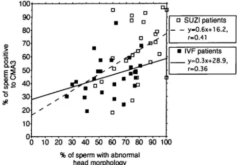
There was no correlation between the percentage of spermatozoa with abnormal head morphology and the percent-age of spermatozoa showing fluorescence after CMA3 staining
(Figure 2). Therefore, although macrocephalic spermatozoa were nearly always fluorescent, spermatozoa with amorphous and normal heads vary in chromatin packaging. Samples
macro amorphous normal macro amorphous normal cephalic cephalic
<73.J±14.7) (48.0±16.2) (38-2±19.2>* <79.2±M.2) (56.4£l9.8> <57.8±27.-)>»
Figure 1. A box plot comparing the different types of sperm morphology to the percentage of spermatozoa positive to the chromomycin A3 (CMA3) fluorochrome. The lower 25th and upper 75th percentiles are shown. Two groups are represented,
normozoospermic patients (n = 31) treated by routine in-vitro fertilization (IVF, open) and male factor patients (n = 30) treated by subzonal insemination (SUZI, shaded). Individual spermatozoa were assessed for both morphology and CMA3 fluorescence. The mean (± SD) percentage of spermatozoa positive for CMA3 fluorescence in each group is given in parentheses. *Percentage of spermatozoa with normal morphology and positive for CMA3 fluorescence was significantly different (P < 0.05) in samples from the IVF and SUZI groups of patients.
prepared from the same ejaculate and evaluated independently gave r values of 0.36 and 0.41 when comparing percentage of cells with abnormal head morphology and showing CMA3
fluorescence for the IVF and SUZI patients respectively (Figure 2).
Chromomycin A3 positivity in relation to fertilization,
cleavage rates and pregnancy
In-vitro fertilization
There was no association between CMA3 fluorescence in all
spermatozoa and fertilizing ability for patients being treated by routine IVF (data not shown). In seven cases where patients had <20% fertilization rates there was no difference in positivity of sperm to the CMA3 fluorochrome when compared
to cases with fertilization rates >20%. As in routine IVF there is a greater competition between spermatozoa, it could be expected that a motile morphologically normal spermatozoon would fertilize the oocyte. However, no relationship was found between fertilizing ability and CMA3 fluorescence in
morphologically normal spermatozoa in patients treated by routine IVF.
Subzonal insemination
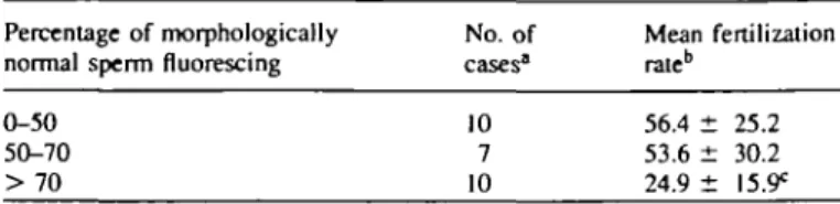
In contrast to the lack of an association in IVF patients, male factor patients treated by SUZI had a significantly lower mean fertilization rate when their spermatozoa showed a high positivity to the CMA3 fluorochrome. Ten SUZI patients had
>70% of spermatozoa positive to the CMA3 fluorochrome
and had a significantly lower (P < 0.02) fertilization rate; CMA3 of >70%: 24.9 ± 15.9 compared with CMA3 of <70%:
55.7 ± 26.3. The same 10 patients also had >70% fluorescence in their morphologically normal spermatozoa, which was indicative of poor fertilizing ability when using SUZI (Table IH).
Embryo cleavage and pregnancies
Once fertilization had occurred, there was no relationship between the ability of a fertilized oocyte to cleave and positivity
| 100 90 80 70 60 | o 40 5 30 20 10 0 10 20 30 40 50 60 70 80 90 100 % of sperm with abnormal
head morphology
Figure 2. Correlation between the percentages of spermatozoa with abnormal head morphology and spermatozoa positive to the chromomycin A3 (CMA3) fluorochrome. Regression equations are given for the normozoospermic patients (n = 31) treated by routine in-vitro fertilization (IVF) and male factor patients {n = 30) treated by subzonal insemination (SUZI).
a a • D D a JBO 1 D 1 D ] • SUZI patients y-0.6x+16.2, r-0.41 • IVF patients y=o.3x+?8 9, r=0.36 i
141
Table III. Mean fertilization rate of sperm samples from men treated by subzonal insemination (SUZI) in relation to positivity of morphologically normal spermatozoa to the CMA3 fluorochrome
Percentage of morphologically No. of normal sperm fluorescing cases1
Mean fertilization rate" 0-50 50-70 > 70 10 7 10 56.4 ± 25.2 53.6 ± 30.2 24.9 ± "Only cases which had more than four oocytes treated were included.
bValues are means ± SD.
CP < 0.02 compared to the 0-50 and 50-70 groups.
to the CMA3 fluorochrome in either treatment group. Five of
the 31 IVF cycles resulted in ongoing pregnancies; of the five pregnancies, four of the male partners showed CMA3
fluorescence values of 20-40%, while one male had 86% of his spermatozoa fluorescing after CMA3 staining. In the two
ongoing pregnancies from the 30 SUZI cycles, the CMA3
fluorescence was 62 and 77%. Although the numbers are limited, the results suggest that CMA3 positivity is not related
to embryo viability.
Discussion
One of the central events of spermiogenesis is the substitution of the chromatin structural proteins by protamines, allowing a different structural organization to take place in the sperm nucleus (Courtens and Loir, 1981; Balhom, 1989; Ward and Coffey, 1991). Indeed, as reviewed by Ward and Coffey (1991), chromatin packaging in spermatozoa is very different from that of somatic cells; for example, the mouse sperm cell nucleus is 40-fold smaller than the nucleus of the mouse somatic cell. Due to the tight packaging afforded by the protamines, any modification or absence of these proteins could lead to an anomaly in the packaging process of sperm nuclei and influence sperm quality and fertilizing capacity.
The different models proposed for the binding of protamines to DNA in spermatozoa (Balhom, 1982; Subirana, 1991) postulate that the polyarginine segment of protamine binds in the minor groove of DNA, cross-linking and neutralizing the phosphodiester backbone. The CMA3 fluorochrome is also
believed to access DNA in the minor groove of the DNA helix
(Behietal, 1969; Berman etal., 1985), a positional preference
that can make it a competitor to protamines. CMA3 has
previously been shown to be a useful tool to indicate protamine-depleted spermatozoa (Bianchi et al, 1993). In contrast to the homogeneity of mature mouse spermatozoa, ejaculated human spermatozoa samples were found to exhibit a wide variation both in their responsiveness to the CMA3 fluorochrome and
in the presence of endogenous nicks in the DNA (Bianchi
et al, 1993; Sakkas et al., 1995).
Treatment of human sperm samples with protamines elimin-ates CMA3 positivity and prevents the identification of
endo-genous DNA nicks by in-situ nick translation, a technique that reveals damaged DNA (Bianchi et al, 1993; Manicardi et al, 1995). We have now investigated the relationship between CMA3 positivity and fertilizing capacity in spermatozoa from 142
two populations of patients undergoing IVF and SUZI treatment and found interesting differences in respect to their response to the CMA3 fluorochrome. In this study we show that the
CMA3 fluorescence provides evidence of: (i) a population of
spermatozoa that exhibit normal head morphology but possess loosely packaged chromatin; and (ii) a positive correlation between fertilization failure and CMA3 fluorescence in
morpho-logically normal spermatozoa in the SUZI treatment, and not in the routine IVF group. Such a difference is most likely related to the different methods of achieving fertilization. In routine IVF, sperm competition is maximal due to the excess number of spermatozoa used; therefore even in patients with a high CMA3 positivity enough normal sperm should be
present to achieve fertilization. In contrast, in semen samples selected for SUZI, where the number of spermatozoa in contact with the oocyte is limited, a high CMA3 positivity yields a
lower chance of achieving fertilization due to the lower probability of selecting a normal spermatozoon.
To this day, sperm quality has mainly been judged by parameters such as number, motility, and morphology. The influence of seminal parameters as a whole or individually on fertility has been a matter of investigation and discussion since the early days of infertility treatment. Literature on the subject is contradictory and results can vary extensively according to methodology of analysis, dyes utilised, interpretation criteria of morphology and technology available. Nonetheless, Bestoffe
etal. (1982), showed an inverse correlation between percentage
of abnormal sperm forms and spontaneous pregnancies in a retrospective study in subfertile couples. This debate has gained new vitality since the new assisted reproduction technologies provided a much more precise tool for evaluation of fertilization rates. Although a proper consensus has not yet been established, it has become widely accepted that normal parameters, defined according to WHO criteria (1989) are associated with high percentages of fertilization and cleavage rates in IVF. Further-more, many authors have concentrated their attention on the predictive value of morphology (Kruger et al, 1986; Pousette
et al, 1986), and it is felt that normal morphology is indicative
of normal function, while high levels of morphologically abnormal spermatozoa in the ejaculate are associated with a reduced fertilizing potential (Oehninger et al, 1988). When we compared the morphology and CMA3 fluorescence in
individual spermatozoa, we found a high frequency of CMA3
positive nuclei among macrocephalic spermatozoa in both treatment groups. This confirms data known on other fluores-cent dyes, in particular Acridine Orange, where access to the DNA was achieved only or mostly in cases of decondensed or damaged chromatin (Evenson et al, 1980; 1991; Foresta
et al, 1992; Molina et al, 1995; Sailer et al, 1995). Our
observations also confirm that macrocephaly is associated with decondensation. Moreover, this is further evidence of the protamine-depleted status of macrocephalic spermatozoa, as CMA3 is unable to access DNA in the presence of protamines
and normally formed disulphide bonds (Bianchi et al, 1993; Kvist, 1982). The fact that fluorescence was still present in the 'amorphous' category but was reduced to 50% probably reflects the fact that this group contained several, and probably
very different head defects, not all of which correspond to a decondensed status of the nuclear chromatin.
In our opinion the most interesting result was the statistically significant difference in fluorescence of normal spermatozoa between the two groups examined, casting a shadow on the value of assessing normal morphology, particularly in the cases of oligoasthenoteratozoospermia (OAT). As proposed by Bartoov et al. (1994) such patients may require a more complete sperm evaluation, as normal forms observed by light microscopy do not fully express the anatomical capability hidden in the sperm morphology. Engh et al. (1992) also found in a cytofluorimetrical evaluation of 100 male patients undergoing investigations because of infertility that patients with severe OAT presented a higher percentage of fluorescent cells in comparison with normozoospermic patients. Our data confirm this in situ with a direct comparison, per individual cell, of fluorescence and morphology. Interestingly, one third of the samples of SUZI patients (usually patients with OAT) presented >80% of fluorescent cells, while only one out of 31 of the FVF patients presented the same features. This suggests that the difference does not only rely on a decrease of the classical sperm parameters but on a defect in the packaging of chromatin, which can affect all the spermatozoa produced by testicular germinal tissue.
This highlights the importance of detecting this population of patients prior to treatment by IVF, as a spermatozoon possessing decondensed chromatin may be unable to achieve syngamy, even upon penetration of the oolemma membrane. When we compared the results of fluorescence to fertilizing capacity in the SUZI patients, we found data to reinforce this hypothesis, as the fertilization rate declines to nearly 20% when a very high number of spermatozoa are fluorescent, a situation that could be taken into account when deciding upon treatment. The fact that cleavage rate is apparently not influenced confirms that the crucial moment is at intra-oocyte decondensation and/or disturbances in its occurrence with all the subsequent processes such as protamine elimination and substitution by histones. Although this result has been observed using the SUZI technique, its relevance to the use of intracyto-plasmic sperm injection (ICSI) as a treatment of male factor infertility awaits resolution.
The results from this study suggest that CMA3 could become
a useful tool for evaluation of male factor patients prior to infertility treatment. CMA3 will distinguish decondensed,
protamine-depleted spermatozoa. Moreover, CMA3 is also
useful in identifying DNA-damaged sperm cells as shown by the presence of endogenous nicks in many decondensed CMA3
positive nuclei (Manicardi et ai, 1995). This would make it possible to screen for those patients whose spermiogenesis has resulted in a sperm population containing intrinsic anomalies in the nuclei, regardless of sperm morphology.
Acknowledgements
We acknowledge the technical assistance of Kirsten Hebin and Nicole Jaquenoud and the statistical advice of Dr Tim Farley, Human Reproduction Program, World Health Organization, Geneva, Switzerland. This work was supported by the Fonds National Suisse
(Grant no. 31.33787.92). P.G.Bianchi was supported by the Fonds National Suisse Score Program.
References
Balhom, R. (1982) A model for the structure of chromatin in mammalian sperm. J. Cell. Biol., 93, 298-305.
Balhom, R. (1989) Mammalian protamines: Structures and molecular interactions. In Adolph, K.W. (ed.), Molecular Biology of Chromosome
Function. Springer Verlag, New York, Berlin, Heidelberg, pp. 366-395.
Bartoov, B., Eltes, F., Pansky, M., Langzam, J., Reichart, M. and Soffer, Y. (1994) Improved diagnosis of male infertility potential via a combination of quantitative ultramorphology and routine semen analyses. Hum. Reprod., 9, 2969-2075.
Behr, W., Honikel, K. and Hartmann, G. (1969) Interaction of the RNA polymerase inhibitor chromomycin with DNA. Eur. J. Biochem., 9, 82-92. Berman, E., Brown, S.C., James, T.L. and Shafer, L.H. (1985) NMR studies
of chromomycin A3 interaction with DNA. Biochemistry, 24, 6887-6893. Bestoffe, E., Serup, J. and Rebbe, H. (1982) Relation between morphologically abnormal spermatozoa and pregnancies obtained during a twenty-year follow-up period. Int. J. Androl., 5, 379-386.
Bianchi, P.G., Manicardi, G.C., Bizzaro, D., Bianchi, U. and Sakkas, D. (1993) Effect of deoxyribonucleic acid protamination on fiuorochrome staining and in situ nick-translation of munne and human spermatozoa.
Biol. Reprod., 49, 1083-1088.
Courtens, J. and Loir, M. (1981) A cytochemical study of nuclear changes in boar, bull, goat, mouse, rat and stallion spermatids. J. Ultrastruc. Res., 74, 327-340.
Engh, E., Clausen, O.P.F., Scholberg, A., Tollefsrud, A. and Purvis, K. (1992) Relationship between sperm quality and chromatin condensation measured by sperm DNA fluorescence using flow cytometry. Int. J. Androl., 15, 407^15.
Evenson, D.P., Darzynkiewicz, Z. and Melamed, M.R. (1980) Relation of mammalian sperm chromatin heterogeneity to fertility. Science, 240, 1131-1133.
Evenson, D.P., Jost, L.K., Baer, R.K., Turner, T.W. and Schrader, S.M. (1991) Individuality of DNA denaturation patterns in human sperm as measured by the sperm chromatin structure assay. Reprod. Toxicol., 7, 297—304. Foresta, C , Zorzi, M., Rossato, M. and Varotto, A. (1992) Sperm nuclear
instability and staining with aniline blue: abnormal persistence of histones in spermatozoa in infertile men. Int. J. Androl., 15, 330-337.
Jager, S. (1990) Sperm nuclear stability and male infertility. Arch. Androl., 25, 253-259.
Jeyendran, R.S., Van der Ven, H.H., Rachagan, S.P., Perez-Peleaz, M. and Zaneveld, L.J.D. (1989) Semen quality and in-vitro fertilization. Aust. N.Z.
Obstet. Gynecol., 29, 168-172.
Kruger, T.F., Menkueld, R., Stander, F.S.H., Lombard, C.J., Van der Merwe, J.P., Van Zyl, J.A. and Smith, K. (1986) Sperm morphology features as a prognostic factor in in vitro fertilization. Fertil. Sterii, 46, 1118-1123. Kvist, U. (1982) Spermatozoa] thiol-disulphide interaction: A possible event
underlying physiological sperm nuclear chromatin decondensation. Ada
Physiol. Scand., 115, 503-505.
Liu, D.Y., Duplessis, Y.P., Nayudu, PL., Johnston, W. and Baker, H. (1988) The use of in vitro fertilization to evaluate putative tests of human sperm function. Fertil. Sterii, 49, 272-277.
Mahadevan, M. and Trounson, A.O. (1984) The influence of seminal characteristics on the success rate of human in vitro fertilization. Fertil.
Sterii., 42, 400-405.
Manicardi, G.C., Bianchi, P.G., Pantano, S., Azzoni, P., Bizzaro, D., Bianchi, U. and Sakkas, D. (1995) The presence of endogenous nicks in DNA of ejaculated human spermatozoa and its relationship to chromomycin A3
accessibility. Biol. Reprod., 52, 864-867.
Molina, J., Castilla, J.A., Gil, T, Hortas, M.L., Vergara, F. and Herruzo, A. (1995) Influence of incubation on the chromatin condensation and nuclear stability of human spermatozoa by flow cytometry. Hum. Reprod., 10, 1280-1286.
Monaco, PJ. and Rasch, E.M. (1982) Differences in staining with DNA-specific fluorochromes during spermiogenesis. J. Hislochem. Cytochem., 30, 585.
Oehninger, S., Acosta, A.A., Morshedi, M., Veek, L., Swanson, R J., Simmons, K. and Rosenwaks, Z. (1988) Corrective measures and pregnancy outcome in
in vitro fertilization in patients with severe sperm morphology abnormalities. Fertil. Sterii., 50, 283-287.
Mini-Percoll: a new method of sperm preparation for IVF in severe male factor infertility. Hum. Reprod., 6, 987-989.
Pousette, A., Akerlof, E., Rosenborg, L. and Fredricsson, B. (1986) Increase in progressive motility and improved morphology of human spermatozoa following their migration through Percoll gradients. Int. J. Androl., 9, 1-13. Sailer, B.L., Jost, L.K. and Evenson, D.P. (1995) Mammalian sperm DNA susceptibility to in situ denaturation associated with the presence of DNA strand breaks as measured by the terminal deoxynucleotidyl transferase assay. /. Androl., 16, 80-87.
Sakkas, D., Jacquenoud, N., Campana, A. and Leppens, G. (1994) Comparison of results after in vitro fertilized human embryos are cultured in routine medium and in coculture on Vero cells: a randomized study. Fenil. SteriL, 61, 521-525.
Sakkas, D., Manicardi, G., Bianchi, P.G., Bizzaro, D. and Bianchi, U. (1995) Relationship between the presence of endogenous nicks and sperm chromatin packaging in maturing and fertilizing mouse spermatozoa. Biol.
Reprod., 52, 1149-1155.
Subirana, J.A. (1991) Protein-DNA interactions in spermatozoa. In
Comparative Spermatology 20 Years Later. Serono Symposia Publications,
Vol 75. Raven Press, New York, pp. 89-92.
Umer, F , Bianchi, P.G., Campana, A. and Sakkas, D. (1993) Evidence of sperm entry into assumed unfertilized human oocytes after sub-zonal sperm microinjection. Hum. Reprod., 12, 2167-2173.
Ward, W.S. and Coffey, D.S. (1991) DNA packaging and organization in mammalian spermatozoa; comparison with somatic cells. Biol. Reprod., 44, 569-574.
Wolf, J.P., Ducot, B., Kunstmann, J.M., Frydman, R. and Jouannet, P. (1992) Influence of sperm parameters on outcome of subzonal insemination in the case of previous IVF failure. Hum. Reprod., 7, 1407-1413.
World Health Organization (1989) Laboratory Manual for the Examination of
Human Semen and Semen—Cervical Mucus Interaction. 3rd edn. Cambridge
University Press, Cambridge.
Received on May 30, 1995; accepted on August 4, 1995