FAM161A, associated with retinitis pigmentosa,
is a component of the cilia-basal body complex
and interacts with proteins involved in ciliopathies
Silvio Alessandro Di Gioia
1, Stef J.F. Letteboer
3, Corinne Kostic
2, Dikla Bandah-Rozenfeld
4,
Lisette Hetterschijt
3, Dror Sharon
4, Yvan Arsenijevic
2, Ronald Roepman
3,{and Carlo Rivolta
1,∗,{ 1Department of Medical Genetics, University of Lausanne, Lausanne, Switzerland,2Unit of Gene Therapy and Stem Cell Biology, Jules-Gonin Eye Hospital, University of Lausanne, Lausanne, Switzerland,3Department of Human Genetics and Nijmegen Centre for Molecular Life Sciences, Radboud University Nijmegen Medical Centre, Nijmegen, The Netherlands and4Department of Ophthalmology, Hadassah-Hebrew University Medical Center, Jerusalem, Israel
Received July 24, 2012; Revised and Accepted August 27, 2012
Retinitis pigmentosa (RP) is a retinal degenerative disease characterized by the progressive loss of photo-receptors. We have previously demonstrated that RP can be caused by recessive mutations in the human FAM161A gene, encoding a protein with unknown function that contains a conserved region shared only with a distant paralog, FAM161B. In this study, we show that FAM161A localizes at the base of the photoreceptor connecting cilium in human, mouse and rat. Furthermore, it is also present at the ciliary basal body in ciliated mammalian cells, both in native conditions and upon the expression of recombinant tagged proteins. Yeast two-hybrid analysis of binary interactions between FAM161A and an array of ciliary and ciliopathy-associated proteins reveals direct interaction with lebercilin, CEP290, OFD1 and SDCCAG8, all involved in hereditary retinal degeneration. These interactions are mediated by the C-terminal moiety of FAM161A, as demonstrated by pull-down experiments in cultured cell lines and in bovine retinal extracts. As other ciliary proteins, FAM161A can also interact with the microtubules and organize itself into microtubule-dependent intracellular networks. Moreover, small interfering RNA-mediated depletion of FAM161A transcripts in cultured cells causes the reduction in assembled primary cilia. Taken together, these data indicate that FAM161A-associated RP can be considered as a novel retinal ciliopathy and that its molecular pathogenesis may be related to other ciliopathies.
INTRODUCTION
Hereditary retinal degenerations (HRDs) consist of a group of diseases that are clinically and genetically heterogeneous. Despite the many differences in progression rates and in sever-ity characterizing each specific form, all patients with HRD lose vision because of the degeneration of rod and cone photo-receptors, and eventually of other retinal cells (1). Retinitis pigmentosa (RP) is the most common form of HRD, affecting more than one million people worldwide (2). Patients with RP usually present with night blindness due to malfunctioning rod photoreceptors that turns progressively into the impairment of
cone photoreceptors and thus daytime vision as well, from the periphery to the center of the visual field (1). Later in life, many patients become legally blind. Genetically, RP can be transmitted as an autosomal dominant, autosomal recessive or X-linked trait; in rare cases, digenic inheritance has also been observed (3,4). Out of the 182 identified genes involved in HRD, 43 have been also associated with non-syndromic RP (https://sph.uth.tmc.edu/retnet/). Interestingly, RP genes display a broad range of expression: some encode proteins that are restricted to rods only, while others are expressed in other retinal cell types or are even ubiquitously active across all tissues and organs of our body (4). Functional categories
†
Equal contribution.
∗To whom correspondence should be addressed at: Department of Medical Genetics, University of Lausanne, Rue du Bugnon 27, 1005 Lausanne,
Switzerland. Tel:+41 216925451; Fax: +41 216925455; Email: [email protected]
#The Author 2012. Published by Oxford University Press. All rights reserved. For Permissions, please email: [email protected]
of RP genes are also very diverse. However, many of them display a clear relationship with retinal biochemistry or have structural roles in retinal cells (5).
In particular, a number of HRD proteins have been found to have important roles in development, maintenance or functioning of the photoreceptor immotile cilium (6). The photoreceptor con-necting cilium (CC) is a particular structure of both photoreceptor types that is located between their inner and outer segments (IS and OS). In addition to its structural role, the CC is also involved in the transport of molecules across these two compartments by intraflagellar transport (6). New evidences also demonstrate its role in cell signaling and signal transmission (7). Disruption of gene-encoding proteins that are structurally and functionally con-nected to primary cilia and their cellular anchoring/nucleation point, the centrosome-derived basal body (BB), causes a broad class of diseases with pleiotropic phenotypes, all classified as ciliopathies (8). Retinal ciliopathies (RCs) include all ciliopathies that cause HRD, exclusively or in conjunction with other systemic defects. For instance, typical RC genes are: the RP-1 gene (RP1) (9), RP GTPase regulator (RPGR) (10), the RPGR-interacting protein 1 gene (RPGRIP1), RPGRIP1-like (RPGRIP1L) (11), Leber congenital amaurosis 5 (LCA5) (12), centrosomal protein 290 (CEP290) (13), Orofacialdigital 1 (OFD1) (14,15) and sero-logically defined colon cancer antigen 8 (SDCCAG8) (16,17). RP1 is associated with autosomal dominant and recessive RP and RPGR is associated with X-linked RP, whereas RPGRIP1, LCA5 and CEP290 are associated with Leber congenital amaurosis (congenital retinal blindness) among other diseases. Mutations in OFD1 and SDCCAG8 cause Joubert syndrome and RP and nephronophthisis with HRD or Bardet –Biedl syn-drome (BBS), respectively.
Recently, we identified FAM161A as the causative gene for RP28-associated RP and one of the major causes of RP in Israel and the Palestinian territories (18,19). The RP28 locus was first mapped on chromosome 2p in 1999, through the genetic analysis of a consanguineous family from India segregating recessive RP (20). In all patients analyzed, mutations in FAM161A lead to null alleles (18,19). FAM161A expression is driven by the photoreceptor-specific transcription factor CRX (18), and its presence is detected mostly in testis and retina. In this latter tissue, standard immunohistochemistry analysis revealed that
FAM161A localized at the level of the IS of photoreceptors and less markedly at the level of the outer plexiform layer (OPL) of the retina (18,19), with a localization pattern similar to USH2A (21). The function of FAM161A or other members of the protein family it belongs to (family FAM161) are unknown.
In this work, we demonstrate that FAM161A is an RC protein that is part of the CC complex and interacts with other proteins associated with RCs. Furthermore, we show that its localization at the base of the cilium is not limited to the photoreceptors, but is also conserved in other ciliated cell types and tissues.
RESULTS
FAM161A is present at the base of the cilium of photoreceptors and in renal cells
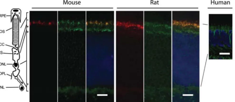
To investigate the localization of FAM161A within the mam-malian retina, we performed indirect immunofluorescence of fresh, unfixed cryosections of adult mouse and rat retinas, as well as paraffin-fixed human retinas with three different primary antibodies. These procedures resulted in specific staining in all species (Fig. 1). FAM161A localized at the base of the photoreceptor cilia, as well as within their IS, where the Golgi, the endoplasmic reticulum and other orga-nelles are found. As previously shown (18), a weak positive staining was detected in the OPL of the retina. Mechanical dis-sociation of the murine retina, allowing photoreceptor isola-tion and the preservaisola-tion of their OS, CC and part of the IS (22) revealed specific FAM161A localization in the region between the IS and the OS (Fig.2). This region corresponded to the CC of photoreceptors.
To define more precisely the distribution of FAM161A within the cilium, we performed immunofluorescence micros-copy by using antibodies against FAM161A and typical ciliary markers. Co-staining with antibodies against RP1, staining the ciliary axoneme (9), revealed that within the same ciliary structure, FAM161A and RP1 were contiguous but not over-lapping (Fig. 3A). RPGRIP1L is a marker of the ciliary BB (11), while pan-centrin localizes within the whole CC (23). Co-staining of FAM161A and each of these two markers
Figure 1. Localization of FAM161A in adult mouse, rat and human retina slices. Murine and rat sections were stained with anti-pan centrin (red), a ciliary marker, and FAM161A (green). The merged images show partial co-localization between the two signals. Human sections were stained with anti FAM161A antibody only (green). Nuclei are stained in DAPI (blue). Scale bars: 10 mm for mouse and rat, 50 mm for human. OS, outer segment; CC, connecting cilium; IS, inner segment; ONL, outer nuclear layer; OPL, outer plexiform layer; INL, inner nuclear layer.
revealed partial co-localization in both cases, according to a pattern indicating its presence between the BB and the CC. Specifically, immunofluorescence with anti-FAM161A and anti-RPGRIP1L antibodies revealed that FAM161A was not present just in the BB, but continued also in the lower part of the CC (Fig. 3B). Simultaneous staining of FAM161A and pan-centrin confirmed the non-complete presence of FAM161A within the whole CC and was indicative of, again, its presence within the BB (Fig.3C).
Although FAM161A mRNA expression seems to be pre-dominant in retina and testis, it is not restricted to these tissues (18,19). Staining of FAM161A in the rat kidney revealed again its presence at the level of cellular cilia. Similar to the results obtained in the retina, FAM161A was present at the level of the BB, as demonstrated by co-staining with RPGRIP1L (Supplementary Material, Fig. S1A), whereas it partially overlapped with the signal produced by anti-polyglutamylated tubulin antibody GT335, a marker of the CC and the BB (Supplementary Material, Fig. S1B).
FAM161A localizes at the level of the BB in ciliated cell lines
Many standard laboratory cell lines develop cilia. To identify a cellular model suitable for studying FAM161A and ciliogen-esis, we assessed 10 human cell lines for FAM161A mRNA expression and corresponding protein detectability (data not shown). We found in retinal pigment epithelium-derived cells hTERT-RPE-1 and ARPE-19 the best models for this purpose. Co-staining with FAM161A and GT335 anti-bodies in hTERT-RPE-1 cells showed partial overlap with the BB and the daughter centriole (Fig. 4A). Similar results were also obtained in ARPE-19 cells (not shown). In addition, co-staining between FAM161A and RPGRIP1L showed perfect co-localization (Fig. 4B). Taken together, these data indicate that FAM161A is present in the BB/centrosomes of ciliated cells of different origins. When we expressed
heterologous fusion FAM161A constructs in these same cell lines, we detected it at the same location, as revealed by using an anti-FLAG antibody (Fig. 4C and D; data not shown for ARPE-19).
Recombinant expression of FAM161A in hTERT-RPE-1 and ARPE-19 cultures allowed some cells to significantly overexpress the FAM161A protein. In these cells, FAM161A decorated the entire microtubule network, with a pattern similar to that observed upon the overexpression of ciliary pro-teins such as lebercilin (12) or CEP170 (24). We therefore tested FAM161A overexpression in non-ciliated cells such as HeLa or mouse photoreceptor-derived 661W cells. Heterol-ogous FAM161A assembled indeed in a web-like structure that partly overlapped with the cytoplasmic microtubule network, revealed by staining with anti-acetylated tubulin (Fig.5A). Supplementation with the microtubule depolymeriz-ing drug nocodazole in the culture medium completely abol-ished this FAM161A organization, indicating its microtubule dependence (Fig.5B).
FAM161A interacts with other ciliary and centrosomal proteins
Given the specific localization of FAM161A, we decided to ascertain its possible interaction with components of the ciliary proteome that are relevant to human diseases. Based on UNIPROT in silico analysis, FAM161A is predicted to have three coiled-coil domains, one at the N-terminus and two located in the well-conserved domain (provisionally called UPF0564 domain) located within the C-terminal part of the protein. We performed a rapid and efficient interaction assay in yeast by using three FAM161A constructs as baits to simultaneously screen a library of 45 unique genes associated with various human ciliopathies or involved in ciliary func-tions. In addition to a FAM161A full-length form, we designed a construct that included its first 217 codons (FAM161A-N-term bait) and another construct encompassing the remainder of the protein and containing the UPF0564 domain (FAM161A-C-term bait, codons 211 – 660) (Supple-mentary Material, Fig. S2A). Several bait-prey combinations induced yeast growth, pinpointing potential interactions (Sup-plementary Material, Table S1). However, colonies were observed only when the full FAM161A or FAM161A-C-term sequences were used to assess a potential interaction, while FAM161A-N-term produced no detectable growth. Among all tested interactions, the strongest signals were observed between both the FAM161A full length and FAM161A-C-term preys and baits expressing SDCCAG8, OFD1, CEP290 and lebercilin, or parts thereof (Fig.6).
To validate the interactions detected by yeast assays, we performed pull-down experiments in bovine retinal extracts, by using recombinant constructs containing the same parts of FAM161A (full form, N-term and C-term) used before but fused with the glutathione S-transferase (GST) protein (Supplementary Material, Fig. S2B). Following an overnight incubation of these chimeric proteins and retinal lysates, we could efficiently co-purify CEP290 and lebercilin, the only two proteins for which reliable antibodies were accessible to us. Again, positive signals were detected only for the full-length FAM161A protein and the construct presenting its
Figure 2. FAM161A localizes at the level of the CC in dissociated murine photoreceptors. Dissociated photoreceptors were co-stained with antibodies against rhodopsin (red) and FAM161A (green). During the dissociation process only part of the IS of photoreceptors is preserved (white arrowhead). BF, bright field. Scale bar: 10 mm.
C-term part (Fig.7A). It should be noted that the signal corre-sponding to lebercilin in co-purified extracts ran at a lower position than its counterpart in total retinal lysates. This was likely caused by specific proteolytic cleavage occurring during the overnight incubation with sepharose beads in the pull-down experiments. In support of this hypothesis, the same difference in size between lebercilin from total lysates and lebercilin after overnight incubation was consistently observed by using two other antibodies against lebercilin, tar-geting different epitopes (not shown).
We then overexpressed recombinant lebercilin, SDCCAG8 and OFD1 fused to a hemagglutinin (HA) tag together with GST-fused FAM161A recombinant proteins (FAM161A-GST, GST-FAM161A-N-term and GST-FAM161A-C-term) in monkey kidney-derived COS-7 cells. In GST-driven pull-down assays, FAM161A-GST and GST-FAM161A-C-term could capture lebercilin, revealed with an anti-HA antibody from the lysate of transfected COS-7 cells, whereas GST alone or GST-FAM161A-N-term could not (Fig. 7A). Furthermore, FAM161A-GST, GST-FAM161A-N-term and GST-FAM 161A-C-term could pull-down 3xHA-SDCCAG8, although the signal was significantly lower than observed with lebercilin (Fig.7A). In the reciprocal experiments, GST-lebercilin could pull down both FLAG-tagged full-length and C-terminal FAM161A, but not its N-terminal fragment (Fig.7B). Converse-ly, GST-SDCCAG8 could capture only full-length
3xFLAG-FAM161A (Fig. 7B). No signals were observed in pull-down experiments performed with OFD1 constructs, probably because of the very low level of expression of the plasmid carry-ing the relevant sequence in COS-7 (data not shown).
These same results were confirmed by microscopy. Following co-expression of FLAG-FAM161A and any of HA-lebercilin, HA-SDCCAG8 or HA-OFD1 in hTERT-RPE-1 cells, we observed co-localization at the level of the endogenous cilium BB (Fig.8). Once again, no co-localization could be observed by co-expressing lebercilin and the FAM161A-N-term construct (Supplementary Material, Fig. S3).
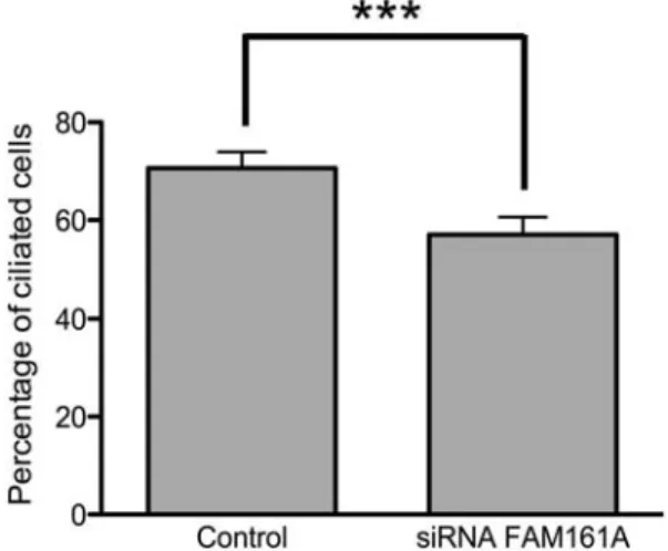
FAM161A is involved in the assembly of the cilium To investigate whether FAM161A had a direct role in cilium assembly, we knocked down FAM161A transcripts in hTERT-RPE-1 cells by a pool of three different small interfer-ing RNAs (siRNAs) targetinterfer-ing its sequence. Followinterfer-ing a .80% silencing of FAM161A expression [revealed by quanti-tative polymerase chain reaction (qPCR), Supplementary Ma-terial, Fig. S4A], we observed a significant reduction in the number of ciliated cells (Supplementary Material, Fig. S4B and C). Specifically, the percentage of cells displaying a com-pletely formed cilium dropped from 71% in controls (cells treated with siRNA against the non-human gene luciferase) to 57%, as assessed 48 h after siRNA supplementation in
Figure 3. Co-staining of FAM161A in rat retina sections with different ciliary markers. Retinal sections were stained with an anti-FAM161A antibody (green) and antibodies against axonemal or ciliary markers (red). Co-staining with RP1, and axonemal marker, reveals no overlap (A). Conversely, co-localization is observed with RPGRIP1L (a marker for the BB) and centrin (a marker for the whole CC), according to specific patterns (B and C). The insets are magnifications of the merged image (scale bars: 10 mm). The cartoons at the left side of each panel summarize the localization of the signal. Ax, Axoneme; CC, connecting cilium; BB, basal body.
three independent experiments, each considering 300 cells per condition (x2¼ 36.38, P , 0.0001, Fig.9).
DISCUSSION
More than one quarter of all genes that are mutated in HRD encode proteins that are responsible for cilium functions or de-velopment (25), highlighting the importance of this organelle for photoreceptor cell homeostasis and thus for vision in general. In this work, we demonstrate that FAM161A is a ciliary protein, interacts with other proteins involved in cilio-pathies and plays a role in cilium assembly.
Previous localization experiments revealed that, within the mouse retina, FAM161A is present in photoreceptors, mostly at the level of their ISs (18,19). Co-immunostaining of
FAM161A and a battery of IS markers revealed the absence of co-localization (not shown) and prompted us to investigate a smaller structure lying between the IS and the OS, the photo-receptor CC. Immunofluorescence performed with antibodies against centrin, a typical marker of the CC, revealed partial overlap with FAM161A. Additional FAM161A staining was also present in the IS and the OPL, as reported previously (18). Although we can speculate that the portion of FAM161A that is present within the IS could simply represent the nascent and immature form of this protein, at the moment we have no explanations for its localization within or in the vicinity of the OPL.
The ciliary localization of FAM161A was confirmed by the analysis of dissociated murine rod photoreceptors, providing the unique level of resolution typical of single-cell analyses. Co-staining with markers that are specifically marking
Figure 4. FAM161A in ciliated hTERT-RPE-1 cell lines. Endogenous FAM161A (green) localizes at the base of the cilium (stained with GT335, red), at the level of the BB (A). The BB localization is also shown by co-staining of FAM161A and RPGRIP1L (red) (B). These results are confirmed by co-localization of recombinant FAM161A (revealed by a FLAG antibody, green) with acetylated tubulin (red) (C), and gamma tubulin (red) (D) in the same cell lines. Insets present a magnified area of the merged picture. DAPI (blue) was used to stain the nuclei. Scale bars: 10 mm. In panel A, the patchy green staining of the nucleus represents background signal that is typical of the antibody used.
subciliary locations indicated that FAM161A exerts its func-tion at the BB and the basal side of the CC of photoreceptors and is not detected at the photoreceptor axoneme. These data are consistent with previous work reporting the presence of FAM161A peptides in low amounts within the ciliary prote-ome (22).
Based on the relatively widespread mRNA expression of FAM161A (18,19) and on the structural conservation of the primary cilium across different cell types, we reasoned that FAM161A could be present in other tissues than the retina and in other ciliated cells than photoreceptors. Staining of rat kidney sections revealed indeed the presence of FAM161A. Re-markably, its localization within the cilium of renal cells corre-sponded perfectly to that observed in photoreceptors and specifically corroborated the presence of FAM161A within BB structures. The same was true when the presence of FAM161A was investigated in cultured ciliated cells. Moreover, the single-cell level of resolution that was possible by the analyses of cul-tured hTERT-RPE-1 and ARPE-19 allowed detecting the pres-ence of FAM161A within centrosomal structures, as it is the case for many proteins that are component of sensory cilia (26) and in agreement with the concept that the BB is a modified centrosome of the photoreceptor cilium (27). Furthermore, several proteins involved in HRD have been shown to be directly or indirectly linked to the BB (11,13,28), the components of which are also thought to regulate the transport of molecules from the IS to the OS, across the CC (29).
In non-ciliated cell lines, overexpressed FAM161A orga-nized in defined structures that partly overlapped with the cellular microtubule network. Such localization has also been observed for other ciliary proteins such as lebercilin (12), especially upon severe overexpression. This likely pin-points the capacity of these centrosomal proteins to bind to acetylated microtubules and partly affect their dynamics. The effect is generally even more pronounced in non-ciliated cells that often induce significant overexpression, such as COS cells. We think that this marks the potential of ciliary/
microtubule binding proteins to bundle microtubules by recruiting other microtubule-binding proteins that decorate this cytoskeletal structure (while it is not clear whether they do this natively). In support of this, treatment of cells overex-pressing FAM161A with the microtubule depolymerizing drug nocodazole destroyed such FAM161A organization, indicating the need of an intact microtubule network for FAM161A to as-semble in such web-like structures. Recent data by Zach et al. (30), available on-line a few days before the present work was submitted, described findings similar to ours on FAM161A lo-calization. Following its overexpression in cultured cells, the authors hypothesized that FAM161A can have a stabilizing effect on microtubules in virtue of its interactions with them. Based on our collective findings, it is clear that FAM161A has the ability to interact with the cytoskeleton. However, as in our experiments, the presence of FAM161A within the microtubule network was never observed in physiological con-ditions, indicating that putative non-ciliary functions of FAM161A would require additional investigation.
Targeted yeast two-hybrid analysis revealed that FAM161A directly interacts with lebercilin. Similar to FAM161A, leber-cilin localizes within the CC of photoreceptors and at the base of the cilium and centrosomes in ciliated cells (12). This inter-action was confirmed by copurification and immunoprecipita-tion experiments. Very likely, the FAM161A – lebercilin interaction is achieved via the two C-terminal coiled-coil domains that are present within the UPF0564 domain, or alter-natively via other undetected structures lying in the C-terminal part of FAM161A. In turn, lebercilin interacts with proteins that are important for centrosome and cilium functions, such as RPGR (12) and OFD1 (14,31). Recently, Boldt et al. (32) demonstrated the direct interaction of lebercilin with the intra-flagellar machinery (IFT) and described a possible role of lebercilin in the protein transport across the cilium. However, since inactivation of LCA5 did not interfere with cilium assembly or localization of other IFT proteins, leberci-lin is probably not a structural component of the IFT
Figure 5. Overexpressed FAM161A associates with the microtubule network. When overexpressed in HeLa cells, flagged FAM161A (green) forms a web-like structure in correspondence of the cell microtubule network (red) (A). Treatment with 10 mM nocodazole, an inhibitor of microtubule polymerization, destroys such structure, highlighting the non-autonomous nature of this particular arrangement (B). Scale bars: 10 mm.
machinery. This fact could explain the absence of systemic phenotypes in patients with LCA5 mutations, in contrast to the syndromic manifestations of IFT mutations, despite the presence of lebercilin in all ciliated tissues (32).
Interestingly, FAM161A interacts also with OFD1 and SDCCAG8, two proteins that were already observed to be part of the LCA5 complex, localizing as expected at the level of the BB. The interaction with SDCCAG8 was con-firmed also in GST pull-down assays, although the level of the pulled-down protein was lower compared with lebercilin. We were unable to confirm the interaction between FAM161A and OFD1 using the same biochemical approach. Mutations in SDCCAG8 were linked to both a retinal-renal ciliopathy (16) and to 1 – 2% of patients with BBS (33). In addition, mutations in OFD1 were identified in patients with Joubert syndrome, nephronophthisis-like ciliopathies with retinal degeneration and RP (14,15,34).
Another important interactor of FAM161A is CEP290. This direct interaction, first observed in yeast binary assays, was also confirmed by pull-down experiments in retinal lysates. Mutations in CEP290 were identified in patients with Joubert syndrome-associated ciliopathies (35,36). Additional-ly, mutations in CEP290 are a major cause of Leber congenital amaurosis (13). CEP290 localizes at the CC in the murine photoreceptor, where FAM161A also partially localizes. In particular, the localization of CEP290 in the transition zone seems to be important for its function, possibly dealing with the docking and the transport of molecules inside the cilium (37). Furthermore, the depletion of CEP290 prevents cilium
Figure 6. FAM161A interacts with different proteins involved in ciliopathies, as revealed by yeast two-hybrid direct binding assay. Constructs expressing FAM161A full form (Full), FAM161A-N-term (N-term) and FAM161A-C-term (C-term) fused to a GAL4-BD protein were co-transformed in yeast with a set of pAD vectors expressing different ciliary genes. Selective medium depleted for relevant amino acids was used to isolate the hybrids. The strength of the inter-action was tested by blue staining of the colony following a-galactosidase (a) and b-galactosidase (b) activity. FAM161A showed the strongest interaction with lebercilin, OFD1, SDCCAG8 and CEP290, as assessed by both colony size and colorimetric assays. Prey constructs showed here included full-length sequences, except for OFD1 and CEP290. These were cloned as fragments and are listed in Supplementary Material, Table S1 as entries ‘OFD1 328’ and ‘CEP290 cc4+5+6’, respectively.
Figure 7. FAM161A interacts with RC proteins in vivo and in cultured cells. (A) Western blots of ciliopathy proteins after pull-down with GST-fused FAM161A constructs. The top two panels show pulled-down proteins from bovine retinal total lysates (TL) incubated with sepharose beads containing FAM161A-GST full length (FL), GST-FAM161A-N-term (N), GST-FAM161A-C-term (C), or GST alone (GST). GST pull-down allows FAM161A-GST and GST-FAM161A-C-term to reveal native retinal lebercilin and CEP290, whereas GST-FAM161A-N-term and GST alone do not. 2% of the total retinal lysate was loaded as input control. The third and fourth panels illustrate the same experi-ments performed in COS-7 cells, transfected with lebercilin (LCA5) and HA-SDCCAG8. GST pull-down of FAM161A-GST and GST-FAM161A-C-term reveals binding to HA-LCA5, whereas the pull down of GST-FAM161A-N-term and GST alone does not. All FAM161A constructs can pull-down recombin-ant SDCCAG8. 10% of the cell lysate was loaded as input control. The fifth panel shows the GST-purified protein used for pull-down assays (pur. GST prot.). (B) Western blots of different FAM161A constructs after pull-down with GST-fused lebercilin (LCA5) and SDCCAG8 (S8) constructs in COS-7 cells. Signals corresponding to FLAG-FAM161A and to FLAG-FAM161A-C-term, but not to FLAG-FAM161A-N-term or GST alone, are revealed. Again, the Coomassie-stained gel image shows the GST-purified protein used for these assays. 10% of the cell lysate was loaded as input control. MW, molecular weight (kDa).
formation and assembly (38,39), as we also observed after FAM161A silencing. Because of its interactions with CEP290 and lebercilin, as well as its subciliary localization, an intriguing possibility is that FAM161A could be involved in protein transport and docking between the IS and the OS.
A strong evidence of the importance of FAM161A for the cilium comes from the negative effects of its depletion in cili-ated cells, for which ciliary assembly itself is compromised. Mutations in FAM161A produce a retina-restricted phenotype and, compared with mutations in its interactors, a milder one. A possible explanation is that the FAM161A role could be hierarchically subordinated to that of its associated proteins and critical only for photoreceptors, where it is expressed in large amounts. Although further interaction experiments are needed to establish a solid mechanistic model, another fas-cinating possibility is that FAM161A, in virtue of its CRX-mediated expression, may help driving the organization of ciliary proteins having ubiquitous expression in rods and cones. In light of the phenotypic heterogeneity underlying its interactors and the current absence of an animal model, we also cannot rule out that variations in FAM161A are also modi-fying more complex syndromic ciliopathy phenotypes.
In conclusion, in this work we show that FAM161A is a functional protein of the cilium. It localizes at the base of the cilium in photoreceptors as well as in other ciliated cell types and tissues and prominently associates with a specific subset of proteins associated with RCs. Based on these find-ings, FAM161A-associated RP should be classified as a novel retinal ciliopathy.
MATERIALS AND METHODS DNA constructs
Full-length FAM161A cDNA, obtained from human retina, was cloned into a pENTR vector (Invitrogen). Similar cloning was performed also for two deletion sequences, obtained by PCR. These generated plasmids pFAM161A-N-term, comprising codons 1 – 219 and containing the first of three coiled-coil domains, and pFAM161A-C-term, spanning codons 211–660 and including the UPF0564 domain. Expression plasmids (GST, FLAG and yeast clones containing the activation domain, AD or the binding domain, BD) were obtained starting from these entry clones using the Gateway cloning technology (Invitrogen). The specificity and quality of the fragments was assessed by re-striction analysis and direct sequencing. Although a difference with respect to the reported reference sequence was identified, this corresponded to the annotated polymorphism rs17513722, having a 0.5 allele frequency within the European population.
Expression plasmids for LCA5, SDCCAG8 and OFD1 were described previously (12,14,16,21).
Antibodies
Custom polyclonal rabbit antibodies against FAM161A (used for the immunostaining depicted in Fig. 3A) included those described previously (18) as well as a new one (used for the staining of human retina sections), raised against a synthetic peptide corresponding to the C-terminal moiety of the murine protein (EKLQKRPMLFERVT) and used at a 1:100 dilution.
Figure 8. Recombinant lebercilin/LCA5, SDCCAG8, OFD1, and FAM161A co-localize in hTERT-RPE-1 cells after cilium induction. FLAG-FAM161A (green) co-localizes at the level of the BB with HA-LCA5 (red, A), SDCCAG8 (red, B), and OFD1 (red, C). Scale bars: 10 mm.
Another commercial polyclonal rabbit anti-FAM161A antibody was purchased from Sigma-Aldrich and used to a final dilution of 1:100 in all other instances. Mouse monoclonal anti-FLAG antibody (1:1000) was obtained from Invitrogen, whereas rabbit polyclonal anti-HA antibody (1:1000) was obtained by Bethyl antibodies. Mouse and rabbit anti-g-tubulin (1:1000) were obtained from Abcam, and mouse and rabbit anti acety-lated a-tubulin (1:1000) from Sigma-Aldrich. Chicken anti-RP1 (1:2000) antibody was a kind gift of Dr E. Pierce (Harvard Medical School, Boston, USA), whereas anti-RPGRIP1L (1:1000) was obtained as reported previously (11). Mouse monoclonal anti-rhodopsin (Ret-1 clone, 1:200) antibody was purchased from Santa Cruz Biotechnology. Mouse monoclonal anti-centrin clone 20H5 (1:100) was obtained from Millipore. Mouse monoclonal antibody against polyglutamylated tubulin (GT335, 1:1000) was provided by Dr C. Janke (Institut Curie, Orsay, France). Polyclonal anti-bodies against lebercilin were obtained and used as reported before (12,32). Secondary goat antibodies mouse, anti-rabbit, anti-chicken and anti-guinea pig were conjugated with Alexa Fluor 488 and Alexa Fluor 568 dyes (1:500). Secondary antibody anti-mouse and anti-rabbit conjugated with IRDye 680 and IRDye 800 for detection with the Odyssey system were purchased from LI-COR Biosciences and used at a 1:5000 final dilution.
Immunohistochemistry of retinal tissues and isolation of photoreceptors
Unfixed eyes of P20 Wistar rats and P20 C57BL/6J mice were isolated and frozen in melted isopentane. Cryosections of 10 mm were permeabilized in 0.01% Tween-20 in phosphate buffered saline (PBS) and then blocked in 0.1% ovoalbumin and 0.5% fish skin gelatin in PBS (blocking solution). Primary antibodies were diluted in blocking solution and then incubated overnight with the sections. After three washing steps with PBS, slides were incubated with
Alexa-conjugated secondary antibodies and mounted with Prolong Gold Antifade (Invitrogen) mounting media to preserve fluor-escence. Slides were then observed using direct fluorescence with the microscope Axioskop 4 (Carl Zeiss) and confocal scanning with the microscope Zeiss LSM 710 Quasar Con-focal (Carl Zeiss). Images were processed using the Axiovi-sion software (Carl Zeiss) and Adobe Photoshop CS5 (Adobe Systems). For paraffin sections (human), a standard immunohistological analysis protocol for the paraffin section was carried out as described previously (40), by using a sec-ondary antibody the DyLight 488 goat anti-rabbit (Jackson ImmunoResearch) at a 1:200 dilution.
Isolated photoreceptors were obtained by disruption of fresh retinas from adult mice, according to the protocol by Liu et al. (22), modified as follows. The intact retina was resuspended in PBS and then gently vortexed for a few seconds, twice. The suspension was then spread on the surface of a microscope slide and let dry for 1 h, before proceeding to IF analysis. Cell growth and transfection
Human immortalized retinal pigmented epithelium cells ARPE-19 and hTERT-RPE-1 were grown at 378C in 5% CO2
and Dulbecco’s modified Eagle’s medium/nutrient mixture F-12 (DMEM/F-F-12) (Gibco) media (1:1 ratio), supplemented with 10% fetal calf serum (FCS), 100 IU/ml penicillin and 100 mg/ml streptomycin. Human-derived HeLa, HEK293 and monkey-derived COS-7 cells were cultivated with DMEM con-taining 10% FCS, 100 IU/ml penicillin, 100 mg/ml streptomycin and 1% Glutamax (Gibco) under the same growth conditions. Cells were transfected using Lipofectamine 2000 (Invitrogen) for 24–48 h according the company specifications. Plasmids containing FAM161A sequences were used in co-localization studies on cells that were fixed 24 h after transfection.
For the microtubule disassembly assay, nocodazole (Sigma-Aldrich) was added directly to the growing medium at a 10 mMfinal concentration and cells were fixed in 2%
par-aformaldehyde (PFA) after 2 h. They were then stained according to the published protocols (41).
Immunocytochemistry
hTERT-RPE-1 and ARPE-19 cells were cultured, seeded on coverslips and serum starved for 24 h (0.2% FCS). Cells were then washed twice in PBS and fixed in 2% PFA for 20 min. HeLa cells were seeded on a coverslip and directly fixed in 2% PFA for 20 min. After fixation, they were treated with PBS plus 1% Triton X-100 for 5 min and then blocked in 2% bovine serum albumin in PBS for 1 h. Staining was achieved following overnight incubation with relevant antibodies. After incubation, fixed cells were washed twice in PBS and stained with a fluorescent secondary antibody for 1 h. Coverslips were then washed first with PBS, briefly with MilliQ water, and then mounted with Vectashield and DAPI (Vector Laboratories).
Direct yeast two-hybrid interaction assay
The direct interaction between FAM161A and other ciliary proteins was tested using a GAL4-based yeast two-hybrid
Figure 9. FAM161A knock down causes a reduction in the number of ciliated cells in hTERT-RPE-1 cell lines. The graph shows the percentage of ciliated cells after cilium induction in samples treated with FAM161A-specific siRNA and in the control group, treated with non-targeting siRNA. N ¼ 900 counts per group, sampled in 3 different experiments. Error bars indicate standard errors. P , 0.0001, shown by the 3 asterisks.
GST pull down
To produce the GST proteins, BL21(DE3) cells were trans-formed with FAM161A-GST, GST-FAM161A-N-term, GST-FAM161A-C-term, GST-LCA5 or GST alone constructs and induced with 0.5 mM isopropyl
b-D-1-thiogalactopyrano-side (IPTG) at 308C overnight. Cells were then lysed in sodium chloride-Tris-EDTA (STE) buffer [10 mM Tris – HCl
(pH 8), 1 mM ethylenediaminetetraacetic acid (EDTA),
150 mM NaCl] supplemented with 10 mg/ml lysozime, 0.5%
sarkosyl, 1% Triton X-100 and Complete Protease Inhibitor Cocktail (Roche). Overnight incubation with glutathione-sepharose 4B beads (Roche) was performed to isolate the GST-fused proteins. This was followed by three washes with STE buffer and four washes with TBSDT buffer [Tris – HCl 25 mM, pH 7.4, 150 mM NaCl, 1% Triton X-100 and fresh
2 mM dithiothreitol (DTT)]. The amount of produced GST
protein was assessed by sodium dodecyl sulfate polyacrylamide gel electrophoresis (SDS – PAGE) and blue Coomassie staining. COS-7 cells seeded in 10 cm plates were transfected using Lipo-fectamine 2000 with 3×FLAG-FAM161A, 3×FLAG-FAM161A-N-term, 3×FLAG-FAM161A-C-term and 3×HA-LCA5. Subsequently, proteins were extracted using the previously described lysis buffer and incubated with the iso-lated GST-fused protein sepharose beads from 5 h to overnight and finally washed five times with the protein lysis buffer used for Co-IP. The beads were then resuspended in loading buffer and analyzed by SDS – PAGE. FLAG or HA tags were revealed by using anti-FLAG or anti-HA antibodies.
Retinas were dissected from two fresh bovine eyes and put directly in Lysis Buffer for the Cytosolic Fraction (10 mM
HEPES, pH 7.9, 10 mM NaCl, 3 mM MgCl2, fresh 1 mM
DTT and 1 mM sodium orthovanadate) and in lysis buffer
for the membrane fraction (50 mM HEPES, pH 7.4, 150 mM
NaCl, 10% glycerol, 0.5% Triton X-100, 1.5 mM MgCl2,
1 mM ethylene glycol tetraacetic acid and 1 mM EDTA)
sup-plemented with protease inhibitors. Retina extracts were then incubated overnight with FAM161A-GST, GST-FAM161A-N-term, and GST-FAM161A-C-term fusion proteins and with GST alone as a control. The beads were then washed five times with a mix of 50% cytosolic and 50% membrane lysis buffers. The beads were then incubated in loading buffer for 5 min at 958C and the supernatant processed by SDS – PAGE. The signal of native LCA5 was revealed using a rabbit polyclonal antibody, previously described by den Hol-lander et al. (12), whereas CEP290 was revealed using the rabbit antibody obtain from Bethyl.
from transfection and the relative expression of FAM161A was evaluated by qPCR. The number of the ciliated cells was evaluated after 2% PFA fixation of transfected cells and following staining with the anti-acetylated tubulin antibody. Statistical analysis was performed by using the on-line x2 cal-culator available at http://vassarstats.net/newcs.html.
SUPPLEMENTARY MATERIAL
Supplementary Material is available at HMG online.
ACKNOWLEDGEMENTS
We want to kindly acknowledge T.A. Peters for providing rat retinal slides, H. Koskiniemi for providing the FAM161A-GST plasmid, as well as D. Mans, E. van Wijk and other members of the R. Roepman/H. Kremers lab for useful advice and helpful discussions. We are grateful to A. Obolensky, E. Banin and G. Tanackovic for providing important sugges-tions about the experimental procedures.
Conflict of Interest statement. None declared.
FUNDING
This work was supported by the Swiss National Science Foun-dation (310030-138346 to C.R. and Sinergia #CRSII3_141814 to C.R., D.S., and Y.A.), the Israeli Ministry of Health (3-7261 to D.S.), a grant from the Netherlands Organization for Scientif-ic Research (NWO Vidi-91786396 to R.R.) and the European Community’s Seventh Framework Programme FP7/2009 under grant agreement number 241955, SYSCILIA to R.R.
REFERENCES
1. Berson, E.L. (1993) Retinitis pigmentosa. The Friedenwald Lecture. Invest. Ophthalmol. Vis. Sci., 34, 1659– 1676.
2. Hartong, D.T., Berson, E.L. and Dryja, T.P. (2006) Retinitis pigmentosa. Lancet, 368, 1795– 1809.
3. Rivolta, C., Sharon, D., DeAngelis, M.M. and Dryja, T.P. (2002) Retinitis pigmentosa and allied diseases: numerous diseases, genes, and inheritance patterns. Hum. Mol. Genet., 11, 1219– 1227.
4. Berger, W., Kloeckener-Gruissem, B. and Neidhardt, J. (2010) The molecular basis of human retinal and vitreoretinal diseases. Prog. Retin. Eye Res., 29, 335 – 375.
5. den Hollander, A.I., Black, A., Bennett, J. and Cremers, F.P. (2010) Lighting a candle in the dark: advances in genetics and gene therapy of recessive retinal dystrophies. J. Clin. Invest., 120, 3042 – 3053.
6. Adams, N.A., Awadein, A. and Toma, H.S. (2007) The retinal ciliopathies. Ophthalmic. Genet., 28, 113 – 125.
7. Novarino, G., Akizu, N. and Gleeson, J.G. (2011) Modeling human disease in humans: the ciliopathies. Cell, 147, 70 – 79.
8. Hildebrandt, F., Benzing, T. and Katsanis, N. (2011) Ciliopathies. N. Engl. J. Med., 364, 1533 – 1543.
9. Liu, Q., Zhou, J., Daiger, S.P., Farber, D.B., Heckenlively, J.R., Smith, J.E., Sullivan, L.S., Zuo, J., Milam, A.H. and Pierce, E.A. (2002) Identification and subcellular localization of the RP1 protein in human and mouse photoreceptors. Invest. Ophthalmol. Vis. Sci., 43, 22 – 32. 10. Hosch, J., Lorenz, B. and Stieger, K. (2011) RPGR: role in the
photoreceptor cilium, human retinal disease, and gene therapy. Ophthalmic. Genet., 32, 1 – 11.
11. Arts, H.H., Doherty, D., van Beersum, S.E., Parisi, M.A., Letteboer, S.J., Gorden, N.T., Peters, T.A., Marker, T., Voesenek, K., Kartono, A. et al. (2007) Mutations in the gene encoding the basal body protein RPGRIP1L, a nephrocystin-4 interactor, cause Joubert syndrome. Nat. Genet., 39, 882 – 888.
12. den Hollander, A.I., Koenekoop, R.K., Mohamed, M.D., Arts, H.H., Boldt, K., Towns, K.V., Sedmak, T., Beer, M., Nagel-Wolfrum, K., McKibbin, M. et al. (2007) Mutations in LCA5, encoding the ciliary protein lebercilin, cause Leber congenital amaurosis. Nat. Genet., 39, 889 – 895.
13. den Hollander, A.I., Koenekoop, R.K., Yzer, S., Lopez, I., Arends, M.L., Voesenek, K.E., Zonneveld, M.N., Strom, T.M., Meitinger, T., Brunner, H.G. et al. (2006) Mutations in the CEP290 (NPHP6) gene are a frequent cause of Leber congenital amaurosis. Am. J. Hum. Genet., 79, 556 – 561. 14. Coene, K.L., Roepman, R., Doherty, D., Afroze, B., Kroes, H.Y.,
Letteboer, S.J., Ngu, L.H., Budny, B., van Wijk, E., Gorden, N.T. et al. (2009) OFD1 is mutated in X-linked Joubert syndrome and interacts with LCA5-encoded lebercilin. Am. J. Hum. Genet., 85, 465 – 481.
15. Webb, T.R., Parfitt, D.A., Gardner, J.C., Martinez, A., Bevilacqua, D., Davidson, A.E., Zito, I., Thiselton, D.L., Ressa, J.H., Apergi, M. et al. (2012) Deep intronic mutation in OFD1, identified by targeted genomic next-generation sequencing, causes a severe form of X-linked retinitis pigmentosa (RP23). Hum. Mol. Genet., 21, 3647– 3654.
16. Otto, E.A., Hurd, T.W., Airik, R., Chaki, M., Zhou, W., Stoetzel, C., Patil, S.B., Levy, S., Ghosh, A.K., Murga-Zamalloa, C.A. et al. (2010) Candidate exome capture identifies mutation of SDCCAG8 as the cause of a retinal-renal ciliopathy. Nat. Genet., 42, 840 – 850.
17. Billingsley, G., Vincent, A., Deveault, C. and Heon, E. (2012) Mutational Analysis of SDCCAG8 in Bardet-Biedl Syndrome Patients with Renal Involvement and Absent Polydactyly. Ophthalmic. Genet., 33, 150 – 154. 18. Langmann, T., Di Gioia, S.A., Rau, I., Stohr, H., Maksimovic, N.S.,
Corbo, J.C., Renner, A.B., Zrenner, E., Kumaramanickavel, G., Karlstetter, M. et al. (2010) Nonsense mutations in FAM161A cause RP28-associated recessive retinitis pigmentosa. Am. J. Hum. Genet., 87, 376 – 381.
19. Bandah-Rozenfeld, D., Mizrahi-Meissonnier, L., Farhy, C., Obolensky, A., Chowers, I., Pe’er, J., Merin, S., Ben-Yosef, T., Ashery-Padan, R., Banin, E. et al. (2010) Homozygosity mapping reveals null mutations in FAM161A as a cause of autosomal-recessive retinitis pigmentosa. Am. J. Hum. Genet., 87, 382 – 391.
20. Gu, S., Kumaramanickavel, G., Srikumari, C.R., Denton, M.J. and Gal, A. (1999) Autosomal recessive retinitis pigmentosa locus RP28 maps between D2S1337 and D2S286 on chromosome 2p11-p15 in an Indian family. J. Med. Genet., 36, 705 – 707.
21. van Wijk, E., van der Zwaag, B., Peters, T., Zimmermann, U., Te Brinke, H., Kersten, F.F., Marker, T., Aller, E., Hoefsloot, L.H., Cremers, C.W. et al. (2006) The DFNB31 gene product whirlin connects to the Usher protein network in the cochlea and retina by direct association with USH2A and VLGR1. Hum. Mol. Genet., 15, 751 – 765.
22. Liu, Q., Tan, G., Levenkova, N., Li, T., Pugh, E.N. Jr., Rux, J.J., Speicher, D.W. and Pierce, E.A. (2007) The proteome of the mouse photoreceptor sensory cilium complex. Mol. Cell Proteomics., 6, 1299– 1317. 23. Wolfrum, U. (1995) Centrin in the photoreceptor cells of mammalian
retinae. Cell Motil. Cytoskeleton, 32, 55 – 64.
24. Guarguaglini, G., Duncan, P.I., Stierhof, Y.D., Holmstrom, T., Duensing, S. and Nigg, E.A. (2005) The forkhead-associated domain protein Cep170 interacts with Polo-like kinase 1 and serves as a marker for mature centrioles. Mol. Biol. Cell, 16, 1095– 1107.
25. Wright, A.F., Chakarova, C.F., Abd El-Aziz, M.M. and Bhattacharya, S.S. (2010) Photoreceptor degeneration: genetic and mechanistic dissection of a complex trait. Nat. Rev. Genet., 11, 273 – 284.
26. Jakobsen, L., Vanselow, K., Skogs, M., Toyoda, Y., Lundberg, E., Poser, I., Falkenby, L.G., Bennetzen, M., Westendorf, J., Nigg, E.A. et al. (2011) Novel asymmetrically localizing components of human centrosomes identified by complementary proteomics methods. EMBO J., 30, 1520 – 1535.
27. Moser, J.J., Fritzler, M.J., Ou, Y. and Rattner, J.B. (2010) The PCM-basal body/primary cilium coalition. Semin. Cell Dev. Biol., 21, 148 – 155. 28. Chakarova, C.F., Khanna, H., Shah, A.Z., Patil, S.B., Sedmak, T.,
Murga-Zamalloa, C.A., Papaioannou, M.G., Nagel-Wolfrum, K., Lopez, I., Munro, P. et al. (2011) TOPORS, implicated in retinal degeneration, is a cilia-centrosomal protein. Hum. Mol. Genet., 20, 975 – 987.
29. Roepman, R. and Wolfrum, U. (2007) Protein networks and complexes in photoreceptor cilia. Subcell Biochem., 43, 209 – 235.
30. Zach, F., Grassmann, F., Langmann, T., Sorusch, N., Wolfrum, U. and Stohr, H. (2012) The retinitis pigmentosa 28 protein FAM161A is a novel ciliary protein involved in intermolecular protein interaction and microtubule association. Hum. Mol. Genet., in press.
31. Singla, V., Romaguera-Ros, M., Garcia-Verdugo, J.M. and Reiter, J.F. (2010) Ofd1, a human disease gene, regulates the length and distal structure of centrioles. Dev. Cell, 18, 410 – 424.
32. Boldt, K., Mans, D.A., Won, J., van Reeuwijk, J., Vogt, A., Kinkl, N., Letteboer, S.J., Hicks, W.L., Hurd, R.E., Naggert, J.K. et al. (2011) Disruption of intraflagellar protein transport in photoreceptor cilia causes Leber congenital amaurosis in humans and mice. J. Clin. Invest., 121, 2169– 2180.
33. Schaefer, E., Zaloszyc, A., Lauer, J., Durand, M., Stutzmann, F., Perdomo-Trujillo, Y., Redin, C., Bennouna Greene, V., Toutain, A., Perrin, L. et al. (2011) Mutations in SDCCAG8/NPHP10 Cause Bardet-Biedl Syndrome and Are Associated with Penetrant Renal Disease and Absent Polydactyly. Molecular syndromology, 1, 273 – 281. 34. Zullo, A., Iaconis, D., Barra, A., Cantone, A., Messaddeq, N., Capasso,
G., Dolle, P., Igarashi, P. and Franco, B. (2010) Kidney-specific inactivation of Ofd1 leads to renal cystic disease associated with upregulation of the mTOR pathway. Hum. Mol. Genet., 19, 2792– 2803. 35. Sayer, J.A., Otto, E.A., O’Toole, J.F., Nurnberg, G., Kennedy, M.A.,
Becker, C., Hennies, H.C., Helou, J., Attanasio, M., Fausett, B.V. et al. (2006) The centrosomal protein nephrocystin-6 is mutated in Joubert syndrome and activates transcription factor ATF4. Nat. Genet., 38, 674 – 681.
36. Valente, E.M., Silhavy, J.L., Brancati, F., Barrano, G., Krishnaswami, S.R., Castori, M., Lancaster, M.A., Boltshauser, E., Boccone, L., Al-Gazali, L. et al. (2006) Mutations in CEP290, which encodes a centrosomal protein, cause pleiotropic forms of Joubert syndrome. Nat. Genet., 38, 623 – 625.
37. Garcia-Gonzalo, F.R., Corbit, K.C., Sirerol-Piquer, M.S., Ramaswami, G., Otto, E.A., Noriega, T.R., Seol, A.D., Robinson, J.F., Bennett, C.L., Josifova, D.J. et al. (2011) A transition zone complex regulates mammalian ciliogenesis and ciliary membrane composition. Nat. Genet., 43, 776 – 784.
38. Tsang, W.Y., Bossard, C., Khanna, H., Peranen, J., Swaroop, A., Malhotra, V. and Dynlacht, B.D. (2008) CP110 suppresses primary cilia formation through its interaction with CEP290, a protein deficient in human ciliary disease. Dev. Cell, 15, 187 – 197.
39. Kim, J., Krishnaswami, S.R. and Gleeson, J.G. (2008) CEP290 interacts with the centriolar satellite component PCM-1 and is required for Rab8 localization to the primary cilium. Hum. Mol. Genet., 17, 3796– 3805. 40. Banin, E., Obolensky, A., Idelson, M., Hemo, I., Reinhardtz, E., Pikarsky,
E., Ben-Hur, T. and Reubinoff, B. (2006) Retinal incorporation and differentiation of neural precursors derived from human embryonic stem cells. Stem Cells, 24, 246 – 257.
41. Liu, Q., Zuo, J. and Pierce, E.A. (2004) The retinitis pigmentosa 1 protein is a photoreceptor microtubule-associated protein. J. Neurosci., 24, 6427– 6436.
42. Letteboer, S.J. and Roepman, R. (2008) Versatile screening for binary protein-protein interactions by yeast two-hybrid mating. Methods Mol. Biol., 484, 145 – 159.