Oxidative and Anti-Oxidative Stress Markers
in Chronic Glaucoma: A Systematic Review
and Meta-Analysis
Ce´dric Benoist d’Azy1,2, Bruno Pereira3, Fre´de´ric Chiambaretta1, Fre´de´ric Dutheil2,4,5,6,7* 1 University Hospital of Clermont-Ferrand (CHU), Ophthalmology, Clermont-Ferrand, France, 2 University
Hospital of Clermont-Ferrand (CHU), Preventive and Occupational Medicine, Clermont-Ferrand, France,
3 University Hospital of Clermont-Ferrand (CHU), Clinical Research Direction, Clermont-Ferrand, France, 4 CNRS Physiological and Psychosocial Stress, LAPSCO, University Clermont Auvergne,
Clermont-Ferrand, France, 5 Australian Catholic University, Faculty of Health, School of Exercise Science, Melbourne, Australia, 6 University Clermont Auvergne, Laboratory of Metabolic Adaptations to Exercise in Physiological and Pathological conditions EA3533, Clermont-Ferrand, France, 7 Research Centre in Human Nutrition (CRNH) Auvergne, Clermont-Ferrand, France
*frederic.dutheil@acu.edu.au
Abstract
Chronic glaucoma is a multifactorial disease among which oxidative stress may play a major pathophysiological role. We conducted a systematic review and meta-analysis to evaluate the levels of oxidative and antioxidative stress markers in chronic glaucoma compared with a control group. The PubMed, Cochrane Library, Embase and Science Direct databases were searched for studies reporting oxidative and antioxidative stress markers in chronic glaucoma and in healthy controls using the following keywords: “oxida-tive stress” or “oxidant stress” or “nitra“oxida-tive stress” or “oxida“oxida-tive damage” or “nitra“oxida-tive damage” or “antioxidative stress” or “antioxidant stress” or “antinitrative stress” and “glau-coma”. We stratified our meta-analysis on the type of biomarkers, the type of glaucoma, and the origin of the sample (serum or aqueous humor). We included 22 case-control studies with a total of 2913 patients: 1614 with glaucoma and 1319 healthy controls. We included 12 studies in the meta-analysis on oxidative stress markers and 19 on antioxida-tive stress markers. We demonstrated an overall increase in oxidaantioxida-tive stress markers in glaucoma (effect size = 1.64; 95%CI 1.20–2.09), ranging from an effect size of 1.29 in serum (95%CI 0.84–1.74) to 2.62 in aqueous humor (95%CI 1.60–3.65). Despite a decrease in antioxidative stress marker in serum (effect size = –0.41; 95%CI –0.72 to – 0.11), some increased in aqueous humor (superoxide dismutase, effect size = 3.53; 95% CI 1.20–5.85 and glutathione peroxidase, effect size = 6.60; 95%CI 3.88–9.31). The dif-ferences in the serum levels of oxidative stress markers between glaucoma patients and controls were significantly higher in primary open angle glaucoma vs primary angle closed glaucoma (effect size = 12.7; 95%CI 8.78–16.6, P<0.001), and higher in pseudo-exfolia-tive glaucoma vs primary angle closed glaucoma (effect size = 12.2; 95%CI 8.96–15.5, P<0.001). In conclusion, oxidative stress increased in glaucoma, both in serum and aqueous humor. Malonyldialdehyde seemed the best biomarkers of oxidative stress in
a11111
OPEN ACCESS
Citation: Benoist d’Azy C, Pereira B, Chiambaretta
F, Dutheil F (2016) Oxidative and Anti-Oxidative Stress Markers in Chronic Glaucoma: A Systematic Review and Meta-Analysis. PLoS ONE 11(12): e0166915. doi:10.1371/journal.pone.0166915
Editor: Reza Khodarahmi, Kermanshah University
of Medical Sciences, ISLAMIC REPUBLIC OF IRAN
Received: July 15, 2016 Accepted: November 7, 2016 Published: December 1, 2016
Copyright:© 2016 Benoist d’Azy et al. This is an open access article distributed under the terms of theCreative Commons Attribution License, which permits unrestricted use, distribution, and reproduction in any medium, provided the original author and source are credited.
Data Availability Statement: All relevant data are
within the paper and its Supporting Information files.
Funding: The authors received no specific funding
for this work.
Competing Interests: The authors have declared
serum. The increase of some antioxidant markers could be a protective response of the eye against oxidative stress.
Introduction
Chronic glaucoma is one of the most frequently established diseases in ophthalmology and represents a growing public health concern, with consequences that lead to blindness [1,2]. This pathology causes significant impact on visual function that may affect the quality of life and work productivity [3,4]. Due to its long time asymptomatic evolution, this disease remains under-diagnosed and the irreversible loss of optic nerve fibers leads to important visual field loss [5]. Chronic glaucoma is a multifactorial disease implicating divers factors such as intraoc-ular pressure, familial history, myopia, corneal thickness and ethnicity [6,7,8,9,10]. Extensive research over the last three decades has demonstrated that oxidative stress can cause peroxida-tion of nucleic acids, bases, lipids, proteins and carbohydrates, thus resulting in their damage. At the ocular level, oxidative stress has been postulated to promote chronic glaucoma [11]. Conversely, antioxidants could protect against glaucoma [12].
However, oxidative stress is not assessed in daily clinical practice [13]. Reactive oxygen spe-cies are so reactive and so short-lived that they are difficult to measure directly [13]. There are numerous markers of oxidative stress, and several methods of measurement. In glaucoma, oxi-dative stress can be assessed both in the serum or the aqueous humor. Although the evaluation of different markers of oxidative and antioxidative stress in glaucoma has been assessed in sev-eral studies and discussed in sevsev-eral reviews [14,15,16,17], the results are under debate. We hypothesized that glaucoma patients would exhibit an increased level of oxidative stress and a decreased level of antioxidative markers.
Thus, we aimed to conduct a systematic review and meta-analysis to summarize all studies reporting the level of oxidative and antioxidative markers in aqueous humor or serum samples of glaucoma patients compared to controls.
Methods
Literature search
We reviewed all studies measuring oxidative or antioxidative stress markers in chronic glau-coma patients and controls. Animal studies were excluded. The PubMed, Cochrane Library, Science Direct and Embase databases were searched on May 10th2016, with the following key-words: “oxidative stress” or “oxidant stress” or “nitrative stress” or “oxidative damage” or “nitrative damage” or “antioxidative stress” or “antioxidant stress” or “antinitrative stress” and “glaucoma”. The search was not limited to specific years. No minimal sample size and no lan-guage restrictions were applied. To be included, articles needed to be case-control studies describing our primary outcome variable, which was the measurement of oxidative or antioxi-dative stress markers in glaucoma patients and healthy controls. We imposed no limitation on the regional origin or the nature of the control group. Studies needed to be primary research. In addition, reference lists of all publications meeting the inclusion criteria were manually searched to identify any further studies that were not found with the electronic search. Ances-try searches were also completed on previous reviews to locate other potential eligible primary studies. The search strategy is presented inFig 1. One author (CBDA) conducted all literature searches and collated the abstracts. Two authors (CBDA and FD) separately reviewed the abstracts and based on the selection criteria, decided the suitability of the articles for inclusion.
A third author (BP) was asked to review the articles where consensus on suitability was debated. All authors then reviewed the eligible articles.
Quality of assessment
Although not designed for quantifying the integrity of studies [18], the “STrengthening the Reporting of OBservational studies in Epidemiology” (STROBE) criteria were used to check the quality of the reporting [19]. The STROBE Statement consists of a checklist of 22 items, which relate to the title, abstract, introduction, methods, results and discussion sections of arti-cles. Eighteen items are common to cohort studies, case control studies and cross-sectional studies and four are specific to each of the three study designs. Among the 22 items, six are split into several sub-items. One point was attributed per item or sub-item when the study ful-filled the criteria. The maximum score achievable was 32, then converted into percentage. Fig 1. Search strategy.
Statistical considerations
Statistical analysis was conducted using Comprehensive Meta-analysis software (version 2, Biostat Corporation) [20,21,22,23] and Stata software (version 13, StataCorp, College Station, US). Baseline characteristics were summarized for each study sample and reported as mean (standard-deviation) and number (%) for continuous and categorical variables respectively. Heterogeneity in the study results was evaluated by examining forest plots, confidence inter-vals (CI) and using formal tests for homogeneity based on the I statistic, which is the most common metric for measuring the magnitude of between-study heterogeneity and is easily interpretable. I values range between 0% and 100% and are typically considered low for <25%, modest for 25–50%, and high for >50%. This statistical method generally assumes heterogene-ity when the p-value of the I test is <0.05. For example, a significant heterogeneheterogene-ity may be due to the variability between the characteristics of the studies such as those of the participants (age, sex, etc), the type of biomarkers, the type of glaucoma, or the origin of the sample (aque-ous humor or serum). Random effects meta-analyses (DerSimonian and Laird approach) were conducted when data could be pooled [24]. P values less than 0.05 were considered statistically significant.
We conducted meta-analysis on the levels of oxidative and antioxidative stress markers in chronic glaucoma patients and healthy controls. We stratified these meta-analyses on the type of biomarkers, the type of glaucoma, and the sample origin (aqueous humor or serum). We described our results by calculating the effect size (ES, standardized mean differences—SMD) of the oxidative and antioxidative stress markers for each dependent variable [24]. An ES is defined as a unitless measure of the levels of the stress markers centered at zero if the stress marker levels in chronic glaucoma patients are not different from those in healthy controls. A positive ES denoted improved performance. A scale for ES has been suggested with 0.8 reflect-ing a large effect, 0.5 a moderate effect, and 0.2 a small effect [25].
For rigor, funnel plots of these meta-analyses were used to search for potential publication bias. In order to verify the strength of the results, further meta-analyses were then conducted excluding studies that were not evenly distributed around the base of the funnel [26].
When possible (sufficient sample size), meta-regressions were proposed to study the rela-tionship between the levels of oxidative and antioxidative stress markers and clinically relevant parameters such as the participants gender, age, the type of biomarkers, the type of glaucoma, and the sample origin (aqueous humor or serum). Results were expressed as regression coeffi-cients and 95%CI.
Results
An initial search produced a possible 9,058 articles (Fig 1). Removal of duplicates and use of the selection criteria reduced the number of articles reporting the evaluation of oxidative or antioxidative markers in aqueous humor or blood to 22 articles [27–48]. All articles were writ-ten in English.
Quality of articles
The assessment of the quality of the 22 studies that were included was performed using the STROBE criteria, with the results varying from 68.8 [45] to 90.6% [32,46], with a mean score of 80.3±6.3. Overall, the studies performed best in the methods section and worst in the discus-sion section. All studies except one mentioned ethical approval [43].
Method of sampling for markers analysis
Levels of oxidant or antioxidant markers were evaluated in serum in 12 studies
[27,28,29,31,32,34,35,39,40,43,45,48], in aqueous humor in six studies [30,36,37,38,44,47], or both in three studies [33,41,42]. One study measured the markers in blood hemolysate [46]. Blood samples were collected in ethylenediaminetetraacetic acid tubes. The tubes were centri-fuged and the plasma layer was separated and stored until analysis. At the beginning of the surgery, 0.1–0.2 ml of aqueous humor samples were obtained from each patient through a paracentesis using a 27 gauge needle. Patients were those requiring either glaucoma surgery (glaucoma group) or cataract surgery (control group) in six studies [30,36,37,42,44,47] or only before cataract surgery (glaucoma and control group) in three studies [33,38,41]. Aqueous humor samples were immediately frozen at -80˚C until processing for the subsequent bio-chemistry techniques.
Inclusion criteria for glaucoma patients
The markers were measured in different types of glaucoma: only in primary open angle glau-coma (POAG) [29,30,36,37,39,41,43,44,45,46,48], only in primary angle closure glaucoma (PACG) [27,31], only in pseudoexfoliation glaucoma (PEG) [28,32,33], in both POAG and PACG [38], in both POAG and PEG [35,42,47], in both POAG, PACG and PEG [40], or it was not specified [34]. Inclusion criteria for glaucoma patients were similar for most studies [27– 33,35–39,41,43,46,47]. Patients had to have a vertical cup/disc ratio of 0.5 or higher and a typical glaucomatous defect at the visual field (standard automatized visual field analyser). Some specificities were required for each type of glaucoma, such as an open angle for the gonioscopy for POAG [29,30,36,37,38,39,41,43,46,48], a close angle for PACG [27,31,38], or clinical evidence of exfoliative material on the pupil margin or anterior lens surface for PEG [28,32,33,35,47]. Some studies required an elevation of the intraocular pressure for glaucoma patients [27,28,32,33,35,37,38,43,48]. Criteria for glaucoma were not defined in five studies [34,40,42,44,45].
Exclusion criteria
Eight studies excluded patients with hypertension, dyslipemia, endocrinopathy, autoimmune disease, liver or kidney or heart failure, malignant tumor, or metabolic diseases such as diabe-tes [31,36,37,39,40,41,43,44,46,47,48]. Seven studies excluded smokers or patients with a his-tory of smoking [32,37,38,40,41,43,44]. Six studies excluded medications with non-steroidal anti-inflammatory agent [32,37,38,39,43,44]. Eight studies excluded patients on special diets with antioxidant vitamins such as vitamin C and E [32,37,38,41,43,44,47,48]. Patients with pre-vious intraocular surgery were excluded in eight studies [31,36,37,39,45,46,47,48].
Population
Sample size. Population sizes ranged from 20 [30] to 691 [40]. We included 2,913 patients in total: 1,614 with glaucoma and 1,319 healthy controls. The proportion of patients with glau-coma varied between 40.0 [43] to 83.8% [34].
Gender. The proportion of men varied between 33.8 [34] to 68.5% [28] in the glaucoma group and from 25.0 [43] to 68.5% [28] in the control group. One study did not specify the proportion of men [38].
Age. The mean age in the glaucoma group was 59.4±7.5 years (ranging from 51.0±14.2 [34] to 74.9±7.5 [30]) and 63.2±6.2 years in the control group (ranging from 44.9±10.8 [34] to 75.3±9.1 [41]).
Glaucoma characterization. Ten studies reported intraocular pressure in the glaucoma
group [27,29,30,31,36,37,38,39,40,42,46,48], ranging from 13.4±1.6 [39] to 30.45±4.01 mmHg [38]. Five studies reported intraocular pressure in controls, ranging from 14.0±6.0 [36,47] to 18.6±5.3 mmHg [37]. The cup/disc ratio was reported in glaucoma group in 6 studies [27,29,36,37,40,42,46,48], ranging from 0.67±0.22 [29] to 0.90±0.10 [36]. Only one study reported the cup/disc ratio in controls [37]. Three studies reported the number of medications for treating glaucoma [27,29,30], ranging from 1.9±1.6 [27] to 3.0±0.0 [30] medications.
Outcome and aim of the studies
The principal aim of all the studies included was to evaluate the level of antioxidant or oxidant markers in glaucoma patients.
Study designs
All studies described a monocentric, prospective, case-controlled study comparing levels of antioxidative or oxidative stress markers in glaucoma patients with aged and sex-matched con-trols. When aqueous humor sampling was needed, controls were recruited among individuals scheduled for cataract surgery.
Oxidant markers and condition of analysis
Total oxidative stress was reported in four studies. Serum total oxidative stress levels were mea-sured according to the method described by Erel using a commercially available kit (Relassay, Turkey) [32,33,35] or by measuring the organic hydroperoxides that are related to the free rad-icals from which they are formed [48].
Malonyldialdehyde was reported in eight studies. In serum, malonyldialdehyde levels were measured with the thiobarbituric acid reaction as described by Yagi [31,34,35,43], or by high performance liquid chromatography [41]. In aqueous humor, malonyldialdehyde levels were determined using the thiobarbituric acid reaction [37,41,44]. One study assessed malonyldial-dehyde levels in blood hemolysate using the method described by Ohkawa et al [46].
Advanced oxidation protein product was reported in two studies [31,34], and measured with the use of the AU 2700 autoanalyser (Olympus Diagnostics Inc. Melville, NY)[34] or quantified as described by Witko-Sarsatet al [31]
8-hydroxydeoxyguanosin (8-OHdG) was reported in two studies, and measured in serum and aqueous humor using an enzyme-linked immunosorbent assay (8-OHdG check; Japan Institute for the Control of Aging, Shizuko, Japan) [31,42].
Protein carbonyl was reported in two studies and was measured using the spectrophoto-metric assay described by Reznick and Packe [49] or Levineet al [35].
Nitric oxide synthase was reported in two studies and was measured in serum using the Griess reaction [35], and using Clontech Ab Microarray 500 (Clontech, CA, USA) and Explorer Antibody Microarray (Full Moon BioSystems, Inc., Sunnyvale,CA, USA) in aqueous humor [30].
Other markers were reported in only one study.4-hydroxynonenal (4-HNE) was measured
using an enzyme-linked immunosorbent assay method [31].Ischemied modified albumin was
analyzed using the rapid and colorimetric method described by Bar-Oret al [31].Conjugated diene was determined using the method of Ward et al [31]Myeloperoxidase was measured
from the reduction of o-dianozidine by spectrophotometer [43].Glutamine synthase was
ana-lyzed using Clontech Ab Microarray 500 (Clontech, CA, USA) and Explorer Antibody Micro-array (Full Moon BioSystems, Inc., Sunnyvale,CA, USA).[30]
Antioxidant markers and analysis conditions
Total antioxidant status was measured in 14 studies using a Colorimetric-based assay available from Randox (Randox Laboratories Ltd, UK) [27,28,29,34,39,40,42,48], an automated colori-metric measurement method developed by Erel [32,33,35], the oxygen-radical absorbance capacity [41], or chemiluminescence [36,47].
Superoxide dismutase was reported in ten studies. In serum, it was measured using the method described by Sunet al [35,45], the procedure of Misra and Ribovich [39], the Oyanagui method [46], or using the Roche COBAS MIRA Plus Chemistry Analyzer (Roche Diagnostics Ltd. W Sussex, UK) [34]. In aqueous humor, it was determined spectrophotometrically [30,36,37,38,47].
Glutathione peroxidase was reported in nine studies. In serum, it was measured using a cel-lular glutathione peroxidase kit (Cayman Chemical Ann Arbor, MI) modified for the Roche COBAS MIRA Plus analyzer (Roche Diagnostics Ltd. W Sussex, UK) [34], using a spectropho-tometric procedure by Little and O’Brian (1968) [39] or by Sedlak and Lindsay [45], or as described by Pagliaet al [46]. In aqueous humor, glutathione peroxidase was determined by reduced nicotinamide adenine dinucleotide phosphate oxidation at 340 nm [30,36,37,38,47].
Catalase was reported in eight studies. In serum, it was evaluated by the spectrophotometric procedure of Beers and Sizer (1952) [39,45] and the method defined by Beutler [43] or Aebi [46]. In aqueous humor, catalase activity was determined by measuring the decrease in H2O2
absorbance at 240 nm in a reaction mix consisting of 100 mmol/L phosphate buffer (pH 7.2) and 10 mmol/l hydrogen peroxide [36,37,38,47].
Paraoxonase and arylesterase were measured in two studies using methods by Kirbas [33] or commercially available kits (Relassay, Turkey) [32].
Vitamin C was measured in two studies in aqueous humor with a spectrophotometer according to the method described by Kyaw [38,47], and in one study in serum using the dini-trophenylhydrazine method developed by Roe and Kuether [45].
Other markers were reported in only one study. In serum,vitamins A and E were measured
using high performance liquid chromatography [34]. In aqueous humor, vitamin E was esti-mated spectrophotometrically according to the method described by Desai and Kyaw [38].
Transferrin was measured using a Beckman kit [34]. The level ofglutathione S transferase was
determined by the method developed by Habiget al [45].
Meta-analyses of oxidative stress in glaucoma
Overall. Twelve studies were included in the overall meta-analysis of the levels of
oxida-tive stress markers in the serum and aqueous humor of glaucoma patients compared with controls [30–35,37,41–44,46]. The effect size of oxidative stress markers in the glaucoma group was 1.64 (95%CI 1.20–2.09, P < 0.001) with an important heterogeneity (I2= 94.6%, P < 0.001) (S1 Fig).
Stratification of oxidative stress markers in serum. Nine studies were included (Fig 2) [31,32,33,34,35,41,42,43,46]. The overall effect size of serum oxidative stress markers in glaucoma patients compared with controls was 1.29 (95%CI 0.84–1.74, P < 0.001; I2= 93.9%, P < 0.001). More specifically, the effect sizes for total oxidative stress and malonyl-dialdehyde in glaucoma were 3.42 (95%CI 0.50–6.34, P = 0.022; I2= 97.6%, P < 0.001) and 1.97 (95%CI 0.84–3.11, P = 0.001; I2= 96.3%, P < 0.001), respectively. Other effect sizes were not significant.
Stratification of oxidative stress markers in aqueous humor. Six studies were included
compared with controls was 2.62 (95%CI 1.60–3.65, P < 0.001; I2= 91.8%, P < 0.001), with an effect size for malonyldialdehyde of 3.63 (95%CI 1.36–5.90, P = 0.002; I2= 96.1%, P < 0.001).
Funnel plots of previous meta-analyses analyzing for potential publication bias are pre-sented inS2 Fig. For serum oxidative stress, meta-analysis was reperformed after the exclusion of studies that were not evenly distributed around the base of the funnel and which showed similar results (S3 Fig).
Meta-analysis of antioxidative stress in glaucoma
Overall. Nineteen studies were included in the overall meta-analysis of the levels of
anti-oxidative stress markers in the serum and aqueous humor of glaucoma patients compared with controls [27,28,29,30,32,33,34,35,36,37,38,39,40,41,42,43,44,46,47]. The effect size of anti-oxidative stress markers in the glaucoma group was –0.35 (95%CI –0.74 to 0.05, P = 0.089; I2= 97.2%, P < 0.001) (S4 Fig).
Fig 2. Meta-analysis of oxidative stress markers from serum in glaucoma. 95%CI: 95% confidence intervals; -: Unknown; PACG: primary angle
closure glaucoma; PEG: pseudoexfoliation glaucoma; POAG: primary open angle glaucoma; 8 OhdG: 8-hydroxydeoxyguanosin; AOPP: Advanced oxidation protein product; IMA: Ischemied modified albumin.
Stratification of antioxidative markers in serum. Fourteen studies were included (Fig 4) [27,28,29,32,33,34,35,39,40,41,42,43,45,46]. The overall effect size of serum antioxidative stress markers in glaucoma patients compared with controls was –0.41 (95%CI –0.72 to –0.11, P = 0.008; I2= 94.1%, P < 0.001), with an effect size for total antioxidative status of –1.03 (95% CI –1.41 to –0.64, P < 0.001; I2= 91.8%, P < 0.001). Other effect sizes were not significant.
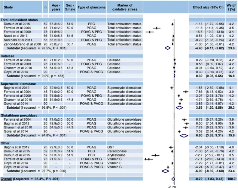
Stratification of antioxidative markers in aqueous humor. Nine studies were included
(Fig 5) [30,33,36,37,38,41,42,44,47]. The overall effect size of serum oxidative stress markers in glaucoma patients compared with controls was non-significant (effect size = –0.70; 95%CI – 1.83 to 0.43, P = 0.227; I2= 98.4%, P < 0.001). However, the effect sizes were significant for total antioxidative status (effect size = –4.40; 95%CI –6.17 to –2.62, P < 0.001; I2= 97.5%, P < 0.001), superoxide dismutase (effect size = 3.53; 95%CI 1.20–5.85, P 0.003; I2= 96.8%, P < 0.001), and glutathione peroxydase (effect size = 6.60; 95%CI 3.88–9.31, P < 0.001; I2= 94.8%, P < 0.001). The effect size of catalase was not significant (effect size = 0.30; 95%CI 0.05–0.55, p = 0.018, I2= 0.0%, P = 0.463).
Funnel plots of previous meta-analyses analyzing for potential publication bias are pre-sented inS5 Fig. For antioxidative stress in both serum and aqueous humor, meta-analyses were reperformed after the exclusion of studies that were not evenly distributed around the base of the funnel and which showed similar results (S6andS7Figs).
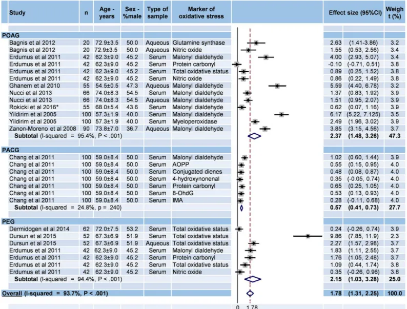
Meta-analysis of oxidative markers for each type of glaucoma
The 2 studies which did not specify the type of glaucoma were excluded.[34,42] Effect sizes were significant for the three types of glaucoma: 2.36 for POAG (95%CI 1.45–3.27, P < 0.001; I2= 96.8%, P < 0.01), 0.55 for PACG (95%CI 0.37–0.72, P < 0.001; I2= 94.8%, P < 0.01), and 2.15 for PEG (95%CI 1.03–3.28, P < 0.001; I2= 97.7%, P < 0.01) (Fig 6).
Metaregressions
Sex. Male glaucoma patients exhibited greater levels of oxidative stress markers in serum
(effect size = 0.94; 95%CI 0.59–1.28, P < 0.001) (Table 1). Levels of oxidative stress markers in aqueous humor and antioxidative stress markers in the serum and aqueous humor of glau-coma patients did not differ significantly between males and females (S1,S2andS3Tables). Fig 3. Meta-analysis of oxidative stress markers from aqueous humor in glaucoma. 95%CI: 95% confidence intervals; -: Unknown; PACG: primary
angle closure glaucoma; PEG: pseudoexfoliation glaucoma; POAG: primary open angle glaucoma; 8 OhdG: 8-hydroxydeoxyguanosin. doi:10.1371/journal.pone.0166915.g003
Age. Levels of oxidative stress markers in the serum of glaucoma patients decreased with
age (effect size = –1.19; 95%CI –1.54 to 0.84, P < 0.001) (Table 1). There was no age effect on the levels of oxidative stress markers in aqueous humor and antioxidative stress markers in the serum and aqueous humor of glaucoma patients (S1,S2andS3Tables)
Fig 4. Meta-analysis of antioxidative stress markers from serum in glaucoma. 95%CI: 95% confidence intervals; -: Unknown; PACG: primary angle
closure glaucoma; PEG: pseudoexfoliation glaucoma; POAG: primary open angle glaucoma; GST: Glutathione S transferase. doi:10.1371/journal.pone.0166915.g004
Markers of oxidative stress. Malonyldialdehyde was higher than other of oxidative stress
markers in the serum of glaucoma patients (effect size = 1.46; 95%CI 0.16–2.77, P = 0.03) (Table 1). Insufficient data precluded further analyses for levels of oxidative stress markers in aqueous humor in glaucoma patients (S1 Table)
Markers of antioxidative stress. No single antioxidative stress marker demonstrated a
greater difference than any other marker in glaucoma patients (S2andS3Tables)
Type of glaucoma. In patients with glaucoma, effect sizes for oxidative and antioxidative
stress markers did not differ significantly between the types of glaucoma (POAG, PACG, and PEG). The differences in serum levels of oxidative stress markers between glaucoma patients and controls were significantly higher in POAG vs PACG (effect size = 12.7; 95%CI 8.78–16.6, P < 0.001), and higher in PEG vs PACG (effect size = 12.2; 95%CI 8.96–15.5, P < 0.001) (Table 1). There were no differences in the levels of oxidative stress markers in aqueous humor Fig 5. Meta-analysis of antioxidative stress markers from aqueous humor in glaucoma. 95%CI: 95% confidence intervals; -: Unknown; PACG:
primary angle closure glaucoma; PEG: pseudoexfoliation glaucoma; POAG: primary open angle glaucoma; GST: Glutathione S transferase. doi:10.1371/journal.pone.0166915.g005
and antioxidative stress markers in the serum and aqueous humor either between patients with different types of glaucoma or between glaucoma patients and controls (S1,S2andS3Tables).
Discussion
The major findings were an overall increase of oxidative stress markers in glaucoma (effect size = 1.64; 95%CI 1.20–2.09), ranging from an effect size of 1.29 in serum (95%CI 0.84–1.74) to 2.62 in aqueous humor (95%CI 1.60–3.65). Malonyldialdehyde seemed the best serum bio-marker of oxidative stress (effect size = 1.46 vs other biobio-markers; 95%CI 0.16–2.77). Despite a decrease in serum antioxidative stress markers (effect size = –0.41; 95%CI –0.72 to –0.11), the level of some aqueous humor antioxidative stress markers increased (superoxide dismutase, effect size = 3.53; 95%CI 1.20–5.85 and glutathione peroxidase, effect size = 6.60; 95%CI 3.88– 9.31), which could be a protective response of the eye against oxidative stress.
Fig 6. Meta-analysis of oxidative stress markers in glaucoma according to the type of glaucoma. 95%CI: 95% confidence intervals; -: Unknown;
PACG: primary angle closure glaucoma; PEG: pseudoexfoliation glaucoma; POAG: primary open angle glaucoma; 8 OhdG: 8-hydroxydeoxyguanosin; AOPP: Advanced oxidation protein product; IMA: Ischemied modified albumin.
Oxidative stress and glaucoma
In our study, we demonstrated an increase in oxidative stress in glaucoma in line with the liter-ature [14,50]. With malonyldialdehyde seeming to be a good marker to evaluate oxidative stress in serum and aqueous humor. Malonyldialdehyde is a product of reactive oxygen species acting on polyunsaturated fat [51]. Evaluation of malonyldialdehyde levels remains a useful indicator of lipid peroxidation linked with oxidative stress [52]. All studies compared the aque-ous humor of glaucoma patients with patients undergoing cataract surgery. Though it has been suggested that cataracts enhance oxidative stress [53,54], our meta-analyses demon-strated significant that the most important effect size for oxidative stress was in aqueous humor. A perturbation of the oxidant/antioxidant balance in the aqueous humor causes an increase in the production of reactive oxygen species which may lead to trabecular meshwork damage [17,55] in predisposed patients [56]. The trabecular meshwork regulates the outflow of aqueous humor via the anterior chamber [57]. However, this structure has the greatest sen-sitivity to the consequences of oxidative stress [14]. Intraocular pressure rises and leads to optic nerve and retinal ganglion cell degeneration, which are already weakened by chronic oxi-dative stress [58].
Antioxidative defense system in glaucoma
We demonstrated a decreased total antioxidant status in serum and aqueous humor in glau-coma. However, the levels of two common antioxidative stress markers, i.e. superoxide dis-mutase and glutathione peroxidase, increased in aqueous humor. This could be a protective response of the eye against oxidative stress [36,59], and may decrease in the long term [37,55]. More interestingly, the trabecular meshwork is a metabolically active tissue containing key Table 1. Meta-regression for oxidative markers in serum.
Covariates Coefficient (95%CI) p-value
Population
Sex (Male as reference) 0.94 (0.59, 1.28) <.001
Age -1.19 (-1.54, 0.84) <.001
Oxidative stress markers
Total oxidative stress vs other ‡ ‡
Malonyldialdehyde vs other 1.46 (0.16, 2.77) .03
Protein carbonyl vs other ‡ ‡
8 OhdG vs other ‡ ‡
Type of glaucoma†
POAG vs PACG 19.2 (-27.3, 65.7) .36
POAG vs PEG 20.2 (-21.5, 62.0) .29
PACG vs PEG 1.04 (-28.1, 26.0) .93
Difference POAG/controls and PACG/controls 12.7 (8.78, 16.6) <.001
Difference POAG/controls and PEG/controls -0.42 (-2.86, 2.00) 0.69 Difference PEG/controls and PACG/controls 12.2 (8.96, 15.5) <.001
95%CI: 95% confidence intervals; PACG: primary angle closure glaucoma; PEG: pseudoexfoliation glaucoma; POAG: primary open angle glaucoma.
†
: Separate models were used to assess all combinations. As coefficient (95%CI) and p-value of other covariates were identical regarding all models, we report all the combinations in the same table in order to avoid duplications.
‡
: Dropped because of collinearity.
enzymes involved in protecting against oxidative stress, in particular the two important enzymes superoxide dismutase and glutathione peroxidase [60]. These two enzymes play a major role by removing the excess H2O2. Despite its toxicity when it exceeds the physiological
values [61], the aqueous humor normally contains H2O2, which is an essential component of
several signal-transduction pathways [62]. The removal of H2O2uses glutathione as a cofactor
and decreases glutathione levels [47]. As glutathione is involved in ascorbic acid metabolism, its depletion produces ascorbyl radicals that cannot be regenerated to ascorbic acid [47]. This might explain the decreased vitamin C levels in serum and aqueous humor found in our meta-analyses. No significant changes were found in the levels of catalase, which could be explain by a change in its absorbance spectrum following its combination with nitric oxide [63]. It has therefore been postulated that some chronic damage in the trabecular meshwork decreases the levels of superoxide dismutase and glutathione peroxidase and leads to increased oxidative stress in the anterior chamber [55].
Type of glaucoma and clinical application
Among the few studies comparing the levels of oxidative or antioxidative stress markers between the different types of glaucoma [35,36,38,40,42], only one study reported a significant difference in malonyldialdehyde serum levels between POAG and PEG [35]. We demonstrated a higher level of oxidative stress markers in patients’ serum compared to controls in POAG vs PACG (effect size = 12.7; 95%CI 8.78–16.6), and in PEG vs PACG (effect size = 12.2; 95%CI 8.96–15.5). Even if glaucoma is a multifactorial disease [7,10], systemic oxidative stress may play an important role in the pathophysiology of POAG and PEG. Despite growing research on antioxidant therapy to prevent glaucoma, in both in vitro [64] and in vivo studies with animals [65] and humans [66], antioxidative supplementation has shown contradictory [66,67,68]. However, there were promising results of supplementation in endothelial function [69], some specific clinical conditions [70] and overall mortality [71]. Therefore, as oxidative stress also contributes to the pathogenesis of systemic clinical conditions [72] and other ocular conditions [73], antioxidative supplementation may not be neglected. Although most hypothe-ses of aging are related to an increase in oxidative stress [74,75,76], we found that levels of oxi-dative stress decreased with aging in glaucoma. Moreover, we found higher levels of oxioxi-dative stress markers in men with glaucoma, compared with women. Even if no study reported this relationship in glaucoma, this finding is in agreement with higher systemic levels of antioxida-tive stress in men in the general population [77,78]. The higher prevalence of glaucoma in men may be linked to increased oxidative stress. These relationships should be further investi-gated, as well as the putative benefits to measure malonyldialdehyde levels in serum in clinical practice.
Limitations
Our study has some limitations. All the studies included were cross-sectional. However, we demonstrated the putative role of oxidative stress in glaucoma. Though there were similarities between the inclusion criteria, they were not identical. In particular for glaucoma patients, some studies did not clearly defined the definition used for glaucoma, and exclusion criteria also differed between studies which may have affected our results. In addition, some controls could have another cause of increased oxidative stress, such as cataract
[30,33,36,37,38,41,42,44,47]. This may have minimized the differences we reported in oxida-tive stress levels between glaucoma participants and controls. Limiting our meta-analysis to studies sharing the same inclusion and exclusion criteria was not feasible due to the limited data. Moreover, all studies were monocentric, limiting the generalizability of our results. Many
markers were reported in only one study precluding further comparisons between markers of oxidative or antioxidative stress levels. However, we demonstrated the potential use of the measure of malonyldialdehyde. Even if several studies assessed the levels of oxidative or anti-oxidative stress within the aqueous humor, it is not necessarily easily applicable in daily clinical practice. For ethical reasons and feasibility, there is no study evaluating in vivo oxidative mark-ers in vitreous, retina and optic nerve. However, our meta-analyses demonstrated the potential for assessing serum oxidative stress levels. Unfortunately, the lack of details surrounding the disease, such as the beginning of glaucoma or the treatment after diagnosis, precluded further analyses.
Conclusion
We demonstrated an overall increase of oxidative stress markers in glaucoma (effect
size = 1.64; 95%CI 1.20–2.09), ranging from an effect size of 1.29 in serum (95%CI 0.84–1.74) to 2.62 in aqueous humor (95%CI 1.60–3.65). Malonyldialdehyde seemed the best biomarkers of oxidative stress in serum. Despite a decrease of antioxidative stress markers in serum, some increased in the aqueous humor which could be a protective response of the eye against oxida-tive stress. Implications of oxidaoxida-tive stress in the pathophysiology of glaucoma and the poten-tially interesting therapeutic approach should be studied further.
Supporting Information
S1 Appendix. PRISMA Checklist.
(DOCX)
S1 Fig. Meta-analysis of oxidative stress markers from serum and aqueous humor in glau-coma. 95%CI: 95% confidence intervals; -: Unknown; PACG: primary angle closure glaucoma;
PEG: pseudoexfoliation glaucoma; POAG: primary open angle glaucoma; 8 OhdG: 8-hydroxy-deoxyguanosin; AOPP: Advanced oxidation protein product; IMA: Ischemied modified albu-min.
(TIF)
S2 Fig. Funnel plots of oxidative stress markers from serum and aqueous humor in glau-coma.
(TIF)
S3 Fig. Meta-analysis of oxidative stress markers from serum in glaucoma after exclusion of studies not evenly distributed around the base of the funnel. 95%CI: 95% confidence
intervals; -: Unknown; PACG: primary angle closure glaucoma; PEG: pseudoexfoliation glau-coma; POAG: primary open angle glauglau-coma; 4NHE: 4-hydroxynonenal; 8 OhdG: 8-hydroxy-deoxyguanosin; AOPP: Advanced oxidation protein product; IMA: Ischemied modified albumin.
(TIF)
S4 Fig. Meta-analysis of antioxidative stress markers from serum and aqueous humor in glaucoma. 95%CI: 95% confidence intervals; -: Unknown; PACG: primary angle closure
glau-coma; PEG: pseudoexfoliation glauglau-coma; POAG: primary open angle glauglau-coma; GST: Gluta-thione S transferase.
(TIF)
S5 Fig. Funnel plots of antioxidative stress markers from serum and aqueous humor in glaucoma.
S6 Fig. Meta-analysis of antioxidative stress markers from serum in glaucoma after exclu-sion of studies not evenly distributed around the base of the funnel. 95%CI: 95% confidence
intervals; -: Unknown; PACG: primary angle closure glaucoma; PEG: pseudoexfoliation glau-coma; POAG: primary open angle glauglau-coma; GPx: Glutathione peroxidase; SOD: Superoxide dismutase; TAS: Total antioxidant status.
(TIF)
S7 Fig. Meta-analysis of antioxidative stress markers from aqueous in glaucoma after exclusion of studies not evenly distributed around the base of the funnel. 95%CI: 95%
con-fidence intervals; -: Unknown; PACG: primary angle closure glaucoma; PEG: pseudoexfolia-tion glaucoma; POAG: primary open angle glaucoma; GPx: Glutathione peroxidase; GST: Glutathione S transferase; SOD: Superoxide dismutase; TAS: Total antioxidant status. (TIF)
S1 Table. Meta-regression for oxidative markers in aqueous humor. 95%CI: 95% confidence
intervals; PACG: primary angle closure glaucoma; PEG: pseudoexfoliation glaucoma; POAG: primary open angle glaucoma.
(DOCX)
S2 Table. Meta-regression for antioxidative markers in serum. 95%CI: 95% confidence
intervals; PACG: primary angle closure glaucoma; PEG: pseudoexfoliation glaucoma; POAG: primary open angle glaucoma.
(DOCX)
S3 Table. Meta-regression for antioxidative markers in aqueous humor. 95%CI: 95%
confi-dence intervals; PACG: primary angle closure glaucoma; PEG: pseudoexfoliation glaucoma; POAG: primary open angle glaucoma.
(DOCX)
Acknowledgments
The authors received no specific funding for this work.
Author Contributions
Conceptualization: FD. Data curation: FD CBDA. Formal analysis: FD BP. Investigation: FD CBDA. Methodology: FD. Project administration: FD. Resources: FD CBDA. Software: FD. Supervision: FD. Validation: FD BP. Visualization: FD.
Writing – original draft: FD CBDA.
Writing – review & editing: FD CBDA BP FC.
References
1. Quigley HA, Broman AT (2006) The number of people with glaucoma worldwide in 2010 and 2020. Br J Ophthalmol 90: 262–267. doi:10.1136/bjo.2005.081224PMID:16488940
2. Bourne RR, Stevens GA, White RA, Smith JL, Flaxman SR, Price H, et al. (2013) Causes of vision loss worldwide, 1990–2010: a systematic analysis. Lancet Glob Health 1: e339–349. doi: 10.1016/S2214-109X(13)70113-XPMID:25104599
3. Hirooka K, Sato S, Nitta E, Tsujikawa A (2016) The Relationship Between Vision-related Quality of Life and Visual Function in Glaucoma Patients. J Glaucoma.
4. Schmidtmann G, Jahnke S, Seidel EJ, Sickenberger W, Grein HJ (2011) Intraocular pressure fluctua-tions in professional brass and woodwind musicians during common playing condifluctua-tions. Graefes Arch Clin Exp Ophthalmol 249: 895–901. doi:10.1007/s00417-010-1600-xPMID:21234587
5. Chua J, Baskaran M, Ong PG, Zheng Y, Wong TY, Aung T, et al. (2015) Prevalence, Risk Factors, and Visual Features of Undiagnosed Glaucoma: The Singapore Epidemiology of Eye Diseases Study. JAMA Ophthalmol 133: 938–946. doi:10.1001/jamaophthalmol.2015.1478PMID:26043441 6. Fingert JH (2011) Primary open-angle glaucoma genes. Eye (Lond) 25: 587–595.
7. Le A, Mukesh BN, McCarty CA, Taylor HR (2003) Risk factors associated with the incidence of open-angle glaucoma: the visual impairment project. Invest Ophthalmol Vis Sci 44: 3783–3789. PMID:12939292 8. Marcus MW, de Vries MM, Junoy Montolio FG, Jansonius NM (2011) Myopia as a risk factor for
open-angle glaucoma: a systematic review and meta-analysis. Ophthalmology 118: 1989–1994 e1982. doi:
10.1016/j.ophtha.2011.03.012PMID:21684603
9. Racette L, Wilson MR, Zangwill LM, Weinreb RN, Sample PA (2003) Primary open-angle glaucoma in blacks: a review. Surv Ophthalmol 48: 295–313. PMID:12745004
10. Thomas R, Walland MJ (2011) Primary angle-closure glaucoma is a multifactorial disease. Clin Experi-ment Ophthalmol 39: 593–594. doi:10.1111/j.1442-9071.2011.02663.xPMID:22452677
11. Sacca SC, Pulliero A, Izzotti A (2015) The dysfunction of the trabecular meshwork during glaucoma course. J Cell Physiol 230: 510–525. doi:10.1002/jcp.24826PMID:25216121
12. Grover AK, Samson SE (2014) Antioxidants and vision health: facts and fiction. Mol Cell Biochem 388: 173–183. doi:10.1007/s11010-013-1908-zPMID:24311110
13. Pryor WA, Godber SS (1991) Noninvasive measures of oxidative stress status in humans. Free Radic Biol Med 10: 177–184. PMID:1650736
14. Izzotti A, Bagnis A, Sacca SC (2006) The role of oxidative stress in glaucoma. Mutat Res 612: 105– 114. doi:10.1016/j.mrrev.2005.11.001PMID:16413223
15. Aslan M, Cort A, Yucel I (2008) Oxidative and nitrative stress markers in glaucoma. Free Radic Biol Med 45: 367–376. doi:10.1016/j.freeradbiomed.2008.04.026PMID:18489911
16. Kumar DM, Agarwal N (2007) Oxidative stress in glaucoma: a burden of evidence. J Glaucoma 16: 334–343. doi:10.1097/01.ijg.0000243480.67532.1bPMID:17438430
17. Zhao J, Wang S, Zhong W, Yang B, Sun L, Zheng Y (2016) Oxidative stress in the trabecular meshwork (Review). Int J Mol Med 38: 995–1002. doi:10.3892/ijmm.2016.2714PMID:27572245
18. da Costa BR, Cevallos M, Altman DG, Rutjes AW, Egger M (2011) Uses and misuses of the STROBE statement: bibliographic study. BMJ Open 1: e000048. doi:10.1136/bmjopen-2010-000048PMID:
22021739
19. Vandenbroucke JP, von Elm E, Altman DG, Gotzsche PC, Mulrow CD, Pocock SJ, et al. (2007) Strengthening the Reporting of Observational Studies in Epidemiology (STROBE): explanation and elaboration. Annals of Internal Medicine 147: W163–194. PMID:17938389
20. Ollier M Jr., Chamoux A, Naughton G, Pereira B, Dutheil F (2014) Chest computed tomography screen-ing for lung cancer in asbestos occupational exposure: a systematic review and meta-analysis. Chest 145: 1339–1346. doi:10.1378/chest.13-2181PMID:24480869
21. Benoist d’Azy C, Pereira B, Naughton G, Chiambaretta F, Dutheil F (2016) Antibioprophylaxis in Pre-vention of Endophthalmitis in Intravitreal Injection: A Systematic Review and Meta-Analysis. PLoS One 11: e0156431. doi:10.1371/journal.pone.0156431PMID:27257676
22. Courtin R, Pereira B, Naughton G, Chamoux A, Chiambaretta F, Lanhers C, et al. (2016) Prevalence of dry eye disease in visual display terminal workers: a systematic review and meta-analysis. BMJ Open 6: e009675. doi:10.1136/bmjopen-2015-009675PMID:26769784
23. Lanhers C, Pereira B, Naughton G, Trousselard M, Lesage FX, Dutheil F (2015) Creatine Supplementa-tion and Lower Limb Strength Performance: A Systematic Review and Meta-Analyses. Sports Medicine 45: 1285–1294. doi:10.1007/s40279-015-0337-4PMID:25946994
24. DerSimonian R, Laird N (1986) Meta-analysis in clinical trials. Controlled Clinical Trials 7: 177–188. PMID:3802833
25. Citrome L (2014) Paging Dr Cohen, Paging Dr Cohen. . .An effect size interpretation is required STAT!: Visualising effect size and an interview with Kristoffer Magnusson. International Journal of Clinical Prac-tice 68: 533–534. doi:10.1111/ijcp.12435PMID:24750523
26. Russo MW (2007) How to Review a Meta-analysis. Gastroenterol Hepatol (N Y) 3: 637–642.
27. Abu-Amero KK, Azad TA, Mousa A, Osman EA, Sultan T, Al-Obeidan SA (2014) Total antioxidant level is correlated with intra-ocular pressure in patients with primary angle closure glaucoma. BMC Res Notes 7: 163. doi:10.1186/1756-0500-7-163PMID:24646376
28. Abu-Amero KK, Kondkar AA, Mousa A, Osman EA, Al-Obeidan SA (2011) Decreased total antioxidants status in the plasma of patients with pseudoexfoliation glaucoma. Mol Vis 17: 2769–2775. PMID:
22065931
29. Abu-Amero KK, Kondkar AA, Mousa A, Osman EA, Al-Obeidan SA (2013) Decreased total antioxidants in patients with primary open angle glaucoma. Curr Eye Res 38: 959–964. doi:10.3109/02713683. 2013.794246PMID:23651069
30. Bagnis A, Izzotti A, Centofanti M, Sacca SC (2012) Aqueous humor oxidative stress proteomic levels in primary open angle glaucoma. Exp Eye Res 103: 55–62. doi:10.1016/j.exer.2012.07.011PMID:
22974818
31. Chang D, Sha Q, Zhang X, Liu P, Rong S, Han T, et al. (2011) The evaluation of the oxidative stress parameters in patients with primary angle-closure glaucoma. PLoS One 6: e27218. doi:10.1371/ journal.pone.0027218PMID:22096540
32. Demirdogen BC, Ceylan OM, Isikoglu S, Mumcuoglu T, Erel O (2014) Evaluation of oxidative stress and paraoxonase phenotypes in pseudoexfoliation syndrome and pseudoexfoliation glaucoma. Clin Lab 60: 79–86. PMID:24600979
33. Dursun F, Vural Ozec A, Aydin H, Topalkara A, Dursun A, Toker MI, et al. (2015) Total oxidative stress, paraoxonase and arylesterase levels at patients with pseudoexfoliation syndrome and pseudoexfolia-tive glaucoma. Int J Ophthalmol 8: 985–990. doi:10.3980/j.issn.2222-3959.2015.05.24PMID:
26558214
34. Engin KN, Yemisci B, Yigit U, Agachan A, Coskun C (2010) Variability of serum oxidative stress bio-markers relative to biochemical data and clinical parameters of glaucoma patients. Mol Vis 16: 1260– 1271. PMID:20664701
35. Erdurmus M, Yagci R, Atis O, Karadag R, Akbas A, Hepsen IF (2011) Antioxidant status and oxidative stress in primary open angle glaucoma and pseudoexfoliative glaucoma. Curr Eye Res 36: 713–718. doi:10.3109/02713683.2011.584370PMID:21780920
36. Ferreira SM, Lerner SF, Brunzini R, Evelson PA, Llesuy SF (2004) Oxidative stress markers in aqueous humor of glaucoma patients. Am J Ophthalmol 137: 62–69. PMID:14700645
37. Ghanem AA, Arafa LF, El-Baz A (2010) Oxidative stress markers in patients with primary open-angle glaucoma. Curr Eye Res 35: 295–301. doi:10.3109/02713680903548970PMID:20373896 38. Goyal A, Srivastava A, Sihota R, Kaur J (2014) Evaluation of oxidative stress markers in aqueous
humor of primary open angle glaucoma and primary angle closure glaucoma patients. Curr Eye Res 39: 823–829. doi:10.3109/02713683.2011.556299PMID:24912005
39. Majsterek I, Malinowska K, Stanczyk M, Kowalski M, Blaszczyk J, Kurowska AK, et al. (2011) Evalua-tion of oxidative stress markers in pathogenesis of primary open-angle glaucoma. Exp Mol Pathol 90: 231–237. doi:10.1016/j.yexmp.2011.01.001PMID:21241689
40. Mousa A, Kondkar AA, Al-Obeidan SA, Azad TA, Sultan T, Osman E, et al. (2015) Association of total antioxidants level with glaucoma type and severity. Saudi Med J 36: 671–677. doi:10.15537/smj.2015. 6.10697PMID:25987108
41. Nucci C, Di Pierro D, Varesi C, Ciuffoletti E, Russo R, Gentile R, et al. (2013) Increased malondialde-hyde concentration and reduced total antioxidant capacity in aqueous humor and blood samples from patients with glaucoma. Mol Vis 19: 1841–1846. PMID:23946639
42. Sorkhabi R, Ghorbanihaghjo A, Javadzadeh A, Rashtchizadeh N, Moharrery M (2011) Oxidative DNA damage and total antioxidant status in glaucoma patients. Mol Vis 17: 41–46. PMID:21245957 43. Yildirim O, Ates NA, Ercan B, Muslu N, Unlu A, Tamer L, et al. (2005) Role of oxidative stress enzymes
44. Zanon-Moreno V, Marco-Ventura P, Lleo-Perez A, Pons-Vazquez S, Garcia-Medina JJ, Vinuesa-Silva I, et al. (2008) Oxidative stress in primary open-angle glaucoma. J Glaucoma 17: 263–268. doi:10. 1097/IJG.0b013e31815c3a7fPMID:18552610
45. Awodele O, Oreagba IA, Olayemi SO, Oladipo I, Iruegbukpe CO, Balogun BG, et al. (2015) Evaluation and Comparison of the Indices of Systemic Oxidative Stress among Black-Africans with Age-related Cataracts or Primary Glaucoma. Middle East Afr J Ophthalmol 22: 489–494. doi:10.4103/0974-9233. 167811PMID:26692723
46. Rokicki W, Zalejska-Fiolka J, Pojda-Wilczek D, Kabiesz A, Majewski W (2016) Oxidative stress in the red blood cellsof patients with primary open-angle glaucoma. Clin Hemorheol Microcirc.
47. Ferreira SM, Lerner SF, Brunzini R, Evelson PA, Llesuy SF (2009) Antioxidant status in the aqueous humour of patients with glaucoma associated with exfoliation syndrome. Eye (Lond) 23: 1691–1697.
48. Tanito M, Kaidzu S, Takai Y, Ohira A (2012) Status of systemic oxidative stresses in patients with pri-mary open-angle glaucoma and pseudoexfoliation syndrome. PLoS One 7: e49680. doi:10.1371/ journal.pone.0049680PMID:23189153
49. Xu L, Chen JH, Li JJ, Luo L, Yang H, Zhang RX, et al. (2004) [The prevalence and its screening meth-ods of primary open angle glaucoma in defined population-based study of rural and urban in Beijing]. Zhonghua Yan Ke Za Zhi 40: 726–732. PMID:15634477
50. Sacca SC, Izzotti A, Rossi P, Traverso C (2007) Glaucomatous outflow pathway and oxidative stress. Exp Eye Res 84: 389–399. doi:10.1016/j.exer.2006.10.008PMID:17196589
51. Pryor WA, Stanley JP (1975) Letter: A suggested mechanism for the production of malonaldehyde dur-ing the autoxidation of polyunsaturated fatty acids. Nonenzymatic production of prostaglandin endoper-oxides during autoxidation. J Org Chem 40: 3615–3617. PMID:1185332
52. Srour MA, Bilto YY, Juma M (2000) Evaluation of different methods used to measure malonyldialdehyde in human erythrocytes. Clin Hemorheol Microcirc 23: 23–30. PMID:11214710
53. Ottonello S, Foroni C, Carta A, Petrucco S, Maraini G (2000) Oxidative stress and age-related cataract. Ophthalmologica 214: 78–85. PMID:10657746
54. Borchman D, Yappert MC (1998) Age-related lipid oxidation in human lenses. Invest Ophthalmol Vis Sci 39: 1053–1058. PMID:9579487
55. Nita M, Grzybowski A (2016) The Role of the Reactive Oxygen Species and Oxidative Stress in the Pathomechanism of the Age-Related Ocular Diseases and Other Pathologies of the Anterior and Poste-rior Eye Segments in Adults. Oxid Med Cell Longev 2016: 3164734. doi:10.1155/2016/3164734PMID:
26881021
56. Wiggs JL (2015) Glaucoma Genes and Mechanisms. Prog Mol Biol Transl Sci 134: 315–342. doi:10. 1016/bs.pmbts.2015.04.008PMID:26310163
57. Alvarado JA, Alvarado RG, Yeh RF, Franse-Carman L, Marcellino GR, Brownstein MJ (2005) A new insight into the cellular regulation of aqueous outflow: how trabecular meshwork endothelial cells drive a mechanism that regulates the permeability of Schlemm’s canal endothelial cells. Br J Ophthalmol 89: 1500–1505. doi:10.1136/bjo.2005.081307PMID:16234461
58. Sacca SC, Izzotti A (2008) Oxidative stress and glaucoma: injury in the anterior segment of the eye. Prog Brain Res 173: 385–407. doi:10.1016/S0079-6123(08)01127-8PMID:18929123
59. Kim KY, Perkins GA, Shim MS, Bushong E, Alcasid N, Ju S, et al. (2015) DRP1 inhibition rescues retinal ganglion cells and their axons by preserving mitochondrial integrity in a mouse model of glaucoma. Cell Death Dis 6: e1839. doi:10.1038/cddis.2015.180PMID:26247724
60. De La Paz MA, Epstein DL (1996) Effect of age on superoxide dismutase activity of human trabecular meshwork. Invest Ophthalmol Vis Sci 37: 1849–1853. PMID:8759353
61. Spector A (1995) Oxidative stress-induced cataract: mechanism of action. FASEB J 9: 1173–1182. PMID:7672510
62. Trachootham D, Lu W, Ogasawara MA, Nilsa RD, Huang P (2008) Redox regulation of cell survival. Antioxid Redox Signal 10: 1343–1374. doi:10.1089/ars.2007.1957PMID:18522489
63. Brown GC (1995) Reversible binding and inhibition of catalase by nitric oxide. Eur J Biochem 232: 188– 191. PMID:7556149
64. Xu P, Lin Y, Porter K, Liton PB (2014) Ascorbic acid modulation of iron homeostasis and lysosomal function in trabecular meshwork cells. J Ocul Pharmacol Ther 30: 246–253. doi:10.1089/jop.2013. 0183PMID:24552277
65. Ozdemir G, Tolun FI, Gul M, Imrek S (2009) Retinal oxidative stress induced by intraocular hyperten-sion in rats may be ameliorated by brimonidine treatment and N-acetyl cysteine supplementation. J Glaucoma 18: 662–665. doi:10.1097/IJG.0b013e31819c46b1PMID:20010244
66. Garcia-Medina JJ, Garcia-Medina M, Garrido-Fernandez P, Galvan-Espinosa J, Garcia-Maturana C, Zanon-Moreno V, et al. (2015) A two-year follow-up of oral antioxidant supplementation in primary open-angle glaucoma: an open-label, randomized, controlled trial. Acta Ophthalmol 93: 546–554. doi:
10.1111/aos.12629PMID:25545196
67. Giaconi JA, Yu F, Stone KL, Pedula KL, Ensrud KE, Cauley JA, et al. (2012) The association of con-sumption of fruits/vegetables with decreased risk of glaucoma among older African-American women in the study of osteoporotic fractures. Am J Ophthalmol 154: 635–644. doi:10.1016/j.ajo.2012.03.048
PMID:22818906
68. Wang SY, Singh K, Lin SC (2013) Glaucoma and vitamins A, C, and E supplement intake and serum levels in a population-based sample of the United States. Eye (Lond) 27: 487–494.
69. Ashor AW, Siervo M, Lara J, Oggioni C, Afshar S, Mathers JC (2015) Effect of vitamin C and vitamin E supplementation on endothelial function: a systematic review and meta-analysis of randomised con-trolled trials. Br J Nutr 113: 1182–1194. doi:10.1017/S0007114515000227PMID:25919436 70. Ji HF, Sun Y, Shen L (2014) Effect of vitamin E supplementation on aminotransferase levels in patients
with NAFLD, NASH, and CHC: results from a meta-analysis. Nutrition 30: 986–991. doi:10.1016/j.nut. 2014.01.016PMID:24976430
71. Jiang S, Pan Z, Li H, Li F, Song Y, Qiu Y (2014) Meta-analysis: low-dose intake of vitamin E combined with other vitamins or minerals may decrease all-cause mortality. J Nutr Sci Vitaminol (Tokyo) 60: 194– 205.
72. Giustarini D, Dalle-Donne I, Tsikas D, Rossi R (2009) Oxidative stress and human diseases: Origin, link, measurement, mechanisms, and biomarkers. Crit Rev Clin Lab Sci 46: 241–281. doi:10.3109/ 10408360903142326PMID:19958214
73. Kruk J, Kubasik-Kladna K, Aboul-Enein HY (2015) The Role Oxidative Stress in the Pathogenesis of Eye Diseases: Current Status and a Dual Role of Physical Activity. Mini Rev Med Chem 16: 241–257. PMID:26586128
74. Schottker B, Saum KU, Jansen EH, Boffetta P, Trichopoulou A, Holleczek B, et al. (2015) Oxidative stress markers and all-cause mortality at older age: a population-based cohort study. J Gerontol A Biol Sci Med Sci 70: 518–524. doi:10.1093/gerona/glu111PMID:25070660
75. Chung HY, Cesari M, Anton S, Marzetti E, Giovannini S, Seo AY, et al. (2009) Molecular inflammation: underpinnings of aging and age-related diseases. Ageing Res Rev 8: 18–30. doi:10.1016/j.arr.2008. 07.002PMID:18692159
76. Salminen A, Ojala J, Kaarniranta K, Kauppinen A (2012) Mitochondrial dysfunction and oxidative stress activate inflammasomes: impact on the aging process and age-related diseases. Cell Mol Life Sci 69: 2999–3013. doi:10.1007/s00018-012-0962-0PMID:22446749
77. Morales RC, Bahnson ES, Havelka GE, Cantu-Medellin N, Kelley EE, Kibbe MR (2015) Sex-based dif-ferential regulation of oxidative stress in the vasculature by nitric oxide. Redox Biol 4: 226–233. doi:10. 1016/j.redox.2015.01.007PMID:25617803
78. Miller AA, De Silva TM, Jackman KA, Sobey CG (2007) Effect of gender and sex hormones on vascular oxidative stress. Clin Exp Pharmacol Physiol 34: 1037–1043. doi:10.1111/j.1440-1681.2007.04732.x