HAL Id: hal-03013448
https://hal.archives-ouvertes.fr/hal-03013448
Submitted on 18 Nov 2020
HAL is a multi-disciplinary open access
archive for the deposit and dissemination of
sci-entific research documents, whether they are
pub-lished or not. The documents may come from
teaching and research institutions in France or
abroad, or from public or private research centers.
L’archive ouverte pluridisciplinaire HAL, est
destinée au dépôt et à la diffusion de documents
scientifiques de niveau recherche, publiés ou non,
émanant des établissements d’enseignement et de
recherche français ou étrangers, des laboratoires
publics ou privés.
Daiki Mori, Claude Gregoire, Guillaume Voisinne, Javier Celis-Gutierrez, Rudy Aussel, et al.. The
T cell CD6 receptor operates a multitask signalosome with opposite functions in T cell activation.
Journal of Experimental Medicine, Rockefeller University Press, 2020, 218, �10.1084/jem.20201011�.
�hal-03013448�
ARTICLE
The T cell CD6 receptor operates a multitask
signalosome with opposite functions in T cell
activation
Daiki Mori1,2*, Claude Gr´egoire1*, Guillaume Voisinne1, Javier Celis-Gutierrez1,2, Rudy Aussel1, Laura Girard1,2, Mylène Camus3, Marlène Marcellin3, J´er´emy Argenty1, Odile Burlet-Schiltz3, Fr´ed´eric Fiore2, Anne Gonzalez de Peredo3, Marie Malissen1,2, Romain Roncagalli1, and Bernard Malissen1,2
To determine the respective contribution of the LAT transmembrane adaptor and CD5 and CD6 transmembrane receptors to
early TCR signal propagation, diversification, and termination, we describe a CRISPR/Cas9
–based platform that uses primary
mouse T cells and permits establishment of the composition of their LAT, CD5, and CD6 signalosomes in only 4 mo using
quantitative mass spectrometry. We confirmed that positive and negative functions can be solely assigned to the LAT and CD5
signalosomes, respectively. In contrast, the TCR-inducible CD6 signalosome comprised both positive (SLP-76, ZAP70, VAV1) and
negative (UBASH3A/STS-2) regulators of T cell activation. Moreover, CD6 associated independently of TCR engagement to
proteins that support its implication in inflammatory pathologies necessitating T cell transendothelial migration. The
multifaceted role of CD6 unveiled here accounts for past difficulties in classifying it as a coinhibitor or costimulator. Congruent
with our identification of UBASH3A within the CD6 signalosome and the view that CD6 constitutes a promising target for
autoimmune disease treatment, single-nucleotide polymorphisms associated with human autoimmune diseases have been
found in the Cd6 and Ubash3a genes.
Introduction
Following TCR triggering, the LAT transmembrane adaptor as-sembles a multimolecular signaling complex known as the LAT signalosome (Balagopalan et al., 2010). Although the LAT sig-nalosome ensures the propagation and diversification of TCR signals, it does not work in isolation, and other T cell surface receptors regulate early T cell activation. Among them stand CD5 and CD6, which belong to the scavenger receptor cysteine-rich superfamily and constitute paralogs that extensively diverged (Gaud et al., 2018;Padilla et al., 2000). Upon TCR-induced tyrosine phosphorylation, CD5 and CD6 assemble poorly defined signalosomes (Burgess et al., 1992;Wee et al., 1993) independently of LAT and with kinetics and in numbers comparable to those of the canonical LAT signalosome (Roncagalli et al., 2014;Voisinne et al., 2019). It thus remains to determine the composition of the LAT, CD5, and CD6 signal-osomes in primary T cells and quantify their respective con-tributions to early TCR signal propagation and termination.
CD5 is expressed on all T cells and on a B cell subset (Brown and Lacey, 2010). On T cells, it colocalizes with the TCR at the
immunological synapse (IS) and negatively regulates TCR sig-nals in response to foreign peptides bound to MHC molecules (Azzam et al., 2001;Brossard et al., 2003;Peña-Rossi et al., 1999;
Tarakhovsky et al., 1995). Although high CD5 expression levels on naive T cells have been correlated with high TCR self-reactivity, whether CD5 also limits TCR self-reactivity remains to be determined (Hogquist and Jameson, 2014). The mechanism used by CD5 to inhibit TCR signaling remains incompletely de-fined (Burgueño-Bucio et al., 2019). Recent data suggest that CD5 constitutes the main T cell–surface receptor capable of recruit-ing the E3 ubiquitin-protein ligases CBL and CBLB in response to TCR stimulation, thereby promoting ubiquitylation of colo-calized signaling effectors (Voisinne et al., 2016).
CD6 is expressed on T cells and recognizes CD166 (also known as Activated Leukocyte Cell Adhesion Molecule [ALCAM];
Chappell et al., 2015) and CD318 (Enyindah-Asonye et al., 2017). The CD6–ALCAM interaction is important for IS stabilization and sustained TCR-induced cell proliferation (Meddens et al., 2018; Zimmerman et al., 2006). Upon TCR triggering, CD6
...
1Centre d’Immunologie de Marseille-Luminy, Aix Marseille Universit´e, Institut National de la Sant´e et de la Recherche M´edicale, Centre National de la Recherche Scientifique,
Marseille, France; 2Centre d’Immunoph´enomique, Aix Marseille Universit´e, Institut National de la Sant´e et de la Recherche M´edicale, Centre National de la Recherche
Scientifique, Marseille, France; 3Institut de Pharmacologie et de Biologie Structurale, Universit´e de Toulouse, Centre National de la Recherche Scientifique, Universit´e Paul
Sabatier, Toulouse, France.
*D. Mori and C. Gr´egoire contributed equally to this paper; Correspondence to Bernard Malissen:bernardm@ciml.univ-mrs.fr.
© 2020 Mori et al. This article is available under a Creative Commons License (Attribution 4.0 International, as described athttps://creativecommons.org/licenses/by/4.0/).
(“the preys”) assembling around proteins (“the baits”) of the TCR-signaling network (Roncagalli et al., 2014). Combining the resulting“interactomes” with the interaction stoichiometry and cellular abundance of the interacting proteins provides quanti-tative parameters for systems-level understanding of TCR signal propagation and diversification (Voisinne et al., 2019). Gene-targeted mice in which T cell proteins are tagged with an affinity Twin-Strep-tag (OST;Junttila et al., 2005) permit gen-eration of primary T cells expressing physiological levels of signalosomes that are amenable to AP-MS (Voisinne et al., 2019). Considering that it takes up to a year to obtain such mice, we describe here a faster approach that uses primary mouse T cells and permits establishment of the composition and dynamics of signalosomes of interest in 4 mo. Accordingly, we improved recent CRISPR/Cas9–based platforms for editing primary T cells (Anderson et al., 2019;Nüssing et al., 2020;Roth et al., 2018) to enable monitoring at the single-cell level that the OST tag is properly inserted in the gene of interest and to sort the low frequency of properly edited T cells before subjecting them to AP-MS analysis. We further used this“fast-track” approach to determine the composition, stoichiometry, and dynamics of the CD5, CD6, and LAT signalosomes that assemble in primary T cells following TCR engagement, and we compared such sig-nalosomes with the transcriptional and functional outcomes resulting from TCR activation of primary T cells lacking LAT, CD5, or CD6.
Results
Primary mouse T cells amenable to fast-track interactomics We used the Lat gene as a proof of concept to develop a CRISPR/ Cas9–based approach permitting determination of the compo-sition of signalosomes assembling in primary CD4+T cells before and after TCR engagement. Our approach preserved cell viability while permitting monitoring at the single-cell level that the OST tag required for AP-MS analysis was properly inserted at the 39 end of at least one allele of the targeted gene. Moreover, it al-lowed sorting of the low frequency of properly OST-edited T cells before subjecting them to in vitro expansion and AP-MS analysis. Accordingly, we designed a single-guide RNA (sgRNA) targeting the 39 coding end of the Lat gene (Fig. 1 Aand Table S1) and a 843-bp-long double-stranded DNA (dsDNA)
6 mice express the CD90.2 allele, permitting ready identification 4 d after nucleofection of the presence of 1.45% ± 1.69% (n = 5) of CD90.1+T cells that retained normal TCRβ and CD4 levels (Fig. 1
C). CD90.1+ T cells were FACS-sorted 5 d after nucleofection (Fig. 1 D) and expanded, and their genomic DNA was analyzed by PCR using primer-pair combinations, permitting validation of the intended HDR (Fig. 1 E). Analysis of the size of the amplicons straddling the 59 and 39 insertion borders showed that HDR occurred correctly (Fig. 1 F), a finding corroborated via DNA sequencing (Fig. S1, C and D). Therefore, CD90.1 expression can be used as a surrogate marker permitting identification and sorting of primary CD4+T cells with a properly inserted OST tag. Edited primary CD4+T cells express LAT molecules suitable for AP-MS
The sorted CD90.1+ CD4+ T cells were expanded for approxi-mately 2 mo to reach substantial cell numbers required for AP-MS and were subsequently denoted as long-term–expanded T cells. WT CD4+T cells were expanded in parallel and used as a control. Immunoblot analysis showed that WT and CD90.1+ OST-edited CD4+T cells expressed comparable levels of LAT and that the addition of the OST sequence resulted, as expected, in LATOSTmolecules with a higher molecular weight than that of WT CD4+T cells (Fig. 1 G). We analyzed next whether the LATOST molecules expressed by the expanded CD90.1+CD4+T cells were amenable to AP-MS. After stimulation with CD3 plus anti-CD4 for 30 and 120 s, the modification introduced in the Lat gene was without measurable effect on the global pattern of TCR-induced tyrosine phosphorylation (Fig. 1 H). LATOST bait pro-teins were efficiently purified using Sepharose beads coupled to Strep-Tactin (Fig. 1 I, lower panel) and underwent TCR-induced tyrosine phosphorylation that peaked 30 s after stimulation (Fig. 1 I, upper panel). As expected, no detectable material was recovered from WT CD4+ T cells. Therefore, upon edition of their genome and expansion in vitro, primary LATOST CD4+ T cells can be used for AP-MS analysis of the LAT signalosome. The LAT interactome of CRISPR/Cas9–edited and long-term–expanded primary CD4+T cells
Long-term–expanded LATOST-expressing CD4+ T cells were lysed with the nonionic detergent n-dodecyl-β-maltoside before or after cross-linkage of the TCR and CD4 molecules for 30 and
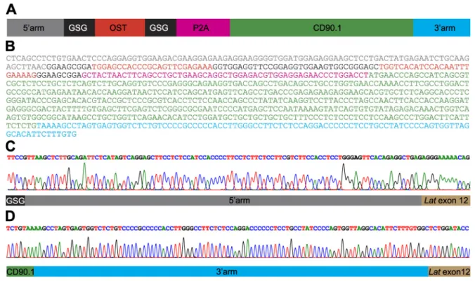
Figure 1. Mouse primary CD4+T cells amenable to fast-track AP-MS characterization of the LAT signalosome. (A) An sgRNA was designed to introduce
a double-strand break 12 bp 39 of the stop codon found in the last exon of the Lat gene, and an 843-bp-long dsDNA was used as a template for HDR (seeFig. S1, A and B). Following HDR, CD4+T cells are expected to coexpress LATOSTand CD90.1 molecules. (B) Workflow used for editing, selecting, and expanding CD4+
T cells expressing LATOSTmolecules amenable to AP-MS. (C) T cells were analyzed for expression of CD90.1 3 d after nucleofection with vehicle alone (None),
sgRNA, or sgRNA plus the HDR template (sgRNA + template). Also shown is the expression of CD4 and TCRβ on CD5+CD90.1+T cells. (D) Sorted CD90.1+CD4+
T cells and WT CD4+T cells were expanded in vitro and analyzed for expression of CD90.1 before AP-MS analysis. Data in C and D are representative of at least
three experiments. (E) PCR genotyping schematics of sorted CD90.1+CD4+T cells expressing LATOSTmolecules. The two specified PCR primer pairs provide
diagnostic bands for each junction. Correct targeting was further confirmed by sequencing the 59 and 39 junction fragments (Fig. S1, C and D). Also shown are the expected sizes of the PCR amplicons. (F) PCR genotyping was performed on WT CD4+T cells (Ctrl) and on sorted CD90.1+CD4+T cells (LATOST) using the
PCR primer pairs specified in E. (G) Immunoblot analysis of equal amounts of proteins from WT and LATOSTCD4+T cell lysates that were either directly
analyzed (Total lysate) or subjected to affinity purification on Strep-Tactin Sepharose beads followed by elution of proteins with D-biotin (Pull down), and both were probed with antibody to anti-LAT or anti-VAV1 (loading control). The long-term expanded LATOSTCD4+T cells showed an even representation of WT and
LATOSTalleles. (H) Immunoblot analysis of equal amounts of proteins from total lysates of WT and LATOSTCD4+T cells left unstimulated or stimulated for 30 s
or 120 s with anti-CD3 and anti-CD4 and probed with antibody to phosphorylated tyrosine (Anti-p-Tyr) or anti-VAV1 (loading control). (I) Immunoblot analysis of equal amounts of lysates of WT and of LATOSTCD4+T cells stimulated as in H and subjected to affinity purification on Strep-Tactin Sepharose beads,
followed by elution of proteins with D-biotin, and probed with antibody to phosphorylated tyrosine (Anti-p-Tyr) or anti-LAT. Left margin in G–I, molecular size in kilodaltons. Data in H and I are representative of at least two independent experiments. PAM, protospacer-adjacent motif. GSG, Gly-Ser-Gly spacer.
second step, we identified among the 10 high-confidence LAT preys those whose interaction stoichiometry changed at least twofold with a P value below 0.05 following TCR plus CD4 stimulation, compared with the nonstimulated condition (Fig. 2 B). All 10 preys passed this second step and corresponded to the cytosolic adaptors SLP-76, GRB2, GRAP, GRAP2, and THEMIS, the protein tyrosine kinase (PTK) ITK, the phospholipase PLC-γ1, the phosphatidylinositol 3,4,5-trisphosphate (PI(3,4,5)P3) 5-phosphatase 1 SHIP1, the guanine nucleotide exchange factor SOS1, and the serine–threonine protein kinase MAP4K1 (HPK1;
Fig. 2 B). The 10 TCR-inducible preys showed a transient pattern of binding to LAT that peaked 30 s after stimulation (Fig. 2 C). By combining the cellular abundances of the protein expressed in long-term–expanded CD4+ T cells (Fig. S2 and Data S1) and in-teraction stoichiometries (Data S2), the LAT signalosome was or-ganized into a“stoichiometry plot” (Fig. 2 D;Voisinne et al., 2019). Accordingly, for each documented LAT–prey interaction, the ratio of bait to prey cellular abundance was plotted as a function of the maximal interaction stoichiometry reached by the considered bait–prey interaction over the course of TCR stimulation (Fig. 2 D). It showed, for instance, that 37% of the pool of SOS1 proteins available in CD4+T cells was mobilized to interact with LAT 30 s after TCR engagement. Therefore, our approach identified in 4 mo the quantitative composition and dynamics of the LAT signal-osome of primary T cells before and following TCR engagement. The CD6 interactome of CRISPR/Cas9–edited and long-term–expanded primary CD4+T cells
Next, we determined the CD6 interactome of long-term– expanded CD4+ T cells edited to express OST-tagged CD6 proteins (Fig. S3, A–C). The appended OST had no detectable effect on CD6 expression (Fig. S3 D) or on the global pattern of TCR-induced tyrosine phosphorylation (Fig. S3 I). Immuno-blot analysis of WT and of CD6OST-expressing CD4+ T cells showed that CD6OSTmolecules were efficiently purified with Strep-Tactin (Fig. S3 J, lower panel). After stimulation with anti-CD3 and anti-CD4, tyrosine phosphorylation of CD6OST molecules reached a maximum 30 s after stimulation and led to their association with tyrosine phosphorylated species (Fig. S3 J, upper panel). The protein complexes that assembled around CD6OSTbait proteins were identified by AP-MS and analyzed as described for the LATOST bait. 18 interacting
distribution than that of the LAT–prey interactions (Fig. 3 D). The previously unreported CD6–UBASH3A and CD6–GRK6 in-teractions were validated by coimmunoprecipitation of proteins in primary CD6OSTCD4+T cells (Fig. 3 E). Consistent with our AP-MS results, UBASH3A interacted with CD6 upon TCR stim-ulation, whereas GRK6 disassembled in part from CD6 upon TCR engagement. Due to its expression on activated CD4+T cells and micromolar affinity for CD6 (Hassan et al., 2004), ALCAM in-teracted with CD6 over all the tested conditions and is thus part of the high-confidence constitutive CD6 interactors (Data S2). Likewise, the RHO GTPase–activating protein ARHGAP45 and theα subunit of casein kinase II (CSNK2A1) were also part of the high-confidence constitutive CD6 interactors (Data S2). The presence of CSNK2A1 supports a former study suggesting that serine residues within the CD6 cytoplasmic segment are con-stitutively phosphorylated by CSNK2 (Bonet et al., 2013). Therefore, the composition and dynamics of the CD6 signal-osome revealed a complexity higher than expected.
The CD5 interactome of short-term–expanded CD4+T cells To identify the composition of the CD5 interactome of primary T cells, we benefited from gene-targeted mice that were avail-able at the onset of our study and that expressed an OST at the C terminus of endogenous CD5 molecules (Fig. S4). Purified pri-mary CD4+T cells from mice expressing endogenous CD5 mol-ecules tagged with an OST tag (CD5OST) were expanded for 4 d in vitro to reach the cell numbers required for AP-MS and are subsequently referred to as short-term expanded. The protein complexes assembling around CD5OSTbait proteins were iden-tified by AP-MS and analyzed as described for LATOST and CD6OST baits. 173 interacting proteins showed a >10-fold en-richment, with a P value below 0.005 in at least one of the four conditions of stimulation (Fig. 4 Aand Data S2). Among them, four interactors showed interaction stoichiometry that in-creased at least twofold, with a P value below 0.05 following CD3 plus CD4 cross-linkage, compared with the nonstimulated con-dition (Fig. 4 B). They comprised the ankyrin repeat domain–
containing protein 13A (ANKRD13A), UBASH3A, and CBLB (Fig. 4, C and D). After the fold enrichment value was slightly relaxed, CD5 was also found to interact with CBL (Data S2), a finding consistent with the reciprocal presence of CD5 among high-confidence CBL interactors (Voisinne et al., 2019).
Function of primary T cells lacking CD5 or CD6 and doubly deficient for CD5 and CD6
The markedly different composition of the CD5 and CD6 sig-nalosomes led us to analyze in parallel and on a homogenous genetic background the functional consequences resulting from their respective ablation. Accordingly, Cd5−/−mice (Tarakhovsky
et al., 1995) were backcrossed on a C57BL/6 background, and we developed C57BL/6 mice lacking CD6 (Cd6−/−) and both CD5 and CD6 (Cd5−/−Cd6−/−;Fig. 5 A). Deletion of CD5 or CD6 had few effects on the selection of thymocytes expressing the polyclonal TCR repertoire (Azzam et al., 2001;Orta-Mascaró et al., 2016). Along that line, the thymus of Cd5−/−, Cd6−/−, and Cd5−/−Cd6−/−
Figure 2. Composition and dynamics of the LAT signalosome of long-term–expanded LATOST-expressing CD4+T cells. (A) Volcano plot showing
proteins significantly enriched after affinity purification in CD4+T cells expressing LATOSTmolecules compared with affinity purification in control CD4+T cells
expressing similar levels of WT (untagged) LAT proteins before (t0s) and at 30 s (t30s) and 120 s (t120s) after TCR plus CD4 stimulation. (B) Volcano plot showing
proteins significantly enriched after affinity purification in LATOST-expressing CD4+T cells 30 and 120 s after TCR engagement compared with affinity
pu-rification in unstimulated LATOST-expressing CD4+T cells. In A and B, the SLP-76, GRB2, GRAP, GRAP2, THEMIS, ITK, PLC-γ1, SHIP1, SOS1, and MAP4K1
proteins are shown in red, and the x and y axes show the average fold change (in log10scale) in protein abundance and the statistical significance, respectively.
(C) Dot plot showing the interaction stoichiometry of LAT with its 10 high-confidence preys over the course of TCR stimulation. For each LAT–prey interaction, the interaction stoichiometry has been row-normalized to its maximum value observed over the course of TCR stimulation (see normalized stoichiometry key). The 10 preys showed maximal binding to LAT after 30 s of activation. Also shown is the P value of the specified interactions (see P value key). (D) Stoichiometry plot of the LAT interactome. The LAT bait is specified by a yellow dot, and its 10 preys are shown as red dots. For each of these LAT–prey interactions, the ratio of prey to bait cellular abundance (“abundance stoichiometry” in log10scale) was plotted as a function of the maximal interaction stoichiometry reached by the
considered LAT-prey interaction over the course of TCR stimulation (“interaction stoichiometry” in log10scale). For instance, LAT (41,443 copies per T cell;
column G of the LAT tab in Data S2) is more abundant than SOS1 (2,562 copies per T cell), giving a ratio of prey to bait cellular abundance of−2.2 in log10scale.
Moreover, the maximum stoichiometry of the LAT–SOS1 interaction is reached at t30sand corresponds to 0.023 (−1.6 in log10scale; column D of the LAT tab in
Data S2). Therefore, 953 (41,443 × 0.023) molecules of LAT are complexed to SOS1 at t30s.As a result, 37% (953/2,562 × 100) of the available SOS1 proteins are
complexed to LAT 30 s after TCR engagement. The area including the LAT–prey interaction involving >10% of the available prey molecules is indicated in light gray and includes SOS1, GRB2, and GRAP. The limit imposed on interaction stoichiometries by the relative LAT–prey cellular abundances is shown by a dashed diagonal line that delimits a“forbidden” area (dark gray). Prey dot size is commensurate to its maximal protein enrichment over the course of stimulation.
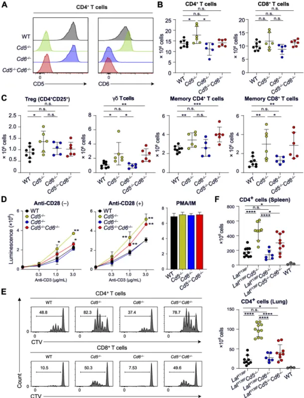
mice showed a normal sequence of T cell development, and slight differences were only noted in the frequency of CD69+CD4+CD8+ cells in the Cd5−/−and Cd6−/−thymus com-pared with the WT thymus (not depicted). The cellularity of the secondary lymphoid organs of Cd5−/−, Cd6−/−, and Cd5−/−Cd6−/−mice was comparable to that of WT counterparts, and their T cells expressed normal levels of CD3, CD4, and CD8 (Fig. 5 B and not depicted). Slightly increased numbers of CD4+ T cells, regulatory T cells (T reg cells),γδ T cells, and memory T cells were found in the secondary lymphoid organs of Cd5−/−mice (Fig. 5 C).
T cells from Cd5−/−, Cd6−/−, Cd5−/−Cd6−/−, and WT mice were activated in vitro with plate-bound anti-CD3 in the presence or absence of anti-CD28. The lack of CD6 had no measurable effect on either TCR- or TCR plus CD28–induced proliferation (Fig. 5 D). In contrast, the lack of CD5 enhanced proliferation at all the tested CD3 concentrations and regardless of CD28 en-gagement (Fig. 5 D). Comparison of Cd5−/−and Cd5−/−Cd6−/−mice showed that the enhanced proliferation resulting from the lack of CD5 was diminished by the absence of CD6. These conclusions were confirmed using the division tracking dye CellTrace Violet (CTV;Fig. 5 E). Deleting CD5 in primary T cells isolated from WT
Figure 3. Composition and dynamics of the CD6 signalosome of long-term expanded CD6OSTCD4+T cells. (A) Volcano plot showing proteins
signif-icantly enriched after affinity purification in CD4+T cells expressing CD6OSTmolecules compared with affinity purification in control CD4+T cells expressing
similar levels of WT (untagged) CD6 proteins before (t0s) and at 30 s (t30s) and 120 s (t120s) after TCR plus CD4 stimulation. (B) Volcano plot showing proteins
significantly enriched after affinity purification in CD6OST-expressing CD4+T cells 30 and 120 s after TCR engagement compared with affinity purification in
unstimulated CD6OST-expressing CD4+T cells. For A and B, see description inFig. 2 B. (C) Dot plot showing the interaction stoichiometry over the course of
TCR stimulation of CD6 with its seven high-confidence preys, the interaction stoichiometry of which changed following TCR engagement. See description in
Fig. 2 C. (D) Stoichiometry plot of the CD6 interactome. The CD6 bait is shown as a yellow dot. Red and blue dots correspond to preys that showed increased or decreased binding following TCR stimulation. The purple dot corresponds to a prey whose association was not regulated by TCR stimulation. See description in
Fig. 2 D. (E) Biochemical validation of the CD6–UBASH3A and CD6–GRK6 interactions predicted on the basis of AP-MS analysis. Long-term–expanded WT (Control) and CD6OST-expressing CD4+T cells were left unstimulated (0) or were stimulated for 30 and 120 s with anti-CD3 and anti-CD4 antibodies and
subsequently lysed. Equal amounts of cell lysates (Total lysate) were immunoblotted with antibodies specific for UBASH3A and GRK6. Equal amounts of lysates were subjected to AP on Strep-Tactin Sepharose beads (Pull down), followed by elution with D-biotin. Eluates were immunoblotted and probed with antibodies specific for UBASH3A and GRK6. Molecular masses are shown. Data are representative of at least two experiments.
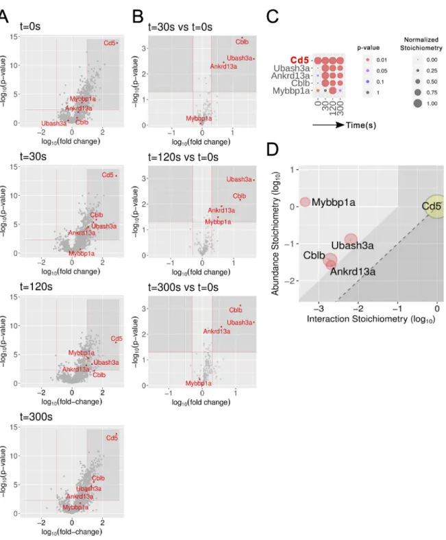
Figure 4. Composition and dynamics of the CD5 interactome of short-term–expanded CD4+T cells from CD5OSTmice. (A) Volcano plot showing
proteins significantly enriched after affinity purification in CD4+T cells expressing CD5OSTmolecules compared with affinity purification in control CD4+T cells
expressing similar levels of WT (untagged) CD5 proteins before (t0s) and at 30 s (t30s), 120 s (t120s), and 300 s (t300s) after TCR plus CD4 stimulation. (B) Volcano
plot showing proteins significantly enriched after affinity purification in CD5OST-expressing CD4+T cells 30, 120, and 300 s after TCR engagement compared
with affinity purification in unstimulated CD5OST-expressing CD4+T cells. For A and B, see description inFig. 2 B. (C) The dot plot shows the interaction
stoichiometry over the course of TCR stimulation of CD5 with its four high-confidence interactors, the interaction stoichiometry of which changed following TCR engagement. See description inFig. 2 C. (D) Stoichiometry plot of the CD5 interactome. It shows that hundreds of copies of CD5–CBLB, CD5–UBASH3A, and CD5–ANKRD13A complexes formed per T cell as early as 30 s of TCR stimulation, mobilizing close to 10% of the cellular pool of ANKRD13A, CBLB, and UBASH3A. The CD5 bait is shown as a yellow dot. Red dots correspond to preys that show increased binding following TCR stimulation. See description in
Fig. 2 D. Mybbp1a; MYB-binding protein 1A.
Figure 5. T cell development and function in CD5-CD6 doubly deficient mice compared with mice lacking either CD5 or CD6. (A) Expression of CD5 and CD6 on CD4+T cells from WT mice and from mice lacking either CD5 (Cd5−/−) or CD6 (Cd6−/−) and both CD5 and CD6 (Cd5−/−Cd6−/−) analyzed by flow
cy-tometry. Note that Cd6−/−T cells expressed levels of CD5 comparable to that of WT mice, whereas increased levels of CD6 were found on Cd5−/−T cells as reported (Orta-Mascaró et al., 2016). (B) Numbers of CD4+and CD8+T cells found in the spleen of the specified mice. (C) Numbers of T reg cells,γδ T cells, and
effector memory CD4+and CD8+T cells found in the spleen of the specified mice. (D) T cells were purified by immunomagnetic negative selection from lymph
nodes of Cd5−/−, Cd6−/−, Cd5−/−Cd6−/−, and WT mice activated in vitro with the specified concentrations of plate-bound anti-CD3 in the absence (−) or presence
(+) of soluble anti-CD28 (1 µg/ml). After 48 h of culture, ATP content was assessed using luminescence as a measure of the extent of cell proliferation. The histogram on the right shows the extent of cell proliferation in response to PMA plus ionomycin (PMA/IM). (E) Profiles of CTV-labeled CD4+(upper panel) and
CD8+(lower panel) T cells isolated from the specified mice and stimulated with anti-CD3 plus anti-CD28 antibodies for 72 h. The percentages of proliferating
CTVlowT cells are indicated. (F) Numbers of CD4+T cells found in the spleen and lungs of WT, LatY136F, LatY136FCd5−/−, LatY136FCd6−/−, and LatY136FCd5−/−Cd6−/−
mice. Data in A–E are representative of at least three independent experiments, whereas data in F correspond to the pool of three independent experiments. In B and C, each dot corresponds to a mouse, and the mean (horizontal bar) is indicated. n.s., nonsignificant; *, P≤ 0.05; **, P ≤ 0.01; ***, P ≤ 0.001; ****, P ≤ 0.0001. Error bars correspond to the mean and SD.
mice enhanced their TCR-induced proliferation compared with that of control cells, whereas CD6 deletion had no measurable effect (Fig. S5). Therefore, the enhanced TCR responses of T cells isolated from Cd5−/−mice did not result from adaptive mecha-nisms set in motion during T cell development to compensate for the lack of CD5.
Mutant mice in which tyrosine at position 136 of LAT is re-placed with phenylalanine (LatY136Fmice) develop a lympho-proliferative disorder involving CD4+T cell effectors that trigger systemic autoimmunity (Aguado et al., 2002). The lack of CD6 was found without measurable effect on the unfolding of the LatY136Fpathology, whereas the lack of CD5 exacerbated it, re-sulting in 2.4-fold increased numbers of pathogenic CD4+T cells in the spleen (Fig. 5 F). In the absence of CD5 and CD6, the numbers of pathogenic LatY136FCD4+ T cells ranked between those observed in Cd5−/−and WT mice. Dense perivascular in-filtrates consisting of pathogenic CD4+T cells are present in the lungs of LatY136F mice (Genton et al., 2006). Although Cd6−/− T cells showed impaired infiltration through brain microvas-cular endothelial cell monolayers (Li et al., 2017), the lack of CD6 was without effect on the magnitude of the lung infiltrate, whereas that of CD5 resulted in fourfold-increased numbers of infiltrating cells (Fig. 5 F). In the absence of CD5 and CD6, in-filtrating CD4+T cell numbers ranked between those observed in Cd5−/−and WT mice. Altogether, these results showed that the absence of CD5 rendered T cells more reactive to TCR and CD28 triggering and increased the numbers of pathogenic LatY136F T cells in both lymphoid and nonlymphoid organs. Therefore, CD5 acts as a negative regulator of T cell activation regardless of the presence of CD6. In contrast, it is only in the context of a CD5 deficiency that we succeeded in documenting a net CD6 cos-timulatory effect (Fig. 5, D–F).
LAT differs from CD5 and CD6 in that it triggers most TCR/ CD28–induced transcription-dependent events
To compare the respective contribution of LAT, CD5, and CD6 with the transcriptional changes elicited by TCR-CD28 stimu-lation, WT, Lat−/−, Cd5−/−, Cd6−/−, and Cd5−/−Cd6−/−naive CD4+ T cells were sorted and subjected to RNA-sequencing analysis before or after stimulation for 20 h with CD3 and anti-CD28. Using principal component analysis (PCA), TCR-CD28 activated Cd5−/−, Cd6−/−, and Cd5−/−Cd6−/−CD4+T cells clustered with TCR-CD28–stimulated WT cells (Fig. 6 A), suggesting that the bulk of TCR-CD28–induced transcriptional changes occurred unabated in the absence of CD5, CD6, or both CD5 and CD6. In contrast, anti-CD3 plus anti-CD28 activated Lat−/−CD4+T cells clustered close to unstimulated WT and Lat−/− CD4+ T cells (Fig. 6 B), indicating that the absence of LAT blunted most TCR-CD28–induced transcriptional events, as previously hinted at using microarray analysis (Roncagalli et al., 2014). Consistent with PCA, the number of genes differentially expressed (fold change >2 and adjusted P value <0.05) between unstimulated and TCR-CD28–stimulated Cd5−/−, Cd6−/−, and Cd5−/−Cd6−/−CD4+ T cells was comparable to that of WT CD4+T cells (Fig. 6 Cand Data S3). In contrast, the number of genes differentially ex-pressed between unstimulated and TCR-CD28–stimulated cells was dramatically decreased in Lat−/−CD4+T cells compared with
WT CD4+ T cells (Fig. 6 Dand Data S3). Therefore, our tran-scriptomics results are consistent with the functional outcomes resulting from the ablation of LAT, CD5, and CD6, in that LAT-deficient T cells failed to proliferate and produce cytokines in response to TCR-CD28 stimulation (Mingueneau et al., 2009) and thus markedly differ from T cells deprived of CD5 and CD6, which responded comparably (CD6−/−) or slightly better (CD5−/−) than WT T cells did (Fig. 5).
Inefficient assembly of high-order LAT signalosomes
The global picture of the LAT signalosome obtained using LAT as a bait (Fig. 7 A) is fully consistent with the“reciprocal” picture inferred from the use of 15 baits corresponding to canonical proteins of the TCR-signaling pathway (Voisinne et al., 2019), and it supports the LAT model obtained by addressing one in-teractor at a time (Balagopalan et al., 2010). However, the pos-sibility of enumerating the number of each LAT–prey interaction that forms per T cell provided an unprecedented quantitative picture of the LAT signalosome (Fig. 7 Aand Data S2). Among the GRB2 protein family, GRAP formed 4,973 in-teractions with LAT 30 s after TCR stimulation, compared with 13,261 and 2,486 for GRB2 and GRAP2, respectively. Although GRAP has received less attention than GRB2 and GRAP2 in the context of LAT (Trüb et al., 1997), its important quantitative contribution to the LAT signalosome emphasizes the pressing need to determine the constellation of molecules it plugs into the LAT signalosome via its SH3 domains. LAT and SLP-76 interact via GRAP2 intermediate, whereas LAT–THEMIS, LAT–SOS1, and LAT–SHIP1 interactions occur through GRB2 intermediate. Prior to TCR engagement, GRAP2–SLP76, GRB2–SHIP1, GRB2–SOS1, and GRB2–THEMIS form stable binary complexes, leaving a large fraction of GRAP2 and GRB2 molecules in a“free” form or in complex with unknown partners (Voisinne et al., 2019). When the LAT signalosome reached its maximal interaction stoichiometry, the sum of the LAT–SLP-76, LAT–SHIP1, LAT– SOS1, and LAT–THEMIS interactions was fourfold smaller than that of LAT–GRAP2 and LAT–GRB2 interactions (Fig. 7 A). The possibility that GRAP2 and GRB2 combine with unknown part-ners might explain the fourfold excess of LAT–GRAP2 and LAT–GRB2 interactions over LAT–THEMIS, LAT–SOS1, and LAT–SHIP1 interactions. However, owing to the advanced in-ventory of GRAP2 and GRB2 interactors, such excess is more likely due to the competition that exists between the GRAP2– SLP-76, GRB2–SHIP1, GRB2–SOS1, and GRB2–THEMIS com-plexes and the larger pool of free GRAP2 and GRB2 adaptors for binding to phosphorylated LAT molecules. Therefore, the for-mation of a larger number of abortive or partially functional LAT signalosomes involving uncomplexed members of the GRB2 protein family likely accompanied that of higher-order and fully functional LAT signalosomes. Interestingly, the maximum numbers of LAT–SLP-76 and LAT–SHIP1 interactions that were reached 30 s after TCR engagement corresponded to 269 and 2,279 copies per T cell, respectively (Fig. 7 A), suggesting that a majority of SHIP-containing LAT signalosomes lack SLP-76 and pointing to the existence of several LAT signalosome isoforms that can further assemble into condensates (Houtman et al., 2006;Huang et al., 2016;Malissen et al., 2014).
Quantitative model of the CD5 signalosome
In the absence of GRB2 in the CD5 interactome (Data S2), CBLB likely binds to tyrosine-phosphorylated CD5 molecules via its tyrosine kinase–binding domain. Upon colocalization with the TCR at IS (Brossard et al., 2003), CD5-bound CBLB molecules are tyrosine phosphorylated by TCR-operated PTK. In turn, their RING-type zinc finger domain becomes available for binding to E2 ubiquitin–conjugating enzymes, leading to K63 ubiquitylation of neighboring substrates that may include CD5 and CBLB them-selves (Demydenko, 2010;Voisinne et al., 2016). The K63 ubiq-uitylated complex assembling around the CD5-CBLB seed allows the recruitment of ANKRD13A via its K63-specific ubiquitin-interacting motifs (Fig. 7 A). ANKRD13A regulates endocytosis of K63-ubiquitylated forms of the epidermal growth factor receptor (Tanno et al., 2012) and of the B cell antigen receptor (Satpathy et al., 2015) through its interaction with the endocytic machinery. Along the same line, ANKRD13A might contribute to the endocytosis of CD5-CBLB–containing vesicles. In addition to its
E3 ubiquitin–protein ligase activity, CBLB possesses numerous interactive elements, including a proline-rich region that con-stitutively binds the SH3 domain of UBASH3A (Feshchenko et al., 2004;Ge et al., 2019;Voisinne et al., 2019) and likely ac-counts in part for the recruitment of UBASH3A to the TCR-induced CD5 signalosome. T cells deficient for both UBASH3A and its UBASH3B paralog (also known as STS-1 or TULA-2) are hyper-responsive to TCR engagement, a finding consistent with their increased ZAP70 activity (Carpino et al., 2004). Although UBASH3A possesses reduced phosphatase activity compared with that of UBASH3B, its negative regulatory function may involve its SH3 and UBA domains (San Luis et al., 2011). Further support for an inhibitory function of UBASH3A in primary T cell activation was recently provided by a study demonstrating that upon association with the CIN85 adaptor, UBASH3A was recruited to TCR microclusters and inhibited their signals (Kong et al., 2019). Therefore, our identification of UBASH3A within the TCR-induced CD5
Figure 6. Transcriptional changes occurring in primary CD4+T cells after engaging the TCR–CD28 pathways in the absence of LAT, of CD5, of CD6, or
of both CD5 and CD6. (A) PCA of gene expression in WT, Cd5−/−, Cd6−/−, and Cd5−/−Cd6−/−primary CD4+T cells that were left unstimulated (0 h) or stimulated
for 20 h with anti-CD3 plus anti-CD28. (B) PCA of gene expression in WT and Lat−/−primary CD4+T cells that were left unstimulated (0 h) or stimulated for
20 h with anti-CD3 plus anti-CD28 (20 h). (C) Quantification of the genes significantly induced (UP) or repressed (DOWN) in WT, Cd5−/−, Cd6−/−, and Cd5−/−Cd6−/−CD4+T cells after stimulation for 20 h with anti-CD3 plus anti-CD28. (D) Quantification of the genes significantly induced (UP) or repressed
(DOWN) in WT and Lat−/−CD4+T cells after stimulation for 20 h with anti-CD3 plus anti-CD28. See Data S3 for details on the induced or repressed genes
quantified in C and D. In A–D, three independent samples were prepared for each condition. In A and B, numbers shown in parentheses on the PCA1 and PCA2 axes indicate the percentage of overall variability in the dataset along each PC axis. Lat−/−CD4+T cells were obtained using Latfl-dtrmaT-Cre mice (Roncagalli
signalosome of primary T cells in addition to that of CBLB further explains its negative regulatory function.
Quantitative model of the CD6 signalosome
Two types of CD6–prey interactions can be distinguished among the CD6 signalosome. Some existed before TCR engagement and persisted (ARHGAP45) or decreased (GRK6) over 120 s of stim-ulation, whereas others occurred in a TCR-inducible manner. Among these last interactions, a first set involved interactors (ZAP70, SLP-76, and VAV1) endowed with positive regulatory functions. ZAP70 is mandatory for the LAT-independent, TCR-inducible assembly of the CD6 signalosome (Roncagalli et al., 2014) and, together with VAV1, likely contributes to IS stabili-zation via integrin activation (Jenkins et al., 2014; Meddens et al., 2018;Roncagalli et al., 2014;Zimmerman et al., 2006). The adaptors GRB2, GRAP2, and SLP-76 also associated with CD6 in a TCR-inducible manner, likely contributing to recruitment of VAV1 and SHIP1. A second set of TCR-inducible CD6 interactors (UBASH3A and SHIP1) is endowed with negative regulatory functions. By catalyzing the hydrolysis of the phosphatidylino-sitol 3-kinase product PI(3,4,5)P3 into PI(3,4)P2, SHIP1 nega-tively regulates positive effectors with pleckstrin homology domain specific for PI(3,4,5)P3. Therefore, negative regula-tory functions can also be conferred to CD6 via its interaction via SHIP1. However, this view needs to be mitigated since SHIP1 can also set in action positive effectors containing a pleckstrin homology domain specific for PI(3,4)P2, including protein kinase B (AKT) isoforms (Liu et al., 2018). Impor-tantly, among the documented CD6–prey interactions, the
CD6–UBASH3A interaction ranked first in terms of numbers of copies per T cell (Fig. 7 A). Therefore, in contrast to the CD5 signalosome, the CD6 signalosome comprised TCR-inducible interactors endowed with both positive and negative func-tions, the dominance of which likely depends on the immune response being measured (Gonçalves et al., 2018). However, our AP-MS analysis does not permit exclusion of the existence of distinct CD6 signalosome isoforms with either a negative or a positive regulatory role.
Discussion
We developed a fast-track interactomics approach permitting assessment of the composition and dynamics of signalosomes assembling in primary T cells in response to TCR stimulation. Here, we focused on the LAT, CD5, and CD6 signalosomes since they coincidently assemble with fast kinetics following TCR engagement (Voisinne et al., 2019). We demonstrated that SLP-76 is part of the CD6 and LAT signalosomes, a finding implying that our former use of SLP-76 as a bait led to the copurification of both CD6 and LAT signalosomes (Voisinne et al., 2019). Based on the present study, SLP-76 interactors such as UBASH3A and PLC-γ1 can be assigned to the CD6 and LAT signalosomes, re-spectively, whereas VAV1 belongs to both of them. Therefore, disentangling the intricacy of the signal-transduction networks involved in T cell activation and of the underlying signalosomes requires conducting an AP-MS analysis at multiple entry points corresponding to both T cell surface receptors and their cytosolic effectors. Note that SLP-76 differently exploits its scaffolding
Figure 7. A quantitative model of early TCR signal diversification integrating inter-actomics and transcriptomics. (A) Tyrosine residues (red dots) present in the intra-cytoplasmic segments of CD5, CD6, and LAT are phosphorylated by the LCK or ZAP70 PTK that associate with active TCR (dashed yellow arrows). Following 30 s of TCR engagement, distinct sig-nalosomes nucleated around the tyrosine phos-phorylated CD5 and CD6 transmembrane receptors and the LAT transmembrane adaptor. The number of copies per T cell of CD5, CD6, and LAT is indi-cated. For instance, 41,443 copies of LAT are ex-pressed per T cell. The maximum copies per T cell of high-confidence bait–prey complexes reached over the course of TCR stimulation is also shown and specified over the arrows connecting the baits and the preys. For instance, the maximum number of CD5–UBASH3A complexes reached per T cell over the course of TCR stimulation is ∼989 (seeFig. 2 Dlegend and Data S2). Solid arrows correspond to TCR-induced interac-tions, whereas dotted arrows correspond to constitutive interactions. Consistent with for-mer studies (Roncagalli et al., 2014;Voisinne et al., 2019), after cutoff values were slightly relaxed, VAV1 was found to interact significantly and in a TCR-inducible manner with both CD6 (5.0-fold enrichment; P value: 3.2 × 10⁻⁵) and LAT
(8.5-fold enrichment; P value: 4.8 × 10⁻⁶), reaching in both cases maximum binding 30 s after TCR engagement (Data S2). CD5 showed a transient binding
with MYB-binding protein 1A (MYBBP1A), a transcriptional regulator that shuttles between the cytoplasm and the nucleus, and the role within the CD5 interactome remains to be determined. Inset: Lifetime of the TCR-induced interactions involving CD5, CD6, and LAT with most of their interactors. (B) The Venn diagram illustrates the commonalities and differences between the CD5, CD6, and LAT signalosomes as well as the functional outcomes expected to result from their engagement at the IS.
LAT–SLP-76 interactions were fully disrupted and further showed that LAT itself was stripped of most of its other in-teractors. Consistent with previous studies (Hashimoto-Tane and Saito, 2016), the LAT signalosomes that are present 30 s after TCR engagement are thus almost gone 90 s later. The present study also allowed analysis of the extent of proteome remodeling that occurs during long-term CD4+T cell expan-sion. Comparing the fraction of cell mass occupied by the proteins quantified in short-term– and long-term–expanded CD4+T cells showed that the fraction of cell mass occupied by 79% of the quantified proteins differed by less than fourfold between short-term– and long-term–expanded CD4+ T cells (Fig. S2).
The numbers of CD6–SLP-76 and LAT–SLP-76 interactions can be used as a proxy of active CD6 and LAT signalosomes, respectively. These numbers peaked 30 s after TCR engagement and differed by approximately twofold. Despite this rather similar quantitative contribution to TCR signal propagation, our transcriptomics analysis demonstrated that LAT markedly differed from CD6 in that it mediated most of the TCR-CD28–induced transcriptional responses, a finding that corre-lates with the presence of SOS1 and PLC-γ1 in and only in the LAT signalosome and, in turn, with their ability to plug into the ERK and NFAT pathways, respectively (Fig. 7 B). Considering that LAT signalosomes are mostly dismantled 120 s after TCR engagement, it is paradoxical that the CD5–CBLB–UBASH3A– ANKRD13A inhibitory complex persisted up to 300 s after TCR engagement. Provided that the antigen-recognition and trig-gering module made of TCR-CD3, LCK, and ZAP70 delivers longer lasting signals than LAT signalosomes (Yudushkin and Vale, 2010), this module might constitute the primary target of the CD5–CBLB–UBASH3A complex (Hu et al., 2016;Huang et al., 2010;Ivanova and Carpino, 2016;Yang et al., 2015), thereby preventing further formation of LAT signalosomes. Although the molecular targets of the TCR-inducible CD5–CBLB–UBASH3A multimolecular complex remain to be determined, the obser-vation that CD4+ T cells deficient in CD5 (Tarakhovsky et al.,
1995; this study), CBLB (Chiang et al., 2007;Paolino et al., 2011), or UBASH3A (Kong et al., 2019) showed enhanced TCR re-sponses strongly supports the negative regulatory function of the CD5–CBLB–UBASH3A signalosome during TCR-mediated T cell activation.
where it regulates chemotaxis of T and B cells in TEM assays (Fong et al., 2002), and ARHGAP45 is a RHO GTPase–activating protein also involved in TEM (de Kreuk et al., 2013;Voisinne et al., 2019). Therefore, the presence of GRK6 and ARHGAP45 in the CD6 interactome irrespective of TCR engagement might explain the role of CD6 in immunopathologies involving TEM and some of the benefits resulting from clinical trials targeting CD6 (Consuegra-Fern´andez et al., 2018).
In conclusion, using a fast-track systems-level approach we retrieved quantitative information on the multiple TCR-dependent signaling processes that unfold simultaneously at the inner face of the plasma membrane of primary T cells. We provided a quantitative model describing the multitude of signals delivered via LAT molecules expressed at physiological levels in primary T cells following TCR engagement and ex-plaining its unique role in ensuing transcriptional activation. T cell–surface receptors are generally categorized as cos-timulators or coinhibitors. Along that line, the CD5 signalosome of primary T cells comprised molecules involved in signal ter-mination and routing through the endocytic pathway and can thus be unambiguously defined as a T cell coinhibitor. In con-trast, the CD6 signalosome comprised effectors involved in TEM as well as positive and negative regulators of T cell acti-vation. Finally, it remains to be determined whether the in-triguing presence of UBASH3A within the CD6 signalosome provides a molecular basis for the observation that single-nucleotide polymorphisms within both CD6 and UBASH3A genes are associated with autoimmune diseases (De Jager et al., 2009;Ge et al., 2019).
Materials and methods
Mice
Mice were on a C57BL/6 background, were 8–10 wk old, and were maintained in specific pathogen–free conditions. Mice constitutively expressing Cas9 (Platt et al., 2014) were pur-chased from the Jackson Laboratory (Gt(ROSA)26Sortm1.1(CAG-cas9*,-EGFP)Fezh/J; JAX stock no. 024858). LatY136Fand Latfl-dtr mice (B6;129-Lattm6Mal) have been described (Aguado et al., 2002;
Mingueneau et al., 2009). CD5-deficient mice (Tarakhovsky et al., 1995) backcrossed on a C57BL/6 background were provided by F. Lozano (Universitat de Barcelona, Barcelona, Spain). Generation of
CD6-deficient mice (C57BL/6JRj-Cd6em1Ciphe), CD5-CD6 doubly de-ficient mice (C57BL/6JRj-Del(Chr19 Cd5-Cd6)em1Ciphe), and CD5OST (C57BL/6NRj-Cd5tm1Ciphe) mice is described below.
Animal experimental guidelines
Mice were handled in accordance with national and European laws for laboratory animal welfare and experimentation (Eu-ropean Economic Community Council Directive 2010/63/EU, September 2010) and protocols approved by the Marseille Eth-ical Committee for Animal Experimentation.
Generation of CD6-deficient mice and of CD5-CD6 doubly deficient mice
Mice doubly deficient in CD5 and CD6 were constructed by using two pairs of sgRNA (Thermo Fisher Scientific) targeting the 59 end of exon 1 of the Cd5 gene (59-CCATGGACTCCCACGAAGTGC TG-39) and the beginning of the intron flanking the 39 end of exon 1 of the Cd6 gene (59-CCATCGGTGGCACAACCGTCTCC-39). Both were designed using the Crispor algorithm (Haeussler et al., 2016). One-cell C57BL/6JRj embryos at day 0.5 (Janvier Labs) were defrosted and subjected to pronuclear microin-jection with the two sgRNA pairs (each used at 5 ng/ml) and 10 ng/ml Cas9 RNA (Thermo Fisher Scientific) in DNase- and RNase-free micro-injection buffer (10 mM Tris and 1 mM EDTA, pH 8; Qiagen). Injected embryos were transferred into the oviducts of day 0.5 pseudopregnant B6/CBA recipient female mice. Genomic DNA from F0 mice was prepared from tail tissue using a solution composed of 100 mM Tris-HCl, pH 7.5, 5 mM EDTA, 200 mM NaCl, 0.2% SDS, 0.5 mg/ml pro-teinase K, and 50 µg/ml RNaseA and analyzed by PCR using a QIAxcel Advanced System. F0 mice with the expected mu-tation were further crossed, and the exact boundary of the intended deletion was determined via PCR amplification and sequencing using 100 ng of genomic DNA. A deletion ex-tending from nucleotide 10738791 to nucleotide 10829931 of chromosome 19 (https://www.ensembl.org/Mus_musculus/) was selected and used to establish mice deprived of both CD5 and CD6. These Cd5−/−Cd6−/−mice (also known as C57BL/6JRj-Del(Chr19 Cd5-Cd6)em1Ciphe) were born at expected Mendelian frequencies and lacked detectable CD5 and CD6 expression (Fig. 5 A). Mice deficient for CD6 were generated as de-scribed above for Cd5−/−Cd6−/−mice. Two sgRNAs were de-signed to target the 59 untranslated region (UTR; 59-CCATCG GTGGCACAACCGTCTCC-39) and the start codon (59-CCA GACATGTGGCTCTTCCTTGG-39) of the Cd6 gene. A resulting 170-bp-long deletion extending from nucleotide 10829689 to nucleotide 10829860 of chromosome 19 (https://www.ensembl. org/Mus_musculus/) and encompassing 3 bp of the 59 UTR and the first 118 bp of exon 1, including the start codon, was se-lected to establish CD6-deficient mice. These Cd6−/−mice (also known as C57BL/6JRj-Cd6em1Ciphe) were born at expected Mendelian frequencies and lacked detectable CD6 expres-sion (Fig. 5 A).
Generation of knock-in mice with OST-tagged CD5 molecules The Cd5 gene was edited using a double-stranded HDR template (targeting vector) with 59 and 39 homology arms of 1,400 bp and
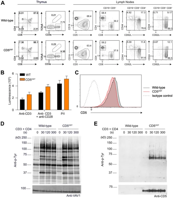
710 bp, respectively. It included an OST coding sequence (Junttila et al., 2005) inserted at the end of the last exon (exon 10) of the Cd5 gene and a self-excising ACN cassette (Roncagalli et al., 2014) that was introduced at the beginning of the 39 UTR sequence. The final targeting vector was abutted to a cassette coding for the diphtheria toxin fragment A (Soriano, 1997). The protospacer-adjacent motif present in the targeting vector was destroyed via a silent mutation to prevent CRISPR/Cas9 cleav-age. Two sgRNA-specifying oligonucleotide sequences (59-CAC CGAGTGGCTCAGAGACTGTAAA-39 and 59-AAACTTTACAGT CTCTGAGCCACTC-39) were annealed, generating overhangs for ligation into the BbsI site of plasmid pX330 (pSpCas9; Addgene; plasmid ID 42230). JM8.F6 C57BL/6N embryonic stem (ES) cells (Pettitt et al., 2009) were electroporated with 20 µg of targeting vector and 2.5 µg of pX330-sgRNA plasmid. After selection in G418, ES cell clones were screened for proper homologous recombination by Southern blot or PCR analysis. A neomycin-specific probe was used to ensure that adventitious nonhomol-ogous recombination events had not occurred in the selected ES clones. Mutant ES cells were injected into BalbC/N blastocysts. Following germ-line transmission, screening for proper deletion of the ACN cassette and for the presence of the sequence coding for the OST was performed by PCR using the following pair of primers: sense 59-GAAGGAGCCCTACACCGA-39 and antisense 59-CTAGGGGCCTCTGTCCAT-39. This pair of primers amplified a 354-bp band and a 164-bp band in the case of the Cd5OSTallele and of the WT Cd5 allele, respectively. Analysis of mice homo-zygous for the Cd5OSTallele showed that their T cells developed properly, yielding normal numbers of mature CD4+and CD8+T cells (Fig. S4 A) that had no defect in proliferation (Fig. S4 B). Mature T cells expressed the CD5 bait at physiological levels (Fig. S4 C) and showed a pattern of TCR-inducible tyrosine phos-phorylation comparable to WT T cells (Fig. S4 D). Affinity pu-rified CD5OST bait proteins showed increased phosphorylation after CD3 plus CD4 stimulation, leading to their binding to ty-rosine phosphorylated species (Fig. S4 E).
Design of sgRNA for editing the Lat and Cd6 genes of primary mouse CD4+T cells
Specific sgRNA of high cutting efficiency were designed using the Crispor algorithm and purchased from Synthego. The se-quences of the sgRNA used to edit the Cd6 and Lat genes are listed in Table S1. They were modified with 29-O-methyl 39 phosphorothioate in the first and last three nucleotides (Hendel et al., 2015). They are intended to introduce a double break lo-cated either 4 bp (Cd6) or 12 bp (Lat) 39 of the corresponding stop codons.
Design of DNA HDR templates for editing the Lat and Cd6 genes of primary mouse CD4+T cells
843-bp-long dsDNA HDR templates (Integrated DNA Technolo-gies) were amplified by PCR to edit the Lat (Fig. S1) and Cd6 (Fig. S3) genes. PCR products were purified with the QIAquick PCR purifi-cation kit (Qiagen) before being used for nucleofection. To prevent CRISPR/Cas9 cleavage of the edited alleles, a silent mutation de-stroying the protospacer-adjacent motif sequence present in the genomic DNA was introduced into the dsDNA HDR templates.
Neon Transfection System (Invitrogen) under the following conditions: voltage (1,500 V), width (20 ms), number of pulses (one), 100 µl tip, and buffer T. Cells were transfected with 5 µg sgRNA and with 10 µg dsDNA HDR template. After electropor-ation, cells were plated in a 24-well plate with mouse IL (mIL)-2 produced by Concanavalin A stimulation of the DC144 T cell hybridoma (Emilie et al., 1991) and of recombinant mIL-7 (1 ng/ ml; Peprotech). Cells were analyzed for the expression of CD90.1 3 d after transfection, and CD90.1+CD4+T cells were sorted on day 5 after transfection using a FACSAria cell sorter (BD Bio-sciences). After cell sorting, CD90.1+T cells were cultured with IL-2 and IL-7 for 1 d. Cells were then restimulated with plate-bound anti-CD3 (1 µg/ml) and soluble anti-CD28 (1 µg/ml), and the medium was supplemented with IL-2 24 h after restim-ulation and with IL-2 and IL-7 2 d after restimrestim-ulation. After 7 d of culture, similar cycles of expansion were repeated until reaching numbers of CD4+ T cells appropriate for AP-MS analysis. WT CD4+T cells were grown under the same conditions and used as control in AP-MS experiments.
Analysis of the 59 and 39 borders of the DNA insertion
Genomic DNA was isolated from the specified CD90.1+ CD4+ T cells using standard methods (DNeasy Blood & Tissue Kit; Qiagen), and the region straddling the 59 and 39 borders of the intended insertion was amplified by PCR using the sets of pri-mers specified in Table S2. The amplified products were se-quenced (Eurofins Genomics) following purification (QIAquick PCR Purification Kit; Qiagen). Individual clones were sequenced and compared with the WT sequence.
Flow cytometry
Stained cells were analyzed using an LSRII system (BD Bio-sciences). Data were analyzed with FlowJo V10 (TreeStar). Cell viability was evaluated using SYTOX Blue or DAPI (4 9,6-dia-midino-2-phenylindole, dihydrochloride; Life Technologies). The following antibodies were used: CD90.1 (HIS51), anti-CD4 (RM4-5), anti-CD8α (53–6.7), anti-CD6 (REA311), anti-TCRβ (H57-597), anti-γδ TCR (GL-3), anti-CD3e (145-2C11), anti-CD44 (IM7), anti-CD25 (PC61), anti-CD45R (RA3-6B2), anti-CD5 (53–7.3), anti-CD62L (MEL-14), and anti-human heparin-binding epidermal growth factor (HB-EGF; BAF259) from BD Biosciences, Miltenyi, R&D Systems, and eBioscience. Note that
Immunoprecipitation and Western blot analysis of OST-tagged CD4+T cells
The specified OST-tagged CD4+T cells were incubated with anti-CD3 (0.2 µg per 106cells; 145-2C11) and anti-CD4 (0.2 µg per 106 cells; GK1.5; Exbio) for 15 min on ice, followed by one round of washing at 4°C. Cells were then incubated at 37°C for 5 min and then left unstimulated or stimulated at 37°C by cross-linking for 30 or 120 s with purified goat anti-rat IgG F(ab9)2(0.4 µg per 106 cells; Jackson ImmunoResearch). Stimulation was stopped by the addition of a twice-concentrated lysis buffer (100 mM Tris, pH 7.5, 270 mM NaCl, 1 mM EDTA, 20% glycerol, and 0.4% n-do-decyl-β-D-maltoside) supplemented with protease and phos-phatase inhibitors. After 10 min of incubation on ice, cell lysates were centrifuged at 21,000 g for 5 min at 4°C. Postnuclear ly-sates were used for immunoprecipitation or as whole-cell lyly-sates for subsequent immunoblot analysis. For immunoprecipitation, equal amounts of cell lysates were incubated for 1.5 h with specified antibodies. Immune complexes were purified with Pansorbin (Calbiochem) and were washed three times before elution in SDS-containing sample buffer. Eluted samples and whole-cell lysates were loaded on 8% SDS-PAGE gel and subse-quently analyzed by immunoblot with specific antibodies. The following antibodies were used for immunoblot analysis: anti-CD5 (SAB4503585; Sigma-Aldrich), anti-CD6 (96123; R&D Sys-tems or H-300; Santa Cruz Biotechnology), anti-LAT (9166; Cell Signaling Technology), anti-phosphotyrosine (4G10; Millipore), UBASH3A (PA5-30637; Thermo Fisher Scientific), anti-VAV1 (2502; Cell Signaling Technology), and anti-GRK6 (5878; Cell Signaling Technology).
CD4+T cell isolation from CD5OSTmice and short-term expansion before AP-MS analysis
CD4+T cells were purified (>95%) from pooled lymph nodes and spleens from CD5OST mice with the Dynabeads Untouched Mouse CD4+T Cell Kit. CD4+T cells were activated with plate-bound anti-CD3 (145-2C11; 5 µg/ml) and soluble anti-CD28 (37–51; 1 µg/ml) antibodies, both from Exbio. After 48 h of cul-ture, CD4+T cells were harvested and grown in the presence of IL-2 (10 U/ml) for 48 h before AP-MS analysis. WT CD4+T cells were subjected to the same expansion protocol and used as controls.
Stimulation and lysis of short-term– and long-term–expanded mouse CD4+T cells before AP-MS analysis
Short-term–expanded CD4+ T cells (100 × 106) from WT and CD5OSTmice or long-term–expanded WT, LATOST, and CD6OST CD4+T cells (100 × 106) were incubated with anti-CD3 (0.2 µg per 106cells; 145-2C11; Exbio) and anti-CD4 (0.2 µg per 106cells; GK1.5; Exbio) antibodies on ice for 15 min, followed by one round of washing at 4°C. Cells were then incubated at 37°C for 5 min and then stimulated at 37°C with a purified rabbit anti-rat Ig (0.4 µg per 106cells; Jackson ImmunoResearch) for 30, 120, and 300 s or left unstimulated. Stimulation was stopped by the addition of a twice-concentrated lysis buffer (100 mM Tris, pH 7.5, 270 mM NaCl, 1 mM EDTA, 20% glycerol, and 0.4% n-dodecyl-β-D-mal-toside) supplemented with protease and phosphatase inhibitors. After 10 min of incubation on ice, cell lysates were centrifuged at 21,000 g for 5 min at 4°C. Postnuclear lysates were then used for affinity purification.
Affinity purification of OST-tagged protein complexes
Equal amounts of postnuclear lysates were incubated with Strep-Tactin Sepharose beads (IBA GmbH) for 1.5 h at 4°C on a rotary wheel. Beads were then washed five times with 1 ml of lysis buffer in the absence of detergent and of protease and phosphatase inhibitors. Proteins were eluted from the Strep-Tactin Sepharose beads with 2.5 mM D-biotin, a ligand that binds to Strep-Tactin with a higher affinity than the OST sequence does.
Tandem MS analysis
Following affinity purification, protein samples were air-dried in a Speed-Vac concentrator and either reconstituted in Laemmli buffer and processed by SDS-PAGE and trypsin in-gel digestion as previously described (Voisinne et al., 2019) or reconstituted in 5% SDS–50 mM ammonium bicarbonate and processed for trypsin digestion using an S-trap micro device (Protifi) accord-ing to the manufacturer’s instructions. Tryptic peptides were resuspended in 17 µl 2% acetonitrile and 0.05% trifluoroacetic acid and analyzed by nano-LC coupled to tandem MS, using an UltiMate 3000 system (NCS-3500RS Nano/Cap System; Thermo Fisher Scientific) coupled to an Orbitrap Q Exactive MS (model Q Exactive Plus or HFX; Thermo Fisher Scientific). 5μl of each sample was loaded on a C18 precolumn (300-µm inner diameter × 5 mm; Thermo Fisher Scientific) in a solvent made of 2% ac-etonitrile and 0.05% trifluoroacetic acid, at a flow rate of 20 µl/ min. After 5 min of desalting, the precolumn was switched on-line with the analytical C18 column (75-µm inner diameter × 50 cm, Acclaim PepMap C18, 2 µM; Thermo Fisher Scientific or in-house packed with 3 µm Reprosil C18) equilibrated in 95% sol-vent A (5% acetonitrile and 0.2% formic acid) and 5% solsol-vent B (80% acetonitrile and 0.2% formic acid). Peptides were eluted using a 10–45% gradient of solvent B over 60 min at a flow rate of 350 nl/min. The MS was operated in data-dependent acquisition mode with Xcalibur software. On the Q Exactive HFX or Q Ex-active Plus MS, MS survey scans were acquired with a resolution of 60,000 or 70,000, respectively, and an AGC target of 3e6. The 12 or 10 most intense ions, respectively, were selected for frag-mentation by high-energy collision–induced dissociation, and
the resulting fragments were analyzed at a resolution of 15,000 or 17,500, respectively, using an AGC target of 1e5 and a maxi-mum fill time of 22 ms or 50 ms, respectively. Dynamic exclu-sion was used within 30 s to prevent repetitive selection of the same peptide.
Protein identification and quantification for interaction proteomics
Raw MS files were processed with MaxQuant software (version 1.5.2.8) for database search with the Andromeda search engine and quantitative analysis. Data were searched against Mus musculus entries of the UniProt KB protein database (release UniProtKB/Swiss-Prot+TrEMBL 2017_01, 89,297 entries includ-ing isoforms) plus the One-Strep-tag peptide sequence, and the set of common contaminants was provided by MaxQuant. Car-bamidomethylation of cysteines was set as a fixed modification, whereas oxidation of methionine, protein N-terminal acetyla-tion, and phosphorylation of serine, threonine, and tyrosine were set as variable modifications. Specificity of trypsin diges-tion was set for cleavage after K or R, and two missed trypsin cleavage sites were allowed. The precursor mass tolerance was set to 20 ppm for the first search and 4.5 ppm for the main Andromeda database search. The mass tolerance in tandem MS mode was set to 0.5 daltons. Minimum peptide length was set to seven amino acids, and the minimum number of unique or razor peptides was set to one for validation. The I = L option of MaxQuant was enabled to avoid erroneous assignation of un-distinguishable peptides belonging to very homologous proteins. Andromeda results were validated by the target decoy approach using a reverse database, with a false discovery rate set at 1% at both peptide sequence match and protein level. For label-free relative quantification of the samples, the match between runs option of MaxQuant was enabled with a match time window of 1 min to allow cross-assignment of MS features detected in the different runs after alignment of the runs with a time window of 20 min. Protein quantification was based on unique and razor peptides. The minimum ratio count was set to 1 for label-free quantification calculation, and computation of the intensity-based absolute quantification (iBAQ) metric was also enabled. Data processing and identification of specific interactors From the proteinGroups.TXT files generated by MaxQuant with the options described above, protein groups with negative identification scores were filtered as well as proteins identified as contaminants. In situations where protein groups corre-sponded to the same gene name, protein intensities in a given sample were summed over the redundant protein groups. Protein intensities were normalized across all samples by the median intensity. Normalized intensities corresponding to different technical replicates were averaged (geometric mean), and missing values were replaced after estimating background binding from WT intensities. For each bait and each condition of stimulation, we used a two-tailed Welch t test to compare normalized log-transformed protein intensities detected in OST-tagged samples across all biological replicates to WT intensities pooled from all conditions of stimulation. To avoid spurious identification of interactors due to missing value imputation, we
OST-tagged samples and stimulation time t averaged (geometric mean) across all biological replicates, and Npepcorresponds to the number of tryptic peptides, theoretically observables as es-timated from iBAQ values. We also computed stoichiometries independently for each biological replicate and used these values to quantify the regulation of bait–prey association following TCR engagement. For a given condition of stimulation, log-transformed stoichiometries were compared with that of the nonstimulated condition using a two-tailed Welch t test. We selected preys whose interaction stoichiometry changed at least twofold with a P value below 0.05 in at least one condition of stimulation compared with the nonstimulated condition.
High-resolution MS characterization of long-term–expanded CD4+T cell proteome
For proteome analysis, cell pellets corresponding to 5 × 10⁶ long-term–expanded CD4+T cells were incubated with 150 µl of lysis buffer containing 50 mM Tris, pH 7.5, 0.5 mM EDTA, 135 mM NaCl, and 1% SDS for 10 min on ice and subjected to sonication with a Bioruptor ultrasonicator. Protein concentration was de-termined using a detergent-compatible assay (DC assay; Bio-Rad), and an aliquot of 100 µg of each sample was migrated briefly on SDS–PAGE gel and processed for in-gel digestion as previously described (Voisinne et al., 2019). Tryptic peptides were resuspended in 125 µl of 2% acetonitrile and 0.05% tri-fluoroacetic acid. 5 µl of each sample was analyzed with nanoLC-MS on the Q Exactive HFX as described above, but with a longer separation gradient (10–45% of solvent B over 120 min). Raw MS files were processed with MaxQuant as described above, except that phosphorylation was not included as a variable modification for the database search.
Calculation of copy numbers per T cell and of fraction of cell mass occupied by a protein
Analysis of the proteome of long-term–expanded CD4+ T cells identified 6,261 protein groups. Protein entries from the Max-Quant proteinGroups.TXT output were first filtered to eliminate entries from reverse and contaminant databases. Cellular pro-tein abundances were determined from raw intensities using the protein ruler methodology (Wi´sniewski et al., 2014), using the following relationship: protein copies per cell = (protein MS signal × NA× DNA mass)/(M × histone MS signal), where NAis
Cd5−/−Cd6−/−mice using the EasySep Mouse Naive CD4 T+cell Isolation Kit. WT (human DTR+) and Lat−/−(human DTR−) naive CD4+T cells were purified from pooled lymph nodes and spleens from Latfl-dtr maT-Cre mice using FACS AriaIII as described (Roncagalli et al., 2014). The various purified naive CD4+T cells were stimulated with plate-bound anti-CD3 (145-2C11; 3 µg/ml) and soluble anti-CD28 (37–51; 1 µg/ml) antibodies for 20 h or left unstimulated. CD25, CD44, and CD69 were up-regulated and CD62L down-regulated on all the CD3 plus anti-CD28–treated T cells, which demonstrated that they had been evenly stimulated. RNA was isolated using an RNeasy Plus mini kit (Qiagen). All samples were analyzed on an Agilent 2100 Bio-analyzer and passed quality control based on the RNA Integrity Number (≥8). DNA libraries were constructed by GenomEast platform using TruSeq Stranded mRNA Library Prep (Illumina). The DNA libraries were subjected to high-throughput sequenc-ing on the Illumina Hiseq 4000 as ssequenc-ingle-read 50-base reads following Illumina’s instructions. The fastq files were assessed with the fastqc program, and trimming was performed with DimerRemover to remove Illumina adapters and low-quality reads. Reads were mapped to the mouse GRCm38 (mm10) ref-erence genome using STAR, and the number of reads mapped to each gene was determined with featureCounts v1.6.0. Raw read counts were combined into a numeric matrix, with genes in rows and experiments in columns, and used as input for dif-ferential gene expression analysis with DESeq2 v1.22.2 after removing genes with less than one total read across all samples. Normalization factors were computed on the filtered data ma-trix using the concept of variance stabilizing transformations. Pairwise comparisons were performed between treatment groups to obtain log2fold change, and adjusted P values were corrected for multiple testing using the Benjamini-Hochberg (BH) method and used to select genes with significant expres-sion differences (q < 0.05).
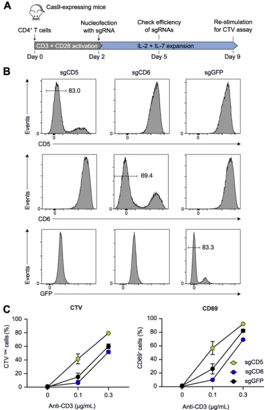
Deletion of the CD5 and CD6 genes in primary CD4+T cells CD4+T cells were purified by immunomagnetic negative selec-tion from mice constitutively expressing Cas9 and edited as described above. The CD5 or CD6 genes were ablated using the sgRNA specified in Table S2. The efficiency of gene deletion was checked by flow cytometry on day 3 after transfection. Edited cells were kept in culture in the presence of IL-2 (10 U/ml) and