Caged Phosphopeptides and Phosphoproteins:
Agents in Unraveling Complex Biological Pathwaysby
Deborah Maria Rothman
S. B., Biochemistry, University of Chicago, 2000 A. B., Biology, University of Chicago, 2000
Submitted to the Department of Chemistry in Partial Fulfillment of the Requirements for the
Degree of Doctor of Philosophy
at the
Massachusetts Institute of Technology
June 2005
© 2005 Massachusetts Institute of Technology All rights reserved
MASSACi.USE_.]S iNSTITTE OF TECHNOLOGY JUN 2 1 2005
LIBRARIES
Signature of Author: Department of Chemistry May 9, 2005 Certified by: Barbara Imperiali Class of 1922 Professor of Chemistry and Professor of Biology Thesis SupervisorAccepted by:
Robert W. Field Haslam and Dewey Professor of Chemistry Chairman, Departmental Committee on Graduate Students
AtmCHlvtS
-This doctoral thesis has been examined by a committee of the Department of Chemistry as follows: Timothy F. Jamison Chairman Professor of Chemistry
6)
Barbara ImDeriali Thesis Supervisor Class of 1922 Professor of Douglas A. LauffenburgerChemistry and Professor of Biology
Whitaker Professor of Biol
oafE r fi s o ei/
-
g/ a Blogical Engineering, Pofessor of themica/ngineering and Biology
Caged Phosphopeptides and Phosphoproteins:
Agents in Unraveling Complex Biological Pathwaysby
Deborah Maria Rothman
Submitted to the Department of Chemistry
on May 9, 2005 in Partial Fulfillment of the
Requirements for the Degree of Doctor of Philosophy
ABSTRACT
Within cellular signaling, protein phosphorylation is the post-translational modification most frequently used to regulate protein activity. Protein kinases and phosphoprotein phosphatases generate and terminate these phosphoryl signals, respectively. Chemical approaches for studying protein phosphorylation and the roles of phosphoproteins include photolabile caged analogs of bioactive species. Caged compounds are ideal chemical probes for studying cellular signaling because they afford researchers spatial and temporal control over the release of targeted effector molecules. Ligands or proteins involved in signal transduction can be chemically caged and subsequently irradiated to release a concentration burst of a specific species, allowing the downstream effects to be monitored without disrupting other aspects of the
system.
The syntheses and applications of caged phosphopeptides and phosphoproteins have been developed and detailed within this thesis. Two methods to synthesize 1-(2-nitrophenyl)ethyl caged phosphopeptides were developed. These peptides demonstrated good quantum yields of uncaging as compared to literature values of other ortho-nitrobenzyl derived caged compounds. A study in which these caged phosphopeptide tools yielded seminal information about the 14-3-3 protein family's involvement in cell cycle control successfully demonstrated the unique utility of these probes. Furthermore, the synthesis that allowed the extension of the nonsense codon suppression methodology to include caged phosphoproteins was developed. The three most commonly phosphorylated amino acids (serine, threonine, and tyrosine) were each incorporated into a test protein in their caged phosphorylated form. Toward studying cell motility, caged
phosphoserine was incorporated into position 153 of mVASP for use in live cell assays.
Thesis Advisor: Barbara Imperiali
Acknowledgements
"I'm gonna tell you a story; I'm gonna tell you about my town
I'm gonna tell you a big bad story, baby; Aww, it's all about my town...
First and foremost, I thank Barbara Imperiali for being nothing less than a wonderful Ph. D. advisor. Thank you for welcoming me to the lab, giving me a place to learn chemistry and science as a whole, teaching me to discover the answers to my many questions, and trusting me to establish a project within the group. It has been a challenging adventure that knowing what I know now, I would choose to do all over again. Also, thank you for being so extremely supportive throughout the post-graduate search.
The Imperiali lab is an incredibly encouraging learning environment; the spectrum of skills members have and their enthusiasm for science and life have turned all the inevitable rough patches of research into learning experiences I won't soon forget. I've seen the lab turnover quite a bit, and I hope I can acknowledge everyone appropriately. From day one, I thank Kathy Franz for introducing me to the lab and getting me started on my first peptide- of course, it was insoluble. I thank Harm Brummerhop for introducing me to the wonderful world of synthetic chemistry, rainbow colored columns, and turning a dark bay into a techno-dance party, liquid nitrogen and all. For answering my thousands of first-year questions, I thank Kevin McDonnell, Dierdre Pierce, Jen Ottessen, Rob Dempski, Mayssam Ali, Mike Torrice, Mark Nitz, and Vladimir Goncharov. ][ thank Carlos Bosques and Maria Ufret for their senior wisdom and other pieces of fun I can't forget: the mad cow on Eugenio's desk, the "laser," and so much time in the dark HPLC room. For creating a lab community that is as good as it gets, I thank the more current lab members: Eranthie Weerapana for being my "twin" all these years; Seungjib Choi for keeping everyone safe; Mary O'Reilly for her ability to wow everyone with her Irish genes; Christina Carrigan for her energy and empathy for bad backs; Jebrell Glover for all the biochemical tips and all the song lyrics; Guofeng Zhang for great political discussions; Elvedin Luckovic for being a fabulous dancing buddy; Langdon Martin for his creative slides and Red Sox spirit; Galen Loving for his genuine curiosity; Christian Hackenberger for all the great chocolate; Bianca Sculimbrene for her eagerness to learn biology, her Red Sox spirit, and her contagious laugh; Matthieu Sainlos for warming up to the crazy Americans in the lab; Dorra Carrico for always bringing a smile to the lab; Debby Pheasant for great lunchroom conversations and a fabulous first name; Ryu Yoshida for his dedication to the lab and learning; Becca Maglathlin for being wise and fun beyond her years. To Mark Chen and Andrew Dutton, best of luck to you newbies. Our lab would not function well if it wasn't for Elizabeth Fong; thank you for answering all the inevitable questions that did not have much to do with chemistry and always sharing a warm smile.
In the realms of Chemistry and Biology labwork, I have been fortunate to have some great collaborators. Thanks to Professor Michael Yaffe for a collaboration that not only provided proof-of-concept for our new tools, but also allowed us to make some new discoveries about the 14-3-3 family. Thank you, Justine Stehn, for being so excited about working on the project and constantly pushing it forward. Thank you, Cokey Nguyen, for taking over from Justine and working with my precious peptide samples, and also being a friend outside the ab. Thanks to Professor Dennis Dougherty and his entire group who welcomed me to CalTech to learn about nonsense codon suppression techniques. A special thanks to James Petersson who worked with me in the trenches to get caged phosphoserine into protein, especially since it seemed to be during the apocalypse. Thanks to Professor Frank Gertler for starting a collaboration with mVASP; I hope that the project continues until cells are walking under the control of light. Also thanks to Melanie Barzik for answering many questions along the way. Of course, I'd like to thank my thesis committee members, Professor Timothy Jamison and Professor Douglas Lauffenburger, for being here on this significant day. A special thank you to Tim for following my progress through the years, and for offering advice when I needed it most.
"Yeah, down by the river; Down by the banks of the river Charles (aw, that's what's happenin' baby)
That's where you'llfind me; Along with lovers, luggers, and thieves (aw, but they're cool people)
Well I love that dirty water; Oh, Boston, you're my home (oh, you're the Number One place)..."
I have been extremeley privaleged to meet three people that have blurred the line between lab-mate and great friend. Eugenio Vdzquez, thank you for being a partner on the caging project, but more than that, thank you for being like a big brother to me. Thanks for always supporting me and encouraging me in lab and
in life, and for telling me stories that maybe I did not need to hear, but always made me smile. Beth Vogel, thank you for being so patient when learning from me, working with me on the caging project, and being a genuinely happy person. I've had so much fun running into lab to talk about the "Sox last night," among many other things. Thanks for being yourself, a definite bright spot in the group. And to my last partner in crime in the lab, thank you, Melissa Shults, for being my comrade in everything else. Thank you for talking world politics, Red Sox, the Beatles and music in general, food and wine, and of course chemistry shop. I can't imagine what the past years would have been like without you. Best of luck on the left coast!
"Frustrated women (I mean they're frustrated); Have to be in by twelve o'clock (oh, that's a shame)
But I'm wishin' and a-hopin, oh; That just once those doors weren't locked (I like to save time for my baby to
walk around); Well I love that dirty water; Oh, Boston, you're my home (oh, yeah)..."
Before I started calling Boston my home, there was Bethesda and Chicago. Thank you, mom and dad, for supporting me throughout the years. From Bradley Hills to Whitman to Chicago and to M. I. T., thanks for allowing me to figure out what I wanted to pursue, and supporting me in every way that you could; I love you. Thank you, Ari, for making the wonderful analogy between chemistry and laundry, but even more for being mny big brother when I needed it most. Momom and Popop, if you were still here, I know you would be proud; thank you for your enthusiasm about everything I ever did. Thank you Aunt Mary, Aunt Sis, and of course the other resident scientist in the family, Uncle Albert, for your devotion and support through all the years.
From Maryland to Boston and a few places in between, I thank Katie Straus who has been a great friend for so many years. Thanks for always being there at the most important times, no matter how different our languages" are and how many miles apart we may have been. Here in Boston, I thank the girls of the ]3A13 for the five fi.un and crazy years we have had. A special thanks to Erika Swanson for understanding me and being a great friend through all the tough times and the great times. I can't wait to see you in my old stomping ground, Chicago! From my days in Chicago, I thank Rachel Knipe, Clara Park, and Meredith Fishbane for being great friends in college and beyond. And a gigantic thank you to Barbara Blank who is the definition of a loyal friend. Thank you for being my "bickering sister" since college and thank you for being a huge support system even from hundreds of miles away for the past five years. All my best to you and JJ, and I hope our visits are more frequent in the coming years.
"Because I love that dirty water; Oh, oh, Boston, you're my home (oh, yeah)..."
I didn't think it was possible to fall in love with a city, but that was my gut reaction to Boston when I hopped on the T at the old Airport station five years ago. "This is where I want to be." So, thank you, Boston, for surpassing my expectations for an amazing city. I plan to be running the bridge loops for years to come. Within Boston, I thank the Red Sox teams through 2003 who taught me what it felt to be a traditional Red Sox fan, and the 2004 team that made the most unbelievable comeback in sports history to then win the World Series while causing massive sleep deprivation throughout Boston proper. Along the sports theme, another thank you to the Patriots and Tom Brady for roping me into football. I may have starting watching for one reason, but now I can say I watch for the game... sort of. In any case, Boston is a great city for many reasons, and I think the Standells sum it up well:
"'Well, I love that dirty water (I love it, baby); I love that dirty water (I love Baw-stun)
I love that dirty wat r (Have you heard about the Strangler?); I love that dirty water (I'm the man, I'm the man); I. love that dirty water (Owww!); I love that dirty water (Come on, come on)"
- Dirty Water
Table of Contents List of Figures ... 7 List of Schemes ... 9 List of Tables ... 10 List of Abbreviations ... 11 List of Spectra ... 13 Chapter 1. Introduction ... 15 References ... 25
Chapter 2. Two Synthetic Methods for Caged Phosphopeptides ... 29
Introduction ... 29
Results and Discussion ... 29
Conclusion ... 38
Experimental ... 39
References ... 48
Chapter 3. Revealing a Global Temporal Role for 14-3-3 in Cell Cycle Regulation ... 49
Introduction ... 49
Results and Discussion ... 50
Conclusion ... 63
Experimental ... 64
Acknowledgements and References ... 69
Chapter 4. Synthesis of Full-length Caged Phosphoproteins ... 72
Introduction ... 72
Results and Discussion ... 73
Conclusion ... 84
Experimental ... 84
Acknowledgements and References ... 99
Chapter 5. Toward Reconstituting Directional Motility in VASP-null Cells ... 102
Introduction ... 102
Results and Discussion ... 104
Conclusion... 110
Experimental ... 110
Acknowledgements and References ... 112
Chapter 6. Conclusions and Future Directions ... 114
References ... 116
List of Figures
Chapter 1
1.1 Phosphoregulation of proteins ... 15
1.2 Mechanical model of the consequences of local photorelease of T[34 compared to unperturbed locomotion ...17
1.3 Caged phosphopeptide allows temporal control over the release of an inhibitory peptide. ... 18
1.4 Caged mutant cofilin (Ser3Cys) reveals a role for cofilin during cell motility ... 19
1.5 Caged thiophosphoproteins mimic phosphoproteins upon uncaging ... 20
1.6 Caged phospho-Smad2 acts like native phospho-Smad2 after UV-irradiation ... 21
Chapter 2 2.1 3 1p Magic Angle Spinning NMR of peptide on AM-PS resin ... 32
2.2 Caged phosphopeptides cpERK and cpPAX were synthesized and characterized for use in biological studies .. ...34
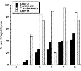
2.3 Peptide synthesized and characterized for studying the 14-3-3 protein family, cpRSLP. 36 2.4 Amount of caged peptide lost by photolysis from various light sources ... 38
Chapter 3 3.1 Peptides synthesized for in vitro binding studies of 14-3-3 ... 51
3.2 Photochemical cleavage of Ac-cpRSLP peptide ... 52
3.3 ITC demonstrates UV-A induced release of high-affinity 14-3-3 peptide ... 52
3.4 Uncaged but not caged phosphopeptide competes with endogenous cellular proteins for binding to 14-3-3 ... 53
3.5 FITC-PTD4 and FITC-3Ah peptides in Ratl fibroblasts ... 55
3.6 Possible f-elimination reactions during longer caged phosphopeptide synthesis ... 56
3.7 Decomposition of FMAQQ-SS-3Ah ... 58
3.8 FRSLP and FRSLP-SS-3Ah peptide series in Ratl fibroblasts ... 58
3.9 UV-A irradiation of cells in the absence or presence of serine-containing control peptides does not affect cell cycle progression .. ... 60
3.10 Coordinated, synchronous loss of 14-3-3 function results in aberrant cell cycle progression and G 1 release .. ... 61
3.11 Coordinated, synchronous loss of 14-3-3 function results in dysfunctional, DNA-damage induced S phase cell cycle checkpoint ... 62
,Chapter 4
4.1 Overview of the nonsense codon suppression method ... 72
4.2 Amino acids activated by cyanomethyl ester for coupling to the dinucleotide ... 73
4.3 Semisynthesis of acylated suppressor tRNA ... 79
4.4 Biochemical synthesis of mutant mRNA ... 80
4.5 Western blot analysis of nAChR-WT and mVASP-WT proteins .. ... 82
4.6 Suppression reaction with 4PO-protected tRNA ... 83
4.7 Suppression of nAChR A122Z ... 83
4.8 Suppression of mVASP S153Z ... 84
Chapter 5 5.1 Ena/VASP activity and protein organization ... 102
5.2 Plasmid maps of original vectors ... 104
5.3 Poor translation efficiency of mVASP-WT-His but not mVASP-S 153Z-His ... 105
5.4 mVASP-WT-His does not fully translate ... 106
5.5 Plasmid maps of modified vectors with N-terminal His-tags ... 106
5.6 His-mVASP-WT and His-mVASP-S153Z translated and purified ... 107
5.7 His-mVASP-WT produced in rabbit reticulocyte lysate treated with PKA or k-Ppase. 107 5.8 Gel shift assay of His-mVASP proteins produced by in vitro translation ... 108
5.9 Purification via Ni-NTA resin of His-mVASP-WT translated in wheat germ extract... 109
List of Schemes
Chapter 1
1.1 Photochemical decomposition of o-nitrobenzyl caged molecules ... 23
Chapter 2
2.1 Synthesis of 0-1-(2-nitrophenyl)ethyl-0O'-[-cyanoethyl-N,N-diisopropylphosphoramidite,
4.304 . .,...,... . . . . . . . . . . . . . . . . . . . . . . . . . . . . . . . . . . . . . . . . . . . . . . .3
2.2 Synthesis of caged phosphopeptide via the interassembly approach ...3 1 2.3 Oxidation of sensitive residues on bead .. ... 33 2.4 Synthesis of Na-Fmoc-phospho-( 1-nitrophenylethyl-2-cyanoethyl)-L-seine, 13 .. ... 34
,Chapter 3
;3.1 Synthesis of FRSLP-SS-3Ah peptide series: peptides for cellular uptake ... 57
Chapter 4
4.1 3-elimination of caged phosphate with transient cyanoethyl protection .. ... 74 4.2 Attempted synthesis of 0-1-(2-nitrophenyl)ethyl-0O'-tertbutyl-N,N-diisopropyl
phosphoramidite, 20 ...74
4.3 Synthesis of 0-1-(2-nitrophenyl)ethyl-O'-tertbutyl-N,N-diethyl phosphoramidite, 24. ..75 4.4 Synthesis of N-4-pentenoyl-phospho(nitrophenylethyl)-L-serine cyanomethyl ester, 32a,
and N-4--pentenoyl-phospho(nitrophenylethyl)-L-threonine cyanomethyl ester, 32b ... 76 4.5 Alternate synthesis of N'-4-pentenoyl-L-Serine cyanomethyl ester, 30a ... 77 4.6 Synthesis of amino acyl-pdCpAs, 36a, 36b, and 37 .. ... 78
List of Tables Chapter 2
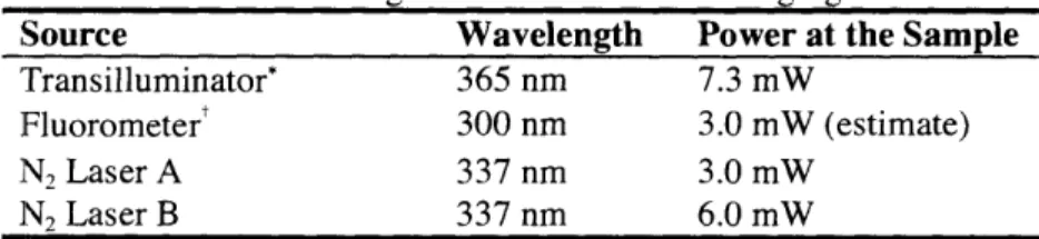
2.1 Quantumr yield of uncaging for various peptides ... 37 2.2 Light sources tested for uncaging ... 37
Abbreviations/Nomenclature
Standard one and three letter codes are used for the naturally occurring amino acids. Standard one-letter codes are used for nucleotides.
Abbreviation Definition 4PO-6XHis-tag ahx AM-PS cp C-terminus DNA ESI-TOF MS FACS FITC Fmnoc G1 phase (32 phase GST HPLC In vitro In vivo ITC M phase MALDI-TOF MS MAS-NMR mRNA mVASP nAChR-a 4-pentenoyl
Protein purification tag comprising 6 histidine residues Aminohexanoic acid
Aminomethyl-polystyrene Caged
phospho-The carboxylic acid end of a polypeptide chain or protein Deoxyribonucleic acid
Electrospray ionization time of flight mass spectrometry Fluorescence-activated cell sorting
Fluorescein-5-isothiocyanate N'-fluorenylmethoxycarbonyl First growth phase in the cell cycle
Second, shorter growth phase in the cell cycle Glutathione S-transferase
High performance/pressure liquid chromatography Performed outside the context of the cell; lit: in glass
Performed in the context of living cells; lit: in life
Isothermal calorimetry
Mitosis; cell phase in which two identical daughter cells divide from one parent cell
Matrix assisted laser desorption ionization time of flight mass spectrometry
Magic angle spinning nuclear magnetic resonance Messenger RNA
Murine vasodilator stimulated phosphoprotein Nicotine acetylcholine receptor ac-subunit
-terminus N-terminus NVOC
srvoc
RNA S phase S DS-PAGE SPPS IRNA UV-A UJV-BThe amino end of a polypeptide chain or protein 6-nitroveratryloxy carbamate
Ribonucleic acid
Synthesis phase; cell cycle phase in which DNA is copied Sodium dodecyl-sulfate polyacrylamide gel electrophoresis Solid phase peptide synthesis
Transfer RNA
Ultraviolet A irradiation, 320 nm 2 X 400 nm
Ultraviolet B irradiation, 280 nm > X a 320 nm
List of Spectra Chapter 2 2.1 'H spectrum of 1-(2-nitrophenyl)ethanol, 2 ... 117 2.2 3C spectrum of 1-(2-nitrophenyl)ethanol, 2 ... 118 2.3 'H spectrum of 0-1-(2-nitrophenyl)ethyl-O'--cyanoethyl-N,N-diisopropylphosphoramidite, 4 .. ... 119 2.4 3]P spectrum of 0-1-(2-nitrophenyl)ethyl-O'--cyanoethyl-N,N-diisopropyl phosphoramidite, 4 ... 120 2.5 31p spectrum of Fmoc-Ser-Pro-Gly-(AM-PS) ... 121 2.6 3 1p spectrum of
Fmoc-phosphi-(O-nitrophenylethyl-O'-cyanoethyl)seryl-Pro-Gly-(AM-PS)
.. ...
122
2.7 31P spectrum of Fmoc-phospho-(0-nitrophenylethyl-O'-cyanoethyl)seryl-Pro-Gly-(AM-PS) ... 123 2.8 3P spectrum of Phospho-(nitrophenylethyl)seryl-Pro-Gly-(AM-PS) ... 1242.9 1H spectrum of N-Fmoc-hydroxytrityl-L-serine tert-butyl ester, 10 .. ... 125
2.10 1 3C spectrum of Na-Fmoc-hydroxytrityl-L-serine tert-butyl ester, 10 .. ... 126
2.11 'H spectrum of N-Fmoc-L-serine tert-butyl ester, 11 .. ... 127
2.12 13C spectrum of N-Fmoc-L-serine tert-butyl ester, 11 ... 128
2.13 'H spectrum of Na-Fmoc-phospho-(O-nitrophenylethyl-O'-j-cyanoethyl)-L-serine tert-butyl ester, 12 ... 129
2.14 13C spectrum of N -Fmoc-phospho-(O-nitrophenylethyl-O '- 1-cyanoethyl)-L-serine tert-butyl ester, 12 .. ...130
2.15 3 1p spectrum of N-Fmoc-phospho-(O-nitrophenylethyl-O'- -cyanoethyl)-L-serine tert-butyl ester, 12 .. ...131
2.16 'H spectrum of N-Fmoc-phospho-(O-nitrophenylethyl-O '- P-cyanoethyl)-L-serine, 13. ... 132
2.17 13C spectrum of N-Fmoc-phospho-(O-nitrophenylethyl-O'- -cyanoethyl)-L-serine, 13. ... .. . . . .. ... . . . .. .. ... 1 3 3 2.18 31p spectrum of N -Fmoc-phospho-(O-nitrophenylethyl-O'- -cyanoethyl)-L-serine, 13. ... 134
2.19 HPLC trace of cpCS: Ac-RRGcpSPG-CONH2... 135
2.20 HPLC trace of cpERK: Ac-PLcpSPAKLAFQFP-CONH 2... 136
2.21 HPLC trace of cpRSLP: Ac-MARRLYRcpSLPAKK-CONH2... 137
Chapter 4
4.1 'H spectrum of 0-1-(2-nitrophenyl)ethyl-O'-tert-butyl-N,N-diethyl phosphoramidite, 24.
... 139
4.2 13C spectrum of 0-1-(2-nitrophenyl)ethyl-O'-tert-butyl-N,N-diethyl phosphoramidite, 24. ... 140
4.3 31 p spectrum of 0-1-(2-nitrophenyl)ethyl-O'-tert-butyl-N,N-diethyl phosphoramidite, 24.
... ...
141
4.4 'H spectrum of N-4-pentenoyl-0-tert-butyl-L-serine, 28a ... 142
41.5 13C spectrum of Na-4-pentenoyl-0-tert-butyl-L-serine, 28a ... 143
4.6 'H spectrum of N-4-pentenoyl-O-tert-butyl-L-serine cyanomethyl ester, 29a ... 144
4.7 13C spectrum of Na-4-pentenoyl-O-tert-butyl-L-serine cyanomethyl ester, 29a ... 145
4.8 'H spectrum of Na-4-pentenoyl-L-serine cyanomethyl ester, 30a ... 146
4.9 13C spectrum of Na-4-pentenoyl-L-serine cyanomethyl ester, 30a ... 147
4.10 'H spectrum of N-4-pentenoyl-L-serine-OH, 34 ... 148
4.11 'H spectrl.lm of Na-4-pentenoyl-L-serine cyanomethyl ester, 30a ... 149
4.12 'H spectru.m of N-4-pentenoyl-phospho(nitrophenylethyl)-L-serine cyanomethyl ester, 32a ... 150
4.13 3C spectrum of N-4-pentenoyl-phospho(nitrophenylethyl)-L-serine cyanomethyl ester, 32a ... 151
4.14 3'P spectrum of N-4-pentenoyl-phospho(nitrophenylethyl)-L-serine cyanomethyl ester, 32a ... 152
4.15 'H spectrum of N-4-pentenoyl-0-tert-butyl-L-threonine, 28b ... 153
4.16 13C spectrum of N-4-pentenoyl-0-tert-butyl-L-threonine, 28b ... 154
4.17 'H spectrum of Na-4-pentenoyl-0-tert-butyl-L-threonine cyanomethyl ester, 29b ... 155
4.18 13C spectrum of N-4-pentenoyl-O-tert-butyl-L-threonine cyanomethyl ester, 29b ... 156
4.19 'H spectrum of Na-4-pentenoyl-L-threonine cyanomethyl ester, 30b ... 157
4.20 13C spectrum of Na-4-pentenoyl-L-threonine cyanomethyl ester, 30b ... 158
4.21 'H spectrum of N-4-pentenoyl-phospho(nitrophenylethyl)-L-threonine cyanomethyl ester, 32b ... 159
4.22 13C spectrum of N-4-pentenoyl-phospho(nitrophenylethyl)-L-threonine cyanomethyl ester, 32b ... 160
4.23 31p spectrum of N-4-pentenoyl-phospho(nitrophenylethyl)-L-threonine cyanomethyl ester, 32b ...161
Chapter 1
Introduction
Portions of this chapter have been submitted for publication to Trends in Cell Biology.
1-1. General Introduction
Cellular signaling pathways integrate the actions of a myriad of ligands, substrates and proteins that comprise extremely complex networks of forward signals, feedback loops, and modulatory actions. Defining the spatial and temporal roles of the participants in the signaling cascades of living systems is a major challenge in cell biology. Since signaling molecules are in a constant state of flux, experimental approaches that afford information on the temporal and spatial dynamics of the components are extremely desirable. In this context, chemically-driven
strategies including small molecule inhibitors and activators as well as biotic and abiotic
chemosensors provide a powerful complement to the robust genetics-based methods that have enabled the identification of numerous pathways in living cells.' Chemical probes for the study of signal transduction networks can be of either exogenous or endogenous origin. Endogenous probes, including designed sensors based on the Aequoria victoria fluorescent proteins (AFPs),
are expressed and used within cells, while exogenous probes are prepared ex vivo and may be
introduced into living cells via microinjection, transfection, or through utilization of protein transduction domains.
Within cellular signaling, protein phosphorylation is the most abundant post-translational modification for the regulation of protein activity.2' 3 Protein kinases and phosphoprotein phosphatases generate and terminate these phosphoryl signals, respectively.4 Chemical approaches for studying protein phosphorylation and the roles of phosphoproteins include photolabile caged analogs of bioactive species (Figure 1.1).
O 0
O-P-O --O-- CAGE
rOH
kinase 0 hv 0... phosphatase ...
Figure 1.1. Phosphoregulation of proteins. Kinases append phosphoryl groups to hydroxyl side chains,
while phosphatases remove them. Alternatively, a caging group on a phosphate can be removed with light to reveal the phosphoprotein.
Caged compounds are ideal chemical probes for studying cellular signaling because they afford researchers spatial and temporal control over the release of targeted effector molecules. Ligands or proteins involved in signal transduction can be chemically caged and subsequently irradiated to release a concentration burst of the specific species, allowing the downstream effects to be monitored without disrupting other aspects of the system. For a brief discussion of caging and the history of the technique, please see Section 1-7. The most frequently implemented caging groups in biological experiments are derived from the ortho-nitrobenzyl protecting group, discussed in further detail below (Section 1-8). Other caging groups used in biological studies include hydroxyphenacyl derivatives, coumarins, and cinammic acid derivatives.5 9 Several examples of recently developed caged signaling molecules are discussed here with an emphasis
on those used for studying phosphorylation in signal transduction.
1-2. Caged Small Molecules, Peptides, and Proteins
More than two decades have passed since the original caging experiments of ATP and clivalent cations were published.0-'2 Currently, multiple reports of caged neurotransmitters, peptides and fulllength proteins are present in the literature. These studies revealed information
in space and time domains inaccessible by other methods. 3 -'7
Both caged L-glutamic acid'8 and caged D-aspartic acid9 have been synthesized and used
to study the effects of each stimulant on its cognate receptor. The molecular mechanism of the high affinity glutamate transport process is difficult to study with traditional rapid solution exchange techniques due to poor time resolution. It was found that L-glutamic acid caged on the y-carboxylate with the c-carboxynitrobenzyl moiety had desirable photochemical properties with a half-life of 21 jis and quantum yield of 0.14.8 Taken together, these parameters allowed the time-resolved study of the activation of glutamate receptors, which have a lifetime for the "open"
species on the 1 ms timescale. In a further example of caged neurotransmitters, D-aspartic acid was caged in order to study the effect of D-aspartic acid on glutamate transport, which is a highly
pH dependent process. The rate and quantum yield of uncaging is also dependent on pH. An -carboxynitrobenzyl caged D-aspartic acid was synthesized that had good uncaging kinetics at various pH values which was valuable for further synaptic studies. 19
Caged peptides can be used as masked inhibitors of protein functions that are regulated by phosphorylation. After introduction of a caged inhibitory peptide, the steady state of the
system is not perturbed; therefore, the immediate downstream effects of sequestering a targeted
protein can be monitored upon uncaging. In a study on eosinophil cell motility two caged
tyrosine peptide inhibitors of myosin light chain kinase (MLCK) were employed to reveal that MNLCK is globally central to whole cell movement.20 Flash photolysis allowed the peptides to rapidly sequester the MLCK in the system, and it was observed that cell motility significantly decreased, thus demonstrating that MLCK is essential in cell migration signaling. Whereas former studies had been performed on the minute to hour timescale, the observations described were made in the previously inaccessible time domain of seconds.
Several amino acid side chains in full-length protein have been caged using various methods. Approaches range from non-specific labeling of nucleophilic residues to cysteine scanning-mutagenesis trials to site-specific incorporation of caged amino acids (see reviews 13,
14, 16, 17). In a more recent study, thymosin 34 (TO4) was non-specifically caged on lysine
residues, and observations from studies with its use were compiled to formulate a mechanical model of cell turning in locomoting keratocytes (Figure 1.2).21 Computer simulations predicted the concentration of locally uncaged T4 necessary within a locomoting cell to cause a biological response without loss of effect due to diffusion. Indeed, upon local photolytic release of TP4, cell movement changed dramatically and appeared temporarily imbalanced relative to control cells. Using caged Tf34 allowed both spatial and temporal release of the protein, yielding new information about its functional role in motile cells.
t:nperturbed ljocorntion Ineduced tJurning behavior
A~. A ^t~, : C C ,ophotr,s, fx,.O' photorelemir Figure 1.2. Schematic of the
Cu~~~
~
t~~~.,/:
=
=~ \
)oFcXrmechanical consequences
*',''ru"$°of
Ol, nl(;e local photorelease of TP4 (C,
D) compared to unperturbed Clockwise torque resulting from ieR reaction locomotion (A, B). Proposed
B D lorce acimgthrough toment r d' model for the turning of
Flinching and Propulsive tractions balancedrtvt in r p t
~ L-or~~~~tnc. v~~~oc ~~n ro:-cnnn co- tn
4 4
l~.,, {tUt/., i,¢o 111 i i.o }/11ol , tsk
photoreleased Tb4 at the wing.
Cell turning is the result of a clockwise torque generated by
c!lhtrItp repar ti n
t-rn Pinching tractio Pivot asa rsultof .LALuJi -r
. Propulisive traction down-regulation of unbalanced propulsive traction.
'_: wsm r Reaction from sustrate pinchinp andprntlsive See reference 21.
1 ¢qua; and oppsite to the tractions
1-3. Caged Phosphopeptides
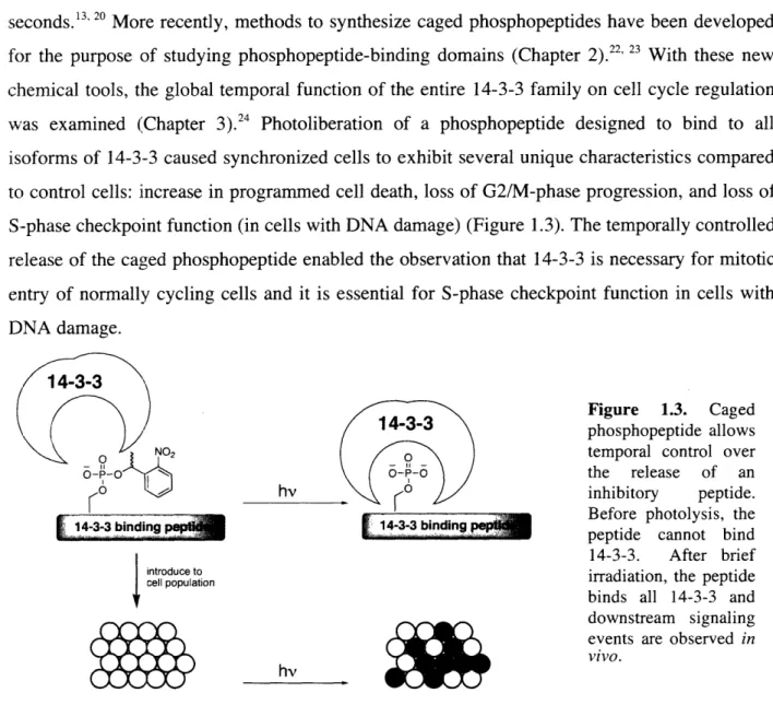
As mentioned above, essential early studies demonstrated that simple caged peptides allow phenotypic observations to be made in the previously inaccessible time domain of seconds.3' 20 More recently, methods to synthesize caged phosphopeptides have been developed for the purpose of studying phosphopeptide-binding domains (Chapter 2).22 23 With these new
chemical tools, the global temporal function of the entire 14-3-3 family on cell cycle regulation was examined (Chapter 3).24 Photoliberation of a phosphopeptide designed to bind to all isoforms of 14-3-3 caused synchronized cells to exhibit several unique characteristics compared
to control cells: increase in programmed cell death, loss of G2/M-phase progression, and loss of
S-phase checkpoint function (in cells with DNA damage) (Figure 1.3). The temporally controlled release of the caged phosphopeptide enabled the observation that 14-3-3 is necessary for mitotic entry of normally cycling cells and it is essential for S-phase checkpoint function in cells with DNA damage.
14-3-3/ '
Figure 1.3. Caged
\'
( )
/ /
14-3-3aphosphopeptide
allows\s fNo2 ( temporal control over
o-o a>I o /the release of an
1K0
hv_________/ inhibitory peptide.4-3-3 binding 433~
_
Before
photolysis,
the
[i!idn 433bnigpeptide cannot bind
14-3-3. After brief
introduce to
cel population irradiation, the peptide
M4~~~~~~~~ r ~~binds all 14-3-3 and
downstream signaling
vivo.
H i hv ( w ;~ events are observed
in
no effect on cells temporal phenotype observed
1-4. Caged Constitutively Active Kinase Substrate
Some kinase substrates are inactivated by phosphorylation and activated by dephosphorylation. The LIM-kinase substrate cofilin is one such protein: in the unphosphorylated form, cofilin binds to F-actin and severs the filaments to create barbed ends and modulate cell motility. LIM-kinase phosphorylates serine-3 of cofilin to render the protein inactive.25 Recently, a constitutively active mutant, cofilin Ser3Cys, was prepared and it was demonstrated that exposure of this mutant to F-actin caused filament severing and
depolymerization in vitro. In order to regulate the cofilin activity, the mutant protein was caged
via selective chemical modification with the a-carboxynitrobenzyl group at the unique cysteine
at position 3. In these studies, the ac-carboxylate substituent in the caging moiety enhanced the
efficiency of uncaging while simultaneously mimicking the inactive phosphorylated cofilin
(Figure 1.4). In the presence of caged cofilin Ser3Cys, F-actin was as stable as in the control
system. However, upon irradiation, 80% of severance activity was restored to cofilin Ser3Cys. This caged mutant protein was further evaluated in live cells and the studies revealed that active cofilin is essential for many aspects of cell motility.26 Cofilin aided in G-actin polymerization in
vivo: global uncaging of cofilin Ser3Cys increased free barbed ends, F-actin content, and cell locomotion, while local uncaging generated lamellipodia and determined the direction of cell migration. Since LIM-kinase cannot phosphorylate the Ser3Cys mutant, the effect of cofilin persisted for the duration of the cell monitoring. For these studies, this effect was advantageous since the active species is the unphosphorylated protein. However, for situations in which the phosphorylated species is the active form of the protein, it is desirable to have access to the caged phosphoprotein.
...-.
O-P-0po
OH LM-kinase
native : olt phosphatase a
active inactive hv
00 NO introduce into cell cell motility affected rSH caging
rs
/'utant.C t * hv ~~hv hu utWC _
constitutively active inactive
Figure 1.4. Caged mutant cofilin (Ser3Cys) reveals a role for cofilin during cell motility.
1-5. Caged Phosphothioproteins
Thiophosphoryl derivatives of serine, threonine and tyrosine can act as phospho-amino acid analogs in biological studies of protein phosphorylation. Thiophosphoryl groups demonstrate distinct properties in cellular systems: they are more nucleophilic and more resistant to hydrolysis by phosphatases compared with native phosphoryl groups.27 Recent work has extended the strategy for caging thiophosphopeptides to the preparation of full-length caged
thiophosphoproteins.6, 7 28 The semi-synthetic approach involves thiophosphorylation of a serine,
threonine, or tyrosine residue using an appropriate kinase in the presence of ATP(y)S and a thiophilic divalent cation, such as Co2+ (Figure 1.5).7 The caging group is then introduced by taking advantage of the nucleophilicity of the thiophosphate sulfur to displace bromide from the electrophile. In these studies, it was found that the hydroxyphenacyl caging group had superior photosensitivity (Box 2) to the o-nitrobenzyl group on thiophosphate.7 At the protein level, a caged thiophosphothreonine-197 variant of PKA was prepared by semi-synthesis and then activated via UV irradiation.6 Upon uncaging, 85-90% of protein activity was restored, corresponding to a 15-fold increase in activity over the caged precursor, which is significant for potential biological studies. Caged thiophosphoproteins are useful reagents for studying signal transduction due to the stability of the thiophosphates towards endogenous phosphatases. However, the enzyme-catalyzed thiophosphorylation reaction is not general and multiple kinases and divalent cations must be screened for the semi-synthesis of each new caged peptide or protein target.
O H O H
-0 0 0 0
ATPI(y)S ATP(y)S O-r-S B--r~jo Br4 o-S-S ,f O O oNp_5 mimic -P-O
OH divalerit cation 0 hv o
r
; kinaser
_ K]Figure 1.5. Caged thiophosphoproteins mimic phosphoproteins upon uncaging.
1-6. Caged Phosphoproteins
Recently, expressed protein ligation and semi-synthesis has been used to prepare the
caged phosphoprotein Smad2, which was caged on two phosphoserines at the C-terminus (Ser
465 and Ser 467) (Figure 1.6).29 Under native conditions, transforming growth factor
3
(TGF-3) phosphorylates two serine residues on Smad2, which then causes disengagement from the cytosolic retention factor SARA and allows homotrimerization of Smad2 and nuclear uptake. The caged phospho-Smad2 binds to SARA and cannot homotrimerize. However, upon brief and intense irradiation, the phospho-Smad2 is uncaged and released from SARA thereby initiating homotrimerization and nuclear localization.29 These experiments demonstrate the application ofnative chemical ligation for the preparation of caged phosphoproteins that are amenable to semi-synthetic strategies.
0
HS cpSer HS pSer ^ ,
native chemical .4$d_ -;/ cpe
ligation .Ser... \
cpSer
I lUi;lt:idr luL;¢1l[lUllur
trimerizationI Ul U d 1
= cpSer = caged phosphoserine
Figure 1.6. Caged phospho-Smad2 acts like native phospho-Smad2 after UV-irradiation. See reference 29.
An alternative general approach for the synthesis of caged phosphoproteins has recently been introduced which allows for the replacement of a single residue in native proteins with a caged phospho-amino acid (Chapter 4).30 A novel phosphitylation reagent enabled the chemical synthesis of an appropriately protected amino acid derivative for the generation of an amino-acyl tRNA charged with the caged phospho-amino acid. Incorporation of the unnatural amino acid into proteins was then accomplished via the nonsense codon suppression methodology using an
in vitro translation system. This strategy was used for the semi-synthesis of the cell motility
protein mVASP with caged phosphoserine at position 153 (Chapter 5). Gel shift analysis demonstrated that the caged phosphoprotein behaves as the unphosphorylated protein, and upon UJV irradiation the uncaged protein migrates adjacent to the phosphorylated control. This
directed approach should allow for the substitution of any serine, threonine, or tyrosine residue in
full-length proteins with caged phosphorylated analogs provided that the in vitro translation efficiency is acceptable.30 The recent developments in the availability of full-length caged
phosphoproteins, in addition to the aforementioned caged analogs, should further advance
studies exploiting the caging technology in the realm of signal transduction.
1-7. History and Requirements of Caging Groups
Caged compounds are molecules that include a photolabile mask on an essential functional group. With UV irradiation the mask is removed and the liberated molecule can exert
its effect on the system.'° Photolabile protecting groups have been in use in organic synthesis for many years.3 Due to the hydrophobicity of photolabile groups, they were first used in biological
systems to aid in cell membrane permeation of caged nucleotide analogs which could subsequently be uncaged in living cells.32 The term "caging" was coined when a photolabile
release ATP upon irradiation (be "uncaged") was characterized by Na,K-ATPase consumption and Na+efflux from cells via the Na:K pump.'0 The caged ATP could not be hydrolyzed by the Na,K-ATPase until after illumination to remove the caging group. Additionally, caged ATP had no effect on the Na:K pump in cells until after photolytic removal of the caging group. This study set the precedent for the future of caging in biological studies. In addition to control over the release of the effector molecule, caged compounds enable complete intracellular distribution of the target compound,4 thus allowing each cell to serve as its own control (before and after photolysis).20 Use of caged compounds circumvents genetic compensation effects often observed
in single knock-out studies.2 4
There are several key requirements of a caged compound to be successful in biological studies: (i) the caged compound should neither agonize nor antagonize the system, (ii) after irradiation, the compound should be released rapidly and in high yield, (iii) the photo-byproducts should be inert within the system, and (iv) the photolytic conversion must be accessible in an intracellular environment such that the downstream events can be immediately monitored.' Other factors to consider include the quantum yield of uncaging, the wavelength of excitation,
and the extinction coefficient of excitation (see below). A caging group can mask a functional group through two different strategies: steric bulk and electronic induction. The steric bulk strategy involves using a caging group to spatially block an interaction prior to photoliberation. The electronic induction strategy employs a caging group to withdraw or distribute electron density such that a key interaction cannot take place before photolysis. A study in which caged cation chelators were synthesized to tightly bind divalent calcium upon irradiation illustrates
these two different strategies.' 2
1-8. Caging Groups and Photophysical Properties
The most commonly used caging groups in biological studies are based on the ortho-nitrobenzyl moiety. The o-ortho-nitrobenzyl group is inert within living cells, has a good quantum yield of uncaging () on the microsecond-millisecond timescale, and can be excited with near-visible UV light (> 350 nm), which does not cause radiative damage to cells. Additionally, when attached to biological molecules, o-nitrobenzyl compounds and the corresponding photo-byproducts are water-soluble at physiological concentrations. Original studies demonstrated that intracellular thiols (e.g., glutathione) act as effective scavenging agents for the nitrososbenzene
photo-byproducts.° 33 The proposed mechanism of photolytic release for o-nitrobenzyl ethers is shown in Scheme 1.1 where X is the key functional group of the biological effector.33
Scheme 1.1. Photochemical decomposition of o-nitrobenzyl caged molecules.
R R R R
- -
X +H+ - R'-
p
R-
+ X
0 ~J-- 105S1 N -0 N=O o Oo 0 5 0 ac-nitro nitrobenzene intermediate photo-byproduct R = H, CH3, CO2 R'= H, OMe 0 O X = -OR, -SR", -N R" -ON-R" H Hor other good leaving group
The cyclization and subsequent breakdown of the aci-nitro intermediate has been spectroscopically monitored to determine the rate of release of the product. More recent research suggests that hemiketal formation may be involved during the photolytic release of 1-(2-nitrophenyl)ethyl ethers.34 The released nitroso-acetaldehyde (R = H) was found to be much more reactive in a biological context than released nitroso-acetophenone (R = CH3),° likely due
to the higher intrinsic reactivity of the aldehyde toward nucleophiles.
It is significant to note that the quantum yield of uncaging is a measure of photoliberation efficiency and not a measure of the rate of uncaging. The quantum yield describes how many caging groups are released for every photon absorbed by the group. The speed of the breakdown
of the excited species to release the effector molecule determines the rate of uncaging. The
o-nitrobenzyl groups have uncaging rates on the microsecond timescale. 8 33
Furthermore, the intensity of the light source can have a significant effect on the apparent rate of uncaging of a sample. For example, a light source sending a large number of photons
focused on the sample, such as a laser, will appear to have a faster rate of uncaging than a more diffuse source, such as a UV lamp. The actual rate of the breakdown of the photo-excited state
does not change between such circumstances; however, the large number of photons from the laser will proportionally uncage more molecules than the UV lamp, and thus analysis on the minute time scale will appear that the laser is uncaging the sample more quickly.
Another important physical characteristic for selecting a caging group is the product of the quantum yield and the extinction coefficient at the excitation wavelength (-E) or the photosensitivity of the caging group.7It is not uncommon for a caging group to have an excellent
A and a poor , and vice versa. Therefore, the product of these values is critical to selecting an efficient caging group.
1-9. Selection of the 1-(2-Nitrophenyl)ethyl-Cage for Phosphopeptides and Phosphoproteins
For the purpose of synthesizing caged phosphopeptides and phosphoproteins, the o-nitrobenzyl derivative 1-(2-nitrophenyl)ethyl moiety was chosen. There was abundant literature precedent for use of the o-nitrobenzyl derivatives at the time of the initial syntheses. The 1-(2-nitrophenyl)ethyl group was selected because it releases a ketone rather than an aldehyde as a photo-byproduct. Groups without any substitution at the benzylic position release an aldehyde, which is highly reactive in the cellular environment (as described in Section 1-8). The 1-(2-nitrophenyl)ethyl group has a maximum absorption at = 259 nm, with a strong extinction coefficient (Em = 5,700 M'cm-1).3 5The absorption spectrum is broad, and thus the aromatic
group still absorbs significantly at high-end UV ( = 365 nm). Caging moieties can been modified on the aromatic ring to include electron donating substituents which help to red-shift the absorption band; for example, the 4,5-dimethoxy-2-nitrobenzyl group can be uncaged with
light as far out as k = 420 nm. However, the 4,5-dimethoxy-2-nitrobenzyl derivative suffers from
a poorer quantum yield. For the studies described in this thesis, uncaging with k = 365 nm was sufficient and thus the 1-(2-nitrophenyl)ethyl-caging group, which had strong literature precedent, was employed.
1-10. Embarkment
As the number of tools for studying signal transduction grows, the amount of information mined from the use of these tools is amplified. Currently available tools for studying signal transduction have already provided significant information about the behavior of signaling molecules in cell lysates and in living cells that would not be possible by other means. Chemical tools have the inherent ability to be modified by the researcher; the growing feasibility of the required manipulations to prepare probes is further increasing their availability. Therefore, the advancement of chemical tools to study signaling networks will lend heavily to the foundation of understanding how cells function.
Caged compounds afford a researcher spatial and temporal control over the release of an effector molecule in a biological system, and studies with these probes have just scratched the
surface of understanding cell signaling pathways. In particular, for the study of phosphorylation regulated signaling, caged phosphopeptides and phosphoproteins are tools that will yield a great amount of information about the function of phosphorylated species in real time. Within this thesis, two methods to synthesize caged phosphopeptides are described (Chapter 2). A seminal example in which these tools yielded information about cell cycle control is then described in detail (Chapter 3). The synthesis that allowed extension of the caged phosphopeptide methodolgy to caged phosphoproteins is also described (Chapter 4). Finally, the progress toward application of caged phosphoproteins in live cell assays is also discussed (Chapter 5).
References
(1) Shogren-Knaak, M. A.; Alaimo, P. J.; Shokat, K. M. "Recent Advances in Chemical
Approaches to the Study of Biological Systems," Annual Review of Cell and
Developmental Biology 2001, 17 405-433.
(2) Hunter, T. "Signaling-2000 and Beyond," Cell 2000, 100 (1), 113-127.
3) Manning. G.; Whyte, D. B.; Martinez, R.; Hunter, T.; Sudarsanam, S. "The Protein
Kinase Complement of the Human Genome," Science 2002, 298 1912-1934.
(4) Hunter, T. "Protein Kinases and Protein Phosphatases: The Yin and Yang of Protein Phosphorylation and Signaling," Cell 1995, 80 (2), 225-236.
(5) In Dynamic Studies in Biology: Phototriggers, Photoswitches and Caged Biomolecules; Goeldner, M. and Givens, R., Eds.; John Wiley & Sons, Inc.: Hoboken, New Jersey,
2005, 584.
(6) Zou, K.; Cheley, S.; Givens, R. S.; Bayley, H. "Catalytic Subunit of Protein Kinase A
Caged at the Activating Phosphothreonine," Journal of the American Chemical Society 2002, 124 (28), 8220-8229.
(7) Zou, K.; Miller, W. T.; Givens, R. S.; Bayley, H. "Caged Thiophosphotyrosine Peptides,"
Angewandte Chemie-International Edition 2001, 40 (16), 3049-3051.
(8) Furuta, T.; Iwamura, M. "New Caged Groups: 7-substituted Coumarinylmethyl
Phosphate Esters," Methods in Enzymology 1998, 291 50-63.
(9) Turner, A. D.; Pizzo, S. V.; Rozakis, G.; Porter, N. A. "Photoreactivation of Irreversibly
Inhibited Serine Proteinases," Journal of the American Chemical Society 1988, 110 (1), 244-250.
(10) Kaplan, J. H.; Forbush, B., III; Hoffman, J. F. "Rapid Photolytic Release of Adenosine
5'-Triphosphate from a Protected Analogue: Utilization by Na:K Pump of Human Red Blood Cell Ghosts," Biochemistry 1978, 17(10), 1929-1935.
(11l) Kaplan, J. H.; Ellis-Davies, G. C. R. "Photolabile Chelators for the Rapid Photorelease of Divalent Cations," Proceedings of the National Academy of Sciences, U. S. A. 1988, 85
(17), 65:71-6575.
(12) Adams, S. R.; Kao, J. P. Y.; Tsein, R. Y. "Biologically Useful Chelators that Take Up
Ca2+Upon Illumination," Journal of the American Chemical Society 1989, 111 (20), 7957-7968.
(13) Shigeri, Y.; Tatsu, Y.; Yumoto, N. "Synthesis and Application of Caged Peptides and Proteins," Pharmacology and Therapeutics 2001, 91 (2), 85-92.
(14) Curley, K.; Lawrence, D. S. "Light-Activated Proteins," Current Opinion in Chemical
Biology 1999, 3 (1), 84-88.
(15) Ellis-Davies, G. C. R. "Development and Application of Caged Calcium," Methods in
Enzymology 2003, 360 226-238.
(16) Marriott, G.; Roy, P.; Jacobson, K. "Preparation and Light-Directed Activation of Caged
Proteins," Methods in Enzymology 2003, 360 (Biophotonics, Part A), 274-288.
(17) Petersson, E. J.; Brandt, G. S.; Zacharias, N. M.; Dougherty, D. A.; Lester, H. A. "Caging
Proteins Through Unnatural Amino Acid Mutagenesis," Methods in Enzymology 2003,
360 (Biophotonics, Part A), 258-273.
(1 8) Wieboldt, R.; Gee, K. R.; Niu, L.; Ramesh, D.; Carpenter, B. K.; Hess, G. P. "Photolabile
Precursors of Glutamate: Synthesis, Photochemical Properties, and Activation of Glutamate Receptors on a Microsecond Time Scale," Proceedings of the National
Academy of Sciences, U. S. A. 1994, 91 (19), 8752-8756.
(19) Grewer, C.; Madani Mobarekeh, S. A.; Watzke, N.; Rauen, T.; Schaper, K. "Substrate
Translocation Kinetics of Excitatory Amino Acid Carrier 1 Probed with Laser-Pulse Photolysis of a New Photolabile Precursor of D-Aspartic Acid," Biochemistry 2001, 40
(1), 232-240.
(20) Walker, J. W.; Gilbert, S. H.; Drummond, R. M.; Yamada, M.; Sreekumar, R.; Carraway,
R. E.; Ikelbe, M.; Fay, F. S. "Signaling Pathways Underlying Eosinophil Cell Motility Revealed by Using Caged Peptides," Proceedings of the National Academy of Sciences,
U. S. A. 1998, 95 (4), 1568-1573.
(21) Roy, P.; Rajfur, Z.; Jones, D.; Marriott, G.; Loew, L.; Jacobsen, K. "Local Photorelease
of Caged Thymosin Beta4 in Locomoting Keratocytes Causes Cell Turning," Journal of
(22) Rothman, D. M.; Vazquez, M. E.; Vogel, E. M.; Imperiali, B. "General Method for the
Synthesis of Caged Phosphopeptides: Tools for the Exploration of Signal Transduction Pathways," Organic Letters 2002, 4 (17), 2865-2868.
(23) Rothman, D. M.; Vazquez, M. E.; Vogel, E. M.; Imperiali, B. "Caged Phospho-Amino
Acid Building Blocks for Solid-Phase Peptide Synthesis," Journal of Organic Chemistry 2003, 68 (17), 6795-6798.
(24) Nguyen, A.; Rothman, D. M.; Stehn, J.; Imperiali, B.; Yaffe, M. B. "Caged
Phosphopeptides Reveal a Temporal Role for 14-3-3 in G1 Arrest and S-Phase Checkpoint Function," Nature Biotechnology 2004, 22 (8), 993-1000.
(25) Ghosh, M.; Ichetovkin, I.; Song, X.; Condeelis, J. S.; Lawrence, D. S. "A New Strategy
for Caging Proteins Regulated by Kinases," Journal of the American Chemical Society 2002, 124 (11), 2440-2441.
(26) Ghosh, M.; Song, X.; Mouneimne, G.; Sidani, M.; Lawrence, D. S.; Condeelis, J. S.
"Cofilin Promotes Actin Polymerization and Defines the Direction of Cell Motility,"
Science 2004, 304 (5671), 743-746.
(27) McMurray, J. S.; Coleman, D. R., IV; Wang, W.; Campbell, M. L. "The Synthesis of
Phosphopeptides," Biopolymers (Peptide Science) 2001, 60 (1), 3-31.
(28) Pan, P.; Bayley, H. "Caged Cysteine and Thiophosphoryl Peptides," FEBS Letters 1997,
405 (1), 81-85.
(29) Hahn, M. E.; Muir, T. W. "Photocontrol of Smad2, a Multiphosphorylated Cell-Signaling Protein, Through Caging of Activating Phosphoserines," Angewandte
Chemie-International Edition 2004, 43 5800-5803.
(30) Rothman, D. M.; Petersson, E. J.; Vazquez, M. E.; Brandt, G. S.; Dougherty, D. A.;
Imperiali, B. "Caged Phosphoproteins," Journal of the American Chemical Society 2005,
127 (3), 846-847.
(31) Barltrop, J. A.; Plant, P. J.; Schofield, P. "Photosensitive protective groups," Chemical Communications 1966, 822-823.
(32) Engels, J.; Schlaeger, E. J. "Synthesis, Structure, and Reactivity of Adenosine Cyclic 3',5'-Phosphate Benzyl Triesters," Journal of Medicinal Chemistry 1977, 20 (7), 907-911.
(33) Walker, J. W.; Reid, G. P.; McCray, J. A.; Trentham, D. R. "Photolabile
1-(2-Nitrophenyl)ethyl Phosphate Esters of Adenine Nucleotide Analogs. Synthesis and Mechanism of Photolysis," Journal of the American Chemical Society 1988, 110 (21),
(34) Corrie, J. E. T.; Barth, A.; Munasinghe, V. R. N.; Trentham, D. R.; Hutter, M. C.
"Photolytic Cleavage of 1-(2-Nitrophenyl)ethyl Ethers Involves Two Parallel Pathways and Product Release is Rate-Limited by Decomposition of a Common Hemiacetal Intermediate," Journal of the American Chemical Society 2003, 125 (28), 8546-8554.
(35) Haugland, R. P. In Handbook of Fluorescent Probes and Research Products; Gregory, J., Eds.; Molecular Probes: Eugene, OR, 2005, 966.
Chapter 2
Two Synthetic Methods for Caged Phosphopeptides
Portions of this chapter have been published in Organic Letters' and Journal of Organic
Chemistry2 as noted in the text. Copyright © 2002 and 2003, respectively, American Chemical
Society.
Introduction
Two synthetic methods that give general access to peptides incorporating the caged phosphoserine moiety are described in this chapter. Both methods make use of a novel phosphitylation reagent incorporating the 1-(2-nitrophenyl)ethyl caging group. The first method is an interassernbly approach that enables the synthesis of caged phosphopeptides without oxidation-sensitive residues C-terminal to the caged phosphoserine.' The second method is a building block approach to synthesize any caged phosphopeptide within the limits of Fmoc-SPPS (N-fluorenylmethoxycarbonyl solid phase peptide synthesis).2While the interassembly approach is limited to laboratories capable of performing organic chemistry manipulations on a solid support, the building block approach is accessible to any scientist with peptide synthesis capabilities. Using these methods, several peptides were synthesized and characterized as targets for biological studies. Furthermore, quantum yield measurements were performed and it was found that all the peptides had quantum yields comparable to literature values for other compounds including similar ortho-nitrobenzyl derived caging groups.
Results and Discussion
2-1. Interassembly Approach
Initially, a "consensus sequence" peptide, CS, was developed to test the chemistry of the interassembly methodology. A literature search was performed on the substrate sequences of the serine kinases. he information was used to develop a CS that incorporates the amino acid residues that most commonly flank the active phosphoserine in the protein. One study attempted to compile such a consensus for sequence recognition by the protein kinases which act upon the serine/threonine and tyrosine kinases.3 The residues surrounding the amino acid that is phosphorylated during signal transduction vary considerably; however, it is known that proline and arginine residues are frequently found proximal to the recognition site. Most often, proline
appears at the (n-tl) site, and arginine appears at the (n-2) and/or the (n-3) site.3 Therefore, the
synthesizing a peptide with caged phosphoserine (Ser is the kinase substrate residue). It is significant to note that several residues commonly employed in Fmoc-SPPS have side chains that require stringent deprotection steps, and it was important to test the stability of the photolabile caging group to these conditions. One of the more difficult residues with which to work in Fmoc-SPPS is arginine: the nucleophilic guanidinium group requires stable protection to prevent side reactions. Of the available arginine protecting groups, the benzyl-sulfonyl- derivatives, such as Pbf (2,2,4,6,7-pentamethyldihydrobenzofuran-5-sulfonyl-), are most commonly used for their relative ease of removal, but even these groups require several hours of agitation in greater than 90% trifluoroacetic acid (TFA) for deprotection. This initially presented the challenge of
selecting a caging group stable to such harsh conditions. In addition to the favorable biological
properties of the ortho-nitrophenylethyl group presented in Chapter 1, the caging group is stable
to strong acid and base conditions, making it ideal for use in Fmoc-SPPS.
Scheme 2.1 Synthesis of 0-1-(2-nitrophenyl)ethyl-O'-l-cyanoethyl-N,N-diisopropylphosphoramidite, 4.
CI
0..5 6 o N
0 -' N N HO 3
MeOH/dioxane NEt3, THF
98% 2 quant 4
The initial method to synthesize caged phospho-peptides was termed an interassembly approach of Fmoc-SPPS. An interassembly approach is one in which a non-native moiety is introduced into a growing peptide chain while it is still attached to the resin. Literature precedent demonstrated that phosphopeptides can be synthesized in such a manner using a mixed phosphoramidite.4 Additionally, it has been reported that mono-protected phosphates are ideal
for Fmoc-SPPS.5 Therefore, a new phosphitylation reagent was synthesized to incorporate a 1-(2-nitrophenyl)ethyl caging group, as seen in Scheme 2.1. Briefly, 2-nitroacetophenone (1) was
reduced with sodium borohydride to the corresponding racemic alcohol, 2. The chloro-substituent of the DNA synthesis reagent 3 was then substituted with the alcohol 2 to afford the mixed phosphorarnidite 4. The racemic benzylic methyl on the caging group should not affect
uncaging. Experiments performed using such a racemic mixture of the caging group form the foundation of uncaging studies with a ortho-nitrophenylethyl caged phosphoryl group.6
Scheme 2.2 Synthesis of caged phosphopeptide via the interassembly approach.
p = acid labile protecting group
a
b
~_.
OH
p
(' - ~ I n rI
PAL-PEG-PS FmocHN .N
or Fmoc-SPPS H1 -H tetrazole AM-PS 0 p.. R2 0 TU/lI Inr/w1n211
2 p 5 6 ,p m-CPBA CH2CI2 0 NO2 OP-O NC o P 0 O p 20 % piperid H o H 0 R ~DMF FmocHN N N DMF ° 0~R2 = 0 pR 7 8
Phosphoramidite 4 was incorporated into a growing peptide chain as shown in Scheme 2.2. A peptide was synthesized on solid support using standard Fmoc-SPPS procedures, and the
target serine residue was incorporated without side chain protection (5). The serine was then phosphitylated with 4 activated by 1-H tetrazole to afford the phosphite peptide 6. The tervalent
phosphorus* species was then oxidized to the pentavalent species 7 using
meta-cholorperoxybenzoic acid (m-CPBA). When the Fmoc-group on the N-terminus of the peptide was removed with basic conditions for chain elongation, the temporary cyanoethyl protecting
group on the phosphate was concurrently removed (8). The caged phosphate was stable to further
Fmoc-SPPS and stringent peptide cleavage conditions: greater than 90 % trifluoroacetic acid (TFA). Peptides with caged phosphothreonine or caged phosphotyrosine were also accessible by using similar methodology.'
* Throughout the text, the term tervalent phosphorus refers to phosphorus with four pairs of electrons, even though it may be tricoordinate. The pentavalent species is that with five pairs of
electrons, and it is often tetracoordinate. See reference 7.
a) a;.A 200 o o 150 10oo0 r ST ' 50 0 -50 -100 b) 140248 139.821 :00 150 100 50 0 -50 -100 -1 677 200 10 100 50s o 50 -100oo
~~~d)
~0.395
200 150 100 50 0 50 -100 Figure 2.1. 3P MAS NMR of peptide on AM-PS resin. Shifts are shown in b (ppm) relative to H3PO4 ( = 0 ppm). See reference 1. a) Free serine, 5. b) Phosphite, 6. c) Phosphate, 7. d) Deprotected phosphate 8.While the peptide depicted in Scheme 2.2 has unspecified side chains flanking the caged phosphoserine residue, the different phosphorus states corresponding to the numbers under each
peptide can be visualized using 3 1
P Magic Angle Spinning Nuclear Magnetic Resonance spectroscopy (MAS-NMR). The peptide cpSer-Pro-Gly was synthesized on high loading aminomethylated polystyrene resin (AM-PS) and the progression of the reactions was monitored
by the phosphorus NMR shifts. As seen in Figure 2.1, the free serine peptide has no 3 1
p signal. Upon phosphitylation, there is a 3 1
p shift characteristic of a tricoordinate, tervalent phosphite
species.7 After oxidation, the 3 1
p signal shifts to the characteristic range of a tetracoordinate, pentavalent phosphate. Finally, upon removal of the cyanoethyl and Fmoc groups, a slight shift
in the 3 1p spectrum is observed, though it remains in the spectral region of a phosphate species.
In addition to the MAS-NMR measurements, final peptide products were cleaved from the resin and their identities were confirmed by reversed phase High Performance Liquid Chromatography
(HPLC) and Electrospray Ionization Mass Spectrometry (ESI-MS). It is significant to note that
only those peptides with tetracoordinate, pentavalent phosphorus could be identified by HPLC and MS. The phosphite intermediate is unstable to the peptide cleavage and analysis conditions, and thus MAS-NMR was employed as a method of monitoring reaction progression.