HAL Id: tel-01567871
https://tel.archives-ouvertes.fr/tel-01567871
Submitted on 24 Jul 2017HAL is a multi-disciplinary open access archive for the deposit and dissemination of sci-entific research documents, whether they are pub-lished or not. The documents may come from teaching and research institutions in France or abroad, or from public or private research centers.
L’archive ouverte pluridisciplinaire HAL, est destinée au dépôt et à la diffusion de documents scientifiques de niveau recherche, publiés ou non, émanant des établissements d’enseignement et de recherche français ou étrangers, des laboratoires publics ou privés.
effector AVR-Pia by the rice immune receptor RGA5
Diana Ortiz
To cite this version:
Diana Ortiz. Study of the molecular basis of recognition of the fungal effector AVR-Pia by the rice immune receptor RGA5. Biochemistry, Molecular Biology. Montpellier SupAgro, 2016. English. �NNT : 2016NSAM0011�. �tel-01567871�
Délivré par
Montpellier SupAgro
Préparée au sein de l’école doctorale GAIA
Et de l’unité de recherche BGPI
Spécialité : Biologie Des Interactions
Présentée par Diana ORTIZ-VALLEJO
Soutenue le 07 novembre 2016 devant le jury composé de
M. Thomas KROJ, DR, INRA Directeur M. Laurent NOËL, DR, CNRS Rapporteur M. Marc-Henri LEBRUN, DR, CNRS Rapporteur Mme Claire NEEMA, Professeur, SupAgro Examinatrice
M. Harald KELLER, DR, INRA Examinateur
Étude des bases moléculaires de la
reconnaissance de l’effecteur fongique
AVR-Pia par le récepteur immunitaire du
Dedicada a
REMERCIEMENTS
« Un ciel, la nuit. La voie lactée scintille au loin. La Terre tourne sur elle-même, poursuivant sa course à travers l’espace, parcourant une parcelle infime d’un univers sans fin. Un cosmos décoré de planètes, d’étoiles, de soleils et de galaxies, qui se déploie, jour après jour. De ses origines nous ne savons rien, ni de son devenir, pourtant nous savons qu’il est habité. La Terre a emporté à travers l’espace et le temps le foisonnement changeant des innombrables engendrements du vivant avec les formes les plus belles et les plus merveilleuses qui ont été composée et recomposée depuis l’origine. Chaque créature vivante peut être considérée comme un microcosme, un infime univers, constitué d’une inimaginable multitude de petits organismes qui se reproduisent sans fin et que sont aussi nombreux que les étoiles dans le ciel ». La sculpture du vivant
Au mystère de l’origine du cosmos qui nous entoure répond le mystère de l’origine du vivant. Tous les deux enlacent pour un fil invisible de molécules qui sont le cœur de ces univers parallèles. Eh bien… c’était l’irrémédiable pulsion de plonger dans l’univers moléculaire qui m’a propulsé à faire une thèse. C’est pour cela qu’avant tout je remercie la vie. C’est grâce à elle que, pour une succession d’imprévisibles accidents terribles et merveilleux je suis arrivée ici, un coin charmant de la France auprès de gens et paysages en qui j’ai énormément appris. Je les garderai pour toujours dans ma mémoire et mon cœur.
A quelques kilomètres d’ici se trouvent les gens à qui je dois la force que j’ai eu quand l’aventure de la thèse a été difficile à traverser. Padresito, alguna vez escuche que los hijos son la prolongación de la vida y yo lo siento así, es por eso que en cada uno de los pasos que he dado lejos de ti he intentado ser feliz, esta es mi única manera de agradecerte y hacerte parte de un sueño que considero mutuo. A mi madre le agradezco por todos los ángeles que pone cada día en mi destino, son muchos!!! Y sé que todos vienen enviados por ti. Mis hermanos, sobrinos, amigos y demás miembros anexos a la familia, quisiera nombrarlos uno a uno pero no con todo lo que podría decirles no acabaría nunca… en estas cortas líneas solo me queda mencionar que ustedes representan la alegría y la luz de esta loca travesía, los amo profundamente. Camilito, gracias por todo el amor compartido, tu de alguna forma fugaz paso por mi vida se ha inscrito en mi como el paso de una ráfaga de dulzura y ternura inolvidable, tu sabes que nos llevaremos por siempre en la eternidad sin tiempo ni formas.
Thomas je te remercie énormément pour m’avoir donné l’opportunité de faire une thèse sous ton encadrement. Ça fait déjà plus de cinq ans qu’on a commencé à travailler ensemble pour faire d’un rêve une réalité. Tu m’as soutenue toujours, même quand j’ai fait des erreurs avec la candidature à Colciencias! Je ne vais jamais oublier que même à ce moment-là tu m’as fait confiance. Ce sont ces beaux actes qui transforment les gens, alors tout comme Jean Valjean à qui la confiance de quelqu’un a transformé, moi je peux te dire que l’encouragement de ta part
a été vital tout au long de cette thèse et que sans cela ce rêve serai une réalité sans saveur. Merci aussi pour toute ta patience et pour tout ce que tu m’as appris et tu m’a poussé à faire. Trois congrès internationaux, un super stage en Angleterre et deux beaux papiers, sont un petit résumé de ton encadrement! En plus je suis très contente d’être ta première thésarde officielle. A Stella d’avoir fait un super travail et faire mon chemin plus facile. Aux membres de mon comité de thèse pour les conseils toujours très constructifs.
Jean Benoit merci beaucoup de m’avoir accueillie dans ton équipe et pour tous tes conseils scientifiques et personnels. Saches que sans le savoir, tes mots d’encouragement sont toujours arrivés dans les moments où j’avais le plus besoin. Merci aussi pour tes gestes qui m’ont fait beaucoup de bien comme par exemple le manioc ou les lumières pour le vélo dans un obscur hiver.
A tout l’ensemble de l’équipe 4 mille et mille mercis pour toute l’aide lors de manips et aussi pour les moments partagés. Véro merci pour toute ton aide avec mes expériences, j’ai trop
adoré travailler avec toi. Merci aussi pour les livres et toutes nos
conversations « philosophiques » tu vas me manquer. Coco, c’était très agréable recevoir tes câlins et partager notre passion pour le tango, j’espère que tu ne t’arrêteras jamais de danser. Aurélie j’espère que tous les souhaites qu’on a envoyé aux étoiles filantes deviendront réalité. Przemyslaw merci pour ton hospitalité en Pologne et pour les vins partagés. Emilie, quelles vagues ont a surmonté ensemble!!!!!! On est souvent passé de l’euphorie aux larmes… mais bon on est toujours là et tu vois tout va bien… particulière époque de nos vies hein ? Je te remercie pour m’avoir fait découvrir de très belles musiques, je suis contente de te voir rapprocher de plus en plus de ton coté vivant et paisible. Celui que j’ai eu l’opportunité de découvrir et apprécier dans les montagnes Polonaises lorsque tu chantes et joues de la guitare. J’espère que la musique et la science continueront à te procurer autant de bonheur. Elsa, Isabelle, Audrey, Loïc, Huichuan, Bastien, Jing-Jing merci beaucoup pour votre courtoisie, votre aide et vos sourires. Paola, c’était cool d’avoir una compatriota au labo! A ton tour de continuer à faire vibrer la Colombie à BGPI.
Romain Ferdinand qu’est-ce que j’aurais fait à BGPI sans toi !!! Je n’ai pas des mots pour te dire combien je suis contente de t’avoir croisé dans ce coin de la terre. Merci énormément pour tout ton soutien. Je sais que je t’ai embêté un peu trop mais tu sais, j’en avais besoin. Merci aussi pour m’avoir appris le français, m’amener quand il « pleuvait » et enfin d’être toujours là. La facture je te la payerai quand je serai une chercheuse milliardaire. Que la lumière de chaque couché de soleil continue à t’émerveiller. Charlotte, à toi aussi je te remercie profondément pour tes justes mots remplis de beauté et de sincérité. Avoir ton amitié c’est un grand cadeau venu du ciel. En fait ce tour en avion c’était une expérience qui représente très bien tout ce que tu m’as apporté. J’espère qu’on fera d’autres voyages ensemble.
Katia, merci pour être à l’écoute et t’investir pour rendre les choses plus simples pour moi et tous les thésards. Merci aussi pour ta joie de vivre, elle est contagieuse!!! Henri, grâce à toi j’ai eu le courage de venir en vélo pendant l’hiver, dans une route sans trop de lumière et avec la menace de sangliers hehe.. quelle aventure !!! mais tu sais ?, pendant que je roulais, je jetais un œil à la lune et les étoiles et ça devenait un expérience agréable. Bon, quand-même je dois avouer que je suis rassurée de pas devoir le faire à nouveau mais mille merci pour me sauver des heures passées dans les transportes publiques. Merci aussi de nous avoir fait découvrir fillols et le Pyrénées-Orientales c’était top.Sonia, Marlène, Romain Gallet, Agnés, Pierre, Didier, merci beaucoup pour être là dans cette dernière ligne droite. De manière plus générale je remercie vraiment toutes les personnes que j’ai croisées à BGPI et qui ont fait partie de cette belle aventure. A Marie Noel pour m’avoir amenée de retour à ma vraie maison et me rendre mes ailes.
A tous mes amis de Montpellier, Boris mon lien Colombie-France, merci pour m’avoir fait rêver d’étudier dans ton pays. Cela a été sans aucun doute le meilleur choix. On pourrait même dire que t’avoir croisé a changé ma vie! Je t’apprécie très fortement. A la bande de intensos Jesus, Nico, Ponchis, Reini, Raul, Nico, Sonia, Thierry, Miguel, Luis, Sheena, Jean-Philippe, Lili, Ryan et Alvaro, j’ai vécu des très bons moments avec vous et je suis sûre qu’on se retrouvera à nouveau parce que les bons amis restent tout au long d’une vie , vous êtes dans mon cœur. A Elisabeth pour toute son hospitalité. A Dani et à Michel mes « parents » en France, quelle chance j’ai de vous avoir rencontré.
A la nouvelle bande de thésardes avec qui j’ai eu la chance de partager ma dernière année de thèse Coralie, Edwige et Maelle trois filles magnifiques et courageuses, toute mon admiration pour vous! Vous m’avez énormément appris et votre amitié c’est l’une des choses les plus jolies que j’ai eu avant partir de ces murs. Je suis sûre que toutes les trois feront de super thèses ! A mes anciens collègues thésardes Pauline, Deborah, Anne, Souhir merci aussi pour les bons moments et les pauses du vendredi. Votre exemple c’était un moteur de motivation. A Claire Neema pour son inconditionnel soutien aux thésards. Merci à Marie-Agnès, Pierre, Léa et Vincent de m’avoir accueillie avec autant de tendresse dans votre famille.
Et bon… moi qui attendais patiemment la voie de retour à mon pays, comme dans les contes de fée, c’est à BGPI que je suis tombée avec un lobo rêveur qui m’a persuadée d’allonger mon chemin pour découvrir d’autres univers. Je te remercie de m’avoir regardée et de m’inviter à faire partie d’une aventure commune, je suis sûre qu’elle sera magnifique.
LIST OF ABBREVIATIONS ABA: Abscisic acid
BAS: Biotrophy-Associated Secreted BIC: Biotrophic Interfacial Complex CC: Coiled-Coil
CEBiP: Chitin Elicitor-Binding Protein CERK1: Chitin Elicitor Receptor Kinase-1 CK: Cytokinin
DAMP: Damage-Associated Molecular Patters EGF: Epidermal Growth Factor
ER: Endoplasmic Reticulum ETI: Effector-Triggered Immunity ETS: Effector-Triggered Susceptibility HMA: Heavy Metal-Associated HR: Hypersensitive Response IP: Invasion Pattern
IPR: Invasion Pattern Receptor
IPTR: Invasion Pattern Triggered Response JA: Jasmonic Acid
LRR: Leucine Rich-Repeats LYK: Lysin motif receptor kinase LysM: Lysin Motif
MAMP: Microbial Associated Molecular Patterns MHD: Methionine-Histidine-Aspartic acid motif
MPSS: Massively Parallel Signature Sequencing NBS: Nucleotide-Binding Site
NLP: Necrosis- and Ethylene-inducing Peptide1-Like Protein NLR: Nucleotide-binding and Leucine-rich repeat protein NO: Nitric Oxide
NPR1: Non-expresser of PR proteins 1
PAMP: Pathogen Associated Molecular Patterns PI3P: Phosphatidylinositol-3-Phosphate
P-loop: Phosphate-binding loop PR: Pathogenesis-related proteins PRR: Pattern Recognition Receptor PTI: Pattern-Triggered Immunity RATX1: Related to ATX1
RLK: Receptor-Like Kinases
RLP: Receptor-Like Kinases and Receptor-Like Proteins ROS: Reactive Oxygen Species
SA: Salicylic Acid
SBS: Sequencing By Synthesis
SNP: Single-Nucleotide Polymorphisms SPD: Suppresors of Plant Cell Death
STAND: Signal Transduction ATPase with Numerous Domains T3SS : Type 3 Secretion System
TABLE OF CONTENTS
List of abbreviations & glossary
INTRODUCTION ... 3
1. Plant immunity ... 3
1.1 Plant defenses and pathogen infection ... 3
1.2 Models to explain plant immunity ... 5
2. Fungal effectors ... 11
2.1 The importance of fungal plant diseases ... 11
2.2 Fungal effectors act either in the plant apoplast or in the cytoplasm ... 11
2.3 Effector delivery and translocation ... 13
2.4 Evolution of fungal effector proteins ... 14
2.5 Localization of effector proteins in the genome ... 16
2.6 Identification of effectors in fungal genomes ... 17
2.7 Validation of fungal effectors ... 19
2.8 Function of fungal effectors ... 21
3. Plant immune receptors ... 23
3.1 Plant surface immune receptors... 23
3.2 Intracellular immune receptors ... 25
3.3 Function and structure of canonical NLR domains ... 27
3.4 Recognition of pathogen attack ... 33
3.5 Frequently, NLRs pairs mediate disease resistance ... 37
3.6 Activation of NLR resistance proteins ... 40
3.7 NLRs and their signaling components ... 45
4. Rice-M.oryzae pathosystem: a model of host-fungal pathogen interactions ... 50
4.1 Rice: an important crop in food security ... 50
4.2 Rice blast disease: a major threat for rice production ... 50
4.3 The fungus: Magnaporthe oryzae ... 52
4.4 Effector proteins in M. oryzae; key determinants of virulence ... 55
4.4 Delivery of M. oryzae effector proteins ... 59
4.5 Rice Immune receptors ... 62
4.7 Downstream responses in blast resistance ... 68
THESIS OUTLINE... 70
General objective ... 70
Research questions and approaches ... 70
CHAPTER I ... 73
Structural analyses of Magnaporthe oryzae effector proteins ... 73
Article I ... 75
SUMMARY POINTS (CHAPTER I) ... 139
Chapter II ... 140
AVR-Pia recognition by the decoy domain of rice the NLR immune receptor RGA5 ... 140
ARTICLE 2 ... 142
CHAPTER III ... 196
Analysis of the interaction of AVR-Pia with RGA5 domains other than RATX1 ... 196
SUMMARY POINTS (CHAPTER II and III) ... 214
CHAPTER IV ... 215
Functional analyses of RGA4-RGA5 interaction ... 215
SUMMARY POINTS (CHAPTER IV) ... 227
GENERAL DISCUSSION AND PERSPECTIVES ... 228
Model of effector recognition and resistance activation by the RGA4/RGA5receptor complex ... 237
PROJET DE THESE ET DISCUSSION GENERALE ... 239
ANNEX 1... 250
Materials & methods chapter III and IV ... 251
BIBLIOGRAPHY ... 254
RESUME ... 271
INTRODUCTION
1. Plant immunity
1.1 Plant defenses and pathogen infection
Plants are essential organisms of any live food chain and are constantly exposed to abiotic and biotic factors that can positively or negatively affect their development. For instance, during their life cycle plants are in intimate association with microorganisms as diverse as bacteria, fungi, nematodes, oomycetes and viruses and interact with them in different ways. In some cases, this association can lead to beneficial interactions in which both organisms benefit to each other, e.g. the interaction between legumes and rhizobium, a bacteria that, after becoming established inside rood nodules, helps the plant to fix nitrogen (Hirsch et al., 2001). However, in other cases microorganism act as pathogens and represent a danger for the plant since their interaction lead to disease. To face this challenge, plants possess preformed
defenses to arrest pathogen infection, such as physical barriers and constitutive production of
chemical compounds with antimicrobial properties (Göhre and Robatzek, 2008). In addition, plants have evolved a sophisticated innate immune system that confers to individual plant cells the capacity to sense and respond to pathogen attack. Indeed, after pathogen recognition, plants induce complex and multilayered defense responses (Figure 1). At sites with contact to the pathogen, the cell wall is reinforced by the production of various cell wall components such as glycoproteins, lignin, callose or suberin. ROS (Reactive Oxygen Species) are produced and act directly by their antimicrobial activities and the crosslinking of cell wall components as well as indirectly by their role as signaling molecules. The production of pathogenesis-related proteins (PR) and secondary metabolites are induced by complex gene regulatory networks and limit pathogen development by their antimicrobial activities (Hammond-kosack & Jones 1996; VanLoon 1997; Torres et al. 2006; Dong & Kahmann 2009; Göhre & Robatzek 2008; Lehmann et al. 2015).
These plant immune responses lead to the establishment of either nonhost or host resistance.
members of a plant species against all isolates of a microorganism that is pathogenic to other plant species whereas host resistance is cultivar or accession specific and is activated only in response to some isolates or races of adapted pathogen species.
Pathogens use specialized structures and molecular arms to overcome plant defenses, colonize plant cells and acquire nutrients. For instance, fungi and oomycetes develop specialized cells named appressoria to break the leaf surface and invasive hyphae or haustoria to feed on living plant cells. Nematodes and insects use styletes to penetrate and feed on plant cells. Bacteria possess macromolecular structures like the type III or IV secretion systems to deliver virulence factors inside host cells. Regardless of life style, infection structures and reproduction mode, all plant pathogens use molecular weapons during plant infection corresponding in particular to secondary metabolites or secreted proteins.
Adapted from (Hammond-kosack & Jones 1996)
1.2 Models to explain plant immunity
In all biological systems, immunity relies on the induction of defense responses upon pathogen attack and this is mediated by the recognition of two types of signals, non-self and modified self either inside cells or on the cell surface. In plants, pathogen perception inside cells is exclusively mediated by NLRs (Nucleotide-binding and Leucine-rich repeat proteins) while two different types of immune receptors perceive signals on the cell surface, RLKs (Receptor-Like Kinases) and RLPs (Receptor-Like Proteins) (Figure 2). A detailed description of NLRs is provided in section 3, while basic knowledge on RLKs and RLPs is summarized in Figure 8.
Figure 2. Schematic view of plant immunity. Plant immunity relays in two different classes of
immune receptors located in the plasma membrane and cytoplasm where they detect virulence factors and modified-self. Adapted from Dodds & Rathjen 2010.
Plasma Membrane PATHOGEN Effector Effector
Host cell
Target proteins Immunity Membrane receptors Intracelular receptors NB LRR CC\TIR PAMPs PTI responses ETI responsesVirulence
factors
Defense
responses
The Zig-zag model
Over the last 10 years, the understanding and interpretation of plant immunity and the action and evolution of the plant immune receptors and their corresponding ligands has been very much influenced by the zig-zag model (Jones & Dangl 2006; Dodds & Rathjen 2010). This model (Figure 3) divides the plant immune systems in two layers.
A first layer relies on the recognition of microbial or pathogen associated molecular patterns
(MAMPs or PAMPs) such as bacterial flagellin or chitin from fungi. MAMPs and PAMPs are
defined as conserved microbial molecules with low variability and wide distribution in a phylogenetically large range of organisms. They allow thus the recognition of wide ranges of potentially deleterious microorganisms by activating pattern-triggered immunity (PTI). This provides basal protection against non-adapted pathogens and potentially damaging microorganisms and attenuates the virulence of adapted pathogens. The recognition of MAMPs or PAMPs in plants is mediated by pattern recognition receptors (PRRs) that are transmembrane receptor proteins of the RLK and RLP type. However, adapted pathogens are able to overcome PTI by the deployment of effectors that suppress or circumvent this first layer of immunity. They are therefore able to infect their host plant and to establish
effector-triggered susceptibility (ETS).
A second layer of immunity that is based on the recognition of effectors by NLR-type immune receptors and therefore named effector-triggered immunity (ETI) allows the control of these adapted pathogens. The ETI response has been described as an amplification of PTI responses and leads to complete disease resistance. It is frequently associated with a localized programmed cell death called the hypersensitive response (HR). Sometimes, pathogens can adapt to ETI by evolving novel effectors that suppress ETI and establish again ETS.
The merit of the Zig-zag model was to provide a first unified view of plant immunity that integrates the phenomena of basal resistance and specific (gene-for-gene type) plant resistance and links it to pathogen effector action. In addition, it provided an evolutionary
explanation for the complex interplay between host resistance and pathogen virulence by nicely illustrating the co-evolution between pathogens and plants that both continually modify their weapons to either avoid or induce resistance. In addition, the zig-zag model also provides a good framework for the understanding of the infection process by phytopathogenic bacteria, in particular Pseudomonas sryingae and many Xanthomonas bacteria under artificial conditions. In infection experiments, these bacteria are generally infiltrated into plant leafs where they instantaneously liberate huge quantities of PAMPs and MAMPs and induce rapid PTI responses (Katagiri et al., 2002; Buell, 2002). Since the type 3 secretion system (T3SS) and T3SS effectors are under transcriptional control and only produced in planta, effector delivery occurs only several hours after infiltration. PTI suppression and ETS establishment is therefore only visible in a second step, typically 3-4 hours after infiltration. Therefore, recognition of effectors and ETI induction also only occurs in a second step (Katagiri et al., 2002; Buell, 2002).
Limitations of zigzag model
Despite its wide popularity and the historical importance of the zig-zag model, research over the last decade showed that major assumptions and conclusions of the zig-zag model miss precision or are erroneous and that there is a need for a novel more unifying model (Thomma et al., 2011). The fundamental conceptual error of the zig-zag model is the separation of plant immunity in two separated layers. This fits badly with experimental data and has led to many misconceptions and wrong assumptions about the evolution of immune receptors and their ligands. In addition, it generates numerous problems in the classification of virulence factors PAMPs vs effector as well as the immune response either PTI or ETI triggered by immune receptors.
Problems become e.g. evident when effectors that are defined in the Zig-zag model as highly variable, rapidly evolving and species or lineage specific proteins behave more like, PAMPs meaning they are broadly distributed and present in large ranges of organisms. It is also problematic, when PAMPs that are defined as pathogen molecules involved in general cellular functions behave like effectors since they are highly polymorphic and trigger differential
resistance responses (Cook et al., 2014). Indeed, research in multiple pathosystems has shown that not only effectors but also MAMPs/PAMPs evolve to escape recognition and are under selection pressure (Michelmore et al., 2013; Vinatzer et al., 2014). The rate of evolution of both types of ligands can vary considerably and their phylogenetic distribution can be more or less wide. For example, there is strong variation in the amino acid sequences of the most-studied PAMPs, the flagellin protein, in the bacterial pathogen Xanthomonas campestris pv.
campestris resulting in variation of defense responses on Arabidopsis thaliana possessing the
flagellin receptor FLS2 (Sun et al., 2006). Similarly, the significant variation in the residues of the flg22 epitope of Ralstonia solanacearum K60 and Pseudomonas cannabina pv. alisalensis ES4326 resulted in a variation in the resistance response triggered by their corresponding receptor proteins in the host (Pfund et al. 2004; Clarke et al. 2013).
Additionally, the discovery of a second epitope, the flagellin flgII-28, that is sufficient to trigger immune responses in tomato, confirmed the dynamic nature of flagellin-host perception and highlight the importance of PAMP diversification as a virulence strategy of the pathogen (Clarke et al., 2013). In addition, PAMPs can have a patchy distribution. An example for this is the case of the sulfated RaxX peptide recognized by the Xa21 RLK in rice. RaxX is present in a limited number of Xanthomonas species and shows presence absence polymorphism in Xoo isolates (Pruitt et al., 2015).
On the contrary, NLPs (Necrosis- and Ethylene-inducing Peptide1-Like Proteins) are an example of effectors with an extremely wide distribution in prokaryotic and eukaryotic phytopathogens that are recognized in Arabidopsis thaliana by a conserved protein pattern (Bohm et al. 2014; Oome & Van den Ackerveken 2014; Oome et al. 2014). Another example of widely distributed effectors, are the LysM domain effector proteins that occur in a broad range of fungal pathogens and that were frequently miss-qualified as PAMPs in the frame of the zig-zag model (Thomma et al., 2011).
Another problem of the zig-zag model is that RLPs and RLKS are categorized as PRRs dedicated to MAMP/PAMP detection. They are predicted to be conserved throughout and between plant species and to evolve at slow rates. NLRs, on the contrary, are viewed as receptors of isolate or pathogen-specific effectors and to be present in only limited numbers of plant accessions. This view is again very biased since RLPs may also recognize isolate-specific effectors and act as full, gene-for-gene type R proteins. For example the race-specific Cf-2 receptor from tomato monitors the presence of the effector protein Avr2 from Cladosporim fulvum strains by guarding the target Rcr3 protease (Rooney et al., 2005). RLKs, show less diversity but possess marked presence-absence polymorphism within and between species. Indeed most MAMP receptors are redistricted to certain species or phylogenetic classes such as EFR that is limited to Brassicaceae (Kunze, 2004; Boller and Felix, 2009). As a consequence, contrary to the view given by the zig-zag model that describes PTI as a stable character, there is clear variability in the detection of MAMPs within and between species (Robatzek et al., 2007; Gómez-Gómez and Boller, 2000). Finally, the view that ETI responses are stronger and more intense than PTI responses is not always verified since various PAMPs, including flagellin induce an HR.
The invasion model
A recent attempt to describe plant immunity in a more unbiased way, to incorporate a large range of mechanistically diverse interactions and to avoid strict divisions is the invasion model (Figure 3). This model states that plant immunity relies on the recognition of invasion
patterns (IP) that can be two different types of ligands, microbe-derived molecules or
modified-self ligands by invasion pattern receptors (IPRs) that may be either PRRs or NLRs. Any molecule that can be recognized by an IPR can act as an IP and induce IP-triggered
responses (IPTRs). An important distinction from the zig-zag model is that IPs are only defined
with respect to their function in host perception without any restriction or prediction of their physiological or biochemical function. Thus, in the invasion model the probability to activate
IPTR depends on several factors such as the ligand’s molecular constrains to retain function
and the variability of IPs and IPRs across organisms. Thereby, the model incorporates on one hand the diversity of ligands that are important in plant immunity and that englobe MAMPs,
damage-associated molecular patters (DAMPs) and effectors and on the other hand since all types of immune receptors fall in the same category, the specificities of recognition are explained in terms of the nature of the response e.g., weak or strong, narrow or broad, specific or common etc., (Figure 3).
Figure 3. Plant immunity explained by Zig-zag model and Invasion model. In the Zig-zag
model plant immunity is divided in two separates branches depending on the biochemical function of the ligand recognized by immune receptors in the host (A). In the invasion model plant immunity result after the stimulation of immune receptors by ligands with any biochemical function (B). The invasion model proposes a more wide view of plant immunity and incorporates diverse interactions that are strictly separate in the Zig-zag model. Adapted
2. Fungal effectors
2.1 The importance of fungal plant diseases
Fungi are one of the most important groups of plant pathogens. They cause devastating diseases and are a permanent threat for food production. The use of resistant crops represents the economically and ecologically most sustainable solution for disease control. However, the durability of crop resistance is frequently broken by the rapid evolution of fungal populations (Brown, 2015). The current knowledge on the molecular bases of fungal pathogenicity and its evolution is still too limited to obtain durably resistant plants by knowledge-driven breeding or engineering. Indeed, resistance break-down frequently occurs only after few cultural cycles reducing the available genetic resistance resources and limiting the utility of natural resistance. Achieving durable crop resistance is a major goal for a sustainable, knowledge-based agriculture and it cannot be achieved without an improved and detailed understanding of the molecular mechanisms underlying plant–fungal interactions (Dangl et al., 2013). Since plant immune receptors and effectors molecules are pivotal components of plant defense responses, in this study our main objective is to better understand the molecular bases of the recognition of fungal effector proteins by plant NLRs.
2.2 Fungal effectors act either in the plant apoplast or in the cytoplasm
Fungi release an arsenal of highly diverse effectors, here defined as any secreted protein that modulates plant immunity to facilitate infection. Fungal effectors can act in the apoplast or in the cytoplasm (Figure 4) of plant cells and are very variable in sequence and distribution.
Adapted from Kamoun 2006
Figure 4. Plant pathogenic fungi secrete apoplastic effectors into the plant extracellular space and cytoplasmic effectors inside the plant cell
While most effectors are species or linege-specific, some, in particular apoplastic ones are widespread and have conserved domains. The already mentioned NLP proteins that induce cell death, necrosis and ethylene production when applied to plant leaves are e.g. present throughout a large range of phytopathogenic bacteria, fungi and oomycetes. Effectors containing a sugar-binding lysin motifs (LysMs) occur in many pathogenic and non-pathogenic fungi (Oome et al., 2014; Kombrink and Thomma, 2013). Some of these LysM effectors such as Ecp6 from the fungal tomato pathogen Cladosporium fulvum and Slp1 from the rice blast fungus Magnaporthe oryzae bind chitin with high affinity and sequester by this cell wall-derived chitin fragments to interfere with chitin detection by immune receptors and
chitin-triggered immunity in the host (Figure 7) (Sánchez-Vallet et al. 2013; Mentlak et al. 2012). Other LysM effectors such as Avr4 from C. fulvum protect fungal hyphae against degradation by hydrolytic enzymes secreted by the host (van Esse et al., 2007) showing that widespread effectors with conserved domains can have versatile functions.
Among the effectors that act in the host cytoplasm, only few show conserved domains that give clues about their function. An example is the secreted chorismate mutase Cmu1 from the maize smut fungus Ustilago maydis that acts as an effector during infection. Chorismate mutase is a key enzyme of the shikimate pathway and catalyses the conversion of chorismate to prephenate the precursor for tyrosine and phenylalanine synthesis. Cmu1 has been shown to be translocated in the host cytoplasm and to reduce levels of the plant defence hormone SA (Salicylic acid) by removing the SA precursor chorismate (Djamei et al., 2011). Secreted chorismate mutases are found in many fungal and nematode plant pathogens suggesting that this virulence mechanism to reduce plant SA levels is widespread (Bekal et al., 2003). Similarly, the effector proteins Psls1 and Vdlsc1 from the two evolutionary distant filamentous pathogens, Phytophthora sojae and Verticillium dahlia, are secreted isochorismatases required for full virulence of the pathogens and acting by reducing the amount of SA in the plant cells. Another example of a cytoplasmic effector with conserved domains is AVR-Pita from M. oryzae which shows homology to fungal zinc-dependent metalloproteases (Jia et al. 2000; Orbach 2000). However, the molecular function and role in virulence of AVR-Pita has not been reported.
2.3 Effector delivery and translocation
Effector delivery from the pathogen is required to allow effector functions either at the interface with the host or inside plant cells. For this, most fungal effectors harbor an N-terminal secretion signal that directs them in the eukaryotic secretory pathway which involves passage through the Endoplasmic Reticulum (ER) and exocytosis of Golgi-derived secretory vesicles. In this way, effectors are secreted and get into the apoplast (Panstruga and Dodds, 2009). However, effectors acting in the host cytoplasm have to enter into plant cells and to
pass the plasma-membrane barrier. To date, this step remains poorly understood. In oomycetes, the conserved motifs RxLR and LxLFLAK are found in a large number of effectors proteins and were shown to be required for host cell translocation (Dou et al. 2008; Schornack et al. 2010). It has been proposed that that RxLR motifs enable oomycete effectors to bind phosphatidylinositol-3-phosphate (PI3P) at the host cell surface and subsequently enter host cells through vesicle-mediated endocytosis (Kale et al., 2010). However, this was largely based on results from lipid binding assays and effector uptake assays that have been shown to be very unspecific. Therefore, this model and the corresponding data are largely debated and it is widely considered that the mechanism of RXLR effector uptake remain to be discovered. In fungi, similar conserved sequence motifs have not been identified with the exception of the powdery mildew fungi where 80% of the effector candidates share a conserved Y/F/WxC motif in the N-terminus. A role of this motif in translocation has however not been demonstrated (Godfrey et al., 2010). A clear limitation for a better understanding of effector translocation is the lack of reliable effector uptake assays. Their development represents a crucial next step and challenge in fungal and oomycete effector research (Lo Presti et al. 2015).
2.4 Evolution of fungal effector proteins
Effectors evolve to optimize the virulence function of the pathogen and to escape from plant recognition. In turn, plants evolve immune receptors that frequently act as resistance proteins to recognize effector and impair pathogen development. This continuous interaction between effectors and plant immune receptors results in certain cases in a boom-and-bust cycle (Brown and Tellier, 2011). On the one hand Avr effectors are selected when resistant genes are in low proportion but are eliminated when resistance genes are abundant, on the other hand resistance genes are selected when avr effectors are common and eliminated when avr effectors are rare. As consequence of this boom-and-bust cycle, co-evolution between effectors and resistance genes may be explained by basically two different models (Lo Presti et al. 2015). The arms race model in which both pathogens and plants will continuously develop new proteins, effectors and immune receptors respectively, that will temporarily fixed in the population (Dawkins & Krebs, 1979) (Figure 5). On the contrary, in the trench warfare model
effectors and immune receptors are maintained in populations and their frequencies oscillate over the time (Stahl et al. 1999) (Figure 5). One of the main signs of an arms race way of evolution is the high variability observed in the interacting proteins. To date, the majority of fungal pathosystems studied in agricultural context support the arms race model what may in part be explained by the constant human intervention on host varieties (Brown & Tellier 2011; Lo Presti et al. 2015).
Figure 5. Co-evolutionary principles driving effector and plant target evolution.
Population-wide allele frequencies of a pathogen-derived effector molecule and a host-derived interactor are represented by red and green lines respectively. Allele fixation and recurrent development of new alleles are indicated by a light-colored line in the arms model (A) and contrast with the fluctuation of allele frequencies in the trench warfare model (B). Adapted from Lo Presti et al. 2015.
Fungal genome-wide analyses have unrevealed a huge repertoire of effector molecules highly polymorphic in their presence or absence and sequence variation e.g., single-nucleotide polymorphisms (SNPs) or insertions and deletions (indels). Fungal effectors have also been shown to exhibit signs of positive selection occurring when the ratio between the number of nonsynonymous substitutions and synonymous substitutions (dN/dS) is greater than one (Joly et al. 2010; Thines 2014; Hacquard et al. 2013). Many effectors belong to multigene families that have diversified from a common ancestor (Nemri et al., 2014; Pendleton et al., 2014; Stergiopoulos et al., 2012). These multigene families are frequently lineage specific but some of them have also been shown to be widespread across the fungal kingdom following multiple expansions (Stergiopoulos et al., 2012). Evolution of pathogen effectors by diversifying
multigene families is according to the birth-and-death model that proposes that new genes are created by gene duplication, and some duplicated genes are maintained in the genome for a long time, whereas others are deleted or become nonfunctional through deleterious mutations (Nei and Rooney, 2006). An example of effector diversification followed of specialization and host adaptation has been recently shown for the oomycetes pathogens
Phytophtrora infestans and Phytophthora mirabilis infecting potato and Mirabilis jalapa
respectively. In this work they showed that diversification of an effector belonging to the cystatin-like protease family changed the specificity of the pathogen toward it associated cysteine protease in the host plant leading to a host jump (Dong et al., 2014).
2.5 Localization of effector proteins in the genome
Many putative fungal effectors have been found to co-localize in distinct genome compartments (Figure 6), such as gene-sparse genomic regions with high genome plasticity and enriched with repetitive elements, accessory chromosomes and AT-blocks (Orbach 2000; Hogenhout et al. 2009). The association of effectors with plastic genomic loci constitute an important strategy to increase the genetic variation and allow an accelerated evolution and adaptation to host resistance without affecting the evolution of essential or housekeeping genes evolving a slow rates (Guttman et al., 2014). The co-localization of effectors with transposons rich regions also promotes the horizontal gene transfer, gene losses and the chimeras making of this genomic context an important hot spot for evolution of virulence traits (Rafaelle & Kamoun, 2012; Lo Presti et al. 2015). The genome of the fungal pathogen
Leptosphaeria maculans represents one of the examples of genome organization and effector
compartmentalization. In this genome e.g. the GC- rich blocks are enriched with housekeeping genes whereas the AT-blocks are gene-sparse, harbors mosaics of transposable elements and are enriched in putative effector genes (Rouxel et al., 2011; Soyer et al., 2014). Effector compartmentalization has also evidenced in the genome of Fusarium oxysporum. In this case it has been found that all known effector genes localize in one of the four F. oxysporum dispensable chromosomes which also contains lineage-specific (LS) genomic regions and transposons. Transferring of LS cromosomes between different F. oxysporum strains conferred
pathogenicity to non-pathogenic strains showing that gene horizontal transfer may also be one of the strategies of fungal pathogens to exchange virulence factors and accelerates evolution (Ma et al., 2010).
Adapted from Lo Presti et al. 2016
Figure 6. Effector genes reside in distinct genome compartments
2.6 Identification of effectors in fungal genomes
Small secreted proteins (<300 amino acids) are considered the most important class of fungal effectors because almost all effectors recognized by either cytoplasmic or cell surface plant immune receptors belong to this class (Hogenhout et al., 2009). The identification of these Avr-effector was achieved in most cases by genetic, map-based cloning strategies and has been accelerated over the last years by the availability of fungal genome sequences and transcriptome data.
With the advent of genome sequencing over the last 10 years, the genomes of many fungal pathogens became available allowing genome-wide searches for effector candidates that resemble these Avr-effectors. The criteria for such effector candidate searches were generally small size, frequently with a cut-off of <300 aa, presence of a secretion signal and lack of homology to other proteins (Wouw and Howlett, 2011). With these criteria, hundreds of effector candidates were identified in the genomes of individual fungal phytopathogens. In the powdery mildew fungus Blumeria graminis, 491 effector candidates were identified (Pedersen et al., 2012) while in the genome of P. graminis f. sp. Tritici, 1106 effector candidates were predicted (Duplessis, 2011). An important additional criterion to refine the search for effector candidates is in planta expression data since effectors show infection specific expression. Indeed, in several cases effector expression is specific to particular infection stages or to the invasion of particular plant tissues. This has e.g. been documented for Colletotrichum
higgensianum and C. graminearum where different waves of stage specific effectors have
been identified (O’Connell et al., 2012) as well as in U. maydis where host tissue specific effector repertoires were described (Schilling et al., 2014).
The recent development of dedicated bioinformatic tools allows now more precise and more reliable identification of fungal effectors. Pipelines using Markov clustering of similar sequences and hierarchical clustering with a set of at least 8 defining features for effectors allowed to identify restricted sets of high confidence effector candidates in different rust fungi and Sclerotinia sclerotiorum (Saunders, et al. 2012; Nemri et al. 2014; Guyon et al. 2014). A machine learning approach based on the extraction of sequence-derived properties characteristic for experimentally validated effectors led to the development of the EFFECTORP pipeline that predicts effector candidates in fungal secretome with high accuracy (Sperschneider, et al. 2015). Features that discriminate fungal effectors from secreted non-effectors are predominantly protein length, weight and net charge, as well as cysteine, serine and tryptophan content. EFFECTORP is particularly powerful when combined with in planta expression data.
Bioinformatic analysis of fungal genome sequences has also provided valuable information e.g. about important features of effector repertoires. The secretome of 84 plant fungi with different lifestyles was stablished from their genome sequence essentially based on the presence of an N-terminal signal peptide, the absence of transmembrane domains and the assignation of functional domains (Lo Presti et al. 2015). Further, fungi were grouped according with their feeding strategies and individual secretomes were classified into three different categories; plant cell wall-degrading enzyme (PCWDE), secreted proteins with functional annotation except PCWDE and secreted proteins without functional annotation. This study led to infer that the effector repertoire was determined by the life fungal style. For example, the proportion of PCWDE was higher in necrotrophs and hemibiotrophs than in biotrophs. This is consistent with the fact that both necrotrophic and hemibiotrophic fungus have to induce plant cell death to colonize plant cells, growth and reproduction. Consequently, hemibiothophs had the largest amount of secreted proteins because they included PCWDEs and putative effectors proteins whereas both biotrophs and necrotrophs had in proportion either putative effectors or PCWDEs respectively
2.7 Validation of fungal effectors
The most straightforward strategy to validate candidate effectors is the creation of loss of function mutants, e.g. by gene disruption or RNAi, and further confirmation that such pathogen mutants have reduced virulence. However, in several cases knock-out of an individual effector has little or no impact on virulence probably due to redundancies in effector function. Indeed, it is believed that multiple effectors in the huge effector arsenals of phytopathogenic fungi target redundantly host cellular pathways important for infection. Loss of individual effectors has therefore in most cases only a small impact on virulence (Guttman et al., 2014). This is e.g. illustrated in a study in M. oryzae where among mutants for 78 different effector candidates expressed during early infection only one, Δmc69, had an infection phenotype (Saitoh et al., 2012).
Other common strategies for the validation and functional characterization of effectors are protein-protein interaction analyses, screening for inducers and suppressors of plant cell death or PTI responses and subcellular localization (Sharpee & Dean 2016; Lo Presti et al. 2015). This have led to the validation of a steadily increasing number of fungal effectors and the elucidation of their role in virulence (reviewed in Jonge et al. 2011; Rovenich et al. 2014; Giraldo & Valent 2013; Wit et al. 2009; Stergiopoulos & de Wit 2009; Lo Presti et al. 2015a; Sharpee & Dean 2016; Selin et al. 2016). In particular large scale screens for effectors in heterologous or homologous experimental systems as cell death inducers or suppressors were quite successful and identified potential cell death inducers or suppressors. However, their localization (apoplasm or host cytoplasm) and their precise molecular activity remain unknown. For example, the transient expression of 70 candidate effector proteins from the necrotrophic apple canker fungus Valsa mali in N. benthamiana identified seven effectors suppressing plant cell death induced by BAX, a pro-apoptotic mouse protein that induces a cell death response similar to the defense-related HR (Lacomme and Santa Cruz, 1999; Li et al., 2015). Knock-out of one of these 7 effectors, VmEP1, resulted in a significant reduction in virulence of the pathogen. This suggests that the suppression of cell death usually associated with ETI, is also important in the interaction of this necrotrophic fungus with its host plant (Li et al, 2015).
Similar large-scale effector analysis revealed that effectors typically act at specific stages during infection. Colletotrichum higginsianum effectors involved in cell death suppression are e.g. specifically expressed during the early stages of disease whereas effectors involved in cell death induction are expressed in the late stages of disease development (Kleemann et al., 2012). Other effectors like Pep1 from U. maydis that accumulates in the apoplastic space, at sites of fungal cell-to-cell passages are needed during penetration into the host tissue (Doehlemann et al., 2009). Indeed, Δpep1 mutant strains were unable to penetrate epidermal cells and elicited a strong plant defense response revealing an important role for Pep1 in U.
2.8 Function of fungal effectors
Overall, only for a limited number of fungal effectors, we have a detailed understanding of their molecular activity and their role during infection (Lo Presti et al. 2015; Selin et al. 2016; Sharpee & Dean 2016). Further optimization of high-throughput approaches and development of new methods for effector analysis are therefore now a crucial challenge to functionally characterize the large effector repertoires identified in plant pathogen fungi and to get a better understanding of their action (Sharpee and Dean, 2016). Our present knowledge on fungal effectors indicates that they employ highly diverse modes of action and that many of them target common effector host targets such as plant proteases, the ubiquitin-proteasome system or phytohormone signaling and homeostasis.
Apoplastic effectors
Most likely the functions of apoplastic effectors are related to enzyme inhibitors as well as prevention of PTI (Lo Presti et al. 2015; Selin et al. 2016; Sharpee & Dean 2016). For example,
C. fulvum secretes Ecp6, an effector containing a LysM chitin-binding domain which binds
selectively to the chitin oligosaccharides, preventing their recognition by host PRRs (de Jonge et al., 2010). To limit the release of chitin oligosaccharides, C. fulvum also secretes the Avr4 effector which binds to chitin in the intact fungal cell wall, preventing its hydrolysis by host chitinases (Figure 7) (van Esse et al., 2007). In other cases, apoplastic effectors act in a more direct manner in the inactivation of plant proteins involved in defense. This is e.g. the case of Avr2 also from C. fulvum that interacts with the plant proteases PIP1 and Rcr3 and inhibits their functions (Figure 7) (Van Esse et al. 2008). Similarly, Pit2, an effector from U. maydis required for virulence directly inhibits a group of apoplastic maize cysteine proteases that act downstream of SA signaling in the activation of maize defense responses (Mueller et al., 2013) and Pep1 also from U. maydis inhibits POX12, a peroxidase protein secreted from maize to counteract pathogen attack (Figure 7) (Doehlemann et al., 2009).
Cytoplasmic effectors
Cytoplasmic effectors employ different strategies to manipulate host cellular functions. An example is the effector protein Tin2 from U. maydis that targets an important secondary
metabolite pathway in plant defense. Tin2 interacts with the cytoplasmic host protein ZmTTK1 that is involved in anthocyanin biosynthesis. Tin2-ZmTTK1 interaction stabilizes ZmTTK1 and increases anthocyanin production provoking a reduction of p-coumaric acid levels required for the lignification of plant cell walls to form physical barriers and avoid pathogen spread (Figure 7) (Tanaka et al., 2014). Cmu1, another effector form U. maydis is a secreted and translocated chorismate mutase that reduces host SA levels by reducing the pool of the biosynthetic precursor chorismate as was mentioned in the previous section (Figure 7). AvrPiz-t an avr-effector from M. oryzae is recognized by the rice resistance gene Piz-t (Figure 7) (Li et al., 2009). In absence of the resistance gene, AvrPiz-t interacts with APIP6, a RING E3 ubiquitin ligase involved in plant immunity, resulting in an APIP6 loss of function (Park et al., 2012).
Adapted from Lo Presti et al. 2015
Figure 7. Effector proteins have different function and different targets in the extra and intracellular space of plant cells.
3. Plant immune receptors
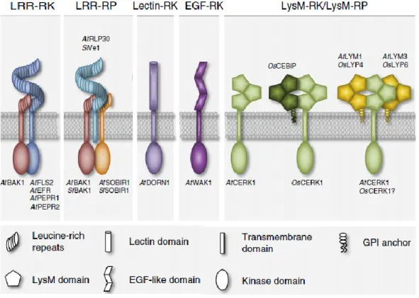
3.1 Plant surface immune receptors
Plants can detect pathogen invasion by the recognition of self or nonself-derived patterns either on the cell surface or in the cytoplasm. Plant cell surface immune receptors, frequently named Pattern Recognition Receptors (PRR) typically recognize small epitopes from invasion patterns present in the plant apoplast (Boller and Felix, 2009; Zipfel, 2008). The two main classes of PRRs are the Receptor Like Kinases (RLKs) composed of an ectodomain, a transmembrane domain and an intracellular kinase domain and the Receptor Like Proteins
(RLPs) that are similar to RLKs in their structural organization but do not possess an
intracellular kinase domain (Macho and Zipfel, 2014; Böhm et al., 2014). The extracellular domains of PRRs are very diverse and can contain e.g. Leucine Rich-Repeats (LRR), lysine motifs (LysMs), lectin motifs or and epidermal growth factor (EGF)-like domains (Figure 8). LRR-type PRRs usually bind to peptides such as flg22, a fragment of bacterial flagellin whereas LysM and EGF-type PRRs recognize carbohydrate-containing molecules such as chitin, bacterial peptidoglycans, extracellular ATP, or plant-cell-wall-derived oligogalacturonides (Brutus et al., 2010; Choi et al., 2014; Kaku et al., 2006; Miya et al., 2007; Willmann et al., 2011).PRRs ligand perception occurs via ectodomains and induces the formation of PRR homo- or hetero-complexes, the activation of intracellular kinase domains and the phosphorylation of substrates that contribute to intracellular signal transduction and activation of plant defense responses.
For instance, in the model plant Arabidopsis thaliana, LYK5 (Lysin motif receptor kinase 5) and CERK1 (Chitin Elicitor Receptor Kinase-1) cooperate in the perception of chitin-oligomers (Miya et al. 2007; Wan et al. 2008; Cao et al. 2014). On the contrary to CERK1 that only possess moderate chitin-binding affinity, LYK5 binds chitin oligomers with very high affinity and acts as the primary chitin receptor. Chitin-binding by LYK5 induces formation of a LYK5/CERK1 hetero-complex resulting in phosphorylation of CERK1 and activation of immune signaling. Interestingly, LYK5 like other LysM RLKs involved in Nod factor receptor (NFR) perception lacks intracellular kinase activity.
In monocotyledonous plants, chitin perception seems to occur by a slightly different mechanism. In rice e.g., CERK1 is recruited by the LysM-RLP CEBiP (Chitin elicitor-binding protein) and LYP4-LYP6 receptors (Couto and Zipfel, 2016). However, in this case CEBiP appears to be the primary high affinity chitin-binding receptor(Kaku et al. 2006; Liu et al. 2012). After chitin binding, CEBiP forms a hetero-oligomeric receptor complex with OsCERK1, the rice ortholog of AtCERK1 which only possess one extracellular LysM domain and does not bind chitin. Subsequently, OsCERK1 activates chitin-mediated signaling and triggers immunity. These two examples illustrate that pathogen ligand perception mediated by PRRs can be different between plants (Shimizu et al., 2010).
Figure 8. Plant surface immune receptors belong to two main different classes: receptor like kinases (RLK) and receptor like proteins (RLP). RLKs are composed of an ectodomain, a
transmembrane domain and an intracellular kinase domain. RLP also have an extracellular and transmembrane domain but lack intracellular kinase domain. After invasion pattern perception both RLKs and RLPs hetero-complexes with downstream acting RLKs. This leads to the
activation of intracellular signal transduction and triggers plant immunity. Adapted from Bohm et al. 2014
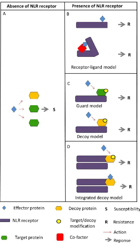
3.2 Intracellular immune receptors
Plants possess intracellular immune receptors that mediate recognition of both modified ‘host-self’ and invasion patterns in the cytoplasm (Jones and Dangl, 2006; Dodds and Rathjen, 2010c; Cook et al., 2014). The largest family of plant intracellular immune receptor proteins is the Nucleotide-binding domain and leucine-rich repeat proteins (NLRs). NLRs belong to the Signal Transduction ATPase with Numerous Domains (STAND) super family of proteins that are regulated by nucleotide-binding and intramolecular domain interactions (Lukasik and Takken, 2009; Danot, 2015). NLRs are present in all eukaryotic organisms and act as molecular switches in the regulation of various processes such as activation of immune responses, regulation of abiotic stresses and apoptosis (Goverse 2012; Jacob et al. 2013; Collier & Moffett 2009; Bernoux et al. 2016; Takken &).
NLRs present a modular architecture with a central nucleotide-binding domain (NB-ARC), a Leucine-rich repeat domain (LRR) at the C-terminus and a coiled-coil (CC) or a Toll Interleukin-1-like receptor (TIR) domain at the N-terminus (Jacob et al., 2013). Typically, NLRs are arranged in TNL or CNL (TIR (T), CC (C), NB-ARC (N), and LRR (L)) configuration but alternative configurations such as “truncate” forms, TNTNL, TNLT, CNNL, TCNL can be also found (Figure 9) (Meyers et al., 2003, 2002; Jacob et al., 2013). In solanaceas an N-terminal domain different to CC and TIR has been frequently found. This domain is called SD (solanacea domain) because is restrict to Solanaceae and is usually found in SDCNL configuration (Lukasik-Shreepaathy et al., 2012).
NLRs can also carry non-canonical domains integrated at low frequencies (Césari, et al. 2014; Sarris et al. 2016; Kroj et al. 2016). These integrated domains correspond to a wide range of molecular and functional categories such as signal transduction proteins, transcription factors or metabolic enzymes. Interestingly, many of them are present in regulators or actors of plant
immunity and/or in targets of pathogen effectors. For instance, RIN4 one of the most studied effector targets which interacts with many NLRs including RPS2 and RPM1 has been found to be fused to different NLRs in different plants such as rice, barley and apple (Sarris et al., 2016). In addition, many of the integrated domains fused to plant NLRs have been found to interact with effectors in targeted studies or effector interactome screens (Sarris et al., 2016). This suggests that effector recognition is a general feature of integrated domains similar to what has been demonstrated experimentally for Pik-1 and RRS1 (Maqbool et al., 2015a; Le Roux et al., 2015; Sarris et al., 2015).
The majority of NLRs present a TIR-NB-ARC-LRR or CC-NB-ARC-LRR configuration but alternative organization of these domain are also found. Many different non-canonical domains can also been fused to NLRs mainly in the C-terminus but also in different other positions such as the N-terminus or between the CC or TIR and the NB-ARC. Adapted from Jacob et al 2013
3.3 Function and structure of canonical NLR domains
CC and TIR – the N-terminal domains
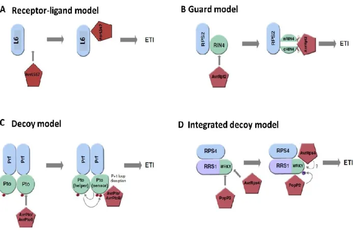
Functional analysis of CC and TIR domains suggest that both domains are involved in downstream signaling and cell death triggering (Qi and Innes, 2013). Transient expression of the TIR1-248 fragment from the flax resistance proteins L10, L6, L2 and L7 triggered effector-independent cell death in flax leaves (Frost et al., 2004; Bernoux et al., 2011b). Point mutations in conserved amino acids of the L6- TIR1-248 domain abolished cell death induction but did not affect the interaction between L6 and its cognate avirulence effector protein AvrL567 from Melampsora lini showing that the TIR domain from L6 is not required for effector binding but is necessary and sufficient for immune signaling (Bernoux et al., 2011b). Furthermore, it was shown that the L6-TIR domain self-associates and forms homodimers that are required for signaling since L6-TIR mutants impaired in self-association lost the ability to trigger cell death (Bernoux, et al. 2011).
Similarly, the CC domain of the barley resistance protein MLA is sufficient to induce cell death and self-associates in vivo, even in the absence of the Avr effector. As with the L6-TIR domain, self-association of the MLA-CC domain is required for immune signaling (Maekawa, et al. 2011). However, isolated CC or TIR domains are not always able to trigger cell death and, in certain cases, others NLR domains seem to activate immune signaling. For example, the overexpression of the of the CC domain of the resistance proteins Rx or RPS5 from potato and
A. thaliana respectively did not induce cell death but the overexpression of the Rx NB domain
did suggesting that in some cases the NB domain is engaging downstream signaling components and triggers immune signaling (Rairdan et al., 2008; Ade et al., 2007). These
different results may reflect important differences about the signaling mechanisms used by NLRs to trigger immunity.
Furthermore, many effector targets such as Pto, RIN4 and PBS1 has been shown to interact with the N-terminal domain of resistance proteins (Mackey et al., 2003; Mucyn et al., 2006; Ade et al., 2007) indicating that this domain is involved in effector recognition. For instance, N-terminal domain SD of the tomato resistance protein Prf interacts with the kinase effector target protein Pto and mediate recognition to the effector proteins AvrPto and AvrPtoB from
Peudomonas syringae (Mucyn et al. 2006; Balmuth & Rathjen 2007; Saur et al. 2015). Similarly,
the CC domain of the resistance gene RPS5 has been shown to associate with PBS1 prior to PBS1 cleavage by the effector protein AvrPphB (Ade et al., 2007; Qi et al., 2012).
To date only the crystal structures of the isolated MLA10 and Rx1 CC domains have been resolved (Maekawa, et al. 2011; Hao et al. 2013). Despite the sequence similarity between these domains, structural analyses revealed that both domains adopt different topologies. The MLA10-CC domain is formed by three different α-helices connected by loops and forms homodimers (Figure 10) whereas CC from Rx1 consists of a more compact fold composed of four different α-helices in a helix bundle (Figure 10) that does not dimerize but instead interact with the conserved domain WPP (Trp-pro-pro) of the RanGAP2 protein which is required for Rx1 function.
The crystal structures of the TIR domain from the plant NLRs L6, RPS4 and RRS1 has been determined (Bernoux, et al. 2011; Williams et al. 2014). These domains consist of a flavodoxin-like fold formed by a five-stranded parallel β-sheet surrounded by five α-helices (Figure 10). Surfaces involved in the formation of L6 and RPS4 TIR domain homodimers and RRS1/RPS4 TIR domain heterodimers has been also identified (Bernoux, et al. 2011; Williams et al. 2014).
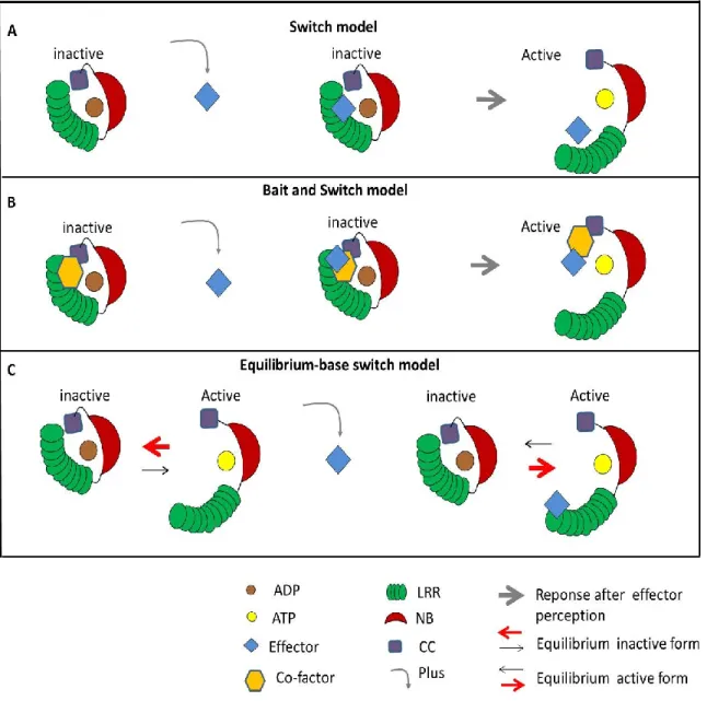
NB-ARC the central switch domain
The NB-ARC domain is the most conserved domain in plant and animal NLRs and acts as a molecular switch that translates the recognition of effector proteins into signal initiation through intra and intermolecular interactions and nucleotide binding (Hu et al. 2013; Takken & Goverse 2012). Indeed, the NB-ARC domain consists of a nucleotide-binding pocket. In the inactive or “off” state, the nucleotide-binding pocket adopts a “closed” configuration where ADP is preferentially bound and stabilizes the closed structure by mediating intramolecular interactions. The activation or “on” state of the NB-ARC is mediated by the release of the ADP which is replaced by ATP (Bernoux et al., 2011a; Williams et al., 2011b). After ATP-binding the intramolecular interactions of the protein are modified and the pocket adopts an “open” configuration that is required to mediate defense responses. Thus, the central NB-ARC domain functions as a molecular switch that fluctuate between an “off” and “on” state depending on ADP- or ATP-binding respectively. The most conserved part of the NB domain is the Walker-A motif, also called phosphate-binding loop (P-loop) which is a glycine-rich flexible loop that is crucial for ATP-binding. Indeed, the p-loop coordinates together with other amino acids a magnesium cation that binds the β and ϒ phosphates and thereby properly positions ATP. In addition, a highly conserved lysine residue in the P-loop interacts directly with the β and ϒ phosphate groups of the nucleotide and is indispensable for its binding (Walker et al., 1982).
Mutations in the NB-ARC domains that weaken ATP binding or stabilize the fixation of ADP result in loss-off function while mutations that weaken ADP-binding or strengthen ATP binding lead to gain of function, autoactive NLR mutants. One of the conserved structural motifs that can be distinguished in the NB-ARC domain is the MHD (Met-His-Asp) motif. Direct mutagenesis in MHD motif of many NLRs result in a spontaneous induction of the defense and cell death responses (Bendahmane et al. 2002; Howles et al. 2005; van Ooijen et al. 2008; Williams, et al. 2011). For instance, site-directed mutation of key residues within the MHD motif of the resistance protein M resulted in autoactivation and ATP binding whereas mutations in important residues within the P-loop motif of M resulted in a loss of nucleotide binding and the inactivation of the resistance protein (Williams, et al. 2011)