HAL Id: hal-02880050
https://hal.archives-ouvertes.fr/hal-02880050
Submitted on 24 Jun 2020HAL is a multi-disciplinary open access
archive for the deposit and dissemination of sci-entific research documents, whether they are pub-lished or not. The documents may come from teaching and research institutions in France or abroad, or from public or private research centers.
L’archive ouverte pluridisciplinaire HAL, est destinée au dépôt et à la diffusion de documents scientifiques de niveau recherche, publiés ou non, émanant des établissements d’enseignement et de recherche français ou étrangers, des laboratoires publics ou privés.
Cultures of Dinophysis sacculus, D. acuminata and
pectenotoxin 2 affect gametes and fertilization success of
the Pacific oyster, Crassostrea gigas
Sylvain Gaillard, Nelly Le Goïc, Florent Malo, Myrina Boulais, Caroline
Fabioux, Lucas Zaccagnini, Liliane Carpentier, Manoëlla Sibat, Damien
Réveillon, Véronique Sechet, et al.
To cite this version:
Sylvain Gaillard, Nelly Le Goïc, Florent Malo, Myrina Boulais, Caroline Fabioux, et al.. Cultures of Dinophysis sacculus, D. acuminata and pectenotoxin 2 affect gametes and fertilization success of the Pacific oyster, Crassostrea gigas. Environmental Pollution, Elsevier, 2020, 265, Part B, pp.114840. �10.1016/j.envpol.2020.114840�. �hal-02880050�
Title
1
Cultures of Dinophysis sacculus, D. acuminata and pectenotoxin 2 affect gametes and 2
fertilization success of the Pacific oyster, Crassostrea gigas 3
4
Authors
5
Sylvain Gaillard1, Nelly Le Goïc2,Florent Malo1, Myrina Boulais2, Caroline Fabioux2, Lucas 6
Zaccagnini2, Liliane Carpentier1, Manoella Sibat1, Damien Réveillon1, Véronique Séchet1, 7
Philipp Hess1, Hélène Hégaret2 8
1 IFREMER, DYNECO, Laboratoire Phycotoxines, F-44000 Nantes, France
9
2 Univ Brest, CNRS, IRD, Ifremer, LEMAR, F-29280 Plouzané, France
10
Author for correspondence: philipp.hess@ifremer.fr; sylvain.gllrd@gmail.com Tel +33 11 (0)2 403 743 76, Fax +33 (0)2 403 742 41 12 13 14 15 16 17 18 19
Highlights
20
Dinophysis sacculus and D. acuminata increased mortality of Crassostrea gigas oocytes 21
Exposure of oocytes and spermatozoa to 0.5 cells mL-1 of D. sacculus decreased subsequent 22
fertilization success 23
Oyster gametes were negatively affected by exposure to whole culture or resuspended cells of 24
Dinophysis spp. 25
5 nM of PTX2 decreased fertilization success of oocytes and 500 nM of PTX2 increased ROS 26
production; OA showed no effect 27
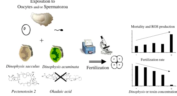
Observed effects may be due to either cell to cell contact or PTX2 or other bioactive compounds 28 or a combination 29 30 Graphical abstract 31 End 32 33 Abstract 34
Harmful algal blooms (HABs) of toxic species of the dinoflagellate genus Dinophysis are a threat 35
to human health as they are mainly responsible for diarrheic shellfish poisoning (DSP) in the 36
consumers of contaminated shellfish. Such contamination leads to shellfish farm closures causing 37
major economic and social issues. The direct effects of numerous HAB species have been 38
demonstrated on adult bivalves, whereas the effects on critical early life stages remain relatively 39
unexplored. The present study aimed to determine the in vitro effects of either cultivated strains 40
of D. sacculus and D. acuminata isolated from France or their associated toxins (i.e. okadaic acid 41
(OA) and pectenotoxin 2 (PTX2)) on the quality of the gametes of the Pacific oyster Crassostrea 42
gigas. This was performed by assessing the ROS production and viability of the gametes using 43
flow cytometry, and fertilization success using microscopic counts. Oocytes were more affected 44
than spermatozoa and their mortality and ROS production increased in the presence of D. 45
sacculus and PTX2, respectively. A decrease in fertilization success was observed at 46
concentrations as low as 0.5 cell mL-1 of Dinophysis spp. and 5 nM of PTX2, whereas no effect 47
of OA could be observed. The effect on fertilization success was higher when both gamete types 48
were concomitantly exposed compared to separate exposures, suggesting a synergistic effect. Our 49
results also suggest that the effects could be due to cell-to-cell contact. These results highlight a 50
potential effect of Dinophysis spp. and PTX2 on reproduction and recruitment of the Pacific 51 oyster. 52 53 Capsule 54
Dinophysis sacculus, D. acuminata and pectenotoxin 2 increase oocyte mortality and ROS 55
production, and decrease fertilization success of the Pacific oyster, Crassostrea (=Magallana) 56 gigas. 57 58 Keywords 59
Dinophysis spp.; okadaic acid; pectenotoxins; oyster gametes; fertilization success 60
Abbreviations
62
DSP, diarrheic shellfish poisoning; DSTs, diarrheic shellfish toxins; DCFH-DA, 2′7’-63
dichlorofluorescein diacetate; DTX1, dinophysistoxin 1; DTX2, dinophysistoxin 2; DTXs, 64
dinophysistoxins; Extra, extracellular; FCM, flow cytometry; FSC, forward scatter; HABs, 65
harmful algal blooms; Intra, intracellular; OA, okadaic acid; PTX2, pectenotoxin 2; PTX2eq, 66
pectenotoxin 2 equivalent; PTXs, pectenotoxins; PI, propidium iodide; ROS, reactive oxygen 67
species; SSC, side scatter; FSSW, filter-sterilized sea water; FSW, filtered sea water; UHPLC-68
LRMS/MS, ultra-high performance liquid chromatography coupled to low resolution tandem 69 mass spectrometry 70 71 Introduction 72
Harmful algal blooms (HABs) of toxic microalgae are increasing in terms of frequency, 73
intensity and duration due, in part, to climate change and eutrophication (Gobler et al., 2017; 74
Hallegraeff, 1993; Wells et al., 2019). Toxins associated with HABs can accumulate in marine 75
bivalves (Landsberg, 2002; Shumway, 1990; Simões et al., 2015), causing a threat to human 76
health through direct contact with toxins or consumption of contaminated organisms (Hallegraeff, 77
2010, 1993; Van Dolah, 2000). Consequently, national surveillance programs monitoring 78
phytoplankton and phycotoxin concentrations in water and bivalves have been implemented (e.g. 79
REPHY in France). The European Council has set a maximum limit of 160 µg OA eq. per kg of 80
fresh whole bivalve meat (EU Commission, 2011), above which shellfish harvesting (farming 81
and recreational) is forbidden in order to protect human consumers (Nielsen et al. 2012). 82
Shellfish farming is an important economic sector worldwide. In France, the Pacific 83
oyster, Crassostrea gigas (= Magallana gigas; Thunberg, 1793) represents the majority of annual 84
shellfish sales (ca. 118,000 tons; France Agrimer, 2018). Shellfish farmers in France annually 85
suffer economic losses due to the presence of several toxic species of the genus Dinophysis 86
(Ehrenberg, 1841; Marcaillou et al., 2005; Trainer et al., 2020). Indeed, D. acuminata and D. 87
sacculus are the main responsible of shellfish farm closures, that can last for several weeks per 88
year (Belin and Soudant, 2018; Marchand et al., 2009). Along French coasts, Dinophysis spp. are 89
regularly observed at a concentration of 102 cells L-1 (Figure S2, REPHY, 2019), which is similar 90
to concentrations typically reported in the literature (Reguera et al., 2012). However, blooms of 91
Dinophysis spp. can occasionally reach cell densities up to 103 – 107 cells L-1 (reviewed in
92
Reguera et al., 2012), including one instance of 8 x 10⁵ cells L-1 reported in France (REPHY,
93
2019). 94
These dinoflagellates can produce two types of lipophilic toxins, okadaic acid (OA) and 95
its analogs dinophysistoxins (DTXs), and pectenotoxins (PTXs; Marcaillou et al. 2005, Reguera 96
et al. 2014). Okadaic acid and DTXs are responsible for diarrheic shellfish poisoning (DSP) in 97
humans following shellfish consumption (Lawrence et al., 2000; Reguera and Pizarro, 2008), 98
with symptoms that include diarrhea, nausea, vomiting and abdominal pain (Yasumoto et al., 99
1978). In contrast, PTXs are not considered diarrheic shellfish toxins (DSTs) as they do not 100
cause diarrhea in humans (Matsushima et al., 2015). However, PTXs are lethal to mice by 101
intraperitoneal injection (Miles et al., 2004). 102
While HABs have mostly been studied in relation to public health, another fundamental 103
issue is the direct effect they have on filter-feeding bivalves (Landsberg, 2002; Matsuyama et al., 104
2001; Shumway and Cucci, 1987; Sandra E. Shumway, 1990). National monitoring programs 105
along the French Atlantic coast indicate that spawning, development and recruitment of larvae 106
may co-occur with Dinophysis spp. (Figure S2; Pouvreau et al., 2016; REPHY, 2019). While 107
adults and juvenile bivalves can mechanically escape toxic microalgae by cessation of filtration 108
and closing their shells (Hégaret et al., 2007), the planktonic early life stages such as gametes and 109
embryos are directly exposed to HABs and their toxins in the water column and appear more 110
sensitive than adults (Castrec et al., 2019; Glibert et al., 2007; Stoecker et al., 2008; Wang et al., 111
2006; Yan et al., 2001). 112
Many studies have focused on the effects of toxic dinoflagellate species on oyster 113
gametes, embryos and larvae, e.g. for the genera Alexandrium (Banno et al., 2018; Basti et al., 114
2015a; Castrec et al., 2019, 2020; Matsuyama et al., 2001; Mu and Li, 2013), Karenia (Leverone 115
et al. 2006, Rolton et al. 2014, 2015, 2016, Basti et al. 2015a), Heterocapsa (Basti et al., 2013, 116
2011), Gymnodinium (Matsuyama et al., 2001), Karlodinium and Prorocentrum (Glibert et al., 117
2007; Stoecker et al., 2008). Nevertheless, due to the mixotrophy of toxic species of the genus 118
Dinophysis and the resulting difficulty in their cultivation until recently (Park et al., 2006), few 119
studies have investigated the effects of Dinophysis spp., their toxins or combinations of both on 120
bivalves, such as oysters. The few available studies indicate that Dinophysis spp. producing PTXs 121
induce hypersecretion of mucus and pseudofeces, paralysis, alteration of the tissues within the 122
digestive gland and reduced escape response in adult scallops (Basti et al., 2015b). Mccarthy et 123
al., (2014) demonstrated that exposure of adult Pacific oysters and blue mussels to OA increased 124
DNA fragmentation. Further studies also highlighted modified hemocyte functions in both 125
Mediterranean mussels (Malagoli et al., 2008; Prado-Alvarez et al., 2012) and carpet shell clams 126
(Prado-Alvarez et al., 2013) exposed to Dinophysis spp. and their toxins. 127
The present study investigated the in vitro effects of whole culture, resuspended cells and 128
culture filtrate of Dinophysis sacculus (Stein, 1883), whole culture of D. acuminata (Claparède 129
and Lachmann, 1859) and certified standards of OA and PTX2 on (i) gamete cellular 130
characteristics (i.e., ROS production, mortality, and morphology), and (ii) fertilization success of 131
oocytes and spermatozoa of the Pacific oyster. 132
133
Materials and methods
134
Microalgal cultures 135
Monoclonal cultures of D. sacculus (Stein, 1883) (strain IFR-DSA-01Lt) and D. acuminata 136
(Claparède and Lachmann, 1859) (strain IFR-DAU-02Ar) were isolated in Arcachon, France, in 137
2015 and 2018, respectively. These mixotrophic species were cultivated in 0.2 µm filter-sterilized 138
natural seawater (FSSW) for D. sacculus and L1/20-Si + K/2-Si (Hernández-Urcera et al., 2018) 139
for D. acuminata at salinity 35 and fed every two days with ciliate prey Mesodinium rubrum 140
(Lohmann, 1908) (strain MBL-DK2009) at a ratio of 1: 1 (predator: prey) according to Park et al. 141
(2006). The ciliate M. rubrum was fed three times a week with the cryptophyte Teleaulax 142
amphioxeia (Conrad) (Hill, 1992) (strain AND-0710). Both M. rubrum and T. amphioxeia were 143
cultivated in flasks respectively in L1/20-Si and L1-Si (Guillard and Hargraves, 1993) and 144
diluted every two days for the ciliates and every week for the cryptophyte. All cultures were 145
maintained at 17.8 ± 0.6 °C, at a light intensity of ~ 100 µmol photons m-2 s-1 provided by cool-146
white and pink fluorescent tubes (fluora and cool-white fluorescent light, Osram, Munich, 147
Germany) and a 12: 12 (L: D) cycle (Table S1). To increase the biomass of Dinophysis spp., 148
cultures were fed at a ratio of 1: 10 (predator: prey) for 4 months before the experiment. One 149
week before the experiment, cultures of Dinophysis spp. were filtered on a nylon sieve (mesh 150
11 µm) and gently rinsed with 75 mL of FSSW to remove any cryptophyte and ciliate. Cultures 151
were then resuspended in 20 mL of FSSW and starved for one week before the experiment to 152
obtain cells from the mid exponential growth phase on the day of the experiment. 153
154
Experimental design 155
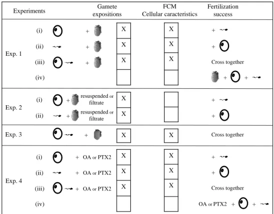
All experiments are summarized in Figure S1 and performed with 3 to 11 replicates. A replicate 156
is either a pool of several females or males, or one male or female, as detailed below. 157
Experiment 1 (Exp. 1) – Effect of the whole culture of D. sacculus upon gametes 158
The aim of Exp. 1 was to determine the effect of whole cultures of D. sacculus on gamete cellular 159
characteristics and fertilization success. In total, four fertilization experiments were performed by 160
crossing exposed or non-exposed oocytes and/or spermatozoa to D. sacculus. 161
- (i) Oocytes exposed to D. sacculus (n = 8 pools of gametes, each from 3 different 162
females) were crossed with non-exposed spermatozoa (a pool of spermatozoa from 5 163
males) 164
-or (ii) Spermatozoa exposed to D. sacculus (n = 8 pools of gametes, each from 3 165
different males) were crossed with non-exposed oocytes (a pool of oocytes from 5 166
females). 167
- (iii) In addition, fertilization success was determined after exposure of both oocytes and 168
spermatozoa to D. sacculus (n = 4 pools of gametes, each from 3 different organisms). 169
- (iv) The effects of whole cultures of D. sacculus on fertilization success was also 170
investigated by exposing gametes only during fertilization. Oocytes, spermatozoa and D. 171
sacculus (at final concentrations of 0 (control), 0.5, 5, 50 and 500 cells mL-1) were put in
172
contact simultaneously in FSSW (n = 11 pools of gametes, each from 3 different 173
organisms). 174
Briefly, for (i), (ii) and (iii); oocytes or spermatozoa were exposed for 2 h in glass vials at 20 ± 1 175
°C to the whole culture of D. sacculus at a final concentration of 0 (control), 0.5, 5, 50 and 500 176
cells mL-1 in FSSW. These D. sacculus cell concentrations were selected to mimic the cell 177
densities in natural blooms occurring in France, from typical (1 x 10² cells L-1) to exceptionally
178
dense blooms (8 x 10⁵ cells L-1, Figure S2, REPHY, 2019). 179
180
Experiment 2 (Exp. 2) – Effect of resuspended cells and culture filtrate of D. sacculus upon 181
gametes 182
Experiment 2 was designed to determine the respective effect of resuspended cells and 183
extracellular medium (culture filtrate) of D. sacculus on gametes. (i) Oocytes or (ii) spermatozoa 184
(n = 5 individual females or males) were exposed for 2 h in glass vials either to the whole D. 185
sacculus culture at 500 cells mL-1 (similar to Exp. 1), or to D. sacculus cells only, obtained by
186
filtration (11 µm-mesh nylon sieve) of a culture at 500 cells mL-1 and resuspended in FSSW (to 187
remove the extracellular metabolites) or to culture filtrate obtained by filtration (0.2 µm-mesh 188
nylon sieve) of the whole culture (500 cells mL-1) to measure the effect of only the extracellular 189
metabolites of living cultures. After exposure, gamete cellular characteristics and fertilization 190
success were determined using gametes exposed to either whole culture or to resuspended cells of 191
D. sacculus or to its culture filtrate and a pool of unexposed spermatozoa or oocytes from 5 192
oysters. 193
194
Experiment 3 (Exp. 3) – Effect the whole culture of D. acuminata upon gametes 195
The aim of Exp. 3 was to determine the effect of a 2 h exposure of whole cultures of D. 196
acuminata (at a final concentration of 0 (control), 0.5, 5, 50 and 500 cells mL-1) on gamete
197
cellular characteristics and fertilization success of both exposed oocytes and spermatozoa (n = 4 198
pools of gametes, each from 3 different organisms), as described in Exp. 1 (iii). 199
200
Experiment 4 (Exp. 4) – Effect of okadaic acid (OA) and pectenotoxin 2 (PTX2) standards on 201
gametes 202
Experiment 4 investigated the effect of 0 (control), 5, 10, 20 and 50 nM solutions of okadaic acid 203
(OA) and pectenotoxin 2 (PTX2) in FSSW and to the corresponding methanol (MeOH) control 204
(2.3 % final) on cellular characteristics and fertilization success of (i) oocytes or (ii) spermatozoa, 205
(iii) both oocytes and spermatozoa exposed to toxins and (iv) gametes exposed to toxins only 206
during fertilization (n = 3 to 4 pools of gametes from 3 different organisms). These 207
concentrations of toxins approximately corresponded to the minimum concentration of OA (i.e. 5 208
nM) found in the studied strains of D. sacculus and D. acuminata (sum of intra and extracellular 209
toxins of 500 cells) to ca. the maximum concentration of PTX2 (i.e. 50 nM; Table 1). All 210
measurements were performed as detailed in Exp. 1. 211
Toxin analyses of Dinophysis spp. strains 213
Toxin analysis was adapted from Sibat et al. (2018) and García-Portela et al. (2018) and 214
performed on 1 mL (n=3) sub samples of Dinophysis spp. cultures collected in exponential 215
growth phase. After centrifugation (3500 g, 4 °C, 15 min), both cells and culture filtrates 216
(supernatants) were extracted. The pellet (intracellular toxins) was extracted with 0.5 mL 217
methanol and sonicated at 25 kHz for 15 min. Extracellular toxins were recovered from the 218
supernatant after liquid-liquid extraction with dichloromethane, which was evaporated under 219
nitrogen and resuspended in 0.5 mL of methanol. Subsequently, samples were filtered (0.2 µm, 220
Nanosep, MF, Pall, Northborough, MA, USA). Analyses were performed using ultra high 221
performance liquid chromatography coupled to low resolution tandem mass spectrometry 222
(UHPLC-LRMS/MS) with a UHPLC system (UFLC XR Nexera, Shimadzu, Tokyo, Japan) 223
coupled to a triple quadrupole/ion-trap mass spectrometer (API 4000 QTrap, ABSciex, Redwood 224
City, CA, USA), equipped with a turboV® ESI source (see details in García-Portela et al. 2018). 225
Certified calibration solutions of PTX2, OA, dinophysistoxin 1 and 2 (DTX1 and DTX2) were 226
obtained from the National Research Council Canada (NRCC, Halifax, NS, Canada). Intracellular 227
(intra) and total (sum of intracellular and extracellular) toxin contents were expressed on a per 228
cell basis (pg cell-1) while extracellular (extra) as equivalent (eq) pg cell-1. Pectenotoxin 2 eq 229
(PTX2eq) was the sum of pectenotoxin 2, pectenotoxin 2b, pectenotoxin 2 seco-acid and 7-epi-230
pectenotoxin 2 seco-acid, all quantified with PTX2 standard by assuming similar molar 231
responses. 232
233
Sampling and maintenance of oysters 234
Sexually mature C. gigas were collected at La Pointe du Chateau-Baie de Daoulas, France 235
(48°20’01.8”N 4°19’02.6”W) in summer 2018 and 2019 or obtained from Ifremer experimental 236
facilities as described in Castrec et al. (2019). Individuals from the field were cleaned with 237
filtered sea water (FSW) to remove sessile organisms. All oysters were maintained in an aerated 238
tank at 16 ± 1 °C with a continuous flow of FSW for one to three days before the experiments. 239
240
Collection of gametes 241
Gonads were dissected and placed in individual Petri dishes to collect gametes according to Song 242
et al., (2009) for oocytes and Boulais et al., (2015) for spermatozoa. Briefly, for each oyster, 243
gametes were collected in 10 mL FSSW and sieved through 100 µm mesh to isolate gametes 244
from gonad debris. Only motile spermatozoa and rounded oocytes were selected for the 245
experiments (Rolton et al., 2015). Spermatozoa and oocyte concentrations were determined by 246
flow cytometry (FCM) according to Le Goïc et al. (2013, 2014) and diluted in FSSW at 107 cells 247
and 105 mL-1, respectively. 248
249
Flow-cytometry analysis – morphology, viability and ROS production 250
Analyses of morphology, viability, and ROS production (i.e. cellular characteristics) of oyster 251
gametes by FCM were adapted from Le Goïc et al. (2013, 2014) and carried out with an 252
EasyCyte Plus cytometer (Guava Technologies, Millipore, Luminex Billerica, USA) equipped 253
with a 488-nm argon laser and three fluorescence detectors: green (525 ± 30 nm), yellow (583 ± 254
26 nm) and red (680 ± 30 nm). 255
For cell morphology measurements, values of the forward scatter (FSC) and side scatter (SSC), 256
respectively proxies of cell size and complexity, were used to estimate cell morphology of 257
spermatozoa and oocytes. Spermatozoa viability was assessed using double staining spermatozoa 258
solution with 2 µL of propidium iodide (PI) and 2 µL of SYBR-14 (Live/Dead® Sperm Viability 259
Kit, Molecular Probes, Eugene, USA) at final concentrations of 2 µg mL-1 and 1 µM during 10 260
min in the dark (Le Goïc et al., 2013). For oocyte viability, PI and SYBR-Green-1 (1/10,000 of 261
the commercial solution; Molecular Probes, Eugene, USA) were used (Le Goïc et al., 2014). 262
ROS production of gametes was measured by staining 200 µL of oocytes or spermatozoa solution 263
with 2 µL (final concentration of 10 µM) of dye 2’7,7’-dichlorofluorescein diacetate DCFH-DA 264
(Sigma, St Quentin Fallavier, France) for 1 h in the dark (Le Goïc et al., 2014; Vignier et al., 265
2017). 266
ROS production was expressed as percentage of control (a.u.). For both viability (expressed as 267
mortality) and ROS production measurements, oocytes and spermatozoa concentrations were 5 x 268
104 and 5 x 10⁶ cells mL-1, respectively. 269
270
Microscopy analysis – fertilization success assessment 271
To assess fertilization success, oocytes and spermatozoa were inoculated in FSSW (20 ± 1 °C) at 272
a ratio of 1: 100 (5 x 10³ oocytes: 5 x 10⁵ spermatozoa) in 12-well plates in a final volume of 4 273
mL FSSW (Boulais et al., 2017). When fertilized oocytes in the control (i.e. non-exposed oocytes 274
and spermatozoa) reached 80 % or, after 2 h of incubation, samples were fixed with 1 % 275
formaldehyde (final concentration). Fertilization success (%) was assessed under an inverted light 276
microscope (Axio observer.Z1, Zeiss, Oberkochen, Germany) by counting fertilized and 277
unfertilized oocytes (polar body extrusion from 2 to 8-cell stages vs. no polar body). For the 278
fertilization success measurements in all experiments except conditions (iv), the concentration of 279
Dinophysis spp. or toxins were minimum 30-fold lower due to dilution in FSSW and their 280
potential contribution to the observed effect were considered not significant. 281
282
Statistical analyses 283
Statistical analyses were performed on RStudio v 1.1.463. After checking the assumptions of 284
independence (Durbin-Watson test), homoscedasticity (Bartlett test) and normality (Shapiro-Wilk 285
test) of the residuals, t-test or one-way ANOVA followed by a Tukey post hoc test were 286
computed. Otherwise, Mann-Withney U or Kruskal-Wallis tests were used, followed by a 287
Conover test. Differences were considered statistically significant when P < 0.05, for a 288
significance level of α = 0.05. Values were expressed as mean ± SD. 289
290
Results
291
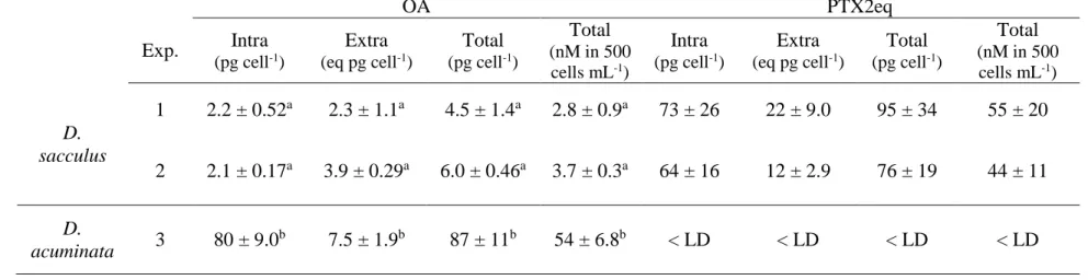
Toxin contents of Dinophysis spp. cultures 292
The D. sacculus strain synthetized OA and three PTX2 derivatives (pectenotoxin 2, 293
pectenotoxin 2b, pectenotoxin 2 seco-acid, 7-epi-pectenotoxin 2 seco-acid) whereas the D. 294
acuminata strain synthetized only OA. Neither of the two algal species produced DTX1 or DTX2 295
(Table 1). 296
For Exp. 1 and Exp. 2, D. sacculus produced similar amounts of total OA (4.5 ± 1.4 and 6.0 ± 297
0.46 pg cell-1) and total PTX2eq, i.e. sum of concentrations (95 ± 36 and 76 ± 19 pg cell-1; Table
298
1). The majority of OA was in the extracellular compartment in contrast to PTX2 which was 299
mainly intracellular. For Exp. 3 with D. acuminata, the total OA content per cell was 16-fold 300
higher (87 ± 11 pg cell-1) than D. sacculus and >90 % was intracellular (P < 0.001; Table 1). 301
302
Exp. 1 – Effect of the whole culture of D. sacculus on gametes 303
None of the tested concentrations affected spermatozoa cellular characteristics. However, 304
mortality of oocytes was 2.9-fold-higher when exposed to D. sacculus at a concentration of 305
500 cells mL-1 compared to the control (P < 0.05), whereas no effect was observed on ROS 306
production. Moreover, a significant increase in FSC was observed for the same exposure 307
condition compared to control (P < 0.05; Table 2). 308
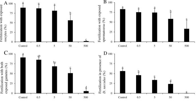
Fertilization success decreased significantly when (i) oocytes or (ii) spermatozoa were exposed to 309
50 and 500 cells mL-1 of D. sacculus compared to their respective controls (89 vs. 56 and 2 % 310
and 83 vs. 58 and 33 %, respectively; P < 0.001; Figure 1 A-B). Interestingly, a 17-fold 311
difference was noted between spermatozoa and oocytes when exposed to 500 cells mL-1 (P < 312
0.001; Figure 1 A-B) with oocytes being more sensitive. When (iii) both gametes were exposed 313
to D. sacculus, fertilization success compared to control was significantly reduced by 25, 44 and 314
93 % at 5, 50 and 500 cells mL-1, respectively (P < 0.001; Figure 1 C). The fertilization success 315
in presence of D. sacculus during the fertilization (iv) was significantly reduced by 18, 39 and 57 316
% when exposed to 0.5, 5 and 50 cells mL-1, respectively (P < 0.001). Fertilization was however, 317
totally impeded at 500 cells mL-1 (Figure 1 D). 318
319
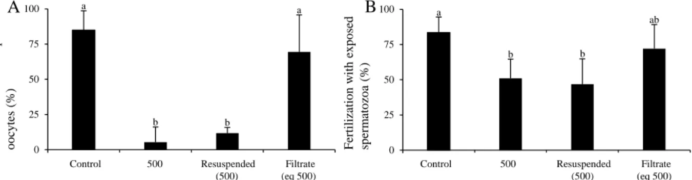
Exp. 2 – Effect of resuspended cells and culture filtrate of D. sacculus on gametes 320
The negative effect of gametes exposed to whole cultures (500 cells mL-1) was confirmed (P <
321
0.001), with fertilization success 10 times lower following exposure of oocytes vs. spermatozoa 322
(P < 0.001; Figure 2 A-B). While similar results were obtained using resuspended D. sacculus 323
cells (P < 0.05), no significant difference was noted between the controls and gametes exposed to 324
culture filtrates (Figure 2 A-B). 325
326
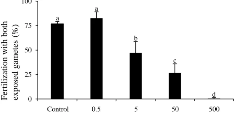
Exp. 3 – Effect the whole culture of D. acuminata on gametes 327
Only oocytes exposed to 500 cells mL-1 of D. acuminata were significantly affected, with a 2.7-328
fold higher mortality (P < 0.001) and a 16 % increase in FSC (P < 0.001; Table 2). Again, 329
spermatozoa were not affected. A significant decrease in fertilization success after exposure of 330
both gametes was observed from as few as 5 cells mL-1 (1.8-fold; P < 0.05), while fertilization
331
was almost completely inhibited at 500 cells mL-1 of D. acuminata (P < 0.001; Figure 3). 332
333
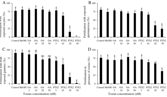
Exp. 4 – Effect of okadaic acid (OA) and pectenotoxin 2 (PTX2) standards on gametes 334
Production of ROS was around twice higher in oocytes exposed for 2 h to 50 nM of PTX2 (P < 335
0.05) compared to the control while spermatozoa were not affected by exposure to OA and PTX2 336
standards (Table 2). 337
A decrease in fertilization success was observed with (i) oocytes exposed for 2 h to 20 and 50 nM 338
of PTX2 compared to control (16 and 0 % vs. 77 %, respectively; P < 0.001; Figure 4 A). 339
Similarly, (ii) spermatozoa exposed to 20 and 50 nM of PTX2 decreased fertilization success 340
compared to control (35 and 25 % vs. 81 %; P < 0.001; Figure 4 B). However, oocytes exposed 341
to 50 nM, exhibited a more pronounced effect than exposed spermatozoa on fertilization (P < 342
0.05; Figure 4 A-B). Exposure of (iii) both oocytes and spermatozoa to 5, 10, 20 and 50 nM of 343
PTX2 reduced the fertilization success by 25, 37, 78 and 97 % compared to control (P < 0.01; 344
Figure 4 C). The fertilization in presence of toxins (iv) was significantly diminished only at a 345
concentration of 50 nM of PTX2 (32 %) compared to control (76 %; P < 0.001; Figure 4 D). 346
Neither OA (at any of the concentrations tested) nor MeOH in the control affected the 347 fertilization success. 348 349 Discussion 350
The present work demonstrated that Dinophysis sacculus and D. acuminata as well as one of 351
their toxins PTX2 impaired cellular characteristics and fertilization success in gametes of the 352
Pacific oyster, Crassostrea gigas, a species of commercial interest. 353
The concentrations of Dinophysis spp. used in this exposure study were selected because of their 354
environmental relevance, since concentrations of > 1000 cells L-1 were frequently observed in the 355
four regions studied on the French Atlantic coast, whereas concentrations of > 10,000 cells L-1
356
were occasionally observed in two of the regions, including major oyster production sites, i.e. the 357
Bay of Arcachon and the Bay of Brest (Figure S2). These concentrations, observed in France 358
over a ten-year period, are moderate compared to other areas affected by Dinophysis spp. blooms, 359
e.g. India or Norway, where concentrations of up 1.5 x 10⁶ and 2.3 x 10⁷ cells L-1 have been 360
observed (Reguera et al., 2012). e 361
The toxin exposure concentrations (i.e. from 5 to 50 nM) corresponded to the maximum amounts 362
of OA and PTX2, respectively, produced by 500 cells of D. sacculus in our experixcment. 363
Noteworthy, PTX2 caused an increase in ROS production of oocytes exposed to 50 nM, which 364
could reflect a stimulation of oocyte metabolism or cellular stress. Indeed, the production of ROS 365
is a key mechanism involved in stress responses (Kadomura et al., 2006), but an excess of ROS 366
could lead to cellular toxic effects such as destruction of membrane integrity by lipid 367
peroxidation, DNA damage and associated alteration of cell functioning and ultimately cell death 368
(Cavallo et al., 2003; Landsberg, 2002; Lesser, 2006). This may explain the observed reduced 369
fertilization success of oocytes exposed to PTX2. Similarly, Le Goïc et al. (2014) observed an 370
increased ROS production of oocytes in the presence of the toxic dinoflagellate Alexandrium 371
minutum, and suggested this increase production may have reduced oocyte quality. 372
Pectenotoxin 2 at concentrations as low as 20 nM reduced fertilization success when either 373
oocytes or spermatozoa were pre-exposed. This is true also at concentration as low as 5 nM of 374
PTX2 when oocytes and spermatozoa were both pre-exposed. This suggests that both oocytes and 375
spermatozoa were negatively affected by PTX2 and that the effect of PTX2 on oyster gametes is 376
cumulative. Secondly, this toxicity is likely mediated by a different mechanism than ROS 377
production since no increase in ROS production was observed below 50 nM concentration of 378
PTX2. 379
Using mammalian and finfish cell lines (e.g. human, rat, rabbit, salmon), PTXs have been shown 380
to interfere with actin assembly/disassembly, thereby affecting cell cytoskeletal functions and 381
leading to cell death, at concentrations ranging from nM to µM (Ares et al., 2005; Dominguez et 382
al., 2010; Spector et al., 1999). It has been shown that PTX2 causes actin depolymerization 383
(Dominguez et al., 2010), sequestration of monomeric actin (at a concentration of 20 nM; Spector 384
et al., 1999), disrupted F-actin (Hori et al., 1999), and inhibited actin polymerization by a capping 385
process at the barbed-end of F- and G-actin (Allingham et al., 2007). The reduced fertilization 386
success observed in this study could be associated with impairment of the oocyte and 387
spermatozoan cytoskeleton by PTX2, as well as fertilization itself since actin polymerization is a 388
crucial mechanism in oysters, involved in spermatozoan motility and the penetration of the 389
oocyte (Ledu and McCombie, 2003). In the literature, the indirect evidence of the involvement of 390
Dinophysis spp. in mortalities observed in the natural environment (reviewed in Landsberg 2002, 391
Basti et al., 2015b) were almost exclusively related to D. caudata, a producer of high cellular 392
contents of PTX2 (Basti et al., 2015b, 2015c; Fernández et al., 2006; Marasigan et al., 2001). The 393
action of PTX2 on actin in oyster gametes would be worthy of investigation in further studies. 394
395
Okadaic acid, in contrast to PTX2, affected neither the gametes nor fertilization success, at 396
concentrations up to50 nM. Okadaic acid is reported to be an inhibitor of serine/threonine protein 397
phosphatase 1 and 2 activities (Bialojan and Takai, 1988; Mccarthy et al., 2014) and is also 398
believed to be a tumor promoter in humans (Lago et al., 2005). This toxin induced chromosome 399
loss, apoptosis and DNA damages in mammalian cell lines (see references in Prado-Alvarez et 400
al., 2013). Okadaic acid has been shown to induce an increase in DNA fragmentation in adult 401
Pacific oyster and blue mussel (Mccarthy et al., 2014) and modified hemocyte functions in 402
several bivalve species (Malagoli et al., 2008; Prado-Alvarez et al., 2013, 2012) at concentrations 403
between 1.2 to 50 and 10 to 500 nM, respectively. The absence of effects of OA on oyster 404
gametes in this study could be due to the relatively short exposure time and low OA 405
concentrations, reaching respectively 2 h and a maximum of 50 nM concentration of certified 406
standard and 4.5 nM concentration of extracellular OA produced by 500 cells of D. acuminata in 407
our experiment. 408
The main structural difference between OA and PTX2 is that PTX2 is a macrocyclic lactone (i.e. 409
cyclic ester). Pectenotoxin 2 biological activity on cytoskeletal dynamics is clearly associated 410
with the macrocyclic ester as the activity disappeared when the esters were hydrolysed and the 411
macrocycle was opened (Allingham et al., 2007; Ares et al., 2007; Miles et al., 2006). The 412
resulting analogue, pectenotoxin 2 seco-acid, is structurally very similar to OA, and is not active 413
on the cytoskeleton (neither is OA). Interestingly, PTX2 has a high structural similarity with 414
goniodomin A, another algal macrocyclic lactone, which has also been reported to affect the 415
cytoskeleton via F-actin (Espiña et al., 2016). In addition, it should be noted that among the three 416
species of Alexandrium that produce goniodomins (Harris et al., 2020), A. monilatum has been 417
clearly associated with fish kills since the middle of the last century (Howell, 1953) and more 418
recently also with shellfish mortalities (Harding et al., 2009; May et al., 2010). 419
Our study also revealed that PTX2 is likely not to be the only bioactive compound responsible for 420
the toxicity of Dinophysis spp. on oyster gametes and fertilization success. Firstly, fertilization 421
success was decreased in the presence of 0.5 cell mL-1 of D. sacculus during fertilization, which 422
corresponded to a non-detectable amount of PTX2, while 50 nM of PTX2 were needed to obtain 423
similar effects. Secondly, our strain of D. acuminata did not produce PTX2 but also caused a 424
decrease in fertilization success, when both gametes were pre-exposed to only 5 cells mL-1, and, 425
as described above, these effects could also not be attributed to OA. 426
Additionally, the present study indicated that the decreased fertilization success, specifically for 427
D. sacculus, was derived from cells and not from the extracellular compartment, as filtrate had no 428
activity, unlike resuspended cells. This observation could be explained by cell-to-cell contact and 429
the effect of (a) mechanical damages and/or (b) surface-bound toxins and/or (c) quick release of 430
intracellular bioactive compounds (Landsberg, 2002). 431
Contact with Dinophysis spp. cells, or by the mean of feeding peduncle (Ojamäe et al., 2016), 432
may have resulted in (a) mechanical damage to the membranes of oyster gametes, as suggested 433
by the increase in FSC morphological parameter of oocytes. This proxy of cell size could indicate 434
a swelling of the cells, when exposed to 500 cells mL-1 of D. sacculus associated to an increase in 435
mortality. 436
Furthermore, it has been hypothesized that (b) the presence of toxins on the cell surface of 437
another HAB species, H. circularisquama can affect pearl oyster larvae after contact (Basti et al., 438
2011). In addition, Mu and Li (2013) suggested that the release by A. catenella of surface-located 439
toxins may affect Pacific oyster egg hatching success. Surface-bound toxins from Dinophysis 440
spp., Alexandrium spp. and Heterocapsa spp. have not been reported yet but could potentially 441
explain the observed effect on gametes and fertilization success. 442
Another explanation could be (c) a rapid release of intracellular compounds, different from the 443
already known and characterized toxins, with activity towards oyster gametes. While most 444
attention has been paid to toxins affecting humans (i.e. DSTs in a sanitary context), other 445
bioactive compounds from Dinophysis spp. have been overlooked despite some interesting 446
observations. Basti et al. (2015b) observed mortality in adult mollusks fed D. caudata 447
independently of PTX2 content, thus they hypothesized the presence of unknown toxins and/or 448
other bioactive compounds. Similarly, Mafra et al. (2016) hypothesized that the mechanism of 449
prey capture of several toxic species of Dinophysis spp. involved uncharacterized allelochemical 450
compounds, other than the known DSTs, which debilitates M. rubrum. The existence of such 451
allelochemicals for Karenia brevis affecting C. virginica larvae (Rolton et al., 2014) as well as 452
for A. minutum affecting early life stages and adults of C. gigas has also been observed (Castrec 453
et al., 2020, 2018). Bioguided-fractionation approaches combining suitable bioassays (Long et 454
al., 2018) as well as chromatography coupled to e.g. high resolution mass spectrometry (Nothias 455
et al., 2018) may be useful to identify these molecules. However, the difficulties inherent to the 456
highly challenging culture of Dinophysis spp. requiring prey organisms may be a limitation in 457
these kinds of studies. 458
Oyster spermatozoa are motile and small (2 µm) cells, which may thus have limited contact with 459
Dinophysis spp. cells or their toxins, as opposed to the immotile and large (75 µm) oocytes, 460
which are more likely to make physical contact with the dinoflagellate or its toxins. Moreover, 461
spermatozoa and oocytes are different in term of composition (e.g. biochemical content), 462
metabolism (e.g. mobility and embryonic development, respectively) and plasma membrane 463
(Boulais et al., 2017, 2015). These observations may explain the different sensitivity between 464
gametes exposed to Dinophysis spp. and PTX2 and the absence of effects observed by flow 465
cytometry for spermatozoa. 466
In addition, the decrease in fertilization success when spermatozoa were exposed to D. sacculus 467
and D. acuminata and the synergistic effect observed when both gametes were exposed, 468
suggested that spermatozoa were indeed impacted by Dinophysis spp.. Further measurements on 469
spermatozoa should be focused on motility and velocity, as well as energetic metabolism and 470
mitochondrial membrane potential, which can affect flagellar movements and ultimately 471
fertilization capacity (Boulais et al., 2017, 2015; Le Goïc et al., 2013). 472
The inhibition of fertilization success was higher with gametes exposed to D. acuminata than to 473
D. sacculus for the same concentration, however whether this species is more toxic or has a 474
different mechanism of action is still unknown. 475
Some preliminary results also indicate that when gametes were exposed to D. sacculus, abnormal 476
development of D-shaped larvae could be observed (personal communications), leading to the 477
question of the effects on early life stages of C. gigas and ultimately, recruitment. Similarly, 478
when C. gigas larvae were exposed to A. minutum, anomalies in swimming behavior, feeding and 479
growth were observed which led to a decrease in survival and settlement of older larvae stages 480
(Castrec et al., 2020). 481
Analysis of the data collected in the REPHY and VELYGER monitoring programs clearly 482
demonstrated that the concentrations of Dinophysis spp. used in this study are environmentally 483
relevant and can occur during spawning of C. gigas (Figure S2). If shifts in climate lead to 484
increased co-occurrence of Dinophysis spp. and oyster spawning periods, effects on reproduction 485
could potentially increase. Economic impact assessment of Dinophysis spp. blooms in French 486
coasts is underway as part of the CoCliME project. Additionally, any significant increase of 487
Dinophysis spp. concentrations, e.g. through increased eutrophication is likely to also amplify 488
such effects. Indeed, Dinophysis spp. blooms have been related to nutrient pollution in France 489
(Souchu et al., 2013) and globally (Hattenrath-Lehmann and Gobler, 2015; Hattenrath-Lehmann 490 et al., 2015). 491 492 Conclusion 493
This study highlighted for the first time that low cellular concentrations (i.e. 5 x 10² to 5 x 10³ 494
cells L-1) of toxic species of the genus Dinophysis, i.e. D. sacculus and D. acuminata, and low 495
PTX2 concentration (5 nM) can interfere with fertilization success of C. gigas and can potentially 496
affect reproduction of this species. 497
The adverse effects observed on oyster fertilization success and gamete cellular characteristics 498
were similar for both Dinophysis species. Whether this activity is a general trait of the genus 499
Dinophysis and results from similar mechanisms requires further investigation. Future studies 500
should also include other Dinophysis spp., as this is a very diverse genus with species showing 501
different toxin profiles, including some that do not produce toxins. It is important to explore the 502
intraspecific and interspecific diversity of this activity and its broader impacts on shellfish 503
reproduction. 504
Therefore, studies focusing on the effects of Dinophysis spp., especially PTX-producers, and 505
their associated allelopathic or bioactive compounds appear fundamental to better assess their 506
effects on marine organisms (i.e. bivalve, fish, zooplankton and phytoplankton). 507
508
Acknowledgements
509
This work was funded by the project CoCliME which is part of ERA4CS, an ERA-NET initiated 510
by JPI Climate, and funded by EPA (IE), ANR (FR), BMBF (DE), UEFISCDI (RO), RCN (NO) 511
and FORMAS (SE), with co-funding by the European Union (Grant 690462). We thank David 512
Jaén (LCCRRPP, Huelva, Spain) for T. amphioxeia and Per Juel Hansen for M. rubrum cultures. 513
We acknowledge Justine Castrec for her valuable help during the experiment, as well as Nadine 514
Neaud-Masson and Marc Sourisseau for extraction of data from Quadrige2 (the database of the 515
REPHY network). 516
References
517
1 Allingham, J.S., Miles, C.O., Rayment, I., 2007. A structural basis for regulation of actin 518
polymerization by pectenotoxins. J. Mol. Biol. 371, 959–970. 519
https://doi.org/10.1038/jid.2014.371 520
2 Ares, I.R., Louzao, M.C., Vieytes, M.R., Yasumoto, T., Botana, L.M., 2005. Actin cytoskeleton 521
of rabbit intestinal cells is a target for potent marine phycotoxins. J. Exp. Biol. 208, 4345–4354. 522
https://doi.org/10.1242/jeb.01897 523
3 Ares, I.R., Louzao, M.C., Espiña, B., Vieytes, M.R., Miles, C.O., Yasumoto, T., Botana, L.M., 524
2007. Lactone ring of pectenotoxins: A key factor for their activity on cytoskeletal dynamics. 525
Cell. Physiol. Biochem. 19, 283–292. https://doi.org/10.1159/000100647 526
4 Banno, K., Oda, T., Nagai, K., Nagai, S., Tanaka, Y., Basti, L., 2018. Deleterious effects of 527
harmful dinoflagellates and raphidophytes on egg viability and spermatozoa swimming velocity 528
in the japanese pearl oyster Pinctada fucata martensii. J. Shellfish Res. 37, 1–8. 529
https://doi.org/10.2983/035.037.0100 530
5 Basti, L., Go, J., Higuchi, K., Nagai, K., Segawa, S., 2011. Effects of the Toxic Dinoflagellate 531
Heterocapsa circularisquama on Larvae of the Pearl Oyster Pinctada Fucata Martensii (Dunker, 532
1873). J. Shellfish Res. 30, 177–186. https://doi.org/10.2983/035.030.0125 533
6 Basti, L., Nagai, K., Tanaka, Y., Segawa, S., 2013. Sensitivity of gametes, fertilization, and 534
embryo development of the Japanese pearl oyster, Pinctada fucata martensii, to the harmful 535
dinoflagellate, Heterocapsa circularisquama. Mar. Biol. 160, 211–219. 536
https://doi.org/10.1007/s00227-012-2079-2 537
7 Basti, L., Nagai, S., Go, J., Okano, S., Nagai, K., Watanabe, R., Suzuki, T., Tanaka, Y., 2015a. 538
Differential inimical effects of Alexandrium spp. and Karenia spp. on cleavage, hatching, and 539
two larval stages of Japanese pearl oyster Pinctada fucata martensii. Harmful Algae 43, 1–12. 540
https://doi.org/10.1016/j.hal.2014.12.004 541
8 Basti, L., Uchida, H., Kanamori, M., Matsushima, R., Suzuki, T., Nagai, S., 2015b. Mortality 542
and pathology of Japanese scallop, Patinopecten (Mizuhopecten) yessoensis, and noble scallop, 543
Mimachlamys nobilis, fed monoclonal culture of PTX-producer, Dinophysis caudata, in: 544
MacKenzie, L.A. (Ed.), Proceedings of the 16th International Conference on Harmful Algae. 545
Cawthron Institute, Nelson, New Zeland and International Society for the Study of Harmful 546
Algae, Wellington, pp. 27–30. 547
9 Basti, L., Uchida, H., Matsushima, R., Watanabe, R., Suzuki, T., Yamatogi, T., Nagai, S., 548
2015c. Influence of temperature on growth and production of pectenotoxin-2 by a monoclonal 549
culture of Dinophysis caudata. Mar. Drugs 13, 7124–7137. https://doi.org/10.3390/md13127061 550
10 Bialojan, C., Takai, A., 1988. Inhibitory effect of a marine-sponge toxin, okadaic acid, on 551
protein phosphatases. Specificity and kinetics. Biochem. J. 256, 283–290. 552
https://doi.org/10.1042/bj2560283 553
11 Belin, C., Soudant, D., 2018. Trente année d’observation des microalgues et des toxines 554
d’algues sur le littoral, PictoSenso. ed. Versailles. 555
556
12 Boulais, M., Soudant, P., Le Goïc, N., Quere, C., Boudry, P., Suquet, M., 2015. Involvement 557
of Mitochondrial Activity and OXPHOS in ATP Synthesis During the Motility Phase of 558
Spermatozoa in the Pacific Oyster, Crassostrea gigas. Biol. Reprod. 93, 7. 559
https://doi.org/10.1095/biolreprod.115.128538 560
13 Boulais, M., Soudant, P., Le Goïc, N., Quéré, C., Boudry, P., Suquet, M., 2017. ATP content 561
and viability of spermatozoa drive variability of fertilization success in the Pacific oyster 562
(Crassostrea gigas). Aquaculture 479, 114–119. 563
https://doi.org/10.1016/j.aquaculture.2017.05.035 564
14 Castrec, J., Hégaret, H., Alunno-Bruscia, M., Picard, M., Soudant, P., Petton, B., Boulais, M., 565
Suquet, M., Quéau, I., Ratiskol, D., Foulon, V., Le Goïc, N., Fabioux, C., 2019. The 566
dinoflagellate Alexandrium minutum affects development of the oyster Crassostrea gigas, 567
through parental or direct exposure. Environ. Pollut. 246, 827–836. 568
https://doi.org/10.1016/j.envpol.2018.11.084 569
15 Castrec, J., Hégaret, H., Huber, M., Le Grand, J., Huvet, A., Tallec, K., Boulais, M., Soudant, 570
P., Fabioux, C., 2020. The toxic dinoflagellate Alexandrium minutum impaired oyster free living 571
stages, embryos and larvae. Harmful Algae. 572
16 Castrec, J., Soudant, P., Payton, L., Tran, D., Miner, P., Lambert, C., Le Goïc, N., Huvet, A., 573
Quillien, V., Boullot, F., Amzil, Z., Hégaret, H., Fabioux, C., 2018. Bioactive extracellular 574
compounds produced by the dinoflagellate Alexandrium minutum are highly detrimental for 575
oysters. Aquat. Toxicol. 199, 188–198. https://doi.org/10.1016/j.aquatox.2018.03.034 576
17 Cavallo, D., Ursini, C.L., Setini, A., Chianese, C., Piegari, P., Perniconi, B., Iavicoli, S., 2003. 577
Evaluation of oxidative damage and inhibition of DNA repair in an in vitro study of nickel 578
exposure. Toxicol. Vitr. 17, 603–607. https://doi.org/10.1016/S0887-2333(03)00138-3 579
18 Claparède, É., Lachmann, J., 1859. Études Sur Les Infusoires Et Les Rhizopodes. Mémoires 580
l’Institut Natl. Genev. 6, 261–482. https://doi.org/http://dx.doi.org/10.5962/bhl.title.29753 581
19 Dominguez, H.J., Paz, B., Daranas, A.H., Norte, M., Franco, J.M., Fernández, J.J., 2010. 582
Dinoflagellate polyether within the yessotoxin, pectenotoxin and okadaic acid toxin groups: 583
Characterization, analysis and human health implications. Toxicon 56, 191–217. 584
https://doi.org/10.1016/j.toxicon.2009.11.005 585
20 Ehrenberg, C.G., 1841. Über noch jetzt zahlreich lebende Thierarten der Kreidebildung und 586
den Organismus der Polythalamien, in: Abhandlungen Der Königlichen Akademie Der 587
Wissenschaften Zu Berlin 1839. pp. 81–174. 588
21 Espiña, B., Cagide, E., Louzao, M.C., Vilariño, N., Vieytes, M.R., Takeda, Y., Sasaki, M., 589
Botana, L.M., 2016. Cytotoxicity of goniodomin A and B in non contractile cells. Toxicol. Lett. 590
250–251, 10–20. https://doi.org/10.1016/j.toxlet.2016.04.001 591
22 EU Commission, 2011. Commission Regulation (EU) No 15/2011 of 10 January 2011 592
amending Regulation (EC) No 2074/2005 as regards recognised testing methods for detecting 593
marine biotoxins in live bivalve molluscs., Official Journal of the European Union. 594
https://doi.org/10.3000/17252555.L_2011.006.eng 595
23 France Agrimer, 2018. The fisheries and aquaculture sector in France. 596
24 García-Portela, M., Reguera, B., Sibat, M., Altenburger, A., Rodríguez, F., Hess, P., 2018. 597
Metabolomic profiles of Dinophysis acuminata and Dinophysis acuta using non-targeted high-598
resolution mass spectrometry: Effect of nutritional status and prey. Mar. Drugs 16. 599
https://doi.org/10.3390/md16050143 600
25 Glibert, P.M., Alexander, J., Meritt, D.W., North, E.W., Stoecker, D.K., 2007. Harmful Algae 601
Pose Additional Challenges for Oyster Restoration: Impacts of the Harmful Algae Karlodinium 602
Veneficum and Prorocentrum Minimum on Early Life Stages of the Oysters Crassostrea 603
Virginica and Crassostrea Ariakensis. J. Shellfish Res. 26, 919–925. 604
https://doi.org/10.2983/0730-8000(2007)26[919:hapacf]2.0.co;2 605
26 Gobler, C.J., Doherty, O.M., Hattenrath-Lehmann, T.K., Griffith, A.W., Kang, Y., Litaker, 606
R.W., 2017. Ocean warming since 1982 has expanded the niche of toxic algal blooms in the 607
North Atlantic and North Pacific oceans. Proc. Natl. Acad. Sci. 114, 4975–4980. 608
https://doi.org/10.1073/pnas.1619575114 609
27 Guillard, R.R.L., Hargraves, P.E., 1993. Stichochrysis immobilis is a diatom, not a 610
chrysophyte. Phycologia 32, 234–236. https://doi.org/10.2216/i0031-8884-32-3-234.1 611
28 Hallegraeff, G.M., 2010. Ocean climate change, phytoplankton community responses, and 612
harmful algal blooms: a formidable predictive challenge1. J. Phycol. 46, 220–235. 613
29 Hallegraeff, G.M., 1993. A review of harmful algal blooms and their apparent global increase. 614
Phycologia 32, 79–99. https://doi.org/10.2216/i0031-8884-32-2-79.1 615
30 Harding, J.M., Mann, R., Moeller, P., Hsia, M.S., 2009. Mortality of the Veined Rapa Whelk, 616
Rapana venosa , in Relation to a Bloom of Alexandrium monilatum in the York River, United 617
States . J. Shellfish Res. 28, 363–367. https://doi.org/10.2983/035.028.0219 618
31 Harris, C.M., Reece, K.S., Stec, D.F., Scott, G.P., Jones, W.M., Hobbs, P.L.M., Harris, T.M., 619
2020. The toxin goniodomin, produced by Alexandrium spp., is identical to goniodomin A. 620
Harmful Algae 92, 101707. https://doi.org/10.1016/j.hal.2019.101707 621
32 Hégaret, H., Wikfors, G.H., Soudant, P., Lambert, C., Shumway, S.E., Bérard, J.B., Lassus, 622
P., 2007. Toxic dinoflagellates (Alexandrium fundyense and A. catenella) have minimal apparent 623
effects on oyster hemocytes. Mar. Biol. 152, 441–447.https://doi.org/10.1007/s00227-007-0703-3 624
33 Hernández-Urcera, J., Rial, P., García-Portela, M., Lourés, P., Kilcoyne, J., Rodríguez, F., 625
Fernández-Villamarín, A., Reguera, B., 2018. Notes on the cultivation of two mixotrophic 626
Dinophysis species and their ciliate prey Mesodinium rubrum. Toxins (Basel). 10, 505. 627
https://doi.org/10.3390/toxins10120505 628
34 Hill, D.R.A., 1992. Teleaulax amphioxeia (Conrad) Hill, comb. nov. (Cryptophyceae). Ann. 629
Bot. Fenn. 29, 175–176. 630
35 Hori, M., Matsuura, Y., Yoshimoto, R., Ozaki, H.Y.T., Karaki, H., 1999. Actin 631
depolymerizing action by marine toxin, pectenotoxin-2. Folia Pharmacol. Jpn. 144, 225–229. 632
36 Howell, J.F., 1953. Gonyaulax monilata sp. nov., the causative dinoflagellate of a red tide on 633
the east coast of Florida in August-September, 1951. Trans. Am. Microsc. Soc. 72, 153–156. 634
37 Ito, E., Suzuki, T., Oshima, Y., Yasumoto, T., 2008. Studies of diarrhetic activity on 635
pectenotoxin-6 in the mouse and rat. Toxicon 51, 707–716. 636
https://doi.org/10.1016/j.toxicon.2007.12.006 637
38 Kadomura, K., Nakashima, T., Kurachi, M., Yamaguchi, K., Oda, T., 2006. Production of 638
reactive oxygen species (ROS) by devil stinger (Inimicus japonicus) during embryogenesis. Fish 639
Shellfish Immunol. 21, 209–214. https://doi.org/10.1016/j.fsi.2005.11.006 640
39 Lago, J., Santaclara, F., Vieites, J.M., Cabado, A.G., 2005. Collapse of mitochondrial 641
membrane potential and caspases activation are early events in okadaic acid-treated Caco-2 cells. 642
Toxicon 46, 579–586. https://doi.org/10.1016/j.toxicon.2005.07.007 643
40 Landsberg, J.H., 2002. The effects of harmful algal blooms on aquatic organisms. Rev. Fish. 644
Sci. 10, 113–390. https://doi.org/10.1080/20026491051695 645
41 Lawrence, J.E., Grant, J., Quilliam, M.A., Bauder, A.G., Cembella, A.D., 2000. Colonization 646
and growth of the toxic dinoflagellate Prorocentrum lima and associated fouling macroalgae on 647
mussels in suspended culture. Mar. Ecol. Prog. Ser. 201, 147–154. 648
https://doi.org/10.3354/meps201147 649
42 Le Goïc, N., Hégaret, H., Boulais, M., Béguel, J.P., Lambert, C., Fabioux, C., Soudant, P., 650
2014. Flow cytometric assessment of morphology, viability, and production of reactive oxygen 651
species of Crassostrea gigas oocytes. Application to Toxic dinoflagellate (Alexandrium 652
minutum) exposure. Cytom. Part A 85, 1049–1056. https://doi.org/10.1002/cyto.a.22577 653
43 Le Goïc, N., Hégaret, H., Fabioux, C., Miner, P., Suquet, M., Lambert, C., Soudant, P., 2013. 654
Impact of the toxic dinoflagellate Alexandrium catenella on Pacific oyster reproductive output: 655
application of flow cytometry assays on spermatozoa. Aquat. Living Resour. 26, 221–228. 656
https://doi.org/10.1051/alr/2013047 657
44 Ledu, C., McCombie, H., 2003. Effects of cytochalasin b on fertilization and ploidy in the 658
pacific oyster Crassostrea gigas. Invertebr. Reprod. Dev. 44, 131–137. 659
https://doi.org/10.1080/07924259.2003.9652563 660
45 Lesser, M.P., 2006. Oxidative stress in marine environments: Biochemistry and Physiological 661
Ecology. Annu. Rev. Physiol. 68, 253–278. 662
https://doi.org/10.1146/annurev.physiol.68.040104.110001 663
46 Leverone, J.R., Blake, N.J., Pierce, R.H., Shumway, S.E., 2006. Effects of the dinoflagellate 664
Karenia brevis on larval development in three species of bivalve mollusc from Florida. Toxicon 665
48, 75–84. https://doi.org/10.1016/j.toxicon.2006.04.012 666
47 Lohmann, H., 1908. Untersuchungen zur Feststellung des vollständigen Gehaltes des Meeres 667
an Plankton, in: Plankton. Wiss. Meeresunters. Abt. Kiel. Schmidt & Klaunig, pp. 131–370. 668
48 Long, M., Tallec, K., Soudant, P., Lambert, C., Le Grand, F., Sarthou, G., Jolley, D., Hégaret, 669
H., 2018. A rapid quantitative fluorescence-based bioassay to study allelochemical interactions 670
from Alexandrium minutum. Environ. Pollut. 242, 1598–1605. 671
https://doi.org/10.1016/j.envpol.2018.07.119 672
49 Luisa Fernández, M., Reguera, B., González-Gil, S., Míguez, A., 2006. Pectenotoxin-2 in 673
single-cell isolates of Dinophysis caudata and Dinophysis acuta from the Galician Rías (NW 674
Spain). Toxicon 48, 477–490. https://doi.org/10.1016/j.toxicon.2006.05.016 675
50 Marchand, M., Amouroux, I., Bédier, E., Belin, C., Claisse, D., Durand, G., Soudant, D., 676
2009. Qualité du milieu marin littoral synthèse nationale de la surveillance. Inst. Français Rech. 677
pour l’exploitation la Mer (IFREMER), Nantes. 678
51 Mafra, L.L., Nagai, S., Uchida, H., Tavares, C.P.S., Escobar, B.P., Suzuki, T., 2016. Harmful 679
effects of Dinophysis to the ciliate Mesodinium rubrum: Implications for prey capture. Harmful 680
Algae 59, 82–90. https://doi.org/10.1016/j.hal.2016.09.009 681
52 Malagoli, D., Casarini, L., Ottaviani, E., 2008. Effects of the marine toxins okadaic acid and 682
palytoxin on mussel phagocytosis. Fish Shellfish Immunol. 24, 180–186. 683
https://doi.org/10.1016/j.fsi.2007.10.012 684
53 Marasigan, A.N., Sato, S., Fukuyo, Y., Kodama, M., 2001. Accumulation of a high level of 685
diarrhetic shellfish toxins in the green mussel Perna viridis during a bloom of Dinophysis 686
caudata and Dinophysis miles in Sapian Bay, Panay Island, the Philippines. Fish. Sci. 67, 994– 687
996. https://doi.org/10.1046/j.1444-2906.2001.00353.x 688
54 Marcaillou, C., Mondeguer, F., Gentien, P., 2005. Contribution to toxicity assessment of 689
Dinophysis acuminata (Dinophyceae). J. Appl. Phycol. 17, 155–160. 690
https://doi.org/10.1007/s10811-005-7907-z 691
55 Matsushima, R., Uchida, H., Nagai, S., Watanabe, R., Kamio, M., Nagai, H., Kaneniwa, M., 692
Suzuki, T., 2015. Assimilation, accumulation, and metabolism of dinophysistoxins (DTXs) and 693
pectenotoxins (PTXs) in the several tissues of Japanese scallop Patinopecten yessoensis. Toxins. 694
7, 5141–5154. https://doi.org/10.3390/toxins7124870 695
56 Matsuyama, Y., Usuki, H., Uchida, T., Kotani, Y., 2001. Effects of harmful algae on the early 696
planktonic larvae of the oyster, Crassostrea gigas, in: Hallegraeff, G.M., Blackburn, S.I., Bolch, 697
C.J.S., Lewis, R.J. (Eds.), Harmful Algal Blooms 2000 Proceedings of the Ninth International 698
Conference on Harmful Algal Blooms. Intergovernmental Oceanographic Commission of 699
UNESCO, Paris, pp. 411–414. 700
57 May, S.P., Burkholder, J.A.M., Shumway, S.E., Hégaret, H., Wikfors, G.H., Frank, D., 2010. 701
Effects of the toxic dinoflagellate Alexandrium monilatum on survival, grazing and behavioral 702
response of three ecologically important bivalve molluscs. Harmful Algae 9, 281–293. 703
https://doi.org/10.1016/j.hal.2009.11.005 704
58 Mccarthy, M., Halloran, J.O., Brien, N.M.O., van Pelt, F.F.N.A.M., 2014. Does the marine 705
biotoxin okadaic acid cause DNA fragmentation in the blue mussel and the Pacific oyster? Mar. 706
Environ. Res. 101, 153–160. https://doi.org/10.1016/j.marenvres.2014.09.009 707
59 Miles, C.O., Wilkins, A.L., Munday, R., Dines, M.H., Hawkes, A.D., Briggs, L.R., Sandvik, 708
M., Jensen, D.J., Cooney, J.M., Holland, P.T., Quilliam, M.A., MacKenzie, A.L., Beuzenberg, 709
V., Towers, N.R., 2004. Isolation of pectenotoxin-2 from Dinophysis acuta and its conversion to 710
pectenotoxin-2 seco acid, and preliminary assessment of their acute toxicities. Toxicon 43, 1–9. 711
https://doi.org/10.1016/j.toxicon.2003.10.003 712
60 Miles, C.O., Wilkins, A.L., Munday, J.S., Munday, R., Hawkes, A.D., Jensen, D.J., Cooney, 713
J.M., Beuzenberg, V., 2006. Production of 7-epi-pectenotoxin-2 seco acid and assessment of its 714
acute toxicity to mice. J. Agric. Food Chem. 54, 1530–1534. https://doi.org/10.1021/jf0523871 715
61 Mu, C., Li, Q., 2013. Effects of the Dinoflagellate Alexandrium catenella on the Early 716
Development of the Pacific Oyster Crassostrea gigas. J. Shellfish Res. 32, 689–694. 717
https://doi.org/10.2983/035.032.0310 718
62 Nothias, L.F., Nothias-Esposito, M., Da Silva, R., Wang, M., Protsyuk, I., Zhang, Z., 719
Sarvepalli, A., Leyssen, P., Touboul, D., Costa, J., Paolini, J., Alexandrov, T., Litaudon, M., 720
Dorrestein, P.C., 2018. Bioactivity-Based Molecular Networking for the Discovery of Drug 721
Leads in Natural Product Bioassay-Guided Fractionation. J. Nat. Prod. 81, 758–767. 722
https://doi.org/10.1021/acs.jnatprod.7b00737 723
63 Ojamäe, K., Hansen, P.J., Lips, I., 2016. Mass entrapment and lysis of Mesodinium rubrum 724
cells in mucus threads observed in cultures with Dinophysis. Harmful Algae 55, 77–84. 725
https://doi.org/10.1016/j.hal.2016.02.001 726
64 Park, M.G., Kim, S., Kim, H.S., Myung, G., Yi, G.K., Yih, W., 2006. First successful culture 727
of the marine dinoflagellate Dinophysis acuminata. Aquat. Microb. Ecol. 45, 101–106. 728
https://doi.org/10.3354/ame045101 729
65 Pouvreau, S., Maurer, D., Auby, I., Lagarde, F., Le Gall, P., Cochet, H., Bouquet, A., Geay, 730
A., Mille, D., 2016. VELYGER Database: The Oyster Larvae Monitoring French Project. 731
SEANOE. https://doi.org/https://doi.org/10.17882/41888
66 Prado-Alvarez, M., Flórez-Barrós, F., Méndez, J., Fernandez-Tajes, J., 2013. Effect of okadaic 733
acid on carpet shell clam (Ruditapes decussatus) haemocytes by in vitro exposure and harmful 734
algal bloom simulation assays. Cell Biol. Toxicol. 29, 189–197. https://doi.org/10.1007/s10565-735
013-9246-1 736
67 Prado-Alvarez, M., Flórez-Barrós, F., Sexto-Iglesias, A., Méndez, J., Fernandez-Tajes, J., 737
2012. Effects of okadaic acid on haemocytes from Mytilus galloprovincialis: A comparison 738
between field and laboratory studies. Mar. Environ. Res. 81, 90–93. 739
https://doi.org/10.1016/j.marenvres.2012.08.011 740
68 Reguera, B., Pizarro, G., 2008. Planktonic Dinoflagellates that contain polyether toxins of the 741
old “DSP complex,” in: Botana, L.M. (Ed.), Seafood and Freshwater Toxins: Pharmacology, 742
Physiology and Detection. London, p. 798. 743
69 Reguera, B., Riobó, P., Rodríguez, F., Díaz, P.A., Pizarro, G., Paz, B., Franco, J.M., Blanco, 744
J., 2014. Dinophysis toxins: Causative organisms, distribution and fate in shellfish. Mar. Drugs 745
12, 394–461. https://doi.org/10.3390/md12010394 746
70 Reguera, B., Velo-Suárez, L., Raine, R., Park, M.G., 2012. Harmful Dinophysis species: A 747
review. Harmful Algae 14, 87–106. https://doi.org/10.1016/j.hal.2011.10.016 748
71 REPHY 2019. REPHY dataset - French Observation and Monitoring program for 749
Phytoplankton and Hydrology in coastal waters. 1987-2018 Metropolitan data 750
https://doi.org/https://doi.org/10.17882/47248 751
72 Rolton, A., Soudant, P., Vignier, J., Pierce, R., Henry, M., Shumway, S.E., Bricelj, V.M., 752
Volety, A.K., 2015. Susceptibility of gametes and embryos of the eastern oyster, Crassostrea 753
virginica, to Karenia brevis and its toxins. Toxicon 99, 6–15. 754
https://doi.org/https://doi.org/10.1016/j.toxicon.2015.03.002 755
73 Rolton, A., Vignier, J., Soudant, P., Shumway, S.E., Bricelj, V.M., Volety, A.K., 2014. 756
Effects of the red tide dinoflagellate, Karenia brevis, on early development of the eastern oyster 757
Crassostrea virginica and northern quahog Mercenaria mercenaria. Aquat. Toxicol. 155, 199– 758
206. https://doi.org/10.1016/j.aquatox.2014.06.023 759
74 Rolton, A., Vignier, J., Volety, A.K., Pierce, R.H., Henry, M., Shumway, S.E., Bricelj, V.M., 760
Hégaret, H., Soudant, P., 2016. Effects of field and laboratory exposure to the toxic dinoflagellate 761
Karenia brevis on the reproduction of the eastern oyster, Crassostrea virginica, and subsequent 762
development of offspring. Harmful Algae 57, 13–26. 763
75 Shumway, S., Cucci, T.L., 1987. The effects of the toxic dinoflagellate Protogonyaulax 764
tamarensis on the feeding and behaviour of bivalve molluscs. Aquat. Toxicol. 10, 9–27. 765
https://doi.org/10.1016/0166-445X(87)90024-5 766
76 Shumway, Sandra E, 1990. A review of the effects of algal blooms on shellfish and 767
aquaculture. J. World Aquac. Soc. 21, 65–104. 768
77 Sibat, M., García-Portela, M., Hess, P., 2018. First identification of a C9-diol-ester of okadaic 769
acid in Dinophysis acuta from Galician Rías Baixas (NW Spain). Toxicon. 770
https://doi.org/10.1016/j.toxicon.2018.08.005 771
78 Simões, E., Vieira, R.C., Schramm, M.A., Mello, D.F., De Almeida Pontinha, V., da Silva, 772
P.M., Barracco, M.A., 2015. Impact of harmful algal blooms (Dinophysis acuminata) on the 773
immune system of oysters and mussels from Santa Catarina, Brazil. J. Mar. Biol. Assoc. United 774
Kingdom 95, 773–781. https://doi.org/10.1017/S0025315414001702 775