824
Electrophoretic Characterization of the Human Sperm–
Specific Enolase at Different Stages of Maturation
ANDRE FORCE,* JEAN-LOUIS VIALLARD,† FABRICE SAEZ,‡ GENEVIEVE GRIZARD,* AND DANIEL BOUCHER*
From *Biologie de la Reproduction, CECOS, and †U.F. Enzymologie CHU, Clermont-Ferrand, France; and ‡UMR CNRS 6547 Equipe e´pididyme et maturation du game`te male, Aubie`re Cedex.
ABSTRACT: The presence of a sperm-specific enolase isoform (ENO-S) in human ejaculated spermatozoa was previously dem-onstrated. The objective of this study was to characterize this ENO-S in spermatozoa at different steps of maturation. Sperm ENO-S was characterized in testicular, epididymal, and ejaculated spermatozoa to determine whether any change occurred in the iso-form patterns of this enzyme during epididymal maturation. In tes-ticular sperm, ENO-S was present under 2 main bands named S1 and S3. In epididymal sperm, S1 and S3 bands and a prominent additional S2 band, with the same electrophoretic properties as the
S isoform of ejaculated sperm, were visualized. In the testicular extracts obtained from testes in which no spermatozoa were vi-sualized by histologic analysis, none of the 3 ENO-S bands was found. ENO-S exists as different isoforms (electrophoretic variants) in the different stages of sperm maturation. Passage through the epididymis seems to play a major role in the maturational process of this sperm-specific enolase.
Key words: Human spermatozoa maturation, enolase isoforms, epididymal sperm, testicular sperm.
J Androl 2004;25:824–829
F
ully differentiated and fertile spermatozoa are the re-sult of a series of events in the male reproductive tract. First, during spermatogenesis, diploid spermatogo-nia will mature to become spermatozoa, highly differen-tiated haploid cells, following a series of biochemical and morphological events (Sharpe, 1994). Then, in the epi-didymis, spermatozoa undergo a number of distinct changes, such as completion of chromatin condensation, stabilization of sperm tail components (Gatewood et al, 1987; Yanagimachi, 1988), increase in membrane fluidity (Haidl and Opper, 1997), and alteration in membrane pro-tein profile (Shivaji et al, 1990). New propro-teins can be added to the sperm surface, whereas preexisting surface proteins can undergo posttranslational modification (Moore, 1996). The maturational changes of surface pro-teins have been well documented (Eddy and O’Brian, 1994; Kirchhoff and Hale, 1996), but few studies have been conducted on internal proteins like energetic meta-bolic enzymes. We have particularly studied the glyco-lytic enzyme enolase (2-phospho-D-glycerate hydrolase,EC 4.2.1.11), which catalyzes the conversion of 2-phos-phoglycerate in a dehydration step to yield phosphoenol-pyruvate in the eighth step of a set of reactions that con-verts 1 molecule of glucose into 2 molecules of pyruvate
Correspondence to: Andre´ Force, Service de Biologie du De´veloppe-ment et de la Reproduction, CHU Hoˆtel-Dieu, Boulevard Le´on Malfreyt, 63003 Clermont-Ferrand, France (e-mail: [email protected]).
Received for publication November 14, 2003; accepted for publication April 21, 2004.
and 2 molecules of adenosine triphosphate (ATP). In ejac-ulated spermatozoa, this glycolytic enzyme is found as enolase aa (ENO-aa), a ubiquitous form, distributed in most adult cell types, and enolase S (ENO-S), a sperm-specific isoform (Edwards and Grootegoed, 1983; Force et al, 2002). Both enolase isoforms seem to reflect op-posite aspects of sperm cell quality: ENO-aa is associated with abnormal and/or immature spermatozoa, and ENO-S is associated with normal spermatozoa. In the present study, sperm ENO-S was characterized in testicular, epi-didymal, and ejaculated spermatozoa to determine wheth-er epididymal maturation provoked any change in the electrophoretic character of this enzyme.
Materials and Methods
Reagents
‘‘ATP Monitoring Kit’’ 5080-200 was provided by Labsystems (Helsinki, Finland). Percoll was purchased from Nidacon (Goth-enburg, Sweden). All other chemicals used were of purest ana-lytical grade and obtained from Sigma Chemical Co (St Louis, Mo).
Patients
Testicular sperm extraction or microsurgical epididymal sperm aspiration was performed during the diagnostic work-up of the azoospermic patients, and a cryoconservation of the retrieved spermatozoa was generally performed for eventual future in vitro fertilization using the intracytoplasmic sperm injection (ICSI)
technique. Our protocol received the patients’ approval, and the enolase evaluation was conducted only when sufficient sperma-tozoa were retrieved without consequence for ICSI success.
Epididymal sperm was retrieved from 11 obstructive azo-ospermic patients, 6 of whom presented with congenital bilateral absence of the vas deferens (CBAVD), 1 with a failed reversal of vasectomy, 1 with an epididymal cyst, and 3 with no defined etiology.
Testicular spermatozoa were obtained from 8 cases of obstruc-tive azoospermia (3 with CBAVD, 3 with infectious syndromes, and 2 with no clear etiology). From 4 patients, both testicular and epididymal spermatozoa were retrieved.
Extracts were also performed from testicular biopsies obtained from 2 patients with secretory azoospermia and elevated plasma follicle-stimulating hormone levels (the absence of spermatozoa observed by histologic examination corresponds to a spermato-genetic arrest at the level of the spermatocyte, and no spermatids were detectable; data not shown).
The ejaculated sperm used for comparison were provided from normospermic fertile patients.
Testicular Sperm Extraction
The surgical technique for testicular biopsy retrieval was previ-ously described (Silber et al, 1995). Briefly, testicular tissue was excised and placed directly in a petri dish with 2 mL of Earle medium. For sperm extraction, the seminiferous tubules were rinsed 2–3 times in Earle medium and gently dissected and minced with a lancet. The preparation was incubated in 5% CO2
at 378C for 3 hours. The supernatant was then collected and centrifuged at 5003 g for 10 minutes. The pellet was suspended in Earle medium, the sperm concentration was estimated, and the cells were finally either cryopreserved according to Grizard et al (1999) or processed immediately (5 cases) for enolase as-say.
Epididymal Sperm Extraction
Epididymal tubule dissection and aspiration were performed us-ing an operatus-ing microscope under magnifications of between 153 and 203. Individual epididymal tubules were entered with microscissors, and fluid with spermatozoa was aspirated into small syringes and subsequently distributed into sterile 15-mL test tubes (Becton Dickinson, Franklin Lakes, NJ) containing Earle medium. After centrifugation, the sperm pellet was either cryopreserved according to Grizard et al (1999) (4 cases) or processed immediately (7 cases) for enolase assay.
Spermatozoa Purification
All sperm samples (of ejaculated, epididymal, or testicular ori-gin) were layered on top of a 2-step discontinuous gradient ob-tained with 1 mL of both 47.5% and 95% Percoll (Nidacon). For epididymal and ejaculated samples, the P95 fraction was collected and processed for protein extraction (see next section). For testicular samples, because the sperm quantity obtained in the 95% Percoll fraction was lower than 100 000 spermatozoa, the enolase analysis was performed using spermatozoa from the 47.5%–95% Percoll interface. When no spermatozoa were ob-served after histologic evaluation, the cell mixture was layered on a 4-step Percoll gradient (30%, 35%, 40%, and 45%) for
separation of the different germ cells according to Gandini et al (1999).
Enolase Extraction and Electrophoretic Determination of
Enolase Isoforms
To extract ENO-S isoforms correctly, the octyl-b-D -glucopyra-noside (OGP; Sigma) detergent was necessary. This nonionic detergent allows a solubilization without the denaturation of hy-drophobic proteins interacting with membrane structures or other proteins.
The sperm samples obtained after Percoll selection (epididy-mal, testicular, or ejaculated) were washed extensively with 20 mM Tris, 150 mM NaCl, and 5 mM MgCl2, pH 7.4, and
cen-trifuged at 12003 g for 10 minutes at 48C. To extract proteins, the pellet was then vortexed for 1 hour at 48C in 500 mL of 20 mM Tris and 5 mM MgCl2 devoid of NaCl and supplemented
with 0.5% OGP. Finally, each extract was stored at2808C until use. Before analysis, each sample was thawed at room temper-ature and then centrifuged at 10 0003 g for 10 minutes at 48C. Total enolase activity was assessed on the supernatant according to a method described by Viallard et al (1985). A minimum of 300 000 spermatozoa were used to perform this assay. The con-tamination by immature germ cells was about 10% and 40% in the epididymal and testicular preparations, respectively. Finally, for the samples showing secretory azoospermia (absence of sper-matozoa in the preparation), the enolase assay was realized from the cells collected in the 40% Percoll fraction. A minimum of 200 000 immature cells were necessary to perform the assay.
Enolase isoforms were separated by electrophoresis on cellu-lose acetate plate ‘‘Titan III Iso-Flur’’ 3905 from Helena (Beau-mont, Tex). To compare different extracts, an identical total eno-lase activity was used in each electrophoretic well. The separated enolase isoforms were detected by overlaying the plate with a specific enolase substrate (2-phosphoglycerate), which, via a multistep reaction, produces NADPH (the reactions involve py-ruvate kinase, hexokinase, and glucose-6-phosphate dehydroge-nase; for details, see Viallard et al, 1986; Force et al, 2002). The bands corresponding to the enolase isoforms were detected by measuring the NADPH-related fluorescence (l 5 340 nm) with a scanning fluorometer, ‘‘Cliniscan II Astron Densitometer,’’ from Helena. In the absence of a specific enolase substrate in the revelation medium, no fluorescent bands were observed.
Results
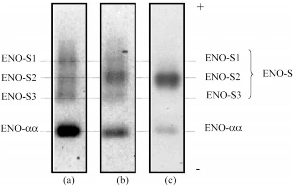
ENO-aa was found in all of the samples (testicular, epi-didymal, and ejaculated) with the same electrophoretic characteristics (Figure 1).
ENO-S was found in all of the samples containing sper-matozoa but with different electrophoretic characteristics, depending on the origin of the spermatozoa (Figure 1).
Percoll-selected ejaculated sperm used as reference from a normozoospermic patient presented a unique S band after electrophoresis (ENO-S2). The electrophoretic profile de-termined on testicular and epididymal spermatozoa clearly demonstrated 2 different patterns of ENO-S (Figure 1a and
Figure 1. Enolase isoform patterns of Percoll-selected sperm fraction from (a) testicular sperm, (b) epididymal sperm, and (c) ejaculated spermatozoa. The testicular and epididymal profiles are representative of 8 and 11 cases, respectively. The ejaculated sperm profile is representative of 10 nor-mospermic patients (revelation under fluorescence, inversed colors).
b). The electrophoretic profile of testicular spermatozoa showed 2 bands, S1 and S3. Such a profile was observed in the 8 analyzed samples. Whatever the epididymal sperm sample, a prominent S2 band appeared, with intermediate electrophoretic mobility, between S1 and S3, whereas the S1 and S3 bands were very slightly detected (Figure 1). This profile is similar to that of ejaculated spermatozoa.
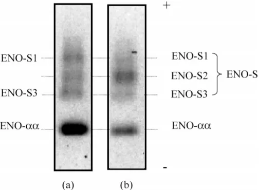
A comparison of the ENO-S electrophoretic profile be-tween the testicular and epididymal sperm of the same patient was made for 4 different patients (Figure 2). These profiles were similar to those obtained from either epi-didymal or testicular samples evaluated from distinct pa-tients. The study performed on the testicular and epidid-ymal spermatozoa from the same patient clearly demon-strated that the differences in ENO-S characteristics were strictly related to the origin of the spermatozoa and did not come from interindividual variations.
Finally, enolase isoform profiles of testicular immature germ cells from an azoospermic patient (nonobstructive azoospermia) were determined (Figure 3). No ENO-S iso-form was found in these extracts, whereas ENO-aa was still present.
Discussion
Edwards and Grootegoed (1983) were the first to describe the enolase pattern of human spermatozoa composed of the common aa isoenzyme and of an unusual isoform
(ENO-S) characterized by its particular electrophoretic migration and sperm specificity. Our results using elec-trophoresis on acetate cellulose confirmed those obtained by Edwards and Grootegoed. Recently, we demonstrated quantitative and qualitative differences in enolase activity between spermatozoa of abnormospermic and normosper-mic men (Force et al, 2002). We suggested that ENO-aa characterizes abnormal and immature spermatozoa, whereas ENO-S characterizes normally developed sper-matozoa.
In the present study, the electrophoretic pattern of eno-lase isoforms in spermatozoa retrieved from different re-gions of the male genital tract was evaluated. As shown previously, the ENO-aa and ENO-S isoforms were pre-sent in ejaculated spermatozoa. Furthermore, 3 variants of the S isoforms, named S1, S2, and S3 according to their pronounced acidic migration characteristics, were identified in epididymal and testicular spermatozoa. In testicular sperm, only 2 bands, S1 and S3, were present, whereas in epididymal sperm, these bands were reduced, and a prominent S2 band appeared. This latter electro-phoretic pattern was similar to the ENO-S pattern of ejac-ulated spermatozoa. Thus, the comparison of electropho-retic ENO-S isoform profiles between testicular and ejac-ulated sperm revealed changes that seemed to take place in the epididymis. The importance of sperm epididymal transport in men is a long-standing question. It is well accepted that spermatozoa are not fully mature when they leave the testis and that they develop the capacity to be
Figure 2. Enolase isoform patterns of Percoll-selected sperm fraction from (a) testicular sperm and (b) epididymal sperm from a man with obstructive azoospermia. The profile is representative of 4 patients (revelation under fluorescence, inversed colors).
Figure 3. Enolase isoform patterns of (a) a Percoll-selected cell fraction (P40) from a testicular extract without spermatozoa and (b) a representative profile of testicular sperm selected on Percoll, interface 47.5%–95% (revelation under fluorescence, inversed colors).
motile along with biochemical transformations during their epididymal transit (Cooper, 1995). The present study shows that, during sperm maturation in the epididymis, S1 and perhaps S3 isoforms are modified in favor of the S2 isoform. In addition, the fact that S2 was the
promi-nent isoform in ejaculated sperm provided evidence that its determination in these spermatozoa was an interesting marker of epididymal sperm maturation. Moreover the presence of ENO-S (S2) in normally developed sperma-tozoa was demonstrated previously (Force et al, 2002).
That we found a majority of S2 isoforms in ejaculated sperm suggests an association of this latter isoform with matured spermatozoa. Most of our results were obtained on mature ejaculated sperm; however, studies of immature ejaculated sperm have shown a heterogeneity in S iso-forms with the presence of S1 and S3 bands (unpublished data).
Different explanations can be proposed concerning the heterogeneity of the S isoforms and/or the remodeling of the ENO-S. Because the S3 isoform is the most neutral isoform, S1 or S2 could derive from this one by a sia-lylation. Sialyltransferases involved in the glycosylation process are present in the epididymis (Singer et al, 1988; Tulsiani et al, 1993). In the rat, a similar variant ofa-L -fucosidase has been found during epididymal maturation (Abascal et al, 1998), and the differentially sialylated iso-forms of a-L-fucosidase indicated a significant trend of increased thermostability with increasing sialylation (Al-hadeff and Andrews-Smith, 1980). Compared to ENO-aa, the increased thermostability of ENO-S (Edwards and Grootegoed, 1983) suggests that this enzyme contributes to the highly specialized performance of mature sperma-tozoa.
The variation in S isoforms could also be due to phos-phorylation. Phosphorylated variants ofbb enolase were observed in muscle (Asaga and Konno, 1975; Nettelblad and Engstrom, 1987). The phosphorylation of glycolytic enzyme in human spermatozoa improves enzyme activity and glycolytic flux (Harrison et al, 1991; Knull and Min-ton, 1996; Ovadi and Srere, 1996; Leclerc and Goupil, 2002).
The presence of theaa isoform in immature testicular cells suggests an early expression in spermatogenesis. That the S isoform was found only in mature testicular sperm and was absent in the testicular extract from one patient showing a spermatogenesis arrest (confirmed by histologic examination) suggest that the S isoform was synthesized in the final stages of spermatogenesis, during spermiogenesis. In mouse sperm, the S isoform is present only in elongating spermatids or in washed spermatozoa and is absent in middle and late pachytene spermatocytes or in round spermatids (Edwards and Grootegoed, 1983). Two hypotheses can be proposed concerning ENO-S ex-pression: 1) ENO-S is the product of a gene locus distinct from those determining the somatic tissue enolases and is expressed in the late stage of spermatogenesis in the hap-loid genome, or 2) the gene locus is the same as that for theaa isoenzyme, and a stable messenger RNA (mRNA) is transcribed premeiotically and stored untranslated until the late spermatid stage of development (Erickson et al, 1980). Enolase studies in different tissues are in favor of 2 genes. Indeed, in brain and muscular tissue, the specific enolase isoforms (g and b enolase genes) were encoded
by a gene distinct from the ubiquitousa gene (Sakimura et al, 1985, 1990).
Another hypothesis concerning the origin of ENO-S variants can be set forth. According to the studies carried out on rat spermatozoa, enolase would be localized to the tail of mature spermatozoa, and an association with microtubules, dependent on the concentration of the me-dium in ATP, cytosine triphosphate, and guanosine tri-phosphate, was demonstrated (Gitlits et al, 2000). These results confirm several reports of glycolytic enzyme as-sociated with microtubules of the flagellum (Storey and Kayne, 1975; Durrieu et al, 1987; Knull and Walsh, 1992). In addition, Westhoff and Kamp (1997) demon-strated the existence of a multienzyme complex on the tail of human spermatozoa. The glyceraldehyde 3-phos-phate dehydrogenase is bound to the fibrous sheath and is associated with other glycolytic enzymes, namely tri-ose isomerase and phosphoglycerate kinase. Together with our results, these reports showing the presence of the ENO-S isoforms probably during spermiogenesis, which correspond to the tail formation of the sperma-tozoa, are in favor of a flagellar localization of ENO-S isoforms.
Moreover, we have indirect evidence of the subcellular localization of enolase isoforms provided by the extrac-tion protocol used. A complete extracextrac-tion of ENO-aa is obtained by a mechanical treatment (high-speed vortex, 1 hour at 48C) in the presence of a Tris buffer without NaCl. By using identical conditions, the lactate dehydrogenase and the creatine kinase, both of which are enzymes with cytosolic localization, are also completely extracted (data not shown). These experimental elements added to the positive correlation found between the ENO-aa activity and the spermatozoa carrying residual cytoplasmic ma-terial (Force et al, 2002) and suggest the cytosolic local-ization of ENO-aa. Contrary to ENO-aa, no relation was found between ENO-S activity and the percentage of spermatozoa carrying residual cytoplasmic material (Force et al, 2002). Moreover, to extract ENO-S isoforms, a detergent (OGP) was necessary to obtain a solubiliza-tion without the denaturasolubiliza-tion of hydrophobic proteins in interaction with membrane structures or other proteins. Thus, it is possible that ENO-S electrophoretic variants are generated from ENO-aa, either by aggregation of the molecules or connection to other glycolytic enzymes or sperm cell protein structures.
In conclusion, we have shown that a heterogeneity in the S isoform existed in testicular sperm with the S1 and S3 variants. This heterogeneity disappeared during epi-didymal maturation with the appearance of a prominent S2 isoform, which was equal in amount to the major S isoform in normally developed ejaculated spermatozoa. These results show that the ENO-S isoforms are inter-esting markers of sperm maturation and clarify the role
of the epididymis in human sperm maturation. In the future, the clinical use of ENO-S profiles could be pro-posed for cases of obstructive azoospermia (infectious syndrome) to help choose the better surgical sperm source for ICSI.
Acknowledgments
The authors thank Dr Jean Hermabessie`re for assistance in the collec-tion of testicular and epididymal spermatozoa. They are also grateful to Mrs Nicole Chardonnel and Monique Farigoule for their technical assistance.
References
Abascal I, Skalaban SR, Grimm KM, Aviles M, Martinez-Menarguez JA, Castells MT, Ballesta J, Alhadeff JA. Alteration of the isoform com-position of plasma-membrane-associated rat sperm a-L-fucosidase during late epididymal maturation: comparative characterization of the acidic and neutral isoforms. Biochem J. 1998;333:201–207. Alhadeff J, Andrews-Smith GL. Thermostability of human
fu-cosidase. Relationship to fucosidosis and low-activity serum alpha-L-fucosidase. Biochim Biophys Acta. 1980;614:466–475.
Asaga H, Konno K. Comparison between muscle and liver enolases and their behavior during differentiation and growth. J Biochem. 1975;77: 867–877.
Cooper TG. Role of the epididymis in mediating changes in the male gamete during maturation. Adv Exp Med Biol. 1995;377:87–101. Durrieu C, Bernier-Valentin F, Rousset B. Binding of glycerate
3-phos-phate dehydrogenase to microtubules. Mol Cell Biochem. 1987;74: 55–65.
Eddy EM, O’Brian DA. The spermatozoon. In: Knobil E, Neill JD, eds.
The Physiology of Reproduction. 2nd ed. New York, NY: Raven
Press; 1994:189–317.
Edwards YH, Grootegoed JA. A sperm-specific enolase. J Reprod Fertil. 1983;68:305–310.
Erickson RP, Kramer JM, Rittenhouse J, Salkeld A. Quantitation of mRNAs during mouse spermatogenesis: protamine-like histone and phosphoglycerate kinase-2 mRNAs increase after meiosis. Proc Natl
Acad Sci U S A. 1980;77:6086–6090.
Force A, Viallard JL, Grizard G, Boucher D. Enolase isoform activities in spermatozoa from men with normospermia and abnormospermia.
J Androl. 2002;23:202–210.
Gandini L, Lenzi A, Lombardo F, Pacifici R, Dondero F. Immature germ cell separation using a modified discontinuous Percoll gradient tech-nique in human semen. Hum Reprod. 1999;14:1022–1027.
Gatewood JM, Cook GR, Balhorn R, Bradbury EM, Schmid CW. Se-quence-specific packaging of DNA in human sperm chromatin.
Sci-ence. 1987;236:962–964.
Gitlits VM, Toh BH, Loveland KL, Sentry JW. The glycolytic en-zyme enolase is present in sperm tail and displays nucleotide-dependant association with microtubules. Eur J Cell Biol. 2000; 79:104 –111.
Grizard G, Chevalier V, Griveau JF, Le Lannou D, Boucher D. Influence of seminal plasma on cryopreservation of human spermatozoa in a biological material-free medium: study of normal and low-quality se-men. Int J Androl. 1999;22:190–196.
Haidl G, Opper C. Changes in lipids and membrane anisotropy in human
spermatozoa during epididymal maturation. Hum Reprod. 1997;12: 2720–2723.
Harrison ML, Rathinavelu P, Arese P, Geahlen RL, Low PS. Role of band 3 tyrosine phosphorylation in the regulation of erythrocyte glycolysis.
J Biol Chem. 1991;5:4106–4111.
Kirchhoff C, Hale G. Cell-to-cell transfer of glycosylphosphatidylinosi-tol-anchored membrane proteins during sperm maturation. Mol Hum
Reprod. 1996;2:177–184.
Knull HR, Minton AP. Structure within eukaryotic cytoplasm and its re-lationship to glycolytic metabolism. Cell Biochem Funct. 1996;14: 237–248.
Knull HR, Walsh JL. Association of glycolytic enzymes with the cyto-skeleton. Review. Curr Top Cell Regul. 1992;33:15–30.
Leclerc P, Goupil S. Regulation of human sperm tyrosine kinase c-yes. Activation by cyclic adenosine 39,59-monophosphate and inhibition by Ca21. Biol Reprod. 2002;67:301–307.
Moore HDM. The influence of the epididymis on human and animal sperm maturation and storage. Hum Reprod. 1996;11:103–110. Nettelblad FA, Engstrom L. The kinetic effects of in vitro
phosphoryla-tion of rabbit muscle enolase by protein kinase C. A possible new kind of enzyme regulation. FEBS Lett. 1987;214:249–252. Ovadi J, Srere PA. Metabolic consequences of enzyme interactions. Cell
Biochem Funct. 1996;14:249–258.
Sakimura K, Kushiya E, Obinata M, Odani S, Takahashi Y. Molecular cloning and the nucleotide sequence of cDNA for neuron-specific eno-lase messenger RNA of rat brain. Proc Natl Acad Sci U S A. 1985; 82:7453–7457.
Sakimura K, Kushiya E, Ohshima-Ichimura Y, Mitsui H, Takahashi Y. Structure and expression of rat muscle-specific enolase gene. FEBS
Lett. 1990;277:78–82.
Sharpe RM. Regulation of spermatogenesis. In: Knobil E, Neill JD, eds.
The Physiology of Reproduction. 2nd ed. New York, NY: Raven
Press; 1994:1363–1434.
Shivaji S, Scheit KH, Bhargava PM. Proteins secreted by the epididymis. In: Shivaji S, Scheit KH, Bhargava PM, eds. Proteins of Seminal
Plasma. New York, NY: John Wiley & Sons; 1990:35–84.
Silber SJ, Nagy Z, Liu J, et al. The use of epididymal and testicular spermatozoa for intracytoplasmic sperm injection: the genetic impli-cations for male infertility. Hum Reprod. 1995;10:2031–2043. Singer R, Levinsky H, Sagiv M, Zukerman Z, Shoenfeld A, Allalouf
D. Sialyl transferase in human semen. Arch Androl. 1988;20:147– 151.
Storey BT, Kayne FJ. Energy metabolism of spermatozoa. V. The Emb-den-Meyerhof pathway of glycolysis: activities of pathway enzymes in hypotonically treated rabbit epididymal spermatozoa. Fertil Steril. 1975;26:1257–1265.
Tulsiani DR, Skudlarek MD, Nagdas SK, Orgebin-Crist MC. Purification and characterization of rat epididymal-fluid alpha-D-mannosidase: similarities to sperm plasma-membrane alpha-D-mannosidase.
Bio-chem J. 1993;290:427–436.
Viallard JL, Murthy MR, Dastugue B. An ultramicro bioluminescence assay of enolase: application to human cerebrospinal fluid.
Neuro-chem Res. 1985;10:1555–1566.
Viallard JL, Ven Murthy MR, Dastugue B. Rapid electrophoretic deter-mination of neuron-specific enolase isoenzymes in serum. Clin Chem. 1986;32:593–597.
Westhoff D, Kamp G. Glyceraldehyde 3-phosphate dehydrogenase is bound to the fibrous sheath of mammalian spermatozoa. J Cell Sci. 1997;110:1821–1829.
Yanagimachi R. Mammalian fertilization. In: Knobil E, Neill J, Ewing LL, Greenwald GS, Markert C, Pfaff W, eds. The Physiology of