University of Montréal
Studies on the role of IL-37 in the pathogenesis of
HIV infection
by Ayoub Abulkhir
Department of Microbiology, Infectiology and Immunology Faculty of Medicine
Master’s Thesis / Mémoire présenté Thesis submitted for the Master’s Degree
In Virology Immunology
September 2018
Résumé
L’IL-37 fait partie de la sous-famille de l’IL-18 et, de façon plus large, se classe avec les cytokines de la famille IL-1. L’IL-18, tout comme l’IL-1β, est une cytokine
pro-inflammatoire puissante, tandis que l’IL-37 a des effets anti-pro-inflammatoires. Les deux cytokines, IL-18 et l’IL37, utilisent la chaîne IL-18Rα pour la liaison initiale à la surface des cellules cibles. Cependant, ces deux cytokines se distinguent par rapport au recrutement et l’utilisation des chaînes secondaires de leurs récepteurs. L’IL-18 utilise la chaîne IL-18Rβ, tandis que l’IL-37 utilise SIGIRR ("Single Immunoglobulin and IL-1 Receptor-Related") pour la signalisation intracellulaire. Jusqu’à récemment, SIGIRR était considéré comme récepteur orphelin. L’IL-37 inhibe l’activation des inflammasomes ainsi que les effets biologiques d’un grand éventail de médiateurs inflammatoires tels que l’IL-1β, l’IL-6 et le TNF-α, parmi d’autres. De plus, ces deux cytokines lient la protéine de liaison à l’IL-18 (IL-18BP) avec des effets différents. Il est connu que la concentration d’IL-18 est plus élevée dans la circulation des personnes VIH-positives. Cette cytokine joue un rôle important dans l’activation aberrante globale du système immunitaire et l’inflammation remarquée chez ses individus. Cependant, très peu est connu sur le rôle de l’IL-37 lors de l’infection au VIH. Ce projet de maîtrise s’est concentré sur cette question.
Les résultats présentés dans ce mémoire démontrent que les niveaux circulants de l’IL-37 sont plus élevés dans le sérum d’individus VIH-positifs sous traitement antirétroviral ainsi que d’individus non-progresseurs à long-terme comparativement aux personnes VIH-positives naïves au traitement antirétroviral. Notamment, les niveaux d’IL-37 sériques sont
significativement plus élevés chez les non-progresseurs à long-terme comparé aux témoins VIH-négatifs. De façon surprenante, nos résultats démontrent aussi une baisse significative de l’expression du SIGIRR à la surface de divers types cellulaires contenus dans la population des cellules mononuclées du sang périphérique chez les personnes VIH-positives. De plus, nous avons observé une augmentation significative de la concentration de SIGIRR soluble dans les individus infectés par le VIH. Fait intéressant, nous avons remarqué que le SIGIRR soluble neutralise les effets anti-inflammatoires de l’IL-37 sur les cellules humaines. En outre, nous démontrons que l’IL-37 humain recombinant cause une baisse de la réplication du VIH dans les lymphocytes humains stimulés à l’aide de la phytohémagglutinine, ainsi que de
l’expression du corécepteur VIH (CXCR-4) et du PD-1, marqueur classique d’épuisement, dans les cellules CD4+ et CD8+ de personnes VIH-positives. Pris ensemble, ces résultats suggèrent que les fonctions anti-inflammatoires de l’IL-37, malgré ses concentrations accrues dans la circulation, sont atténuées chez les personnes qui sont dépisté Séropositives. La restauration de ces fonctions peut pallier l’inflammation et l’activation immunitaire chez ces patients.
Abstract
IL-37 belongs to the IL-18 subfamily and to the larger IL-1 family of cytokines. While IL-18, like IL-1β, is a potent pro-inflammatory cytokine, IL-37 exerts anti-inflammatory effects. Both IL-18 and IL-37 use the IL-18Rα chain for initial binding to the surface of target cells. However, they differ in the recruitment and use of the secondary chains of their
receptors. IL-18 uses the IL-18Rβ chain, whereas IL-37 uses SIGIRR (Single Immunoglobulin and IL-1 Receptor-Related) for transducing intracellular signals. Until recently, SIGIRR was known as an orphan receptor. IL-37 inhibits activation of inflammasomes, as well as the biological effects of a wide range of inflammatory mediators such as IL-1β, IL-6 and TNF-α, etc. Furthermore, the two cytokines bind IL-18 Binding Protein (IL-18BP) with divergent effects. It is now well established that the concentration of IL-18 is increased in the circulation of HIV-infected individuals. It plays an important role in overall aberrant immune activation and inflammation in these individuals. However, little is known about the role of IL-37 in this infection; and thus, is the focus of this master’s thesis.
In this thesis, we demonstrate that circulating levels of IL-37 are higher in the serum of HIV-infected individuals on anti-retroviral treatment (ART) and in HIV-infected long term non-progressors (LNTP), as compared to HIV-infected individuals naïve to ART. More importantly, serum levels of IL-37 are significantly higher in LTNP as compared to healthy controls. Surprisingly, the results also show that a significant decrease in the surface
expression of SIGIRR occurs in different types of peripheral blood mononuclear cells of HIV-infected individuals. Furthermore, we found a significant increase in the circulating
concentrations of soluble SIGIRR in HIV-infected individuals. Interestingly, we also noted that soluble SIGIRR neutralizes the anti-inflammatory effects of IL-37 on human cells. We also show that recombinant human IL-37 reduces HIV replication in human
phytohaemaglutinin blasts. It also reduced expression of the HIV co-receptor CXCR4 and of PD-1, a classical marker of exhaustion, on the surface of CD4+ and CD8+ T cells from HIV-infected individuals. Taken together, these results suggest that anti-inflammatory functions of IL-37, despite its increased concentration in the circulation, are dampened in HIV-infected individuals. Restoring these functions may attenuate inflammation and immune activation in these patients.
Table of Contents
Résumé ... i
Abstract ... iii
Table of Contents ... v
List of Tables ... viii
List of Figures ... ix
List of Abbreviations ... x
Acknowledgement ... xiii
Introduction & Review of the Literature ... 1
1. REVIEW OF LITERATURE ... 2
1.1 Human Immunodeficiency Virus (HIV) ... 2
1.1.1 The virus and related diseases... 2
1.1.2 Transmission of the virus ... 3
1.1.3 HIV-1 structure ... 4
1.1.4 The HIV replication cycle ... 6
1.1.5 Infection with HIV ... 8
1.1.6 Immunopathology ... 9
1.1.7 Specific immune responses to HIV... 11
1.1.8 The effect of antiviral therapy... 14
1.1.9 Consequences of chronic infection ... 16
1.1.10 Immune activation increases susceptibility to HIV infection ... 20
1.1.11 Viral reservoirs... 20
1.1.12 Cytokines and HIV infection ... 22
1.2 IL-37 ... 24
1.2.1 Discovery of IL-37 ... 24
1.2.2 Expression of IL-37 ... 25
1.2.4 Processing of IL-37 ... 27
1.2.5 Secretion of IL-37 ... 27
1.2.6 IL-37 receptor ... 28
1.2.7 IL-37-mediated signaling pathways ... 30
1.2.8 Biological function of IL-37 ... 33
2 HYPOTHESIS & OBJECTIVES ... 32
2.1 Hypotheses ... 32
2.2 Rationale ... 32
2.3 Main objective ... 32
2.4 Specific aims ... 33
3 MATERIALS & METHODS ... 34
3.1 Antibodies and reagents ... 34
3.2 Isolation of peripheral blood mononuclear cells (PBMCs) ... 34
3.3 Cell culture ... 35
3.4 Virus preparation ... 35
3.5 In vitro infection of cells with HIV-1 ... 36
3.6 Measuring IL-37 and SIGIRR concentrations ... 36
3.7 Measuring TNF-α concentrations ... 36 3.8 Flow cytometry ... 37 3.9 Qualitative PCR ... 37 3.10 Study participants... 38 3.11 Statistical analysis ... 40 3.12 Ethical statement ... 40 4 RESULTS ... 41
4.1 Levels of IL-37 in HIV-infected individuals ... 41
4.2 Levels of soluble SIGIRR in HIV-infected individuals ... 43
4.3 Expression of SIGIRR in HIV-infected individuals ... 45
4.4 Soluble SIGIRR neutralizes the function of IL-37 ... 49
4.5 The effect of different concentrations of IL-37 on TNF-α ... 51
4.7 The effect of IL-37 on expression of the HIV chemokine receptor CXCR4 ... 53
4.8 The effect of IL-37 on markers of exhaustion ... 54
4.9 IL-37 isoforms in HIV-individuals ... 56
5 DISCUSSION ... 59
6 CONCLUSIONS... 68
List of Tables
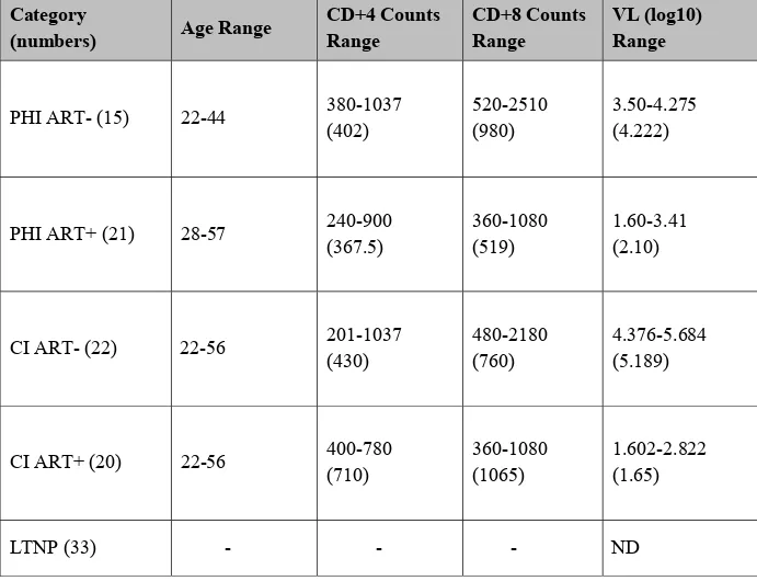
Table 1. The sequences of the primers used in RT-PCR to identify IL-37 isoforms ... 38 Table 2. Demographic and clinical parameters of the study participants ... 39
List of Figures
HIV structure ... 6
Stages of the viral replication cycle ... 8
Clinical stages of HIV infection ... 12
Stages of the viral life cycle which may provide targets for HIV therapy ... 16
Gene structure and splice variants (isoforms) of IL-37 ... 26
Production and secretion of IL-37 ... 28
IL-1 family receptors ... 29
IL-37-induced signaling pathways ... 32
Concentration of IL-37 in the circulation of HIV-infected individuals ... 42
Comparison of SIGIRR concentrations between ART+ and ART- HIV infected individuals 44 Expression of SIGIRR on different immune cells ... 48
The effect of soluble SIGIRR on the anti-inflammatory function of IL-37 in the sera of HIV-infected individuals ... 49
Effect of recombinant SIGIRR on the anti-inflammatory effects of IL-37 ... 50
Effect of different concentrations of IL-37 on the expression of TNF-α ... 51
Effect of different concentrations of IL-37 on HIV replication ... 52
Effect of IL-37 on expression of the HIV chemokine receptor CXCR4 ... 54
Effect of IL-37 on expression of PD-1 in T cells ... 55
List of Abbreviations
AIDS: Acquired immune deficiency syndrome ART: Antiretroviral therapy
cART: Combination anti-retroviral therapy CCR5: C-C chemokine receptor type 5 cDNA: Complementary DNA
CTLA: Cytotoxic T-lymphocyte
CTLA4: Cytotoxic T-lymphocyte associated protein 4 CVD: Coronary vascular disease
CXCR4: CXC chemokine receptor type 4 DCs: Dendritic cells
DNA: Deoxyribonucleic acid dsRNA: Double-stranded RNA EC: Elite controllers
ELISA : Enzyme-linked immunosorbent assay ENV: HIV envelope glycoprotein
Gag: Group-specific antigen, coding for structural proteins GALT: Gut-associated lymphoid tissue
GAPDH: Glyceraldehyde 3-phosphate dehydrogenase GIT: Gastro-intestinal tract
Gp: Glycoprotein
HAART: Highly aggressive anti-retroviral therapy HC: Healthy controls
HIV-1: Human immunodeficiency virus type 1 HIV-2: Human immunodeficiency virus type 2 HIV: Human immunodeficiency virus
HTLF: Helicase-like transcription factor HVL: High viral load
IBD: Inflammatory bowel disease ICI: Immune checkpoint inhibitors
IEC: Intestinal epithelial cells IFN-γ: Interferon gamma Ig: Immunoglobulin IL-1R: IL-1 receptor IL: Interleukin
IL1R: Interleukin 1 family receptor LAG-3: Lymphocyte-activation gene 3 LPS: Lipopolysaccharide
LTNP: Long-term non-progressors MHC: Major histocompatibility complex mRNA: Messenger RNA
ND: Non-detectable Nef: Negative factor
NF-kB: Nuclear factor kappa-light-chain-enhancer of activated B cells NFAT: Nuclear factor of activated T cells
NKs: Natural killer cells NLR: Nod-like receptors
PBMC: Peripheral blood mononuclear cells PCR: Polymerase chain reaction
PD-1: Programmed cell death-1 PHA: Phytohaemaglutinin PHI: Primary HIV infection PI: Protease inhibitor Pol: DNA polymerase
Rev: Regulator of expression of viral proteins RNA: Ribonucleic acid
RT: Reverse-transcriptase
RT-PCR: Real-time polymerase chain reaction
SIGIRR: Single immunoglobulin and toll-interleukin 1 related receptor SIV: Simian immunodeficiency virus
STIs: Sexually transmitted diseases TAT: Trans-activator protein
TGF-β: Transforming growth factor beta TIR-8: Toll interleukin-1 receptor 8 TLR: Toll-like receptors
TNF-α: Tumor necrosis factor alpha Vif: Viral infectivity factor
VIH: Virus de l’immunodéficience humaine Vpr: Viral protein R
Acknowledgement
First and foremost, all the praise and thanks be to Allah, the Almighty, most Gracious, and Merciful for His blessings that directed me to complete this research successfully. I would like to express my deepest and sincerest gratitude to my academic research supervisor, Dr. Ali Ahmad, for giving me the opportunity to work with him and for providing invaluable leadership throughout this research project. He contributed to a rewarding graduate lab experience by giving me intellectual freedom in my research, supporting my attendance at various conferences, and inspiring me with new ideas to produce high quality work in all of my experiments. His dynamism, vision, sincerity, and motivation have deeply inspired me. He has taught me the methodology to carry out and present research as clearly as possible. It has been a great privilege and honor to work and study under his guidance. Additionally, I would like to thank Dr. Louis De Repentingy and Dr. Jean-Francois Gauchat for taking time to serve as my committee members.
I would like to express my appreciation to Suzanne Samarani, for sharing her pearls of wisdom throughout my research. I also thank her for her excellent, valuable, and persistent technical assistance with cell culture, FACS, and analysis of results. I am also immensely grateful to her for believing in my potential and for the valuable time we spent together. She has taught me more than I could ever give her credit for here. She has shown me, through her example, what a great researcher should be.
Nobody has been more important to me in the pursuit of this project than the members of my family. I would like to thank my parents whose affection and guidance are with me in whatever I pursue and who believed in me to produce my best work toward this research. I would like to thank a very special person who contributed to making this work possible, and who provided endless inspiration, support, and encouragement throughout my studies, my beloved fiancé Malak.
Last, but certainly not least, I am especially indebted to the government of my country, Libya. I could not have gone through the master’s program overseas without their financial support. I would like to express my full appreciation to the staff of the Department of
Scholarship, especially Francine Brisebois, the Academic manager for Students in Montreal, for her generous support and for her advice, which were available whenever I needed them.
Introduction & Review of the Literature
The aim of my master’s thesis project was to investigate the role of Interleukin-37 (IL-37) in HIV-1 infection.
1. REVIEW OF LITERATURE
1.1 Human Immunodeficiency Virus (HIV)
1.1.1 The virus and related diseases
HIV-1 is the underlying cause of AIDS and AIDS-related disease; however, its origin remains obscure. Firm serological evidence of the infection can be found on the east and west coasts of the USA as of the mid-1970s, but HIV infection in central Africa may have predated the AIDS epidemic in North America (Clavel, Mansinho et al. 1987). Phylogenetic analysis of the HIV-1 genome suggests it originated in chimpanzees; whereas HIV-2, which shows similarity to the simian immunodeficiency virus (SIV) genome, suggests a source in sooty manage monkeys (Marx, Li et al. 1991, Jaffar, Grant et al. 2004). Confined to West Africa, HIV-2 infection is less virulent than HIV-1 infection (Clavel, Mansinho et al. 1987, Sharp and Hahn 2011). Like some other RNA viruses, HIV appears to have evolved rapidly, mutating and shifting its host range and virulence as a result of its error-prone reverse transcription enzyme (St Clair, Martin et al. 1991). This high rate of mutation explains how a new pathogenic retrovirus could arise in human. The United Nations Programme on HIV/AIDS (UNAIDS) reports that there are currently thirty-six million people living with HIV worldwide (UNAIDS 2017).
Retroviruses are named as such because their genomes encode an unusual enzyme, Reverse Transcriptase, which allows DNA to be transcribed from an RNA template (Preston, Poiesz et al. 1988, Roberts, Bebenek et al. 1988). Thus, HIV can make copies of its genome in host cells, such as the human CD4+ “helper” T lymphocyte, which is the main target of HIV-1 infection (Dragic, Litwin et al. 1996). Other antigen presenting cells (APCs) such as macrophages, dendritic cells (DC), and monocytes, are also targets of HIV infection (Janeway 2001, Stevenson 2003). A hallmark characteristic of HIV infection is the integration of the newly synthesized viral DNA into the genome of the infected lymphocyte. Integration of the viral DNA into the host genome allows the viral DNA to evade immune detection, thereby forming the basis of chronic HIV infection and creating the greatest obstacle for viral
eradication (Schröder, Shinn et al. 2002). Many anti-retroviral treatments suppress replication of the virus but do not eliminate the integrated viral DNA (provirus) from infected cells.
Nonetheless, advanced therapies, which include combinations of nucleoside analogs and protease inhibitors, have transformed the prognosis for carriers of HIV. Treatment with these combination anti-retroviral (cART) therapies often leads to a sustained decrease of virus in the blood, helping to restore levels of the central target cell (CD4+ lymphocytes) to near normal (Sabin and Lundgren 2013, Sabin, Reiss et al. 2016). Despite these efforts, the inherent variability of the HIV genome and failure of the human host to produce sufficient amounts of neutralizing antibodies against the virus, as well as concerns surrounding safety, have
continued to frustrate attempts to develop a reliable vaccine (Titti, Cafaro et al. 2007).
1.1.2 Transmission of the virus
HIV has been isolated from several bodily fluids including semen, cervical secretions, lymphocytes, cell-free plasma, cerebrospinal fluid, tears, saliva, urine, and breast milk (Sabin and Lundgren 2013). However, not all these fluids transmit infection due to the considerable variation in their viral concentration. The predominantly infectious fluids are semen, blood, and possibly cervical secretions. Transmission of HIV-1 and HIV-2, known as the major and minor human AIDS viruses, respectively, is similar to that of other retroviruses and can be classified in two ways: horizontal and vertical. Epidemiological observations have shown that, globally, the most common mode of HIV transmission is through unprotected anal or vaginal intercourse (Pantaleo and Fauci 1995). Transmission through sexual intercourse or through the exchange of infected blood or fluids (e.g. via transfusion, organ donation, sharing or reusing of contaminated needles between infected drug users or during therapeutic
procedures) is classified as horizontal transmission, given the transfer between individuals of the same generation (Lipsitch, Siller et al. 1996). Vertical transmission, on the other hand, occurs across two generations, as it refers to the transmission of the virus from mother to offspring. Vertical transmission occurs in utero and has also been shown to transfer through breast milk (Kreiss 1997).
Several factors affect the transmission of HIV, notably the concentration of HIV in bodily fluids, secondary infection of the genital tract, the efficiency of epithelial barriers, and the efficiency of immune function against HIV infection. The stage of the disease and
associated viral load are essential in determining infectivity (Hollingsworth, Anderson et al. 2008).
1.1.3 HIV-1 structure
HIV is a spherical enveloped virus belonging to the lentivirus subgroup of retrovirus. The lentivirus subgroup includes other retroviruses that cause slow progressing diseases in humans and animals (Cullen 1991, Klimas, Koneru et al. 2008). As illustrated in Figure 1, HIV particles are approximately 110 nm in diameter and consist of a lipid membrane bilayer known as the envelope. The viral envelope is formed from the plasma membrane of the host cell and two viral proteins, the external glycoprotein (gp) 120 and transmembrane gp41, which are non-covalently attached (Chan, Fass et al. 1997, Kwong, Wyatt et al. 1998). The virus uses these glycoproteins to infect target cells. The viral envelope encloses a viral genome comprised of two identical single-stranded positive-sense RNA strands and numerous copies of viral enzymes, which are pivotal for the infection of host cells. In order to complete the life cycle of the virus, the viral genome must be transcribed from RNA to DNA within the infected cells. This process is known as reverse transcription and is catalysed by the viral reverse transcriptase enzyme (Sarafianos, Marchand et al. 2009). It is because of this enzyme that HIV is classified as a retrovirus. In addition to the basic retroviral genes (envelope (env), group antigen genes (gag), and polymerase (pol)), the virus has additional genes that are important for infection. These genes include regulator of expression of virion proteins (rev), viral infectivity factor (vif), viral protein U (vpu), viral protein R (vpr), negative factor (nef), and trans-activator of transcription (tat). Tat and rev are essential regulatory genes coding for accumulating proteins and increase production of the viral genome (Malim and Bieniasz 2012).
Each protein plays an essential role in viral structure and/or infectivity. The Tat protein plays a positive feedback role in the transcription of viral messenger RNA (mRNA), while the Rev protein splices transcripts from the nucleus and ensures their transport from the nucleus to the cytoplasm (Langer and Sauter 2016).
Another important protein in the life cycle of the virus is Nef. The Nef protein sustains T cell activation during the early phases of the viral cycle by decreasing the threshold of T cell activation. It achieves this through the down-regulation of cytotoxic T lymphocyte associated protein 4 (CTLA-4), which is a negative regulator of T cell activation (El-Far, Isabelle et al. 2013). Nef also reduces the expression of both major histocompatibility complex (MHC) class 1 and 2 on the surface of host cells; therefore, cytotoxic T cells and antiviral immune response are less likely to be induced by the actively infected cells. A third function of Nef is to enhance the degradation of surface CD4+ molecules, thereby blocking reinfection of the cell (Piguet and Trono 1999, Pawlak and Dikeakos 2015).
The Vpu protein has a unique role in HIV-1. Its function is to increase the intracellular degradation of CD4 and it is also responsible for releasing newly produced virus particles into the cytoplasm (Pham, Lukhele et al. 2014, Apps, Del Prete et al. 2016). The Gag protein is essential for the formation of the viral core and matrix proteins (PAL, REITZ JR et al. 1990). Vif inactivates the apolipoprotein B mRNA editing enzyme, catalytic polypeptide-like 3G (APOBEC-3G), a cytidine deaminase that catalyzes the conversion of deoxycytidine into deoxyuridine in reverse transcribed viral complementary DNA (cDNA) (Donahue, Vetter et al. 2008). This inactivation triggers ubiquitination and degradation of the deaminase and prevents modifications of the viral cDNA (Nakashima, Ode et al. 2016). Vpr is important for HIV-1, as it allows for the infection of non-dividing cells, causing cell cycle arrest in G2M (Elder, Benko et al. 2002, Richard, Sindhu et al. 2010). Independently, it also downregulates helicase-like transcription factor (HTLF), which is a DNA translocase that functions to repair damaged replication forks (Zhou, DeLucia et al. 2017). The Pol protein, which is arguably the most important structural HIV-1 protein, results in the expression of three enzymes critical for viral replication: reverse transcriptase, integrase, and protease. Reverse transcriptase allows the viral RNA to be transcribed into viral DNA, which then integrates into the host DNA with the help of the integrase enzyme. Viral protease is essential for maturation of the virus
particle, as it cleaves polyproteins into active functional proteins (Baldwin and Linial 1998). Finally, the Env protein precursor, gp160, is cleaved by the cellular enzyme Furin into gp120 and gp41. Gp120/41 use CD4+ as a receptor and both chemokine receptors CXCR4 and CCR5 as co-receptors to infect human cells (Malim and Bieniasz 2012).
HIV structure
This figure illustrates the structure of HIV and describes the enzymes essential for replication. Originally published in (Murphy and Weaver 2016) and reproduced with permission from the publisher.
1.1.4 The HIV replication cycle
The HIV life cycle is comprised of various steps including entry, reverse transcription, integration, transcription, and translation (KARAGEORGOS, LI et al. 1993). The virus targets the CD4 molecule, which is mainly expressed on CD4+ T cells and, to a lesser extent, on macrophages and DCs. As depicted in Figure 2, the entry of HIV into the host cell begins with the high affinity binding of gp120 to the CD4 molecule. Before the virus can enter the host cell, gp120 must also bind to either the CCR5 or CXCR4 chemoreceptor on the host cell surface. CCR5 or CXCR4 serve as co-receptors (Gomez and Hope 2005, Panos and Watson 2015). CCR5 is predominantly expressed on effector memory CD4 T cells, macrophages, and DCs (Zaitseva, Blauvelt et al. 1997). In contrast, CXCR4 is expressed by naïve and central
memory T cells (Nicholson, Browning et al. 2001). After gp120 binds the co-receptors, it undergoes a conformational change which exposes the fugenic peptide of gp41 and then fuses with the plasma membrane of the host cell. This process enables the viral nucleocapsid, viral genome, and associated proteins to enter the target cell’s cytoplasm (Gomez and Hope 2005, Panos and Watson 2015). Once the virus has entered the host cell, the nucleocapsid
disintegrates, releasing two strands of viral RNA and essential viral enzymes into the
cytoplasm and begins to replicate. As a first step, the reverse transcriptase transcribes the viral RNA into cDNA, which encodes nine genes. The newly synthesized cDNA is then
transported into the cell nucleus, where it is integrated into the host genome by the viral integrase enzyme. This integrated viral cDNA is called the provirus (Panos and Watson 2015).
Stages of the viral replication cycle
This figure shows how free virus can fuse with the host cell, where it releases its genetic material. The viral RNA genome is reverse transcribed into DNA by the reverse transcriptase enzyme, after which the newly synthesized viral cDNA inserts into the host cell’s DNA. Integration allows for the production of viral proteins by the host cell’s own transcription/ translation machinery, after which the proteins come together to form an immature virus particle. Finally, this form of the virus is pushed out through the host cell membrane, taking the lipid bilayer with it, enveloping the viral components and creating a mature virus particle. Originally published in (Vanpouille, Arakelyan et al. 2012) and reproduced with permission from the publisher.
In order to begin the replication process and to form infectious virions, HIV requires activation of the target cell. The virus hijacks the target cell’s transcriptional machinery and activates the host cells’ own transcription factors. Two important transcription factors required for virus production are nuclear factor kappa-light-chain-enhancer of activated B cells (NF-kB) and nuclear factor of activated T-cells (NFAT) (Murphy and Weaver 2016). Although NF-kB is expressed in all immune cells infected by HIV, NFAT is primarily activated in CD4 T cells (Murphy and Weaver 2016).
1.1.5 Infection with HIV
Many of the clinical features of HIV infection can be linked to the profound immune deficit that develops in infected individuals. HIV is immunosuppressive, as it infects the CD4+ T cells of the immune system and ultimately destroys them (McCune 2001, Brenchley, Schacker et al. 2004). An understanding of this process is helpful in interpreting tests that monitor the disease and may also explain the failure of immunotherapy, as well as the difficulties in developing vaccines for HIV (Gomez and Hope 2005).
HIV’s primary target is a subset of thymus-derived (T) lymphocytes carrying the CD4 surface molecule receptor. These cells have been shown to bind with gp120 on the HIV envelope, thus beginning the fusion process (Mohan, Bhatnagar et al. 2014). CD4 is also expressed on a large number of monocytes, macrophages, Langerhans cells of the skin, and DCs in all tissues (Bernstein, Plasterer et al. 2006, Zhen, Krutzik et al. 2014). More recently,
it has become clear that entry of the virus also requires the function of co-receptors, most of which are members of the seven transmembrane-spanning G protein-coupled receptor (GPCR) family. In the immune system, GPCRs principally function as chemokine receptors capable of regulating migration, differentiation, and function of leucocytes during immune responses and inflammation (Lodowski and Palczewski 2009). The two co-receptors, CCR5 and CXCR4, are of great importance (Mohan, Bhatnagar et al. 2014). CCR5 (R5) is widely expressed on lymphocytes, macrophages, DCs, and on cells of the rectal, vaginal, and cervical mucosae. Viral strains capable of infecting primary macrophages (M or R5 tropic viruses) use CCR5 as their co-receptor. Only R5 strains are detected in the early stages immediately after infection, while both R5 viruses and strains that infect T cells and use CXCR4 (T or X4 tropic viruses) are found in later-stages of infection (Trkola, Kuhmann et al. 2002, Cicala, Arthos et al. 2011). These data suggest that R5 strains are necessary for the transmission of HIV, while X4
variants evolve in the infected host and may be responsible for T cell loss and disease
progression (Bednar, Hauser et al. 2015, Sironi, Malnati et al. 2015). Even stronger evidence that CCR5-using M-tropic viruses transmit infection comes from the observation that
individuals homozygous for a 32-base pair deletion of CCR5 showed substantially increased resistance to HIV infection (Balotta, Bagnarelli et al. 1997, Suppiah, Armstrong et al. 2013).
1.1.6 Immunopathology
CD4 lymphocytes are known as “leaders of the immunological orchestra” owing to their central role in the induction of an immune response. Stimulation of these cells through antigen presentation results in cell division and in the production of lymphokines such as interferons (IFN), interleukins (ILs), tumor necrosis factor (TNF), and chemoattractant chemokines (Mohan, Bhatnagar et al. 2014). Lymphokines function as local hormones controlling migration, growth, maturation, and behavior of other lymphocytes; especially of the cytotoxic/suppressor (CD8) T cells and of antibody-producing B lymphocytes (B cells). Lymphokines also affect the maturation and function of monocytes, tissue macrophages, and DCs (Corinti, Albanesi et al. 2001).
Macrophages and DCs are antigen-presenting cells that play a critical role in the initiation of the immune response (Banchereau and Steinman 1998, Geissmann, Manz et al. 2010). Monocytes, especially inflammatory ones, are the precursors to some glial cells. After HIV infection, their abnormal lymphokine production may have harmful effects on neural tissue, which may result in encephalopathy (Imran, Manzoor et al. 2016).
Early after HIV infection, antibody responses are not impaired. In fact, the presence of antibodies in the blood, targeting the viral envelope and core proteins, is the crucial evidence required to diagnose HIV infection; the development of these antibodies persists until death (Lange, Paul et al. 1986, Walker and Burton 2010). In adults, enormous activation of B lymphocytes is characterized by an increase in serum immunoglobulin (Ig) levels, perhaps because of the direct and indirect activation of the B cells by HIV (Imran, Manzoor et al. 2016). This massive polyclonal activation explains, in part, the reason for false positive serological tests seen in HIV infection. Interestingly, in young children, the reverse may be seen. Low Ig levels seen in children can sometimes necessitates intravenous replacement therapy (Cheynier, Langlade‐Demoyen et al. 1992). Within days or weeks following
infection, there may be a decrease in the number of CD4+ T lymphocytes and an increase in the number of CD8+ cytotoxic/suppressor T cells (Migueles, Osborne et al. 2008). Depletion of CD4+ T cells can occur via two mechanisms: direct cytopathology of HIV-1 or immune responses. The host immune responses activate the elimination of infected cells through the action of Natural Killer (NK) and cytotoxic CD8+ T lymphocytes (CTL); thus explaining the rise in CTLs (Cloyd, Chen et al. 2001, McCune 2001, Brenchley, Schacker et al. 2004). Following acute infection, healthy seropositive individuals may still have a reasonable level of CD4+ T lymphocytes; however, the number of CTLs frequently remains high (Betts, Nason et al. 2006). This happens, in part, due to reduced production of the lymphokine IL-2. Infected individuals may remain healthy for extended periods of time (several years). A hallmark of disease progression, often before the development of clinical symptoms, is a decrease in the number of CD4+ T lymphocytes. As CD4+ T cells are the main mediators of immune responses, their depletion due to HIV infection increases the vulnerability of infected individuals to opportunistic infection and to death (Okoye and Picker 2013). In AIDS, the number of CTLs is also reduced (Mohan, Bhatnagar et al. 2014, Imran, Manzoor et al. 2016).
1.1.7 Specific immune responses to HIV
Even though HIV-infected individuals show a deficit in their immune function, as described above, their immune system can directly target HIV itself. While serum reactivity to viral proteins is detectable, virus neutralizing titers are low and directed against the immunizing viral strain. Antibodies against HIV might even facilitate infection of new cells via binding to Ig (Fc) receptors on target cells (Mohan, Bhatnagar et al. 2014).
CTLs capable of killing HIV-infected target cells are detected in most HIV-infected individuals and are host-protective. This protective nature of CTLs is evidenced by the observation that viremia declines at the time when CTLs are first detected in the peripheral blood following infection, as well as in patients with stable disease (Borrow, Lewicki et al. 1994, Kiepiela, Ngumbela et al. 2007). Moreover, it is interesting to note that individuals who have been regularly exposed to HIV, but who remain seronegative and without a detectable viral load, do have detectable levels of HIV-specific CTLs (Pereyra, Palmer et al. 2009). In addition to killing the infected cells directly, CTLs may contribute to protection by producing several chemokines such as CD8 T cell antiviral factor (CAF), which actively suppresses viral replication in CD4+ T cells (Levy 2003). All these findings suggest that CTLs are an efficient protective mechanism. Nevertheless, because reverse transcription is highly error-prone, mutations of the viral genome occur frequently and allow the virus to escape the CTL response. Moreover, these mutants themselves not only avoid immune detection, but they may also inhibit the recognition of unmutated virus (Imran, Manzoor et al. 2016) .
A hallmark of HIV infection is the substantial depletion of CD4+T cells. HIV can trigger host cell immune activation in two ways: directly through antigens, and indirectly through the activation of other viruses, such as the cytomegalovirus (Hussey, Richardson et al. 1988, Cloyd, Chen et al. 2001). After an individual is infected with HIV, the primary targets of the virus are the CCR5 CD4+ activated T cells (Siliciano and Siliciano 2000). The majority of these T cells are located in lymphoid tissues, such as the lymph nodes; and in mucosal lymphoid tissues, like the gastrointestinal tract (Reynes, Portales et al. 2001). During the acute infection phase, HIV induces a significant T cell response, especially of CTLs, and the
response can last well into the chronic phase of the disease due to persistent viral replication (Douek, Brenchley et al. 2002). HIV pathogenesis can be divided into three groups based on the duration of the infection, viral load, CD4+ T cell counts, and presenting clinical symptoms. These three groups are illustrated in Figure 3 and are discussed below:
Clinical stages of HIV infection
HIV infection is divided into three different stages: (i) primary/acute stage, (ii) chronic stage, and (iii) AIDS. This division is based on the duration of the infection, the number of
circulating CD4+ T cell, the viral load in the blood, and the clinical symptoms that infected individual’s experience. Chronic infection, also known as the period of clinical latency, is characterized by a period where the infection is asymptomatic. Originally published in (An and Winkler 2010) and reproduced with permission from the publisher .
Group I: Primary HIV infection
Primary HIV infection (PHI) is also known as seroconversion illness or as acute HIV infection (Schacker, Collier et al. 1996, Schacker, Hughes et al. 1998). It represents the stage of infection after initial acquisition of the virus and is characterized by the development of
viral antibodies. It often lasts two to four weeks following initial infection, and between 25% and 65% of infected individuals present with symptoms at the time of seroconversion. The symptoms can be flu-like, with a mild to moderate glandular fever-like illness similar to an encephalopathy. Nevertheless, severe symptoms are rarely seen (Kahn and Walker 1998).
During the acute phase of HIV infection, sometimes there is a high rate of viral replication which leads to a transient rise in viral load due to a temporary fall in the CD4+T cell count. This interplay between increased viral production and decreased immune response might affect the outcome of HIV-related diseases (Meulendyke, Croteau et al. 2014).
The best diagnostic tests used during this stage are those that detect HIV antibodies and antigens. If the results of these tests are negative, but PHI is suspected, a more definitive test is an HIV RNA PCR, as it is the most sensitive test for the detection and quantification of the virus.
Group II: Asymptomatic chronic infection
Following the acute phase, HIV antibodies continue to be detectable in the blood. The amount of virus in the blood and lymphoid tissues decreases to very small amounts and the rate of HIV replication is reduced, but not eliminated (Goodsell 2015). This is also referred to as the chronic, asymptomatic phase. During this stage, CD4+ T cell counts are within normal range, generally above 350 cells/mm3. However, due to immune exhaustion from the major
activity of the acute phase, the immune system becomes persistently activated, leading to high levels of circulating pro-inflammatory cytokines (Brenchley, Price et al. 2006). This chronic immune activation is thought to cause cell death (Douek, Picker et al. 2003, Ford, Puronen et al. 2009). This step can persist for up to ten years or more (An and Winkler 2010).
Group III: Symptomatic HIV infection before the development of AIDS
The progression of HIV infection is a result of the depletion of immune competence, which happens after increased replication of latent HIV virus sites. Occasionally, the latent virus undergoes reactivation; however, the exact mechanism of this reactivation remains
unknown. Because of the variability of disease progression, HIV-infected individuals may exhibit different symptoms that may range from skin disorders and mouth ulcers, to
hematological disorders; many of which are not difficult to treat or alleviate. A drop in viral load due to the introduction of antiretroviral therapy often leads to a complete or partial resolution of such symptoms (Goodsell 2015). Nevertheless, during this period, infected individuals become much more vulnerable to various opportunistic conditions.
1.1.8 The effect of antiviral therapy
Viral replication leads to the depletion of CD4 T cells and to immune dysfunction. If not treated, in most infected individuals, this process gradually and slowly results in fatal immunodeficiency. Thus, many drugs have been developed to inhibit HIV replication and they have led to significant improvements in the outcome of this deleterious infection. As illustrated in Figure 4, there are different mechanisms through which these drugs inhibit viral replication. Antiretroviral therapies can target the viral life cycle at five different steps. The first wave of successful drugs was developed to interfere with the reverse transcriptase enzyme, which functions to convert viral RNA to cDNA (De Clercq 1998, Butanda-Ochoa, Hernández-Espinosa et al. 2017). These nucleoside reverse transcriptase inhibitors served as chain terminators of DNA synthesis, prematurely ending transcription of the viral genome. Soon after, non-nucleoside reverse transcriptase inhibitors were introduced (De Clercq 1998). These molecules induced a conformational change in the HIV reverse transcriptase enzyme, preventing its retro-transcription activity. The second generation of medication was developed to inhibit activity of the viral protease required to cleave the precursor Gag-Pol protein (De Clercq 2002, Murphy and Weaver 2016); thus preventing assembly of functional virus
particles. The mechanism of third-generation agents was to block fusion of the virus with the cell membrane, via gp41, thereby preventing viral entry into the host cell (Murphy and Weaver 2016). The fourth group of treatments targeted the CCR5 chemokine receptor
(Lederman and Margolis 2008, Cummins and Badley 2010). By binding to the receptor, these molecules induce conformational changes preventing attachment of gp120, which is required for entry into the host cell. Finally, the latest antiviral group of drugs was developed to
interfere with the viral integrase, which is required for the insertion of the viral genome into the infected host cells’ own DNA (De Clercq 2004, Murphy and Weaver 2016). Integrase inhibitors prevent close interaction with the host DNA, which is required to initiate
integration. Initially, when used separately, these medications were very efficient at inhibiting viral replication; however, with time and due to its mutagenic nature, the virus developed resistance to these drugs (Murphy and Weaver 2016). Resistance significantly hinders the treatment process, rendering it little or completely ineffective. Thus, a new approach known as highly active antiretroviral therapy (HAART) was developed. HAART refers to therapy that combines two or more drug classes previously described. In most cases, cART therapy appears to overcome the ability of the virus to mutate, which is the primary cause of drug resistance (Cao, Mehraj et al. 2015). HAART has been shown to lower the viral load in the plasma to levels that cannot be detected by current tool and methods. The success of using HAART against HIV infection has increased the length of time over which patients progress to the AIDS stage; thus, converting HIV infection into more of a chronic and manageable disease (Estes, Haase et al. 2008). However, in order to maintain this chronic state, infected individuals must take HAART for life.
While antiviral therapies have revolutionized the war against this harmful virus and have suppressed viremia in many infected individuals, they have also markedly reduced the death rate from AIDS and improved the quality of life of HIV-infected individuals. However, abnormalities of the immune system are still observed in virus-infected individuals. These individuals show low-grade chronic inflammation and immune activation, which leads to many side effects including enhanced aging, weakness, cardiovascular disease, bone abnormalities, and cancers to name a few (Schneider, Birger et al. 2016).
Stages of the viral life cycle which may provide targets for HIV therapy As demonstrated in the figure above, there are five different points to target HIV activity: (1) the first, to interfere with co-receptor attachment; (2) the second, to inhibit fusion with the cell membrane; (3) the third, through medication that can interfere with reverse transcription of viral RNA to cDNA; (4) the fourth, through drugs that interfere with viral integrase, necessary for generation of the pro-virus; (5) finally, through medicines that block the viral protease that cleaves precursor proteins into the peptides needed to assemble new virions. Originally published in (Owen, Punt et al. 2013) and reproduced with permission from the publisher.
1.1.9 Consequences of chronic infection
Decreased numbers of CD4+ T cells
A hallmark of HIV infection is the depletion of CD4+ T cells. The mechanism by which CD4+ T cell numbers are decreased has been investigated extensively, revealing two reduction mechanisms: (i) directly, by the virus itself, and (ii) indirectly by the non-specific
response to infection and immune activation (Douek, Picker et al. 2003, Ford, Puronen et al. 2009).
Lymphoid tissue pathology
Persistent antigen stimulation and inflammation lead to enlargement of the lymphoid tissues, ultimately changing the architecture of the lymph nodes (Orenstein, Feinberg et al. 1999, Biancotto, Grivel et al. 2007, van Grevenynghe, Halwani et al. 2008). Chronic
activation also promotes the deposition of collagen in lymphoid tissues, disrupting their fibro-reticular network (Ulrickson, Press et al. 2012). These effects reduce proliferation of naïve T cells by lowering access to IL-7 and by leading to both apoptosis and to the depletion of T cells (Gannon, Khan et al. 2011).
Lymphoma associated with HIV
Importantly, HIV per se is not known to be a cancer-causing virus, but it has been shown to facilitate the development of lymphoma through the indirect mechanisms of chronic infection, aberrant immune activation, inflammation, and immunodeficiency (Adle-Biassette, Chretien et al. 1999). Furthermore, HIV can reactivate other directly oncogenic viruses, such as Epstein-Barr virus and Human Herpes-virus 8. Fortunately, cART leads to a significant decrease in the incidence of lymphoma linked to HIV infection (Bonnet and Cheˆne 2008).
HIV and neurological problems
HIV is associated with minor to severe neurocognitive disorders (Heaton, Franklin et al. 2011, Zanni and Grinspoon 2012). Local chronic infection of the neural tissues results in ongoing activation of the macrophages and microglia, which can lead to severe neurological disorders (Cota-Gomez, Flores et al. 2002, Heaton, Franklin et al. 2011).
HIV and cardiovascular disease (CVD)
The incidence of CVD is significantly increased in HIV-infected individuals and is one of the main causes of death among these patients. Moreover, such individuals have also demonstrated an increase in the traditional factors that predispose to CVD, such as dyslipidemia, diabetes, hypertension, and central obesity (Freiberg, Chang et al. 2013). Immune activation stimulates the endothelial cells of the coronary artery to produce more chemokines and adhesion molecules, which facilitate the development of atherosclerosis (Woollard, Haqshenas et al. 2008). The Tat proteins can also upregulate the expression of adhesion molecules (Funderburg, Mayne et al. 2010). Furthermore, the incidence of acute myocardial infarction is 50% higher in HIV-infected patients (Giralt, Domingo et al. 2011).
HIV and thrombosis
Ongoing infection leads to an imbalance between pro- and anti-coagulation factors, which results in the increased incidence of thrombosis. Moreover, another study has shown that monocytes of HIV infected individuals upregulate the expression of pro-coagulating agents (Atta 2010).
Diabetes mellitus
Chronic infection with HIV is associated with lipodystrophy, which involves the redistribution of adipose tissue, mitochondrial dysfunction, and increased lipolysis due to apoptosis of adipocytes (Barbaro and Iacobellis 2009, Caron-Debarle, Lagathu et al. 2010). Consequently, adipokine secretion is altered, resulting in the release of pro-inflammatory cytokines and free fatty acids, which exacerbate chronic inflammation, dyslipidemia, and insulin resistance (Vescini, Cozzi-Lepri et al. 2011). Additionally, HIV patients receiving antiretroviral treatment, particularly protease inhibitors, are likely to become insulin resistant (Aboud, Elgalib et al. 2007).
Renal and hepatic complications
HIV infection, itself, can cause renal disorders (Glassock, Cohen et al. 1990); and hepatic cells are a direct target of HIV infection, leading to chronic liver inflammation. Furthermore, kidney and liver problems are associated with the use of antiviral therapies (Palella Jr, Baker et al. 2006, Deeks and Phillips 2009). Renal disease might affect vitamin D metabolism, resulting in reduced activation of vitamin D, which may then lead to bone disorders (Borderi, Gibellini et al. 2009).
HIV and gastrointestinal tract
Gut-associated lymphoid tissue (GALT) is a site of massive HIV replication in infected patients (Nilsson, Kinloch-de-Loes et al. 2007, Chun, Nickle et al. 2008). Replication of the virus is accompanied by a drastic depletion of CD4+ T cells in the gut, leading to increased intestinal permeability (Brenchley and Douek 2008). Consequently, bacterial products such as lipopolysaccharide (LPS), bacterial fragments, and even intact bacteria can gain access to the intestinal tissue and systemic circulation (Canny and McCormick 2008), causing intense activation of the immune system. This immune activation further enhances viral replication. HIV-associated intestinal pathology is a common finding in HIV-infected individuals,
especially during later stages of the infection. Increased intestinal permeability, microbial translocation, and enteropathy are also seen in the simian model of SIV infection (Brenchley, Price et al. 2006, Marchetti, Tincati et al. 2013). It is evident that persistent immune
activation and inflammation are two fundamental driving forces in the loss of CD4 T cells, and in the progression to AIDS and other HIV-related complications (Brenchley and Douek 2008).
Therefore, new immunotherapeutic strategies are needed to seek a cure or, at least, to normalize the immune status (Lafeuillade 2011, Lewin and Rouzioux 2011, Deeks, Autran et al. 2012). Two suggested approaches to achieve this normalization of the immune system are described here. The first approach suggests a sterilizing cure by eradicating the virus. This proposed strategy is based upon observations following the transplantation of CD34+ hematopoietic stem cells into a so-called ‘Berlin’ patient from a donor homozygous for the
CCR5 Δ32 gene (Allers and Schneider 2015). In this situation, the HIV-infected patient was also suffering from leukemia. Transplantation resulted in the apparent eradication of HIV, as the virus was no longer detectable in the blood of the patient, without use of any anti-retroviral therapy (Palella, Delaney et al. 1998). A second approach is known as a functional cure. The purpose of this approach is not to eliminate the virus, but rather to develop active host
immunity to keep viral replication under control so that it does not cause any pathology. The concept of this approach came from studies of elite controllers and long-term non-progressors (LTNP) who were infected with HIV and who continued to show an effective anti-viral response, remaining healthy for more than seven years and maintaining undetectable viral loads and normal CD4+ T cell counts, without taking any anti-retroviral therapy (Chomont, El-Far et al. 2009).
1.1.10 Immune activation increases susceptibility to HIV infection
It is well known that sexually transmitted infections (STIs), which lead to micro-lesions caused by the pathogen and to recruitment of activated immune cells to the site of the infection, increase susceptibility to HIV infection (Stoiber, Banki et al. 2008). On the other hand, natural protection against HIV infection in HIV-exposed seronegative sex workers has been linked to a lower level of immune activation and decreased expression of
pro-inflammatory cytokines (Kaul, Pettengell et al. 2008, McLaren, Blake Ball et al. 2010). Based on these findings, inducing immune quiescence, thereby decreasing the presence of HIV target cells in the genitourinary tract, has been suggested as a novel concept for the prevention of HIV infection (Lajoie, Mwangi et al. 2017).
1.1.11 Viral reservoirs
In ART-treated individuals, latent infection of a small population of quiescent cells persists and these cells are resistant to anti-viral therapy (Archin, Liberty et al. 2012). These latently infected quiescent cell populations are called ‘viral reservoirs’ (Siliciano and Siliciano 2004, Swiggard, Baytop et al. 2005) and are found in immune privileged sites within the body
called ‘viral sanctuaries’. These reservoirs can reinitiate new rounds of infection if anti-retroviral therapy is interrupted or stopped. These viral reservoirs, pools of potentially active virus, are established within days following primary infection and are unaffected by the antiviral immune response or current treatment regimens (Chun, Garrett et al. 2002). Thus, the complete eradication of HIV through the use of antiretroviral drugs seems like an
impossible task because the virus persists, lying dormant in these viral reservoirs (Strain, Little et al. 2005).
The central dynamics of seeding the viral reservoir have not been identified but are thought to occur at peak viremia during acute HIV-1 infection (Cockerham and Deeks 2014). Resting memory CD4+ T cells, especially central and transitional memory CD4+ T cells, are considered to be the primary source of the latent viral genomes. Hence, they represent the main viral reservoirs (von Stockenstrom, Odevall et al. 2015). It is believed that CD4+ T cell depletion is significantly associated with the size of the viral reservoir (Chomont, El-Far et al. 2009). After infection, when HIV RNA becomes detectable, the number of virus particles increases dramatically. As do the viral reservoirs, demonstrating about a 100-fold rise occurring in the subsequent two weeks, and peaking approximately four weeks after initial infection (Fromentin et al., 2016). However, early initiation of HAART is associated with a decrease in HIV reservoirs (Jain, Hartogensis et al. 2013). Several studies have shown that the size of the viral reservoir is strongly associated with the low-grade immune activation that persists during ART, suggesting that HIV persistence and residual inflammation are unlinked (An and Winkler 2010). Interestingly, one study reinforced the idea of memory CD4 T cell proliferation as the mechanism of viral persistence in the blood and lymphoid tissues (Chun, Carruth et al. 1997, Finzi, Blankson et al. 1999). This study suggested a connection between HIV persistence and immune checkpoints such as programmed cell death-1 (PD-1),
lymphocyte-activation gene 3 (LAG-3), and CTLA-4 (Surh and Sprent 2008). More studies are needed to identify and to validate this connection.
1.1.12 Cytokines and HIV infection
Cytokines are small molecular weight gps that are secreted by cells to communicate with other cells in the body. They also exert biological effects in an autocrine fashion. Dysregulated production of several cytokines has been reported in HIV infection (Catalfamo, Le Saout et al. 2012, Reuter, Pombo et al. 2012). In general, HIV infection causes an increase in the expression of pro-inflammatory cytokines such as TNF-α, IL-6, IL-32, and IL-18, etc. (Kedzierska and Crowe 2001, Torre and Pugliese 2006, Lester, Yao et al. 2008, Nold, Nold-Petry et al. 2008, Desai and Landay 2010). Additionally, there is a decrease in the production of other immune cytokines such as IL-2, IL-21, IL-13, IL-27 and IFN-1(Kedzierska and Crowe 2001, Iannello, Tremblay et al. 2008). Interestingly, while the production of some cytokines (IL-7, IFN-γ and TGF-β) may be increased, this increased production is often accompanied by resistance to the cytokine. For example, although IL-7 concentrations are usually increased in the tissues and circulation of HIV-infected individuals, the cells do not respond to it. Interestingly, the level of this cytokine was found to have a strong correlation with the depletion of CD4+ T cells (Torheim, Ndhlovu et al. 2009). The cytokine
dysregulation observed in HIV infection is very complex and does not involve a simple T helper (TH) 1- to TH2-type cytokine switch. More importantly, dysregulation plays an important role in the pathogenesis of HIV-induced AIDS through several mechanisms:
1. It increases HIV replication by activating cellular transcription factors such as NF-κB, Activator Protein-1 (AP-1), and Activating Transcription Factor-2 (ATF-2), etc. (Copeland 2005);
2. It causes intense immune activation and inflammation in the body, resulting in immunosuppression and metabolic changes, and predisposes infected
individuals to opportunistic infections and rare cancers. The γ-chain using cytokines such as IL-7, IL-15, and IL-21, etc., induce expression of PD-1 and programmed cell death ligand-1 (PDL-1) on CD4+ and CD8+ T cells, causing their exhaustion (Boasso, Hardy et al. 2008, Vandergeeten, Fromentin et al. 2012). Exhausted T cells lose their antiviral effector functions. Once PD-1 binds to PDL-1, it induces and upregulates the expression of IL-10, resulting in reduced T cell proliferation (Boasso, Hardy et al. 2008). Furthermore, cytokine
dysregulation also has an effect on differentiation of different TH subsets. For example, an imbalance in TH17/Treg cell ratios in the gut has been reported in HIV-infected individuals (Bettelli, Oukka et al. 2007);
3. Cytokine dysregulation contributes towards the establishment and persistence of viral reservoirs, i.e. the presence of latently infected cells in immune-privileged sites in the body (Vandergeeten, Fromentin et al. 2012). These reservoirs present a major obstacle to developing an effective ‘cure’ for the infection using powerful anti-inflammatory drugs;
4. Anti-retroviral drugs suppress viral replication to undetectable levels, restoring cytokines to their physiological levels. However, this restoration is not
complete. The levels of several pro-inflammatory cytokines remain above or within upper limits of their physiological ranges. Consequently, the virus-infected individuals suffer from chronic low-grade inflammation, which predisposes them to immune-senescence, accelerated aging, metabolic syndrome, and cancers (Pawelec, Goldeck et al. 2014).
Several members of the IL-1 family such as IL-1α, IL-1β, IL-18, and IL-33 have been studied in HIV infection (Catalfamo, Le Saout et al. 2012). Among these cytokines, IL-18 has consistently been shown to be increased in HIV-infection. Increased levels of IL-18 in the circulation of HIV-infected individuals, even after ART, suggests treatment failure (Iannello, Tremblay et al. 2008). Formerly known as IFN-γ inducing factor, IL-18 is produced by an array of immune and non-immune cells in the body in response to microbial infections and stress. IL-18 stimulates IFN-γ production via NK cells and can stimulate the development and differentiation of both TH1- and TH2-type cells in a context dependent fashion (Iannello, Samarani et al. 2009).
There has only been one study investigating the regulation of IL-37 in HIV-infected individuals (Hojen, Rasmussen et al. 2015). It is worth noting that IL-37 is closely related to 18 structurally, and that the two cytokines are members of the 18 subfamily. Unlike IL-18, IL-37 exerts anti-inflammatory effects on human cells and tissues and attenuates immune
activation. Given that inflammation and immune activation are the main processes that drive disease progression in HIV infection, and that IL-37 could potentially oppose these processes, it would be very important to understand how this anti-inflammatory cytokine is regulated in HIV-infected individuals. In the following section, a brief overview of IL-37 biology is provided.
1.2 IL-37
1.2.1 Discovery of IL-37
IL-37, formally known as interleukin-1 family member 7 (IL-1F7), was discovered in silico by three groups in 2000 (Kumar, McDonnell et al. 2000, Nold, Nold-Petry et al. 2010). It is a member of the IL-1 family, which is comprised of pro-inflammatory cytokines IL-1α, 1β, 18, 33, 36 α, β, and γ, as well as anti-inflammatory members such as the IL-1R antagonist, IL-36Ra, IL-37, and IL-38 (Garlanda, Dinarello et al. 2013). All members of the IL-1 family share the same β-trefoil structure consisting of 12 β-strands. IL-37 is a member of the IL-1 family due to its function as a broad and general inhibitor of innate immunity. It can reduce activation of and cytokine production in different innate immune cells. IL-37 also exerts immune deviatory, immunosuppressive, and metabolic effects (Chen and Fujita 2015, Dinarello, Nold-Petry et al. 2016). Members of the IL-1 family are known to be produced as precursor proteins, which contain pro-piece/pro-domains of different lengths. The pro-piece is cleaved in the cytoplasm by a protease, which converts the precursor into its biologically active mature form. IL-1F members are divided into three sub-families based on the length of the pro-piece: IL-1, IL-18, and IL-33. The IL-18 subfamily contains IL-18 and IL-37. These two cytokines are closely related to one another in their amino acid sequences and three-dimensional protein structures (Dinarello, Nold-Petry et al. 2016). It is also worth noting that IL-37 is the only gene of the IL-1 family that is represented by a pseudogene in mice. No functional homolog of human IL-37 has yet to be discovered in this species.
1.2.2 Expression of IL-37
IL-37 is expressed in small quantities in different tissues and cells in the body. Tissues in which IL-37 is expressed include the lymph nodes, thymus, bone marrow, intestines,
placenta, airways, uterus, testis, prostate, breast, and kidney, etc. (Nold, Nold-Petry et al. 2010, Dinarello, Nold-Petry et al. 2016). In these tissues, IL-37 is expressed in a wide range of cells such as monocytes, activated B cells, plasma cells, DCs, macrophages, epithelial cells, CD4+ regulatory T cells (Tregs), and in keratinocytes (Fonseca-Camarillo,
Furuzawa-Carballeda et al. 2015, Shuai, Wei-Min et al. 2015, Dinarello, Nold-Petry et al. 2016). Among those cells, the myeloid (m)DC constitutively secrete IL-37 upon activation, while monocytes mainly store it and release it rapidly upon encountering inflammation (Rudloff, Cho et al. 2017). Despite widespread expression of the IL-37 gene, healthy individuals have less than 100 pg/ml of IL-37 in their circulation. Due to the presence of an instability motif in exon five, the IL-37 mRNA has a short half-life and is rapidly degraded (Bufler,
Gamboni-Robertson et al. 2004). Stabilization of this mRNA occurs after deletion of exon 5. Toll-like receptor (TLR) stimulation by specific ligands such as LPS, flagellin, CpG, PAM3CSK4, etc., activates myeloid cells. This activation results in stabilization of the IL-37 mRNA and in increased production of the cytokine. Thus, treatment of human cells with these ligands leads to enhanced production of IL-37 (Bufler, Gamboni-Robertson et al. 2004). Interestingly, TGF-β is a significant inducer of IL-37 in human cells and tissues (Chen and Fujita 2015).
1.2.3 Alternate splice variants and isoforms of IL-37
In humans, like other members of the IL-1 family, the IL-37 gene is located on chromosome 2q12.21 and has six exons as described in Figure 5 (Busfield, Comrack et al. 2000, Taylor, Renshaw et al. 2002). Due to alternate splicing of its pre-mRNA, IL-37 exists in five different isoforms, named IL37a-e. Among these variants, IL-37b is the most studied and most abundant isoform. As illustrated in Figure 5, IL-7b contains five of the six exons that comprise the gene, while exon 3 is spliced out. It is interesting to note that exons 4, 5, and 6 are predicted to form a β-trefoil structure, which is characteristic of the IL-1 family. The isoforms that lack any of these exons, for example isoforms c and e, are predicted to be
non-functional proteins. While each isoform does not have a signal peptide, which is needed for secretion, they do contain a pre-domain encoded in exon 1 and 2. This pre-domain is cleaved by proteases intra- and extra-cellularly (Boraschi, Lucchesi et al. 2011). The only isoform that lacks exon 1 and 2 is IL-37a, but it is also the only isoform that has exon 3. The protein sequences encoded by exon 3 contain the cleavage site for elastase (Boraschi, Lucchesi et al. 2011).
Gene structure and splice variants (isoforms) of IL-37
This figure shows the exon-intron structure of the IL-1F7 gene and its different isoforms (a to e). NLS stands for nuclear localization signal. Originally published in (Boraschi, Lucchesi et al. 2011) and reproduced with permission from the publisher.
1.2.4 Processing of IL-37
As described in section 1.2.3 Alternate splice variants and isoforms of IL-37, IL-37 is produced as a precursor protein with no signal peptide. Due to the absence of this signal peptide, the precursor protein cannot enter the endoplasmic reticulum and thus cannot be secreted through the classical secretory pathway.
Caspase-1 and caspase-4 can process the precursor IL-37 into its mature form (Kumar, Hanning et al. 2002). However, caspase-3 cleaves and inactivates it. Although, the IL-37 precursor is biologically active, the mature IL-37 is biologically more active than the precursor form of the cytokine.
1.2.5 Secretion of IL-37
Both the mature and immature (precursor) forms of the cytokine are secreted from the cells through non-classical mechanisms. However, these mechanisms are still not fully understood. The release of the mature form, but not of the precursor variant, requires
activation of Caspase-1, which in turn requires assembly of an inflammasome (Bulau, Nold et al. 2014, Dinarello, Nold-Petry et al. 2016). Interestingly, the immature form can be released either upon cell death or loss of membrane integrity. It is worth noting that about a quarter of the cleaved mature form of IL-37 binds with SMAD-3 in the cytoplasm, translocate to the nucleus, and reduce the transcription pro-inflammatory cytokine and chemokine genes (Figure 6) (Sharma, Kulk et al. 2008, Bulau, Nold et al. 2014).
Production and secretion of IL-37
Pro-inflammatory stimuli, like those induced by TLR agonists, inhibit the degradation of cytokine mRNA and promote production of the precursor form. The immature cytokine is processed by Caspase-1 and by other proteases inside the cytoplasm. Both the precursor and mature form of IL-37 are secreted from the cell via non-classic secretory pathways. A fraction of the cleaved form binds phosphorylated SMAD-3, which translocates it to the nucleus, and inhibits transcription of the genes of the pro-inflammatory mediators. Originally published in (Abulkhir, Samarani et al. 2017) and reproduced with permission from the publisher.
1.2.6 IL-37 receptor
It is well known that cytokines of the IL-1 family bind to different members of the IL-1 receptor family (IL-1RF). Figure 7 describes the IL-1F members and their specific receptors.
IL-1 family receptors
This figure shows the different members of the IL-1FR and their respective ligands. As illustrated above, TIR-8 has a single Ig-like domain in its extra-cellular region and an atypical intracellular TIR domain with a long cytoplasmic tail, giving this receptor a unique structure. As it can be appreciated in this figure, all but two members of the IL-1RF have three Ig domains in the extracellular region. After binding with IL-18Rα, IL-37 recruits TIR-8. Originally published in (Garlanda and Mantovani 2013) and reproduced with permission from the publisher.
A typical IL-1RF usually has three Ig-like domains in the extracellular region, which bind its cytokine. It also has a Toll-IL-1R (TIR) domain in the cytoplasmic region to mediate intracellular signals. IL-37 was shown to bind the IL-18Rα chain, also known as IL-1RF5 (Kumar, Hanning et al. 2002, Boraschi and Tagliabue 2013). Interestingly, IL-18 also binds to IL-18Rα. However, the affinity of IL-18 for IL-18Rα is much higher than that of IL-37 for this chain (Novick, Kim et al. 2013, Nold-Petry, Lo et al. 2015). After binding 18Rα, IL-18 recruits the IL-IL-18Rβ chain (IL-1RF7) and exerts its pro-inflammatory functions. On the other hand, when IL-37 binds to IL-18Rα, it does not recruit IL-18Rβ, but instead recruits
another member of the IL-1RF known as TIR-8 (Lunding, Webering et al. 2015, Nold-Petry, Lo et al. 2015). TIR-8 is also known as IL-1R8 and Single Ig and IL-1R Related (SIGIRR). First identified in 1999, in humans, the TIR-8 gene is located on chromosome 11p15.5 (Thomassen, Renshaw et al. 1999). Surprisingly, in humans, the genes for other members of the IL-1R family are located on chromosome 2. TIR-8 is a unique member of the IL-1RF that has only one Ig domain in the extracellular region and a long cytoplasmic tail that contains an atypical TIR domain (Thomassen, Renshaw et al. 1999).
TIR-8 is expressed in a broad range of tissues and cells in the human body, which include DC, T cells, B cells, NK cells, neutrophils, macrophages, neurons, astrocytes,
epithelial cells of the airways, gastro-intestinal tract, and renal tube, etc. (Molgora, Barajon et al. 2016). It is worth noting that the inflammatory stimuli that stabilize mRNA and enhance the production of IL-37 also reduce the expression of TIR-8 on human and murine cells by preventing the binding of specificity protein-1 to the TIR-8 promoter (Ueno-Shuto, Kato et al. 2014).
The expression of TIR-8 in human tissues and cells plays an important physiological role. For instance, its downregulation via TLR-2 activation in microglia has been implicated in the development of Alzheimer disease (Costello, Carney et al. 2015). On the other hand, its increased expression in DC causes tolerance to gut-dwelling bacteria (Davies, MacSharry et al. 2010).
1.2.7 IL-37-mediated signaling pathways
IL-37 is a major anti-inflammatory cytokine of the IL-1 family. There are two mechanisms of its unique functions and they are shown in Figure 8, below. The first
mechanism involves binding of the secreted IL-37 to its receptor IL-18Rα/SIGIRR (Lunding, Webering et al. 2015, Nold-Petry, Lo et al. 2015, Dinarello, Nold-Petry et al. 2016). Although both mature and immature forms of the cytokine are biologically active, the mature form binds to its receptor more efficiently than the full-length precursor form (Nold-Petry, Lo et al. 2015). The second mechanism involves binding of the cleaved IL-37 to SMAD-3 in the cytosol and