Université de Montréal
Exploring the role of IL-32 in premature age-related
cardiovascular diseases in HIV-infected individuals
By Sarah Muftah Ali Zaidan
Department of Microbiology, Infectiology and Immunology Faculty of Medicine
Master’s Thesis / Mémoire de maîtrise Thesis submitted for the Master’s degree
in Microbiology and Immunology
April 2018
Université de Montréal
Faculté des études supérieures
Ce mémoire intitulé:
Exploring the role of IL-32 in premature age-related cardiovascular diseases in HIV-infected individuals
Présenté par: Sarah Muftah Ali Zaidan
A été évalué par un jury composé des personnes suivantes:
Dr. Hugo Soudeyns, Président-rapporteur Dre. Cécile Tremblay, Directrice de recherche
Dr. Daniel Lamarre, Membre du jury Dr. Mohamed El-Far, Co-directeur de recherche
i
Résumé
Malgré l’introduction de la thérapie antirétrovirale combinée (cART), l’infection par le VIH continue à accélérer les comorbidités non infectieuses telles que les maladies cardiovasculaires (MCV). Cependant, seule une sous-population de patients infectés par le VIH et traités est sujette à ces complications. Par conséquent, il est important de comprendre la pathogenèse de ces maladies cardiovasculaires précoces afin d'identifier les marqueurs prédictifs pouvant être utilisés pour le diagnostic précoce et les stratégies de prévention. Dans cette étude, nous investiguerons l’utilisation potentielle de la cytokine inflammatoire IL-32 comme biomarqueur des maladies cardiovasculaires. L'IL-32 humaine est une cytokine polyfonctionnelle clé. Des études antérieures ont attribué à cette nouvelle cytokine des fonctions intracellulaires antivirales contre le VIH-1 mais aussi des propriétés immunosuppressives dues à l'induction d'interférons de type I et de type III et à la création d’un environnement favorable à la réplication du VIH-1, respectivement. L'IL-32 est impliquée dans plusieurs pathologies infectieuses et non infectieuses avec une expression accrue dans diverses maladies auto-immunes inflammatoires, certains cancers, maladies pulmonaires, ainsi que dans le cas d’infections virales. Elle peut induire d'autres cytokines inflammatoires telles que l'IL-6, l'IL-8 et le TNF-α. Elle est exprimée à la fois dans les cellules immunitaires et non immunitaires. Actuellement, aucun récepteur membranaire cellulaire d’IL-32 n'a été identifié. L'expression de l'IL-32 peut être induite par les mitogènes, les bactéries et les virus. Un seul gène code pour l’IL-32 et un épissage alternatif conduit à plusieurs isoformes d'ARN messager : pro-inflammatoire (α & γ), anti-pro-inflammatoire (β & θ) et régulatoire (δ). Il est intéressant de noter qu’IL-32δ est connue pour sa capacité à se lier à IL-32β et inhiber ses fonctions anti-inflammatoires. La contribution de ces isoformes aux maladies inflammatoires et infectieuses est mal comprise. Nous avons préalablement démontré qu’IL-32 est augmentée dans l’infection au VIH-1 et corrèle avec l’inflammation persistante et la progression des maladies. L’inflammation chronique associée aux MCV représente un des phénotypes de vieillissement prématuré associés au VIH, justifiant ainsi notre intérêt pour l’étude de l'IL-32 dans la progression des MCV.
ii
Nos résultats montrent une augmentation significative des niveaux d’IL-32 dans le plasma des patients VIH+ (traités avec cART) par rapport aux contrôles non infectés. Nous avons également montré pour la première fois une modulation dans l’expression différentielle d’IL-32 basée sur l’âge et le sexe chez les personnes infectées par le VIH. Les femmes infectées présentent des niveaux d’IL-32 plasmatique supérieurs aux hommes. Cependant, les femmes dont l’âge est inférieur à 50 ans ont des niveaux nettement plus élevés par rapport à celles de plus de 50 ans alors que l’inverse a été observé chez les hommes. Au niveau transcriptionnel, toutes les isoformes d’IL-32 sont exprimées à des niveaux plus élevés chez les individus VIH+ par rapport aux contrôles non infectés. En mesurant les isoformes d’IL-32 des patients traités pour lesquels nous disposons d’imagerie cardiaque, nous avons montré que le ratio entre certains isoformes d’IL-32, particulièrement les isoformes D et beta (ratio D/β), corrèle positivement et de manière significative avec le volume total de plaque d’athérome. Étant donné le rôle de l’IL-32β dans l’induction de la cytokine anti-inflammatoire IL-10, nos données suggèrent un rôle protecteur d’IL-32β contre les maladies cardiovasculaires mais un rôle délétère de l’IL-32D. Nos données montrent que l'infection par le VIH augmente sélectivement l'IL-32D, tôt après l'infection.
Enfin, le résultat de cette étude montre pour la première fois que le ratio des isoformes IL-32D/β pourrait être utilisé comme un biomarqueur de la formation des plaques coronariennes et des maladies cardiovasculaires chez les sujets VIH+ traités par cART.
iii
Abstract
Despite the introduction of combined antiretroviral therapy (cART), HIV infection continues to accelerate non-infectious comorbidities such as premature-age associated cardiovascular disease (CVD). However, only a subpopulation of HIV-infected and treated patients is prone to these complications. Therefore, it is important to understand the pathogenesis of these accelerated disorders and to identify predictive markers of premature CVD that can be used for early diagnosis and prevention strategies. In the current study, we investigated the potential use of the pro-inflammatory cytokine IL-32 as a biomarker for CVD. Human IL-32 is a key polyfunctional cytokine. Previous studies have attributed both intracellular anti-viral functions against HIV-1 but also immunosuppressive properties to this novel cytokine due to the induction of type I and type III interferons and through the creation of an environment conducive to HIV-1 replication, respectively. IL-32 is involved in several infectious and non-infectious pathologies with increased expression in various inflammatory autoimmune diseases, certain cancers, pulmonary diseases, and viral infections. It can induce other inflammatory cytokines such as IL-6, IL-8 and TNF-α. It is expressed in both immune and non-immune cells. However, IL-32 cell surface receptors have yet to be identified. IL-32 expression can be induced by mitogens, bacteria and viruses. IL-32 is encoded by a single gene that produce several isoforms by multiple splicing. These isoforms were recently described to exhibit distinct immune-regulatory potential: pro-inflammatory (α & γ), anti-inflammatory (β & θ), and regulatory (δ). Of note, IL-32δ is known to bind IL-32β and inhibits its anti-inflammatory functions. The contribution of these isoforms to infectious and anti-inflammatory disease is poorly understood. Our group has previously shown that IL-32 is upregulated during HIV-1 infection and that it correlates with persistent inflammation and disease progression. Given that chronic inflammation is associated with CVD, a condition with increased prevalence in the HIV-infected population, we sought to characterize the role IL-32 in CVD progression.
Our results showed significantly high levels of total IL-32 (pool of all isoforms) in the plasma of HIV+ (cART-treated) individuals compared to non-infected controls. We were the first to show that there is a differential expression of IL-32 levels depending on sex and age in HIV infected individuals. Infected women were found to have higher levels of IL-32 in their
iv
plasma, when compared to men. Furthermore, women under the age of 50 had significantly higher levels of IL-32 compared to women over 50, whereas the opposite was observed in men (although age-associated difference in this group did not reach statistical significance). At the transcriptional level, all IL-32 isoforms were highly expressed in HIV+ individuals compared to non-infected controls. By comparing IL-32 isoforms from treated subjects with cardiovascular imaging, we show that the ratio between certain IL-32 isoforms, particularly the D and beta isoforms (D/β ratio), positively and significantly correlates with the total atherosclerotic plaque volume of the coronary artery. Given the role of IL-32β in the induction of the anti-inflammatory cytokine IL-10, our data suggests a protective role for IL-32β against CVD but a deleterious role for IL-32D. Furthermore, our data showed that HIV infection selectively upregulates IL-32D early post-infection.
Overall, the results of this study are the first to show that the ratio IL-32D/β can be used as a predictive biomarker for coronary artery plaque formation and CVD in HIV+ ART-treated subjects.
v
Table of contents
Résumé ... i
Abstract... iii
Table of contents ... v
List of tables ... viii
List of figures ...ix
List of abbreviations ...xi
Acknowledgments ... xvi
Chapter 1: ... 1
LITERATURE REVIEW ... 1
1.1. Epidemiology of HIV-AIDS ... 2
1.2. The origin and discovery of HIV ... 2
1.2.1. Origin of HIV ... 2
1.2.2. Discovery of HIV ... 3
1.3. Morphological structure of HIV-1... 4
1.4. HIV-1 genome ... 5
1.4.1. Structural genes ... 6
1.4.2. Auxiliary genes ... 6
1.5. HIV-1 replication cycle ... 8
1.5.1. Early phase ... 8
1.5.2. Late phase... 8
1.6. Anti-retroviral therapy to HIV-1 infection ... 10
1.6.1. Importance of using combined anti-retroviral therapy ... 12
1.7. Natural history of HIV-1 infection ... 13
1.7.1. Acute phase ... 14
1.7.2. Chronic phase ... 15
1.7.3. AIDS phase ... 16
1.8. Immune response to HIV infection ... 16
1.8.1. Innate immune response ... 16
vi
1.9. Immunopathogenesis of HIV-1 ... 22
1.10. Restoration of immune response with ART ... 24
1.10.1. Timing of HAART initiation ... 24
1.10.2. Anti-retroviral therapy as prevention against HIV ... 25
1.11. HIV Reservoirs and Other Obstacles to the Treatment of HIV-1 ... 25
1.12. Consequences of chronic HIV infection ... 27
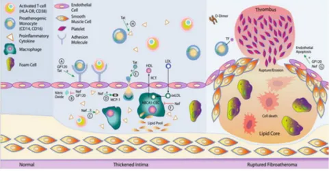
1.13. Inflammation and cardiovascular disease ... 31
1.13.1. Endothelial cell dysfunction and association with carotid intima media thickness and arteriosclerosis ... 31
1.13.2. Cardiac imaging for coronary heart disease ... 33
1.14. Highly expressed inflammatory factors in HIV infection... 34
1.15. IL-32 as a potential player in CVD in HIV infection ... 36
1.15.1. Discovery and general properties of IL-32 ... 36
1.15.2. IL-32 receptors ... 38
1.15.3. IL-32 induction mechanisms ... 39
1.15.4. Downstream effects of IL-32 ... 40
1.15.5. Role of IL-32 in Pathogen Infection ... 41
1.15.6. Effect of HIV infection on the induction of IL-32 ... 42
1.15.7. IL-32 in HIV-1 infection ... 42
Chapter 2:... 46
RATIONALE, HYPOTHESIS AND OBJECTIVES ... 46
2.1. RATIONALE ... 47
2.2. HYPOTHESIS: ... 47
2.3. OBJECTIVES: ... 47
Chapter 3: ... 49
MATERIAL & METHODS ... 49
3.1. Study population and biological specimens ... 50
3.2. Sample preparation for ELISA ... 51
3.3. ELISA technique ... 51
3.4. Non-quantitative RT-PCR ... 53
vii
3.6. In vitro infection of cells with HIV-1 ... 56
3.7. Bradford Technique ... 57
3.8. FACS cell sorting: ... 57
3.9. Statistical analysis ... 58
Chapter 4: ... 59
RESULTS ... 59
4.1. Schematic representation of baseline samples ... 60
4.2. IL-32α and IL-32αβδ isoforms ... 61
4.3. Total levels of IL-32 in HIV-infected and non-infected subjects. ... 62
4.4. Correlations between IL-32, common CVD markers, and viral load. ... 64
4.5. Age- and sex-associated differential expression of IL-32 in HIV+ aviremic subjects ... 65
4.6. Correlation of total IL-32 with the CVD markers D-dimer and LDL/HDL ratio ... 66
4.7. PCR Amplification of IL-32 isoforms ... 67
4.8. Differential expression of IL-32 isoforms ... 70
4.9. Individual expression of IL-32 isoforms in PBMCs from HIV+ with coronary artery atherosclerosis ... 72
4.10. Higher IL-32D levels positively correlate with the coronary artery plaque volume .... 73
4.11. Differential expression of IL-32 isoforms in immune cells ... 74
4.12. In vitro infection of PBMC ... 76
Chapter 5: ... 78
DISCUSSION AND CONCLUSIONS ... 78
5.1. Discussion ... 79
5.2. Strengths and Limitations ... 89
5.3. Conclusion ... 90
Chapter 6: Appendix ... 91
Article: Differential expression and functions of IL-32 isoforms in HIV+ individuals under cART ... 91
viii
List of tables
Table I. Demographics and clinical data of the study participants ... 50
Table II. Composition of Disruption buffer (5X) and preparation of (1X) DB ... 51
Table III. Different Oligonucleotide primers (forward / reverse) for IL-32 isoforms ... 54
Table IV. Run protocol of non-quantitative PCR. ... 55
Table V. Reaction mixture for quantitative PCR. ... 56
ix
List of figures
Figure 1. Structure and proteins of HIV-1. ... 5
Figure 2. Genomic organization of HIV-1 proviral DNA. ... 5
Figure 3. The Lifecycle of HIV. ... 9
Figure 4. Clinical Stages of HIV Infection. . ... 14
Figure 5. Rates of AMI among the HIV-infected and non-infected population. ... 30
Figure 6. HIV-mediated changed in arterial structure and function leading to atherosclerotic plaque formation. ... 32
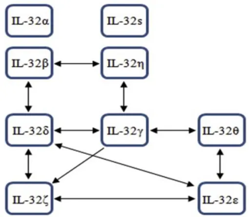
Figure 7. The IL-32 Genome and Isoforms. ... 37
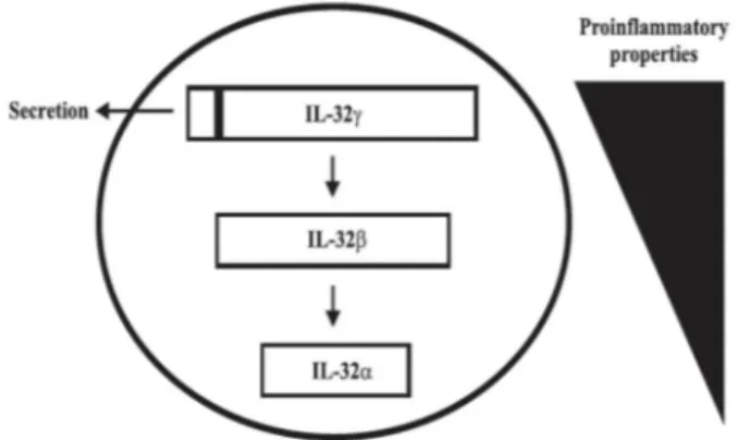
Figure 8. IL-32 isoform splicing. Splicing of IL-32γ generates two isoforms (IL-32α, IL-32β) with diminished pro-inflammatory properties. ... 38
Figure 9. Inducers of IL-32. ... 40
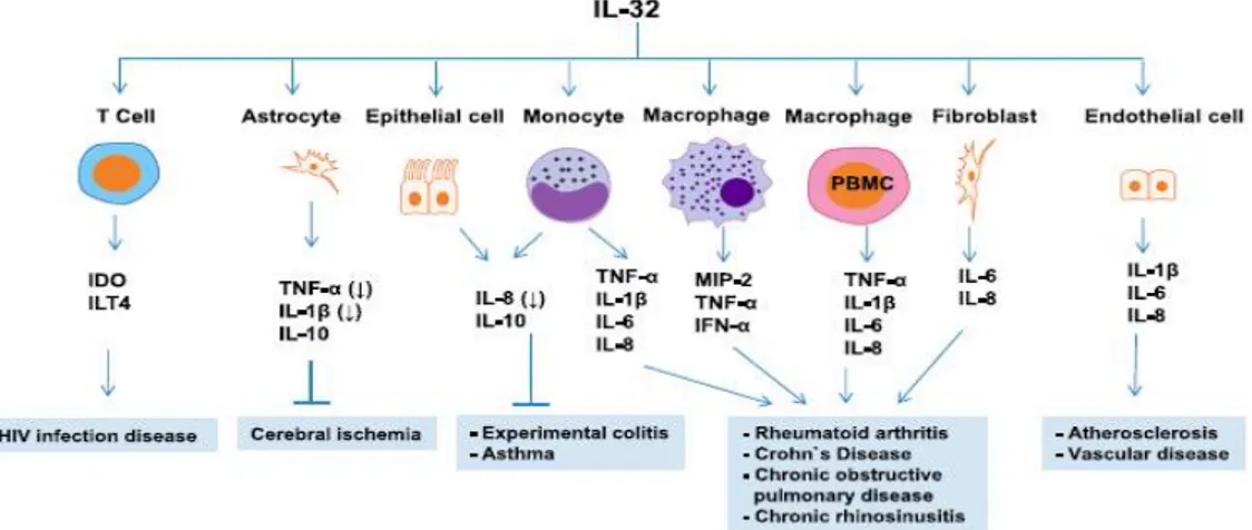
Figure 10. Roles of IL-32 in Different Inflammatory Diseases. ... 41
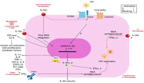
Figure 11. The role of 32 in HIV Infections. HIV induce the expression of endogenous IL-32 in T-lymphocytes. . ... 42
Figure 12. Role of several inflammatory diseases where IL-32 may play a central role in the development of CVD... 44
Figure 13. Model for IL-32 isoform-interaction mapping. m... 45
Figure 14. Gating strategy for cell sorting of the major immune cell types in PBMCs... 58
Figure 15. Schematic representation for the number of available samples at baseline visits used in the current study.. ... 60
Figure 16. Significant levels in experiment-to-experiment variations in measures of IL-32α but not total IL-32.. ... 61
Figure 18. Comparison of fold changes in experimentto-experiment variations between measurements of IL-32α and total IL-32. ... 63
Figure 19. Plasmatic levels of total IL-32 protein in HIV+ subjects. ... 63
Figure 20. Correlations among IL-32 levels, CD4/CD8 ratio, and viral load (VL)... 64
Figure 21. Plasmatic levels of total IL-32 protein (pool of all isoforms) in ART-treated HIV+ men and women (aviremic) in association with age. ... 65
x
Figure 23. Correlation between total IL-32 levels and CVD biomarkers (LDL/HDL ratio and D-dimer). ... 67 Figure 24. Isoform-specific PCR amplification of IL-32 isoforms (α, β, γ, D, ε, and θ) in addition to the housekeeping gene β-glucuronidase. ... 68 Figure 25. Amplification curves and cycle threshold for IL-32 isoforms and housekeeping genes produced by LightCycler 480. ... 69 Figure 26. Correlation between cell-associated total IL-32 protein (measured by ELISA) and total IL-32 mRNA isolated from the same PBMCs. ... 69 Figure 27. IL-32 mRNA expression in HIV+ and HIVneg subjects. ... 71 Figure 28. Comparison between the level of each individual isoform of IL-32 mRNA in HIV+ subjects and HIVneg controls. ... 72 Figure 29. Relative expression of IL-32α, β, γ, , ε and ɵ mRNA in total PBMCs isolated from HIV+ male aviremic subjects with or without TAPV. ... 73 Figure 30. The ratio between IL-32D and IL-32β predicts the total coronary artery plaque volume (TAPV) in HIV+ ART-treated subjects. ... 74 Figure 31. IL-32 isoform expression by different immune cells.. ... 75 Figure 32. IL-32D expression by immune cells in HIV positive and HIV negative donors. .. 76 Figure 33. Infection rate in total PBMCs using the dual tropic virus p89.6. ... 77 Figure 34. Differential expression of IL-32 isoforms in response to HIV infection. ... 77 Figure 35. A hypothetical model for the role of L-32 in CVD in aging HAART-treated HIV+ individuals... 88
xi
List of abbreviations
ABC: Abacavir
ADCC: Antibody-dependent cellular cytotoxicity ART: Anti-retroviral therapy
AIDS: Acquired immunodeficiency syndrome Akt: Protein Kinase B
ALG-2: Apoptosis-linked gene 2
APOBEC-3G: Apolipoprotein B mRNA Editing Enzyme, Catalytic Polypeptide-like 3G AZT: Azidothymidine (Zidovudine)
bDNA: branched DNA)
cART: Combination anti-retroviral therapy CCR5: C-C chemokine receptor type 5 cDNA: Complementary DNA
CI: Chronically infected CTL: CD8+ T lymphocytes
CTLA-4: Cytotoxic T-Lymphocyte Associated Protein 4 CVD: Cardiovascular Disease
CXCL: Chemokine (C-X-C motif) ligand CXCR: Chemokine C-X-C Motif Receptor d4T: Stavudine
xii
ddI: Didanosine, marketed under trade name Videx, used to treat HIV/AIDS d4T: Dstavudine, used to treat HIV/AIDS
ddN: 2’,3’dideoxynucleoside DNA: Deoxyribonucleic Acid ENV: HIV envelope glycoprotein
ESCRT: Endosomal sorting complexes required for transport FDA: Food and Drug Administration
Gag: Group-specific antigen, coding for structural proteins GI: Gastro-intestinal
Gp: Glycoprotein
HAART: Highly aggressive anti-retroviral therapy HC: Healthy controls
HDL-C: High density lipoprotein- cholesterol HIV-1: Human immunodeficiency virus type 1 HIV-2: Human immunodeficiency virus type 2 HTLV-I: Human T-Lymphotropic Viruses IBD: Inflammatory bowel disease
IDO: Indoleamine 2,3-Dioxygenase IgG1: Immunoglobulin G1
IFN: interferon
xiii IL: Interleukin
INI: Integrate inhibitors JAK-2: Janus kinase-2 JNK: c-Jun N-terminal kinase
LAV: Lymphadenopathy-Associated Virus LDL-C: Low density lipoprotein- cholesterol LPS: Lipopolysaccharide
LTNP: Long-term non progressors LTR: L-terminal repeat
MAPKs: Mitogen-activated protein kinases mCD14: Membrane CD14
mRNA: messenger RNA
NEF: Negative Regulatory Factor
NF-κB: Nuclear factor kappa-light-chain-enhancer of activated B cells NLR: Nod-like receptors
NNTRI: Non-nucleotide reverse transcriptase inhibitors
NOD: nucleotide-binding oligomerization domain-like receptors NRTI: Nucleotide reverse transcriptase inhibitors
PBMC: Peripheral blood mononuclear cells PCR: Polymerase chain reaction
xiv pDC: Plasmacytoid dendritic cells
PHA: Phytohaemagglutinin PHI: Primary HIV infection PI: Protease inhibitor
PI3K: Phosphatidylinositol 3-kinase PKA: Protein kinase A
PKC: Protein kinase C Pol: DNA polymerase PR: Protease
PRR: Pattern recognition receptors RA: Rheumatoid arthritis
RER: Rough endoplasmic reticulum
REV: Regulator of expression of viral proteins RLH: RIG-like helicases
RNA: Ribonucleic acid
RRE: Rev Responsive Element RT: Reverse-transcriptase
RTI: Reverse-transcriptase inhibitor sCD: soluble cluster of differentiation SiRNA: Small interfering RNA SIV: Simian immunodeficiency virus
xv
SIVcpz: Simian immunodeficiency virus infection of Chimpanzees SIVcpzPt: SIV of chimpanzees Pan troglodyte troglodytes
SIVgor: SIV of gorilla
SNP: single-nucleotide polymorphism
STAT-3: Signal transducer and activator of transcription-3 SU: Surface unit
TAT: Trans-activator protein TH: T helper
TLR: Toll-like receptors TCR: T cell receptor TM: Transmembrane
TNF-α: Tumor necrosis factor alpha Vif: Viral infectivity factor
Vpr: Viral protein R Vpu: Viral protein U
xvi
Acknowledgments
It is a genuine pleasure to express my deepest appreciation and thanks to my research supervisor, Dre. Cecile Tremblay, for having me in her research team and giving me the chance to work on this master’s project. Her timely advice, meticulous scrutiny, scholary advices and scientific approach have helped me to a very great extent to accomplish this task. It is my great privilege to have accomplished this thesis under her guidance. I would also like to express my sincerest gratitude to my co-director and mentor Dr. Mohamed El-Far, who was a real fountain of infinite knowledge throughout the process. His high expectations and exceptional leadership were my pillars of success. I express my sincerest appreciation for his assistance in any way that asked; he is indeed a true gentleman. Thank you for your unwavering support, advice and availability.
I would also like to express my gratitude to my committee members Dr. Hugo Soudeyns and Dr. Daniel Lamarre. The time they spent providing their comments is greatly appreciated and have contributed to the improvement of this thesis.
I would like to extend huge, warm thanks to my previous supervisor Dr. Ali Ahmad for accepting me in his lab when I had no previous lab experience; and to his research team Suzzane Samarani, Zainab Aldbah and Ayoub Abualkhir, who were generous enough to share their knowledge, experience and information with me.
I also had the chance to meet a lot of incredible people during my stay in the lab at crCHUM. I would like to extend my deepest gratitude to all my lab members Mohamed Sylla, Etienne Larouche-Anctil, Annie Chamberland and Remi Bunet and to my colleagues Amelie Cattin, Laurence Raymond Marchand, Annie Gossline, Hawley Rigsby for their help, support and advice.
xvii
For the ancestors who paved the path before me, upon whose shoulders I stand – a special Thank you to my family members, especially my parents, my father Muftah and my mom Khadija, for their unconditional love and endless dua (prayers). I thank them for their constant and reassuring presence in my life, and for instilling in me the desire to always look further, which helped keep my feet on the ground during the more difficult times. A special appreciation goes to my brothers and sisters for their support and for being a source of inspiration to me.
My heartfelt thanks go to my beloved husband, Mokhtar, for his patience, great support and encouragement, and for waving away my frustrations through the stressful journey of life. Thank you to my sons Mohammed, Ayham and Yamen who have given me much happiness and keep me hoping; who have grown up watching me study and juggle family and work – thank you for your presence in my life. I hope I have been a good mother and that I have not missed too much during the tenure of my study.
Great appreciation goes to my uncle Abdulmotaleb and his wife Zenap and to their children Hadeel, Shahed, Sarah, Ali and Manal who have always been a great source of love, support and encouragement since I first arrived in Canada.
A special appreciation goes to my second family (my husband’s family) for their prayers and moral support. They have always supported me and encouraged me with their best wishes.
The work done here could not have been completed without the help of various collaborators and financial organizations. In this sense, a special thank you to the funding agencies that funded my master’s
xviii
project (CIHR, CTN, NIH and FRQS). Thank you to each individual donor for their generous donation of time and blood.
I am especially indebted to my government, the Libyan government, for the scholarship. I could not have completed my master’s program overseas without their financial support.
Finally, I thank all those who have helped me directly or indirectly in the successful completion of my thesis. Anyone missed in this acknowledgement are also thanked.
Chapter 1:
1.1. Epidemiology of HIV-AIDS
HIV-1 and HIV-2 are both members of the Retroviridae family and have the capacity to infect and replicate in the human body. However, HIV-1 is the etiological agent responsible for the majority of acquired immunodeficiency syndrome (AIDS). According to the most recent United Nations Programme on HIV/AIDS (UNAIDS), there are currently 36 million people living with HIV worldwide, among whom 34.5 million are adults (UNAIDS, 2017). Each year, there are more than 1.8 million new infections worldwide, which stresses the fact that HIV/AIDS remains a global concern (WHO, 2018). The highest population of individuals living with the viral infection are native to sub-Saharan African communities. Epidemiological observations have shown that the most common and causative factor for the transmission of novel HIV infection is through unprotected sexual intercourse (HIV.gov, 2017). However, HIV can also be transmitted by other horizontal means such as through transfusions with infected blood or through the sharing of infected needles (Day et al., 2008). Additionally, vertical transmission of HIV, from mother to child during pregnancy, labour, delivery or breastfeeding, has been on the rise in the absence of anti- retroviral therapy (King et al., 2013). Over the years, the rate of people with access to anti-retroviral therapy has increased to reach 19.5 million in 2016, compared to 17.1 million in 2015 and just 7.7 million in 2010. This encouraging data reveals that access to effective medications is improving and is, in part, responsible for the 11% decrease in new HIV infections reported among adults in 2016. More encouragingly, the number of deaths due to AIDS-related illnesses has fallen by 48%, reaching one million in 2016 compared to 1.9 million in 2005 (UNAIDS, 2017). However, global efforts are still needed to help make anti-retroviral treatments more accessible to infected people in countries with limited resources.
1.2. The origin and discovery of HIV
1.2.1. Origin of HIV
HIV is thought to have originated in monkeys, resulting from adaptation of the simian immunodeficiency virus (SIV) following transmission to humans (Sharp et al., 2010).
3 Interestingly, there are four distinct groups of HIV-1, which have been classified based on four independent events resulting in transmission to humans. These groups are the M (Major), O (Outlier), N (New) and P (Putative) (Charneau et al., 1994; Wainberg, 2004). The most common group worldwide, the M group, was discovered in 1981 at the origin of the pandemic. It has 9 subtypes (A, B, C, D, F, G, H, J, K), of which subtype B is the most prevalent in North America (Plantier et al., 2009). With only 10-1000 reported cases, Group O was discovered in 1990 in Central Africa (De Leys et al., 1990). In 1998, the N group was identified in Cameroon, reporting only a dozen known cases (Simon et al., 1998). Similarly, the P group was recently identified after being isolated from two individuals in Cameroon (Plantier et al., 2004).
However, phylogenetic studies of these groups suggest two possible origins of HIV. Evolutionary relationships trace the origin of the HIV-1 genome to the Pan troglodyte troglodytes SIV (SIVcpzPtt) in chimpanzees (Gao et al., 1999), whereas HIV-2 appears to be derived from the SIV of sooty mangabey (SIVSM) lineages (Sharp et al., 2010, 2011). HIV-2 is confined to West Africa and is less virulent compared to HIV-1 infection (Sharp et al., 2011). These phylogenetic analyses have also allowed researchers to approximate how long the virus made its transmission to humans. The transmission of the first three groups (M, N & O) took place at the beginning of the 20th century. In contrast, the date the P group was introduced in humans remains unknown (Wertheim et al., 2009). Regardless of the origin, HIV evolves rapidly due to a high rate of mutation resulting from the error-prone nature of its reverse transcriptase enzyme and to recombination events in its human host.
1.2.2. Discovery of HIV
During the Summer of 1981, clinicians in New York and California were reporting high incidence of unusual cancers such as Kaposi's sarcoma and of other opportunistic infections such as Pneumocystis jiroveci, among the homosexual community (Gottlieb et al., 1981). Some of these patients were also affected by persistent lymphadenopathy and had significantly low CD4+ lymphocyte counts. These observations led clinicians to agree that these patients suffered from a common immunodeficiency and prompted the quest to identify the etiologic agent responsible for this disease. Because of its high prevalence among gay men, it was first hypothesized to be a result of sexual transmission (Brennan et al., 1981; Friedman-Kien et al.,
4 1981). However, this theory came into question with a study in the United States where the syndrome was observed in various social groups, suggesting transmission through blood (Masur et al., 1981)(Masur et al., 1981). In 1983, the virus in question was isolated from an infected lymph node by a group of researchers led by Françoise Barré-Sinoussi and her mentor Luc Montagnier at Institut Pasteur in France (Barré-Sinoussi et al., 1983). This infectious pathogen was named lymphadenopathy-associated virus (LAV). Interestingly, this virus exhibited a striking similarity to the Type I and Type III Human T-Lymphotropic Viruses (HTLV-I and III) previously identified by Robert Gallo in the United States (Poiesz et al., 1980; Gallo et al., 1983). It turned out that both LAV and HTLV-III were actually the same virus and it was renamed human immunodeficiency virus (HIV) in 1986 (Coffin et al., 1986).
1.3. Morphological structure of HIV-1
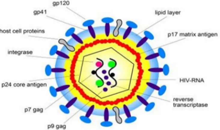
HIV-1 viral particles measure around 80-120 nanometers (nm) in diameter and are enclosed by a lipoprotein membrane made up of 72 glycoprotein (gp) heterodimers that are integrated into the lipoprotein membrane (Murray et al., 2009). The membrane heterodimers are comprised of gp120 and transmembrane gp41. During the budding process, HIV incorporates various host cell membrane proteins into its lipoprotein envelope. Among these host cells proteins are the intercellular adhesion molecule 1 (ICAM-1), which accelerates adhesion to the target cells, and the human lymphocyte antigen class I and II proteins (Paquette et al., 1998). The viral matrix consists of p17 protein and links the lipoprotein membrane of the retrovirus to its viral core (Dolcetti et al., 2015), which is comprised of the primary antigen (p24). The p24 core houses two copies of single-stranded HIV-1 RNA and the p7 nucleoprotein. The core also contains the enzymatic makeup of the virus; notably protease p11, integrase p32, and the p66 reverse transcriptase (RT). These enzymatic elements are essential for early viral replication. Figure 1 illustrates the structure of the HIV-1 virus and its key replication factors (Hill et al., 2005).
5 Figure 1. Structure and proteins of HIV-1. Reproduced from (Behrens, 2011), no
permission required.
1.4. HIV-1 genome
The HIV-1 genome, which is about 9 kb of RNA, encodes for 15 proteins (Frankel et al., 1998). HIV replication relies on two types of genes: structural and auxiliary genes. The three distinct structural genes necessary for building a new virus particle include the envelope gene (env), the polymerase gene (pol), and the group antigen genes (gag). The auxiliary genes include two essential regulatory genes: the trans-activator of transcription (tat) and the regulator of expression of virion proteins (rev), as well as four important accessory genes: viral infectivity factor (vif), viral protein U (vpu), viral protein R (vpr), and negative factor (nef) (Behrens, 2011; Karn et al., 2012). Figure 2 illustrates the HIV-1 genome and the critical genes it codes for.
Figure 2. Genomic organization of HIV-1 proviral DNA. Reproduced from (Behrens, 2011), no permission required.
6
1.4.1. Structural genes
The classical framework of the viral genome is represented by 5’LTR-gag-pol-env-3’LTR structure. The long-terminal repeats (LTRs), which do not code for any retroviral proteins, flank genome and contain the HIV promoters required for viral expression (Frankel et al., 1998). Both gag and env code for the glycoprotein and nucleocapsid components of the retroviral membrane. During maturation, activated viral proteases cleave the Gag polyprotein into the matrix protein (p17), core proteins such as the capsid protein (p24), the spacer peptides 1 and 1 (sp1 and sp2), the nucleocapsid protein (p7), and the late-assembly p6 protein which is involved in virion budding (Votteler et al., 2011). The env gene encodes the gp160 homotrimer, which is then cleaved into gp120 and gp41. These active fusion proteins are critical for internalization of the virus. Gp120 binds to C C chemokine receptor 5 (CCR5) and to cluster of differentiation 4 (CD4) on the host cell, while gp41 plays a key role in the fusion and internalization process (Morikawa et al., 1993; Alkhatib, 2009). Arguably the most important structural HIV-1 gene is the pol gene, which codes for key enzymes that are critical for viral replication, reverse transcriptase (RT), and protease enzymes. These viral enzymes are produced following cleavage of the Gag-Pol precursor polyprotein and are translated following a shift in the reading frame of the ribosome, near the 3' end of the gag gene. RT is critical for the retro-transcription of viral RNA into DNA, which then integrates into the host genome through the action of the viral integrase. HIV protease is equally important, as it cleaves other polyproteins such a Gag into active and functional proteins essential for the virion's maturation and infection (Jacks et al., 1988; Greene et al., 2002; Jeang et al., 2007).
1.4.2. Auxiliary genes
The HIV-1 genome codes for six other genes: nef, vif, vpr, rev, tat, and vpu. Although vpu, vif, nef, and vpr are considered to be accessory genes because they are not needed for viral replication in vitro, their functions and regulations are significantly important for HIV pathogenesis in vivo (Malim et al., 2008). The two regulatory genes, tat and rev, code for accumulating proteins that regulate the expression of other viral proteins by binding to the retroviral RNA genome. The Tat protein plays a substantial role in the transcription of viral
7 messenger RNA (mRNA) by binding to the transactivation-response element (TAR) located in LTR. In the complex containing the transcriptional positive elongation factor b (P-TEFb), through the help of cyclin T1, binding of Tat onto TAR allows for the recruitment of the CDK9 cell cyclin on the LTRs. Through the kinase activity of CDK9, the C-terminal phosphorylation of the large subunit of RNA polymerase II, allows for initiation of genomic transcription. When Tat is not present, RNA polymerase II is unable to synthesize full-length viral transcripts (Fisher et al., 1986; Berkhout et al., 1989; Bannwarth et al., 2005). On the other hand, Rev expression activates expression of structural and enzymatic genes, including gag, pol and env of proviral DNA. It binds the RRE (Rev responsive element) and ensures the transport of partially spliced and un-spliced HIV-1 mRNA from the nucleus to the cytoplasm during viral replication, where it allows for the translation and stabilization of the mRNA. Rev is also responsible for the transport of the gag transcript to the cytoplasm (Malim et al., 1988; Malim et al., 1989; Zapp et al., 1989).
Accessory gene, vif, encodes a 23 kilo Dalton (kDa) Vif protein, which is transcribed during the late stage of the virus life cycle. Vif binds the apolipoprotein B mRNA editing enzyme, catalytic polypeptide-like 3G (APOBEC-3G) and induces degradation of the enzyme via the proteasomal pathway. This degradation prevents the hypermutation effects of APOBEC-3G and leads to the subsequent enhancement of viral replication (Miyagi et al., 2014). Vpu is a specific protein involved in the degradation of the CD4 molecule in the endoplasmic reticulum (Willey et al., 1992). Vpu is also involved in releasing the newly produced virions attached to Tetherin protein (BST-2) into the cytoplasm by inducing the formation of pores in the cell membrane (Strebel, 2014). This BST-2-induced host-protective mechanism can be overcome by 1 via ubiquitination (Laplana et al., 2012). Nef represents a critical protein in the HIV-1 life cycle. In addition to its key role in the down-regulation of CD4 and MHC-I molecules to protect infected cells from cytotoxic killing by CD8 T-cells (Schaefer et al., 2008), the Nef protein also sustains T-cell activation during the initial stages of the HIV life cycle (Das et al., 2005). Nef does this by decreasing the threshold of T-cell activation through, among other mechanisms, down-regulation of the negative regulators of T-cell activation such as cytotoxic T-lymphocyte-associated protein 4 (CTLA-4) (El-Far et al., 2015). Nef also downregulates CD4 molecules on the surface of infected host cells, thereby blocking re-infection of the cell and intracellular binding of newly synthesized viral Env proteins, allowing for proper viral assembly
8 (Li et al., 2005a). Finally, Vpr plays a pivotal role in transcription activation of the LTR (Kogan et al., 2011). Vpr also promotes viral replication by allowing the viral transactivation of cellular genes and cell differentiation (Romani et al., 2009).
1.5. HIV-1 replication cycle
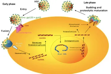
1.5.1. Early phase
The initial stage of the HIV replication cycle involves binding of the virus to the surface of the host CD4+ T-lymphocyte via the CD4 receptor and fusing with the cellular membrane (Goodsell, 2015). As illustrated in Figure 3, the HIV gp120 attaches to the CD4 molecules, as well as to the CCR5 or CXCR4 co-receptors on the host cell before initiating the fusion process. Following penetration of the fusion peptide into the cell membrane, the two helices of gp41 fold back onto themselves, allowing fusion of cellular and viral membranes (Weissenhorn et al., 1997), and releasing the nucleocapsid into the host-cell’s cytoplasm. Once inside the cell, the nucleocapsid disintegrates, liberating two strands of viral RNA and essential viral enzymes. Using its own reverse transcriptase and by hijacking some of the host cell’s replication machinery, the viral RNA is reverse-transcribed into single-stranded and then into double-stranded DNA (Farnet et al., 1996). More specifically, transcription is initiated using a host cell-derived transfer RNA (tRNA) as a primer. The viral RNA is then reverse transcribed into the first strand of cDNA. During cDNA synthesis, the template RNA is completely degraded by RNAse H, except for two short purine-rich sequences (PPT for poly-purine tracts). These PPTs serve as a primer for synthesis of the second DNA strand, which is also carried out by the reverse transcriptase (Basu et al., 2008). The newly synthesized viral DNA is then transported into the cell nucleus, where it integrates into the host genome through the action of the viral integrase enzyme. This integrated DNA is referred to as the provirus, which can remain inactive for several years, forming a few to no replicates of the virus (Miller et al., 1997).
1.5.2. Late phase
The transcription of mRNAs by the RNA polymerase enzyme takes place during the late stage of the viral replication cycle. In the host cell nucleus, viral mRNA is spliced and exported to the cytoplasm for translation by the ribosomes. This process results in the synthesis of the
9 first viral proteins Nef, Tat, and Rev. Gag, Gag-Pol and Env (gp160) precursor proteins are later transcribed from an unspliced or mono-spliced mRNA, as their export is dependent upon the expression of Rev (Suhasini et al., 2009; Checkley et al., 2011). Integrase, protease, and reverse transcriptase enzymes are translated from the same Gag-Pol transcript, using a frame-shifting process.
Virion assembly is mediated by conformational changes within Gag. The Gag domain binds to the plasma membrane, through interaction of its amino-terminal domain with the lipid bilayer of the cell membrane (Sundquist et al., 2012), where it recruits the viral Env protein. The Env proteins traffic through the secretory pathway of the rough endoplasmic reticulum (RER), to the Golgi body and vesicles, until they reach the plasma membrane. There, the Env protein induces endosomal sorting, which is needed for viral budding and transport, while Gag engages both the endosomal sorting complex required for transport (ESCRT), and the apoptosis-linked gene 2 (ALG2)-interacting protein (Usami et al., 2009; Sundquist et al., 2012). These glycoproteins play a key role in enabling budding of the virus, so the new particles can bind co-receptors and infect CD4+ T-cells (Figure 3). Also, at the plasma membrane, small protein chains associate with copies of the HIV RNA and enzymes to form new virus particles ready for release during the budding stage (Sundquist et al., 2012). During the budding process, the new virus emerges from the cell, taking with it part of the host cell membrane and incorporating expression of certain co-stimulatory molecules such as CD40L from the viral envelope membrane (Imbeault et al., 2011).
Figure 3. The Lifecycle of HIV. Reproduced from (Greene et al., 2008) with permission from Elsevier: Anti-viral Research, copyright 2008.
10
1.6. Anti-retroviral therapy to HIV-1 infection
HIV treatment experienced a breakthrough in the mid-1990s and has since become easier to manage with the emergence of combined therapies. There are currently six classes of anti-viral drugs classified based on how they affect the anti-viral life cycle.
i) Nucleoside reverse transcriptase inhibitors
Nucleoside reverse transcriptase inhibitors (NRTIs) were the first drugs available against HIV infection. In 1986, the United States Food and Drug Administration (FDA) approved the first ever anti-retroviral medicine, zidovudine (AZT), which targets replication of HIV (Broder, 2010) by suppressing the activities of the reverse transcriptase.
Various nucleoside analogs such as zalcitabine (ddC), didanosine (ddI), lamivudine (3TC), stavudine (d4T), abacavir (ABC), and tenofovir (a nucleotide RTI) were added to the anti-retroviral arsenal. They are 2’, 3’dideoxynucleoside (ddN) analogues that compete with normal cellular deoxynucleoside triphosphates during transcription. They act as chain terminators of DNA synthesis by incorporating into the ongoing synthesis of the DNA strand, leading to pre-mature termination of the reaction and subsequent blocking of viral replication (Broder, 2010; Arts et al., 2012).
ii) Non-nucleoside reverse transcriptase inhibitors
The class of non-nucleoside reverse transcriptase inhibitors (NNRTIs) was introduced in 1996. First generation NNRTIs include nevirapine, delavirdine, and efavirenz. The second generation has an improved resistance profile and includes etravirine and rilpivirine (Usach et al., 2013). These inhibitors function by inducing a conformational change in the HIV reverse transcriptase enzyme by binding to the hydrophobic pocket of its P66 subunit, away from the active site of the enzyme (Sluis-Cremer et al., 2004).
11 Protease inhibitors (PIs) are different from reverse transcriptase inhibitors. Instead of interfering with transcription, PIs are used to inhibit the activity of HIV protease in host cells already infected with the virus, thereby preventing further viral replication (Lv et al., 2015). They prevent the proteolytic cleaving function of the protease enzyme, preventing the generaton of functional protein units (Lv et al., 2015) from their large non-functional polyproteins precursors (Gag and Gag-Pol). This category of drugs includes ritonavir, indinavir, lopinavir, nelfinavir, saquinavir, amprenavir, atazanavir, and darunavir.
iv) Integrase strand transfer inhibitors
Integrase strand transfer inhibitors (INSTIs) include Raltegravir and Elvitegravir introduced in 2007, as well as Dolutegravir introduced in 2013 (Cooper et al., 2008; Cahn et al., 2013; Gallant et al., 2013). Additionally, there are two new INSTIs (bictegravir and cabotegravir) that are currently in clinical trials. Bictegravir can be given once a day, while cabotegravir is a long-acting agent that can be dosed once a month because of its exceptionally long half-life (Han et al., 2017). INSTIs prevent integration of the viral genome into the host DNA of infected cells (Dow et al., 2014). INSTIs achieve this by binding metallic ions in the active site of the integrase, preventing the formation of a covalent bond with host DNA, and further inhibiting the strand transfer reaction (Hazuda et al., 2000).
v) Viral entry inhibitors
The entry of HIV-1 into the target cell can be blocked at several stages during HIV entry into cells. These inhibitors include CD4-binding inhibitors, co-receptor inhibitors, and fusion inhibitors (Wilen et al., 2012).
A) Attachment inhibitors
Also known as CD4-gp120 inhibitors, attachment inhibitors interfere with the viral gp120. Four types of drugs with different mechanism of action belong to this category of inhibitors: PRO-542 (CD4-IgG2) which acts by mimicking the CD4
12 receptor, TNX-355 which acts by competing with HIVgp120 for CD4 binding, CADA which downregulates CD4 receptor expression and, lastly, BMS-806 which prevents the conformational changes of gp120 that take place after binding of gp120 to the CD4 receptor (Briz et al., 2006). Fostemsavir is a gp120 attachment drug that has recently shown promise in clinical trials involving heavily treated individuals.
B) Co-receptor binding inhibitors
Another name for this class of drugs is chemokine receptor antagonists (CCR5 antagonists). This class is comprised of small molecules that, by binding to the receptor, induce conformational changes of the extracellular loops (allosteric mechanism) in the protein, preventing attachment of gp120. It includes Maraviroc, which interferes with viral entry into susceptible cells by targeting the CCR5 chemokine co-receptors. Maraviroc works well; however, resistance mechanisms are known. The most common type of resistance is the emergence of X4 virus (Westby et al., 2006). The second mechanism of resistance developed by HIV is the ability to use the co-receptor in its compound-bound form (Westby et al., 2007). It is only effective against R5 viruses.
C) Fusion inhibitors
Fusion inhibitors (FIs) were approved in 2003. They are directed against viral gp41, specifically the formation of the 6HB structure responsible for fusion. FI drugs such as Enfuvirtide (the only fusion inhibitor currently marketed) function by blocking viral fusion and subsequent entry into the cell (Lalezari et al., 2003; Lazzarin et al., 2003). Although Enfuvirtide is the only FDA approved fusion inhibitor, its use is limited because of the side effects such as severe local injection site reaction (Ball et al., 2003).
1.6.1. Importance of using combined anti-retroviral therapy
Since the mid-90s, Highly Active Anti-Retroviral Therapy (HAART) has been the standard of care to block the active replication of HIV (Arts et al., 2012). HAART refers to
13 combination therapy, which includes the use of protease inhibitors, integrase inhibitors, non-nucleoside, and nucleoside analogue drugs, given in a wide range of combinations (Arts et al., 2012). It had previously been shown that treatment with a single protease inhibitor and two other anti-retroviral therapies had a ripple effect in reducing viral replication and copies of HIV in the blood. Thus, combination therapy has become the standard of care for patients living with HIV/AIDS as it targets multiple stages of the HIV-1 replication cycle and reduces the chances of the virus developing resistance to the various treatments (Staszewski et al., 1996; Arts et al., 2012).
Viral replication occurs at a high rate. Several billion copies of the retrovirus are created and destroyed on a daily basis. This high turnover is one of the mechanism by which HIV develops drug resistance. Due to the high rate of replication and a highly error-prone HIV-reverse transcriptase, random mutations are introduced into the HIV RNA. These genetic mutations lead to changes in viral proteins targeted by anti-retrovirals, decreasing their efficacy. Resistance significantly complicates the treatment process, rendering the medication provided either less or completely ineffective. Once resistance develops to a particular drug, the virus is likely to become resistant to drugs in the same class. Thus, it has become clear that a single-agent against HIV would have limited ability to control such a rapidly replicating virus. Expansion of the anti-retroviral drug classes has made it possible to combine treatments. Combination therapy can more effectively suppress the mutated forms of the retrovirus.
1.7. Natural history of HIV-1 infection
HIV infection is predominantly characterized by the depletion of CD4+ T lymphocytes (Février et al., 2011). Since these cells are one of the main mediators of immune responses, HIV-mediated depletion of CD4+ T-cells increases the vulnerability of infected individuals to opportunistic infections and death (Okoye et al., 2013). As with other lentiviruses, HIV infection can have slow periods of replication, leading to chronic disease (Okoye et al., 2013). Some individuals may show significant progression throughout the course of the disease; However, less than 5% of infected individuals do not show disease progression over several decades. These individuals are called long term non-progressors (LTNP) (Kumar, 2013; Gurdasani et al.,
14 2014). The course of HIV-1 infection can be divided into three phases: acute, chronic, and AIDS phase (Figure 4). Each phase is determined by its duration and specific clinical characteristics.
Figure 4. Clinical Stages of HIV Infection. The clinical features of the different phases of the pathogenesis of HIV-1. Reproduced from (Bashirova et al., 2011) with permission from Elsevier: Cytokine, copyright 2012.
1.7.1. Acute phase
The acute phase occurs within the first two to four weeks of HIV-1 infection. This stage is characterized by a high plasma viral load due to the significant amount of virus being produced. The body naturally responds to this vigorous HIV replication, leading to flu-like symptoms which include but are not limited to swollen glands, asthenia, joint aches, weight loss, and sore throat (Kahn et al., 1998). Most of the virus particles identified in this phase show R5 tropism (i.e. binding to CCR5 co-receptor), whereas switching to CXCR4 viruses may occur during the later-stage of infection (Scarlatti et al., 1997).
During the acute phase of HIV infection, activated target host cells (CD4+ CCR5+ T lymphocytes) begin to overexpress early tissue-resident marker CD69 and activation markers such as CD25 and HLA-DR receptors (Kunkel et al., 2002; Douek et al., 2003). At the same time, viral accessory proteins Nef, Vif, Vpr and Vpu, bypass typical T cell receptor (TCR)-mediated activation and contribute to the activation of these cells (Unutmaz et al., 1999;
15 Simmons et al., 2001). As HIV infects and depletes CD4+ T cells, CD4 counts are significantly reduced, particularly in the lymphoid organs associated with the genitals and gastrointestinal tract (Kahn et al., 1998). Two mechanisms are responsible for the depletion of CD4+ CCR5+ T cells: direct cytopathology of HIV-1 or immune responses. The host immune responses lead to destruction of infected cells through the action of natural killer (NK) cells and cytotoxic CD8+ T lymphocytes (Douek et al., 2003; Vieillard et al., 2005), and work to reduce the viral load to a stable level (Meulendyke et al., 2014). At the viral set point (i.e. the end of the acute phase, which is characterized by a marked reduction in viral load), the CD4+ T cell count starts to increase again. This viral set point varies from one individual to another, which will remain the same throughout the chronic phase (Koup et al., 1994; Safrit et al., 1994; Huang et al., 2012).
1.7.2. Chronic phase
The chronic phase of HIV infection is the longest and can take place over several years or decades. It starts after HIV-specific antibodies are developed. Due to the exhaustion of the immune system and bacterial translocation at the gastrointestinal mucosal barriers, the immune system becomes persistently activated; leading to high levels of circulating pro-inflammatory cytokines (Brenchley et al., 2006). HIV infection per se is not solely responsible for the massive depletion of CD4+ T cells. Chronic immune activation is largely believed to lead to compromised cellular metabolism and, consequently, to cell death (Douek et al., 2003; Ford et al., 2009). Several mechanisms are involved in the events leading to depletion of CD4+ T cells. These mechanisms include (i) attrition of the memory cell pool due to persistent immune activation, (ii) extensive activation of naïve T cells entering the pool of memory cells, (iii) reduced number of reserve cells in steady state due to recurrent stimulation, and (iv) lower supply of naïve T cells due to HIV-mediated decreased thymic output (Douek et al., 2003). Infected patients with a high level of HIV RNA proceed to the symptomatic stage at a faster rate than patients with low RNA levels (Watanabe et al., 2015). At this stage, CD4 cells continue to decline at a progressive rate (Streeck et al., 2009). Some patients experience organ dysfunction as a direct result of the virus rather than as a consequence of defective cell-mediated immunity (Naif, 2013); while other individuals may develop generalized lymphadenopathy.
16
1.7.3. AIDS phase
The AIDS stage of the HIV-1 infection occurs once the human immune system is completely damaged and the infected individual becomes vulnerable to various opportunistic infections, most notably Tuberculosis, Pneumocystis jiroveci, Cryptococcal meningitis, and Cytomegalovirus infections (Jung et al., 1998; Corbett et al., 2003). It is also possible to observe neoplasias such as Kaposi’s sarcoma (Chu et al., 2011). During this stage, CD4+ T cell counts fall below 200 cells/µl and there is an increase in viremia (Leng et al., 2001). There can also be a change in viral tropism from R5 to X4 (Douek et al., 2003), which promotes rapid spreading of the virus. Without anti-retroviral therapy (ART) medication, HIV-1 infected individuals can only survive for a few years.
1.8. Immune response to HIV infection
1.8.1. Innate immune response
Innate immune responses represent the first line of defense of the human immune system. The primary components of the innate immune system include the skin, mucosal lining, and epithelial layers of the gastrointestinal tract (GI). These elements act as either chemical or physical barriers to illnesses (Riera Romo et al., 2016). The innate immune response is largely based on the detection of pathogen-associated molecular patterns (PAMPs) by pattern recognition receptors (PRR)s. Thus, during viral evasion, the inflammatory response causes the recruitment of innate immune system cells, including neutrophils, NK cells, monocytes, and plasmacytoid dendritic cells (pDCs). These cells express several PRRs, which act as sensors capable of detecting viral nucleic acids (PAMPs) and initiating a response to type 1 interferon (α and β); thereby preventing spreading of the virus. Recognition of HIV by host cells, primarily CD4+ T cells, can occur at many stages of the replicative cycle. Recognition can occur through detection of the products of reverse transcription, and also through interaction of the virion, capsid, or viral RNA with the different PRRs. Among other things, two types of intracellular PRRs allow for the detection of retroviral DNA in the cytoplasm: IFI16 (interferon inducible protein 16) (Jakobsen et al., 2013) and cGAS (cyclic GMP-AMP synthetase) (Gao et al., 2013). TLRs (toll-like receptors), receptors on the cell surface and in endosomes, are also involved in
17 the recognition of HIV. As a result, gp120 is recognized by TLR2 and 4 on the surface of mucosal epithelial cells (Nazli et al., 2013) and viral genomic RNA is recognized by endosomal TLRs 7 and 8 (Schlaepfer et al., 2006). Neutrophils express TLR 1-9 and respond to pathogens by phagocytosis, producing oxidative compounds and releasing antimicrobial peptides such as Trappin, α-1, -2 and -3 defensins (Chang et al., 2004; Klotman et al., 2006). These defensins are also known as human neutrophil peptides (HNP 1, -2 and -3). They interact directly with the virus and inhibit viral replication (Wang et al., 2004; Klotman et al., 2006). As a result of this recognition, activation of the transcription factor and nuclear factor kappa B (NFκB) signaling cascade occurs, leading to the expression of costimulatory molecules and to the downstream production of several pro-inflammatory cytokines (Kawai et al., 2007; Henrick et al., 2015). During the innate immune response, NK cells have the ability to recognize infected cells with weak MHC class I (MHC-I) expression. Through their cytotoxic function, NK cells lyse infected cells detected via killer immunoglobulin receptor (KIR) inhibitors that interact with MHC-I (Alter et al., 2009). NK cells can also kill virus-infected cells in the absence of antibodies through an antibody-independent mechanism. This mechanism causes NK cells to express the Fas Ligand molecule (CD 178) on their surface and can induce the death of Fas-expressing target cells (CD95) as soon as they adhere to it (Yoon et al., 2015).
1.8.2. Adaptive immune response
Adaptive immune responses are mediated by both B and T lymphocytes. 1.8.2.1. Humoral immunity (B lymphocytes)
The humoral immune response is mediated by antibody-producing B cells. During HIV-1 infection, these cells control the infection by producing and secreting a variety of HIV-specific antibodies (IgA, IgD, IgG, and IgM) (Moir et al., 2009). The first sign of B cell response detected in the plasma are complexes of antibody-virions (Tomaras et al., 2008). A few days later, specific antibodies against gp41 are detected, followed by specific antibodies for gp120 (mainly against the V3 loop) and antibodies specific to other viral proteins such as p24 (Derdeyn et al., 2004; Gray et al., 2007). During primary HIV-1 infection, recent studies have shown that gp41-specific IgM and IgD initial antibodies do not significantly affect the dynamics of viral
18 load (Tomaras et al., 2008). Moreover, during the acute phase, IgA antibodies specific to gp41 can be detected in mucosal secretions. However, it appears that the initial IgA response is non-neutralizing (McMichael et al., 2009).
Owing to the frequent mutations in the variable region of the envelope, HIV-1 escapes detection by the humoral antibodies, thereby preventing neutralization of the pathogen. While the conserved regions of the envelope, such as the CD4 attachment site, do not undergo mutation (Burton et al., 2015), neutralizing antibodies specifically directed against the constant regions are only produced 20-30 months after infection (Burton et al., 2015). Thus, antibodies produced at a given moment do not neutralize the contemporary virus particles, but rather are directed against earlier variants. Consequently, newer virus particles are capable of escaping the humoral anti-viral response (Albert et al., 1990; Moog et al., 1997). Therefore, fast-progressing individuals rarely have these antibodies during the course of infection (Tomaras et al., 2008; Kwong et al., 2013).
There are five different epitopic regions of the HIV envelope spike identified by broadly neutralizing antibodies 1) the CD4-binding site (CD4bs), 2) the V2 proteoglycan moiety loop, 3) the V3 proteoglycan moiety loop, 4) the membrane proximal external region (MPER) of the Env transmembrane domain, and 5) the gp120-gp41 interface epitopes (Sahay et al., 2017). These neutralizing antibodies are generated from multiple hypermutations (Kwong et al., 2013), and their proper production remains the major obstacle for inducing protective immunity by vaccination.
During the chronic phase, persistent immune activation, antigenic pressure, and the progressive decline of CD4+ T cells (less help from CD4) can lead to B cell dysfunction (e.g hyper-gammaglobulinemia). This immune dysfunction eventually leads to B cell depletion, compromiseing the humoral response with loss of immunological memory of the B lymphocytes (De Milito, 2004).
Studies are currently underway to develop a vaccine against HIV-1 by generating specific neutralizing antibodies against both the conserved and variable regions of the HIV-1 genome (Virgin et al., 2010; Haynes et al., 2012; Burton et al., 2015).
19 HIV-1-specific CD4+ T cells
CD4 T cells are the primary target of HIV-1 infection. They play a critical role in the establishment and maximization of the immune system's ability to control a pathogen by serving as helper cells. CD4+ T lymphocytes are divided into different populations based on their location, function, and the type of cytokines they secrete. The activation and differentiation of a naive CD4+ T lymphocyte in either of these populations will depend on the environment of cytokines and costimulatory molecules produced by DCs. The first two types of CD4+ T cells discovered are T helper (Th) 1 and Th2 (Mosmann et al., 1986). CD4+ Th1 T cells produce INF-γ and are involved in cytotoxic cellular responses, as well as in the effector functions of NK, CD8+ T cells and macrophages (Younes et al., 2003; Crotty, 2015). Therefore, they promote a response to intracellular pathogens. CD4+ Th2 T cells secrete IL-4 and IL-13. This provides a second signal for the activation of B cells and promotes their production of IgG4 and IgE. Upon viral antigen stimulation from either the envelope or by the Nef, Gag and Pol proteins, the viral antigen peptide is presented to T cells via the MHC-II complex. The Th cells recognize and bind to the pathogen presented by the MHC-II complex through their T cell receptors (TCRs) (Mohan et al., 2014) and express the surface protein CD40L and several cytokines such as IL-4 and IL-21. Expression of these factors provides the necessary co-stimulation to activate B cells by binding to their CD40 cell surface receptor (Crotty, 2015). It is now known that there are two other major types of CD4+ T cells. The Th17-type expresses the IL-23 receptor (IL-23R) and CCR6. These CD4+ T cells produce IL-17, IL-1, IL-6, IL-21, IL-22, and TNF-α, and are essential in maintaining intestinal homeostasis. As a result, they are involved in the control of the integrity of the intestinal epithelium and in the production of pro-inflammatory cytokines that counteract the invasion of extracellular bacteria and fungi (Bettelli et al., 2008). Finally, Tregs have anti-inflammatory properties and participate in the control of immune tolerance (Costantino et al., 2008). These cells secrete IL-10 and TGF-β (Sempere et al., 2007; Février et al., 2011), which inhibit the proliferative response of HIV-1-specific CD4+ T cells. Although all types of CD4+ T cells can be infected with HIV, HIV preferentially infects activated CD4+ T cells or memory CD4+ T cells expressing the CCR5 receptor (Grossman et al., 2006).
20 documented. These viral epitopes from the Gag-Pol polyprotein, Nef, and Env protein (Zaunders et al., 2006; Yue et al., 2008) lead to clonal expansion and heterogeneity based on the stage of infection. During the acute phase, HIV-1 specific CD4 T cells are compromised and express low levels of the anti-apoptotic B-cell lymphoma 2 (Bcl-2) protein. Decreased expression of Bcl-2 makes them more vulnerable to apoptosis, thus contributing to the overall decline of the total CD4 T cell population (Zaunders et al., 2005). As previously mentioned, HIV infection is accompanied by the significant depletion of mucosal CD4+ T cells, particularly of Th17 lymphocytes, which express high levels of CCR5 (Brenchley et al., 2008b). This depletion occurs not only at the level of peripheral CD4 + T cells but mainly in lymphoid tissue associated with the gastrointestinal tract (GALT, gut-associated lymphoid tissue) (Brenchley et al., 2004), where nearly 30 to 60% of CD4 + T cells become productively infected and are eliminated in just four days in in-vivo studies in a model of SIV infection in macaques (Li et al., 2005b; Mattapallil et al., 2005). Th17 infection cannot fully explain this depletion. However, the pro-inflammatory environment and the presence of viral proteins is likely to contribute to apoptosis of these cells (Douek et al., 2003). Moreover, because of their important role in maintaining the integrity of the mucosal epithelial barrier, loss of Th17 cells leads to an increase in microbial translocation (Eggena et al., 2005).
In addition to viral- and apoptosis-mediated cell death, pyroptosis, another form of programmed cell death, contributes to the depletion of CD4+ T cells during HIV infection. Pyroptosis accounts for approximately 95% of CD4+ T cell death localized in the lymphoid tissues (Doitsh et al., 2014). It occurs due to release of several inflammatory cytokines such as IL-1β and IL-18 (Fink et al., 2005). Release of these cytokines leads to cellular swelling, rupture, and to the subsequent release of intracellular contents, thereby creating an environment conducive to the death of surrounding cells (Doitsh et al., 2014). During abortive HIV-1 infection, which represents the majority of infected cells in vivo, incomplete reverse transcription products are released in the cytosol. As a result, the cytosolic DNA sensor and interferon-γ-inducible protein 16 (IFI16) detect these products and lead to activation of caspase-1 and to the assembly of an inflammasome (Monroe et al., 20caspase-14). Activation of caspase-caspase-1 causes cell death by pyroptosis (Bergsbaken et al., 2009). Factors that bias CD4 responses include the upregulation of negative immune regulators such as Cytotoxic T-Lymphocyte Antigen 4
21 (CTLA) and Programmed cell death-1 (PD1) on HIV-1 specific CD4+ T cells (Kaufmann et al., 2007; El-Far et al., 2008).
HIV-1 specific CD8 + T lymphocytes
Cytotoxic CD8+ T lymphocytes play an important role in the control of HIV-1 infection (Migueles et al., 2008). CD8+ T cells recognize specific antigens (small viral peptides 8-13 amino acids in length) with the help of MHC-I present on the surface of all nucleated cells, with the exception of germ cells (Lieberman, 2003). Once activated, antigen-specific cytotoxic CD8+ T lymphocytes lyse infected cells through the action of various enzymes (perforin, granzyme A and granzyme B). This enzyme-mediated process leads to apoptosis of target cells through activation of the cell death pathway known as the caspase cascade (Mahajan et al., 2003). It is believed that failure of CD8+ T cell maturation has a direct effect on the production of perforin (Migueles et al., 2002; Halwani et al., 2006).
HIV-1 specific CD8+ T cells have non-cytolytic anti-viral properties. Like NK cells, CD8+ T cells can produce chemokines that sequester the CCR5 ligand, such as β-chemokines macrophage-inflammatory proteins (MIP-1α, MIP-1β) and RANTES. Since these suppressive factors compete to bind to CCR5 and CCR3, they block the entry of R5 HIV into the target cell, resulting in the subsequent reduction of viral replication (Le Borgne et al., 2000). Another non-cytolytic anti-viral activity of CD8+ T cells involves secretion of the thermostable soluble cell anti-viral factor (CAF) (Walker et al., 1986). CAF can block HIV transcription, thus inhibiting viral replication (Shridhar et al., 2014). Upregulation of immunosuppressive PD-1 on the surface of HIV-1 specific CD8+ T cells is an indicator of chronic activation of the immune system. Subsequent cell exhaustion and lack of specific CD8+ T cell function differentiation, correlates with accelerated disease progression (Petrovas et al., 2006; El-Far et al., 2008; Kaufmann et al., 2009).
In the acute phase of HIV infection, the CD8 + T response is restricted to very few epitopes, mostly present in Env and Nef. Nevertheless, this narrow response gives rise to an effective response, causinga significant decrease in viremia (Goonetilleke et al., 2009). During the chronic phase, it is still possible to detect a CD8 + T response that is extended to a wider variety of epitopes (Addo et al., 2003). However, this response results in selective