HAL Id: tel-00695336
https://tel.archives-ouvertes.fr/tel-00695336
Submitted on 7 May 2012HAL is a multi-disciplinary open access
archive for the deposit and dissemination of sci-entific research documents, whether they are pub-lished or not. The documents may come from teaching and research institutions in France or abroad, or from public or private research centers.
L’archive ouverte pluridisciplinaire HAL, est destinée au dépôt et à la diffusion de documents scientifiques de niveau recherche, publiés ou non, émanant des établissements d’enseignement et de recherche français ou étrangers, des laboratoires publics ou privés.
Elisabetta Iessi
To cite this version:
Elisabetta Iessi. TRAIL signalling regulation by ezrin. Agricultural sciences. Université de Bourgogne, 2011. English. �NNT : 2011DIJOS055�. �tel-00695336�
Ecole doctorale Environnement - Santé - STIC
Année 2011 N° ATTRIBUÉ PAR LA BIBLIOTHEQUE
THÈSE
Pour obtenir le grade de
DOCTEUR DE L’ UNIVERSITE DE BOURGOGNE
Discipline: SCIENCES DE LA VIE
Présentée et soutenue publiquement par
Elisabetta IESSI
Le 29 novembre 2011
TRAIL signalling regulation by ezrin
Régulation de la signalisation TRAIL par l'ezrine
Directeur de thèse: Dr. Olivier MICHEAU Co-directeur de thèse: Dr. Eric SOLARY
MEMBRESDUJURY
Dr. Bruno SEGUI Rapporteur Dr. Laurent POULAIN Rapporteur
Dr. Patrick LEGEMBRE Examinateur externe Dr. Stefano FAIS Examinateur externe Dr. Naim KHAN Examinateur interne
3
Acknowledgements
Firstly, I would like to express my sincere gratitude to my supervisor Dr. Olivier Micheau for giving me the opportunity to hold a Ph.D. position in his group. Thank you very much for your support, advice, guidance, and openness in sharing your life experiences with me, which helped me all the way through this thesis.
I am grateful to Dr. Stefano Fais, who proposed to me this interesting research project and convinced me to leave Italy to embark on this fantastic experience in France.
My appreciation goes to the past and present heads of the INSERM U866.
I thank all my colleagues: Sarah Shirley, Najoua Lalaoui, Alexander Morizot,
Jacqueman Guillume, Virginié Granci, Aurelié Boussel, Aymeric Morlé, Elise Rabaub and Aurelié Etringer for being much more than just co-workers. You made
daily life in the laboratory extremely nice and smooth and you were very good friends! I feel truly blessed to have been part of your team.
I wish to thank Sarah for the love, support and understanding you’ve given me throughout the project and for the difficult times you’ve helped me get through. Your enthusiasm and optimism have repeatedly inspired me and have given me confidence. Your trust and constant support helped me pass all difficulties and complete this work. ‘Thank you, darling!’ for correcting so many spelling mistakes. I would like to thank my Italian friend Alessandra for your support, advice and friendship which has made the last part of my thesis much more enjoyable. I really appreciate that you let me invade and spend hours upon hours with you.
Special thanks to Najoua. You always had an open ear and an open mind for me and spent your time helping me with constructive discussions about the discouraging, unclear and confusing data I obtained during the first part of my thesis. I enjoyed the time we spent together. Take care of yourself, my darling, in Australia. I hope we can meet again in the future.
My gratitude also goes to Alex. Thank you very much for your support, critical point of view and useful advice. I expecially thank you for teaching me the protocols for viruses production and infection. I wish you a wonderful career in Canada.
I am particularly thankful to my office neighbor Guillaume for your help and for being my personal “computer specialist”. Thanks for your technical assistance. It was a pleasure for me to be your friend. Take care of Virginié and Lilian.
I would like to express my gratitude to Virginié, who was like a second mother to me through her endless personal support and assistance with managing my life in France.
4
I thank Aurelié Beltz for the excellent help with cloning and for the technical assistance.
Thanks to Aymeric for your effort and for your excellent homemade chocolate cakes which supported me throughout the last and very hard months of my thesis. I wish you the best for your thesis and a successful working life.
I wish to express my sincere thanks to Aurelié Etringer for all the help in the Hoechst counting. Good luck for your thesis and future.
Lot of thanks to Marion for the excellent help you gave me with the last experiments needed to complete the paper. We met just one time but I will never forget your contribution to my paper.
I wish Elise and Romain all the best for their future. The time we spent together was very short but enough to appreciate what kind and gently people you are! I am also grateful to Arlette and Annabelle for the help you gave me with the flow cytometry, and Lydie and André for their effort.
Special thanks to Alexandrine for all the help.
I also thank all past and present members of the INSERM U866 Laboratory, who made the lab such fun to be a part of and provided me with encouragement and laughter.
Finally and most importantly, I would like to thank my husband Paolo for his patience, love, and for being close to me during these three years although we were very far from each other. I also thank my family for their constant encouragement and support. I feel truly blessed.
Funding for this study has been generously provided by the European Marie Curie foundation, which give me the opportunity to be a member of the Marie Curie research training network “Apoptrain” and of this laboratory.
5
Résumé
Objectifs: La cytokine TRAIL (TNF Related Apoptosis Inducing Ligand) suscite un
intérêt majeur en thérapie anti-cancéreuse grâce à sa capacité à induire l’apoptose des cellules cancéreuses tout en épargnant les cellules saines. L’association du récepteur Fas et de l’actine via l’ezrine, une protéine de la famille ERM (Ezrin, Moesin, Radixin), régule les premières étapes de l’induction de l’apoptose par FasL. Au cours de mon projet de thèse, nous avons voulu déterminer le rôle que pouvait jouer l’ezrine au cours de l’apoptose induite par TRAIL, dans des lymphomes B ou des cellules cancéreuses adhérentes (HeLa, HCT116 et SW480).
Matériel et Méthodes: Des approches biochimiques et moléculaires nous ont
permis d’étudier et de déterminer l’implication de l’ezrine et sa phosphorylation dans la régulation de la mort induite par TRAIL.
Résultats: Ce travail démontre que l’ezrine peut réguler de manière négative
l’apoptose induite par TRAIL et FasL. Cette activité inhibitrice est régulée par la phosphorylation/déphosphorylation sur la serine 66 ainsi que sur la tyrosine 353. Néanmoins cette régulation n’affecte ni la formation, ni l’activation du DISC (Death Inducing Signalling Complex). Des mutations de ces résidus par une alanine (S66A) ou un acide aspartique (Y353D) augmente sélectivement la capacité de TRAIL à induire l’apoptose. Au contraire, des mutations ponctuelles de ces résidus permettant de mimer la phosphorylation de l’ezrine sur la serine 66 (S66D) ou l’expression d’un variant non phosphorylable sur la tyrosine 353 (Y353F) protègent les cellules cancéreuses de l’apoptose induite par TRAIL. De manière concordante, l’utilisation du H89, un inhibiteur de PKA, kinase responsable de la phosphorylation de la serine 66 augmente la sensibilité des cellules cancéreuses à TRAIL, alors qu’au contraire, un activateur de PKA (8bromocyclic AMP) rend ces
6
mêmes cellules plus résistantes à TRAIL. Enfin, l’association de TRAIL et du cisplatine permet de dépasser l’inhibition de l’apoptose par l’ezrine.
Conclusion: L’ensemble de nos résultats démontre que la phosphorylation de
l’ezrine sur la serine 66 ou la tyrosine 353 module la sensibilité des cellules cancéreuses à l’action cytotoxique de TRAIL. Cette résistance induite par l’ezrine peut cependant être dépassée en associant TRAIL au cisplatine.
Discipline: Sciences de la vie
Mots clés: Cancer, TRAIL, TRAIL-R, ezrine, actine, cytosquelette, phosphorylation, chimiothérapie
Laboratoire: INSERM U866, Faculté de médecine, 7 Bd Jeanne d’Arc, 21 079 Dijon Cedex.
7
Summary
Background and Aim: TRAIL has sparked a growing interest in oncology due to its
ability to selectively trigger cancer cell death while sparing normal cells. The Fas/actin association through ezrin, a member of the ERM protein family, has been reported to regulate early steps of Fas-mediated apoptosis. In this project, we addressed the role of ezrin regarding TRAIL-induced cell death in B lymphoma cell lines, or adherent cancer cell lines (HeLa WT, HCT116, SW480).
Methods: Molecular and biochemical approaches were employed to study the
relevance of ezrin and its phosphorylation status in TRAIL signaling.
Results: We found that ezrin displays a negative function towards TRAIL- and
Fas-mediated apoptosis and that the ezrin-Fas-mediated TRAIL-induced cell death inhibition led to ezrin activation through phosphorylation/dephosphorylation events at serine 66 and tyrosine 353, but is mainly independent of TRAIL DISC (Death Inducing Signalling Complex) formation or activation. Mutations of these residues to alanine (S66A) or aspartic acid (Y353D) selectively enhanced TRAIL-induced cell death, whereas point mutations mimicking ezrin phosphorylation on S66 (S66D) or a nonphosphorylable variant on Y353 (Y353F) strongly protected cancer cells from apoptosis induced by TRAIL. Moreover, inhibition of the ezrin serine 66 PKA target site, using H89, increased cancer cell sensitivity to TRAIL, while treatment with 8bromocyclic AMP, a PKA activator, decreased TRAIL-induced cell death. In addition, combined TRAIL/cisplatin treatments abrogated ezrin-mediated inhibition of TRAIL-induced apoptosis.
Conclusions: Altogether our findings show that ezrin phosphorylation at serine 66
or tyrosine 353 differentially modulates cancer cell sensitivity to TRAIL-induced cell death. We also provide evidence that ezrin-mediated resistance to TRAIL can be overcome by a combined treatment cisplatin and TRAIL.
8
Discipline: Life Sciences
Keywords: Cancer, TRAIL, TRAIL-R, ezrin, actin cytoskeleton, posphorylation, chemotherapy
Laboratory: INSERM U866, Faculté de médecine, 7 Bd Jeanne d’Arc, 21 079 Dijon Cedex
9
Contents
List of figures ... 13 List of tables... 17 List of abbreviations ... 19 1. Introduction ... 25 1.1. Cancer ... 25 1.2. Apoptosis ... 301.3. The TRAIL pathway ... 37
1.3.1. TRAIL ... 37
1.3.2. TRAIL receptors ... 38
1.3.3. The apoptotic extrinsic pathway and the DISC formation ... 43
1.3.4. The apoptotic intrinsic pathway ... 47
1.3.5. Nonapoptotic signaling pathways ... 59
1.3.6. Regulation of the TRAIL-induced cell death ... 61
1.3.7. Biological role of TRAIL ... 67
1.3.8. TRAIL as anti-cancer drug ... 70
1.4. Ezrin ... 76
1.4.1. Biochemical structure ... 77
1.4.2. Mechanism of activation... 81
1.4.3. Mechanisms of regulation ... 85
1.4.4. Physiological roles of ezrin in normal development ... 90
1.4.5. Role of ezrin in cancer ... 93
1.5. Role of ezrin in the Fas-mediated cell death ... 97
2. Aim of the thesis ... 99
3. Materials and methods ... 101
3.1. Material ... 101
10
3.1.2. Buffers and solutions ... 101
3.1.3. Culture Media ... 103
3.1.3.1. Media for culturing bacteria: LB ... 103
3.1.3.2. Media for culturing eukaryotic cells ... 103
3.1.4. Biological material ... 103
3.1.5. Antibodies ... 104
3.1.6. Materials for molecular biology ... 107
3.2. Methods ... 109
3.2.1. Cell biological methods ... 109
3.2.1.1. Cell culture conditions ... 109
3.2.1.2. Preparation of frozen stocks ... 109
3.2.1.3. Starting cultures from frozen stocks ... 110
3.2.1.4. Passaging of cells ... 110
3.2.1.5. Transient transfection of 293T cells ... 110
3.2.1.6. Retrovirus production and cell transduction ... 110
3.2.1.7. Analysis of TRAIL receptor expression by FACS ... 111
3.2.1.8. Analysis of TRAIL receptor internalization by FACS ... 111
3.2.1.9. Measurement of cell viability ... 111
3.2.1.10. Hoechst analysis ... 112
3.2.1.11. Apo 2.7 staining ... 112
3.2.1.12. Analysis of Bax activation by flow cytometry ... 112
3.2.1.13. Gene silencing using small interfering RNA ... 113
3.2.2. Molecular biological methods ... 113
3.2.2.1. Cloning of ezrin wyld-type and mutant ... 113
3.2.2.2. Site-directed mutagenesis ... 113
3.2.3. Protein biochemical methods... 114
3.2.3.1. Preparation of cell lysates ... 114
11
3.2.3.3. Western blot analysis ... 115
3.2.4. Statistical analysis ... 115
4. Results ... 117
4.1. TRAIL-R2-ezrin association in the TRAIL pathway in SKW6.4 cells ... 117
4.2. Ezrin over-expression in tumour cells ... 123
4.3. Ezrin depletion by siRNAs had no significant effect in the TRAIL pathway .... 132
4.4. TRAIL-R1/R2-ezrin association in the TRAIL pathway in HCT116 and SW480 cells ... 134
4.5. TRAIL induces ezrin phosphorylation ... 141
4.6. Characterization of ezrin phosphorylation mutants ... 145
4.7. Ezrin inhibition could act downstream of the TRAIL DISC ... 155
4.8. PKA inhibition enhanced TRAIL-induced apoptosis ... 162
4.9. WWOX depletion protects against TRAIL-induced apoptosis ... 167
4.10. Effect of cisplatin and ezrin treatments in the pathway ... 171
4.11. Manuscript: Ezrin phosphorylation at serine 66 and tyrosine 353 regulates TRAIL-induced apoptosis in human colon cancer cells. ... 175
5. Discussion ... 177
5.1. Ezrin, moesin and actin bind in a nonspecific manner protein G-sepharose used to perform immunoprecipitations ... 178
5.2. Is ezrin a positive or negative regulator in the TRAIL signalling pathway? .... 181
5.3. Ezrin phosphorylation in the TRAIL pathway ... 184
5.4. WWOX in the TRAIL pathway ... 188
5.5. The effect of combined treatments on TRAIL-induced cell death ... 191
5.6. Conclusions ... 194
6. Annexes ... 197
13
List of figures
Figure 1.1. Schematic representation of the apoptotic process. ... 31
Figure 1.2. Scheme of the intrinsic and extrinsic pathways of apoptosis. ... 35
Figure 1.3. TRAIL receptors. ... 38
Figure 1.4. Inhibition of TRAIL-induced apoptosis by TRAIL-R4 and TRAIL-R3. ... 41
Figure 1.5. The TRAIL apoptotic extrinsic pathway. ... 46
Figure 1.6. The TRAIL apoptotic intrinsic pathway. ... 51
Figure 1.7. Schematic representation of the Bcl-2 family. ... 52
Figure 1.8. Schematic representation of the interaction between the BH3-only proteins and the anti-apoptotic partners. ... 56
Figure 1.9. Direct model for Bax/Bak activation. ... 57
Figure 1.10. Indirect model for Bax/Bak activation. ... 58
Figure 1.11. cFlip can block the apoptotic pathway. ... 64
Figure 1.12. IAPs proteins can block the apoptotic pathway. ... 66
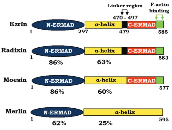
Figure 1.13. Domain organization of ERM proteins. ... 78
Figure 1.14. Three dimensional structure of the ezrin FERM domain.. ... 79
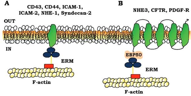
Figure 1.15. Model for ERM association to membrane proteins. ... 80
Figure 1.16. Model for ERM activation. ... 84
Figure 1.17. Fas linkage to F-actin through ezrin... 98
Figure 4.1. HeLa WT, HCT116, SW480 and SKW6.4 cells are sensitive to TRAIL and express TRAIL-R1 and TRAIL-R2 on the cell surface. ... 118
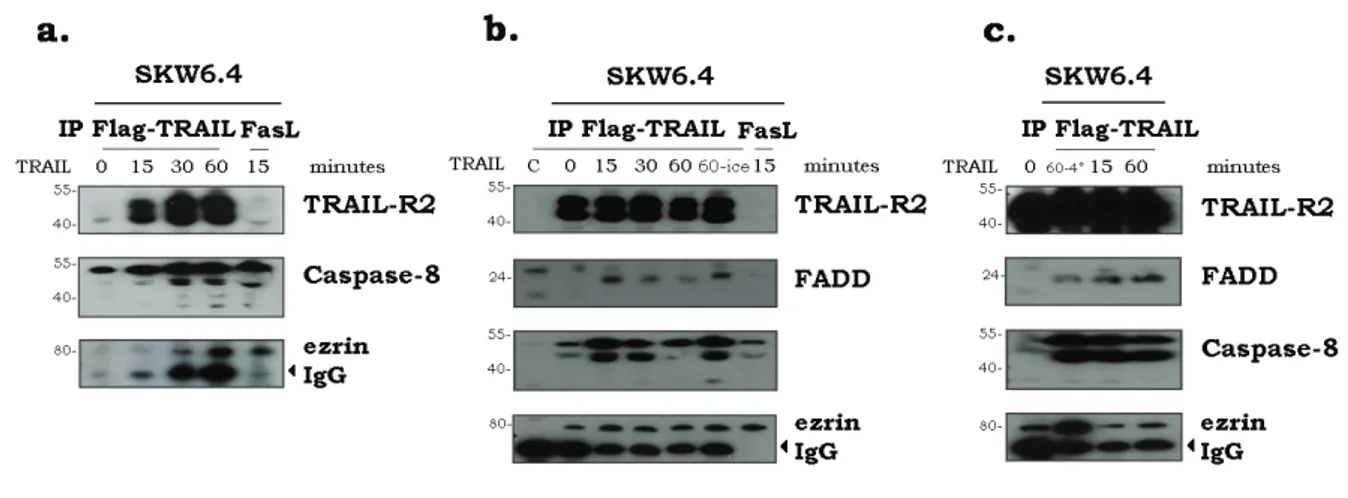
Figure 4.2. Ezrin is present in TRAIL immunoprecipitates of SKW6.4 cells... 119
Figure 4.3. ZVAD doesn’t interfere with the TRAIL-R2 association with ezrin. .... 120
Figure 4.4. Ezrin recruitment to TRAIL-R2 in SKW6.4 and HeLa cells. ... 121
Figure 4.5. Analysis of ezrin tyrosine phosphorylation in the TRAIL DISC. ... 122
Figure 4.6. Schematic representation of the two chimeric ezrins. ... 123
Figure 4.7. Protection against TRAIL and Fas ligand-induced cell death in HeLa cells over-expressing the mutant ezrin. ... 125
14
Figure 4.8. The mutant ezrin is not associated with TRAIL-R2. ... 127 Figure 4.9. Protection against TRAIL and Fas ligand-induced cell death in HCT116
cells over-expressing wild-type ezrin. ... 128
Figure 4.10. Protection against TRAIL and Fas ligand-induced cell death in SW480
cells over-expressing wild-type ezrin. ... 129
Figure 4.11. The new HeLa cells over-expressing the mutant ezrin are not
protecting against TRAIL and Fas ligand-induced cell death. ... 131
Figure 4.12. Ezrin depletion by siRNA slightly enhances TRAIL and Fas
ligand-induced cell death in HCT116 and SW480 cells. ... 133
Figure 4.13. Ezrin is present in a nonspecific manner in TRAIL
immunoprecipitates. ... 135
Figure 4.14. Ezrin was not found associated with TRAIL-R1 and TRAIL-R2 in
co-expression experiments. ... 136
Figure 4.15. Analysis of ERM association with caspase-8 and GAPDH. ... 137 Figure 4.16. Ezrin binds the sepharose and agarose polimers crosslinked to
proteins G or A. ... 138
Figure 4.17. Nonspecific binding of ezrin to the proteins G-sepharose saturated
with BSA that are used to immunoprecipitate. ... 140
Figure 4.18. Phosphorylation of ezrin after TRAIL, Fas ligand and EGF stimulation.
... 143
Figure 4.19. Analysis of ezrin phosphorylation in ezrin immunoprecipitates. ... 144 Figure 4.20. Ezrin phosphorylation sites. ... 146 Figure 4.21. The ezrin phosphorylation variants are well expressed in SW480 cells
except the Y145F mutants. ... 147
Figure 4.22. Ezrin Y145F is expressed in a lesser extent than the other mutants.
... 148
Figure 4.23. The phosphorylation variants of ezrin did not change the TRAIL-R1
and TRAIL-R2 expression at the membrane level. ... 149
Figure 4.24. TRAIL-R1 and TRAIL-R2 internalization in SW480 cells. ... 150 Figure 4.25. TRAIL-R1 and TRAIL-R2 internalization in ezrin phosphorylation
mutants-expressing SW480 cells. ... 151
Figure 4.26. Expression of ezrin S66A variant in SW480 cells enhanced TRAIL but
not Fas ligand-induced cell death. ... 152
15
Figure 4.28. Table of TRAIL inhibitory concentration. ... 154 Figure 4.29. Ezrin S66A sensitize SW480 cells to TRAIL- but not CDDP-induced
cell death. ... 156
Figure 4.30. Caspase 2 and 3 are more activated in ezrin S66A-expressing SW480
cells. ... 157
Figure 4.31. Caspase 2, 9 and 3 are more activated in ezrin S66A-expressing
SW480 cells. ... 158
Figure 4.32. Mutations on serine 66 do not affect TRAIL DISC formation ... 159 Figure 4.33. Phosphorylation variants of ezrin are not present in the DISC. ... 160 Figure 4.34. There is more active Bax in SW480 cells expressing ezrin S66A than
in control or ezrin WT-expressing cells. ... 161
Figure 4.35. Effect of PKA inhibition on TRAIL-induced cell death. ... 162 Figure 4.36. Effect of PKA inhibition or activation on TRAIL-induced cell death in
SW480 and HCT116 cells. ... 163
Figure 4.37. Effect of PKA inhibition or activation on TRAIL-induced cell death in
SW480 cells. ... 164
Figure 4.38. Phospho-p70 is phosphorylated upon TRAIL stimulation. ... 164 Figure 4.39. TRAIL induced activation by phosphorylation of CREB. ... 165 Figure 4.40. CDDP inhibits PKA by indirectly blocking the phosphorylation of
CREB, p70 and Bad. ... 166
Figure 4.41. WWOX depletion by siRNA significant protects mock-infected and
ezrin S66A- and Y353D-expressing SW480 cells against TRAIL-induced cell death. ... 170
Figure 4.42. Effect of CDDP treatment followed by TRAIL stimulation on ezrin
phosphorylation variants-expressing SW480 cells. ... 172
Figure 4.43. Cisplatin restores TRAIL sensitivity in ezrin S66D and Y353F
mutants. ... 173
Figure 5.1. Proposed model of the ezrin-mediated inhibition of TRAIL-induced cell
death. ... 190
Figure 5.2. Proposed model of cisplatin-mediated sensitization to TRAIL-induced
17
List of tables
Table 1-1. Physiological role of the ezrin phosphorylation sites. ... 89
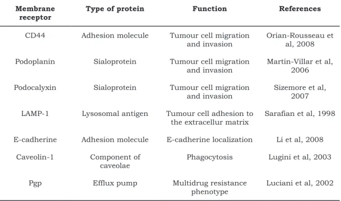
Table 1-2. Ezrin-associated membrane receptors related to the formation of tumour metastasis. ... 96
Table 3-1. Chemicals ... 101
Table 3-2. Bacterial strains. ... 103
Table 3-3. Eukaryotic cell lines and growth media. ... 104
Table 3-4. Primary antibodies used for immunoblotting ... 104
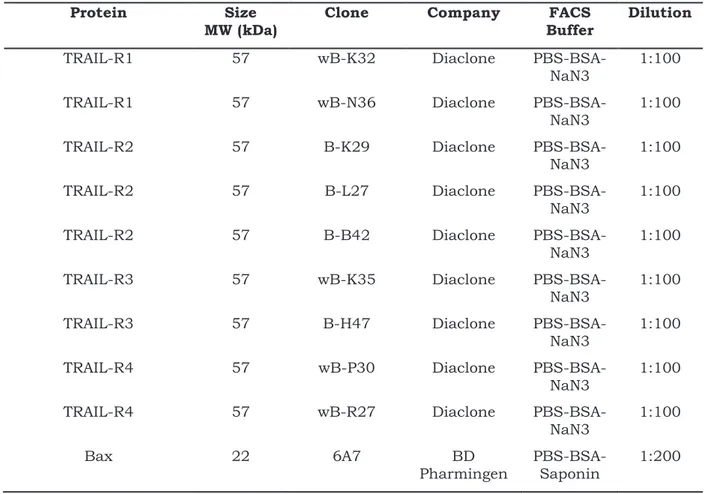
Table 3-5. Primary antibodies used for flow cytometry ... 106
Table 3-6. HRP conjugated secondary antibodies ... 106
Table 3-7. Antibodies used for immunoprecipitation ... 106
Table 3-8. TNF-superfamily ligands ... 107
Table 3-9. Vectors ... 107
Table 3-10. Oligonucleotides ... 108
19
List of abbreviations
A
β2A: beta 2 adrenergic receptor AIDS: acquired immune deficiency syndrome
AIF: apoptosis inducing factor AKT: activated protein kinase AMP: adenosine monophosphate APAF: apoptotic protease activating factor
APC: adenomatous polyposis coli APS: ammonium persulfate ATP: adenosine-5’-triphosphate
B
8B: 8-bromoadenosine 3′,5′-cyclic monophosphate
BAD: Bcl-2 antagonist of cell death BAK: Bcl-2 antagonist killer 1 BAX: Bcl2-associated X protein Bcl-2: B cell lymphoma 2
Bcl-XL: Bcl-2 related gene X, long
isoform
BH: Bcl-2 homology
BID: BH3-interacting domain death agonist
BIK: Bcl-2 interacting killer
BIM: Bcl-2 interacting mediator of cell death
BIR: baculovirus IAP repeat BOK: Bcl-2 related ovarian killer BSA: bovine serum albumin
C
CD: cluster of differentiation Cdc: cell division control
CDDP: cisplatin, cisplatinum or cis-diamminedichloroplatinum
Cdk5: cyclin-dependent kinase 5 cDNA: complementary DNA
C-ERMAD: carboxy-terminal Ezrin Radixin Moesin associated domain c-FLIP: cellular FLICE like inhibitory protein
CFTR: cystic fibrosis transmembrane conductance regulator
CREB: cAMP response element binding
D
DD: death domain
DED: death effector domain
DIABLO: direct inhibitor of apoptosis-binding protein with low pI
DISC: death inducing signalling complex
DMEM: dulbecco’s modified eagle’s medium
20
DMSO: dimethyl sulfoxide DNA: deoxyribonucleic acid DR: death receptor
DTT: dithiothreitol
E
EBP50: Ezrin Radixin Moesin binding protein 50
ECL: enhanced chemiluminescence EDTA: Ethylene diamine tetraacetic acid
EGF: epidermal growth factor
E3KARP: sodium hydrogen exchanger type 3 kinase A regulatory protein Endo G: endonuclease G
ERK: extracellular signal-regulated kinase
ERM: Ezrin Radixin Moesin
F
FACS: fluorescence-activated cell sorter
FADD: Fas-associated protein with death domain
FasL: Fas ligand
FBS: fetal bovine serum FCS: fetal calf serum
FERM: four point one Ezrin Radixin Moesin
G
GAPDH: glyceraldehyde 3-phosphate dehydrogenase
GPI: glycosyl phosphatidyl inositol GRK2: G protein-coupled receptor kinase 2
H
HDAC: histone-deacetylase HGF: hepatocyte growth factor His: histidine
HIV: human immunodeficiency virus HRP: horseradish peroxidise
HUVEC: human umbilical vein endothelial cells
I
IAP: inhibitor of apoptosis protein ICAM: inter-cellular adhesion molecule
IFN: interferon
IgG: immunoglobulin G IkB: inhibitor of kB
IKK: inhibitor of kB kinase IL: interleukine
IP3: inositol 1,4,5-trisphosphate
IS: immunological synapse IZ: isoleucine zipper
21
J
JNK: c-Jun N-terminal kinase
K
KBTBD2: Kelch-repeat and BTB domain containing 2 kDa: kilodaltonL
LB: luria bertaniLOK: lymphocyte-oriented kinase LPS: lipopolysaccharide
LZ: leucine zipper
M
MAPK: mitogen activated protein kinase
Mcl-1: myeloid cell leukemia 1 MDM2: mouse double minute 2 MDR: multidrug resistance MHC: major histocompatibility complex
MOMP: mitochondrial outer membrane permeabilization MP: milk powder
mRNA: messenger RNA
N
NEMO : NFkB essential modifier
N-ERMAD: amino-terminal Ezrin Radixin Moesin associated domain NF2: neurofibromatosis type 2 NFkB: nuclear factor kappa-light-chain-enhancer of activated B cells NHE: sodium hydrogen exchanger NHL: non Hodgkin’s lymphoma NK: natural killer
NP40: noninet P-40
NSCLC: non-small cell lung cancer
O
OMM: outer mitochondrial membrane OPG: osteoprotegerin
ORT: orthovanadate
P
PBS: phosphate buffered saline PCR: polymerase chain reaction PDGF: platelet derived growth factor PE: phycoerythrin
PFA: paraformaldehyde Pgp: P-glycoprotein
PI3K: phosphoinositide triphosphate kinase
PIP2: phosphatidylinositol
4,5-bisphosphate
PKA: protein kinase A PKC: protein kinase C
22
PMA: phorbol 12-myristate 13-acetate PTB: polypyrimidine tract binding PUMA: p53-upregulated modulator of apoptosis
R
Rb: retinoblastoma
RhoGAP: Rho GTPase-activating protein
RhoGDI: Rho GDP-dissociation inhibitor
RhoGEF: Rho guanine nucleotide exchange factor
rhTRAIL: recombinant human TRAIL RIP: receptor interaction protein RNA: ribonucleic acid
ROCK: Rho-associated protein kinase RPMI: roswell park memorial institute
S
SDS: sodium dodecyl sulphate SDS-PAGE: SDS-polyacrylamide gel electrophoresis
SH2: Src homology 2 shRNA: small hairpin RNA siRNA: small interfering RNA
Smac: second mitochondria-derived activator of caspases
Syk: spleen tyrosine kinase
T
TBE: tris borate EDTA TBS: tris buffered saline TCR: T cell receptor TEMED: N, N, N’,
N’-tetramethylethylendiamine THR: Threonine
TNF: tumor necrosis factor TNFR: TNF receptor
TRADD: TNF receptor associated death domain
TRAF: TNF receptor associated factor TRAIL: TNF-related apoptosis inducing ligand
TRAIL-R: TNF-related apoptosis inducing ligand receptor
TWEAK: TNF-like weak inducer of apoptosis
U
UV: ultraviolet
V
VDAC: voltage-dependent anion channel
VSV: vesicular stomatitis virus
W
WT: wild-type
23
WWOX: WW-domain containing oxidoreductase
X
XIAP: X-linked inhibitor of apoptosis protein
Z
ZAP70: zeta-chain associated protein kinase of 70 kDa
25
1. Introduction
1.1. Cancer
Cancer is a general name for a group of over 100 diseases, characterized by abnormal, uncontrolled cell growth, and which is the result of pleiotropic and
multi-factorial events (Hanahan and Weinberg, 2000; Hanahan and Weinberg, 2011).
Normally, cells of a healthy body grow, multiply, and die in a tightly controlled manner. However, when normal processes break down, cancer can develop. When cancer begins, cells grow and divide without any control, develop strategies to inhibit cell death signalling pathways, and ultimately form an abnormal mass of cells called a tumour. Sometimes cells originating from this tumour acquire the capacity either to invade nearby tissues, or to penetrate the blood vessels through which they can move to other parts of the body, where they develop a new mass of tumour cells. This process is termed metastasis and is a characteristic of malignant
tumours (Klein, 2008; Chiang and Massagué, 2008). Most are solid tumours except
leukemia, which starts in blood-forming tissue and causes the formation of abnormal blood cells.
Several stimuli can induce the transformation of normal cells into tumour cells. For instance, DNA damage or alterations in the genetic material of cells by environmental or internal factors can cause cancer (Mena et al, 2009). Likewise carcinogens can promote formation of cancer by directly inducing DNA damage, and can include ultraviolet, gamma, and X-rays, and tobacco (Mena et al, 2009). These mutations can occur in genes responsible for the cell division process, apoptosis, DNA repair, or tumour suppression. Once mutations occur in some of these cryptic genes, cells become unable to control their proliferation, to engage suicide programs and are often resistant to conventional chemotherapies. For instance, p53 can
26
regulate different cellular processes, such as cell cycle arrest, senescence, apoptosis (Lane, 1992; Levine and Oren, 2009), angiogenesis (Teodoro et al, 2007), and autophagy (Maiuri et al, 2010), to name a few. P53 is the most commonly mutated tumour suppressor gene in human cancers (Vazquez et al, 2008). Mutations in the gene coding for p53 leads to the loss of its tumour suppressive function, and then results in its inactivation or malfunction, causing cancer development (Brosh and Rotter, 2009; Goh et al, 2011). The fact that most cancer cells are defective in p53, together with the multiple roles of p53 in cell physiology, have led to the consideration that p53 is one of the most important players in the development of cancer (Bálint and Vousden, 2001; Meek, 2009). Mutations can also induce an increase in the activity of growth factors, growth factor receptors, or transduction pathways, leading to increase in the activation of pro-growth signal pathways, which results in an uncontrolled cell growth (Mendelsohn and Baselga, 2003; Takeuchi and Ito, 2010). Some viruses can also divert the cellular machinery and play a decisive role in cancer development, such as human papillomavirus, hepatitis B and C, Epstein-Barr virus, and human immunodeficiency virus (HIV) (Mena et al, 2009; Moore and Chang, 2010). Last but not least, genetic inheritance can predispose to cancer, such as in the case of Bloom Syndrome (Ding et al, 2009), Fanconi anemia (Garcia et al, 2009), or mutations in BRCA1 (Antoniou et al, 2003; Thompson and Easton, 2004), all of which increase a person’s chance of developing a cancer during their lifetime.
The transformation of normal cells into tumour cells is a complex multistep process, which requires dynamic alteration of the genome and breaking down of intracellular checkpoints (Hanahan and Weinberg, 2000; Hanahan and Weinberg, 2011). This process can be viewed as a “somatic evolution”, because during carcinogenesis normal cells evolve to become malignant, accumulating genetic and epigenetic changes and acquiring new phenotypic properties that confer upon them
27
survival advantages, ultimately leading to emergence of a malignant population (Gillies and Gatenby, 2007; Fang et al, 2008). Several essential alterations in cell physiology, termed the “hallmarks of cancer”, which are found in almost every type of human cancer, have been described by Hanahan and Weinberg, and are involved in the malignant process, including resistance to apoptosis, genomic instability, uncontrolled proliferation, aberrant cell cycle, cellular invasion and metastasis, angiogenesis, inflammation, and abnormal metabolism (Hanahan and Weinberg, 2000; Hanahan and Weinberg, 2011). In addition, during the cell progression towards malignancy, most cancer cells can acquire two more phenotypic properties that confer new survival advantages: the capability to evade immune surveillance, and to suppress immune reactivity (Cavallo et al, 2011). Recently, the role of the tumour microenvironment in determining tumour malignancy has received renewed interest, with the suggestion that environmental conditions may drive the selection
of a cancerous phenotype (Gillies and Gatenby, 2007). The tumour
microenvironment is characterized by hypoxia, low blood supply, and acidity. Under anaerobic conditions, tumour cells produce energy using glycolysis instead of using the aerobic metabolism, and the glycolysis is constitutively upregulated, a
phenomenon known as the “Warburg Effect” (Warburg, 1956; Gillies and Gatenby,
2007). Elevated level of glycolysis in turn induces increased acid production that, together with hypoxia, contributes to create an extracellular environment that is not permissive for the growth of normal cells. In order to survive in the unfavourable environment created by themselves, tumour cells upregulate several proton extrusion mechanisms (Sennoune et al, 2004), such as the V-ATPase (Ma et al, 2011; Fais et al, 2007), the Na+/H+ exchanger (NHE) (Slepkov et al, 2007) and the carbonic anhydrase (Robertson et al, 2004). Therefore, the peculiar features of hypoxia and acidosis characterizing the tumour microenvironment produce a selective pressure that favours the survival of tumour cells, which are resistant to
28
acidosis and have upregulated glycolysis (Gillies and Gatenby, 2007). These observations led Gatenby and Gillies to propose an additional new hallmark of cancer: increased glucose consumption through increased glycolysis, which contribute to confer a metabolic advantage to tumour cells (Gillies and Gatenby, 2007; Fang et al, 2008).
Solid tumours have been treated by surgery for the past 4000 years (Pavet et al, 2011). X-rays were discovered at the end of the nineteenth century (Eisenberg, 1992), and it was observed that cancer cells were susceptible to death by radiations, leading to radiotherapy becoming another major therapeutic approach (Connell and Hellman, 2009). The third main approach that is currently used to treat cancer is chemotherapy, using cytotoxic chemical compounds. At present, surgery, radiation and/or chemotherapy represent the standard treatment for the majority of cancers. Patients often receive a combination of these therapies. However, chemotherapy drugs currently used for treating cancers can be very aggressive and highly toxic for patients. Moreover, a significant proportion of patients are not cured from the disease because of chemoresistance and/or relapse. Thus, novel chemotherapeutic compounds are always being pursued. Alternative approaches are also under investigation, including non conventional chemotherapy (Jacquemin et al, 2010; Mérino et al, 2007; Luciani et al, 2004a; De Milito et al,
2006; De Milito et al, 2010; Marino et al, 2010), immunotherapy (Sato et al, 2009),
or gene therapy (Zhang et al, 2011). Despite the great advances that have been made, and the great efforts of the scientific community in finding effective treatments for cancer, the disease still causes death for millions of people per year worldwide (Thun et al, 2010; American Cancer Society, 2007; IARC, 2008). More than 12 million new cases and 7.6 million cancer deaths have been estimated to have occurred in 2007 (American Cancer Society, 2007; Thun et al, 2010). By 2030, it is projected that there will be 26 million new cancer cases and 17 million cancer
29
deaths per year (IARC, 2008; Thun et al, 2010). New investigations are therefore necessary for the design of novel and innovative therapeutic strategies to cure cancer, and which are less invasive for patients in order to enhance their quality of life.
In light of these considerations, research in the field of apoptosis is of great interest in order to find new ways to kill malignant cells.
30
1.2. Apoptosis
Cells have several means to trigger cell death, and the literature has multiple examples of number of different modes of cell death, including apoptosis, autophagy, necrosis, anoikis, cannibalism, and pyroptosis. Apoptosis is the most renowned of these, being a highly conserved and tightly controlled type of programmed cell death process, first described in 1972 by Currie and colleagues (Kerr et al, 1972), which eliminates superfluous or irreparably damaged cells from multicellular organisms (Danial and Korsmeyer, 2004; Strasser et al, 2011). This mechanism regulates not only the correct development of organs in mammals, but also the homeostasis and integrity of tissues in adult organisms throughout their lifetime (Meier et al, 2000; Chowdhury et al, 2006; Strasser et al, 2011). Aberrant regulation of the cell death process, such as deficiency or excess in apoptosis, can lead to the development of several diseases, such as autoimmune diseases (Oliveira
and Gupta, 2008; Hotchkiss et al, 2009), neurodegenerative disorders (reviewed in
Cavallucci and D’Amelio, 2011), or cancer (Cory and Adams, 2002; Vazquez et al,
2008; Hotchkiss et al, 2009;Hanahan and Weinberg, 2011).
Apoptosis is characterized by reduction in cellular and nuclear volume, condensation and fragmentation of DNA, preservation of organelle structure and plasma membrane integrity, and blebbing of the plasma membrane which generates apoptotic bodies that are engulfed by phagocytes, thereby avoiding an inflammatory response in surrounding tissues (Strasser et al, 2011) (Figure 1.1). Apoptosis is morphologically distinct from the other types of cell death. For example, during necrosis the organelles swell, the cell volume increases, mitochondria produces reactive oxygen species, the ATP is depleted, and the lysosomes and the plasma membrane are disrupted, resulting in the release of cellular contents into the microenvironment, which induces the activation of an inflammatory response (Golstein and Kroemer, 2007). Alternatively, when cell death occurs with the
31
features of autopaghy, the cells are characterized by vesicular accumulation, absence of chromatin condensation and cytoplasmic vacuolization (Amelio et al, 2011).
Figure 1.1. Schematic representation of the apoptotic process.
Apoptosis may be initiated following the activation of one of two pathways, the intrinsic and extrinsic apoptotic pathways (Figure 1.2). Binding of extracellular ligands to death receptors (DRs) at the cell surface stimulates the extrinsic pathway, whereas cellular damage or stress signals for cell death through the intrinsic pathway (Indran et al, 2011). Both pathways converge on the activation of specific proteins, called caspases, which are highly conserved cysteine-aspartate proteases (Lavrik et al, 2005; Li and Yuan, 2008). The first caspases activated following induction of apoptosis are the initiator caspases, which include caspase-2, -8, -9 and -10 (Chen et al, 2002; Boatright et al, 2003; Bouchier-Hayes et al, 2009; Milhas et al, 2005; Lafont et al, 2010). Once these caspases are activated, they in turn activate the effector caspases-3, -6, -7, which start to dismantle the cell by cleaving and then degradating multiple cellular proteins, leading to the execution of the apoptotic process.
32
Cells may undergo apoptosis via engagement of the intrinsic pathway. In this case, apoptosis can be initiated by a variety of receptor-independent stimuli, such as UV- or gamma-irradiation, free radicals, viral infections, cellular stress, serum or growth factor withdrawal, heat shock, chemotherapy, DNA damage, etc (Figure
1.2). Activation of the intrinsic pathway induces the permeabilization of the
mitochondrial outer membrane, leading to the formation of large pores through which soluble apoptogenic factors, such as cytochrome c, Smac-DIABLO and Omi/HtrA2, usually present in the inter-membrane space gain access to the cytosol (Vaux, 2011). Once in the cytosol, they activate the effector caspases in cooperation with cytosolic factors, leading to cell death. Thus, cytoplasmic cytochrome c binds to and activate Apaf-1 (apoptotic protease activating factor 1), and they form a platform termed the apoptosome, which allows recruitment and activation of initiator caspase-9 molecules (Zou et al, 1999; Acehan et al, 2002). Activation of caspase-9 causes the activation of effector caspases, thereby enabling full execution
of cell death. Molecules of Smac (second mitochondrial activator of
caspases)/Diablo (direct IAP binding protein with low pI) (Du et al, 2000; Verhagen
et al, 2000) associate with IAPs (inhibitor of apoptosis proteins) proteins (see paragraph 1.3.6), neutralizing their inhibitory function on caspase-9, -3, and -7 molecules, which are then free to execute apoptosis (Wu et al, 2000; Ekert et al, 2001). In addition to Smac/Diablo, another mitochondrial serine protease protein, HtrA2/Omi, has been identified by virtue of its ability to bind to IAPs (Verhagen et
al, 2007; Martins et al, 2002; Hegde et al, 2002). Moreover, other two apoptogenic
proteins, AIF (apoptosis inducing factor) and endonuclease G (Endo G), were reported to be released from mitochondria during apoptosis. Once released, AIF and Endo G translocate from the mitochondria to the nucleus to cause DNA
fragmentation and cell death in a caspase-independent manner (Joza et al, 2001;Ye
33
reported about HtrA2/Omi, AIF and Endo G, and the physiological significance of their release during apoptosis is still under investigation, and has yet to be established.
The intrinsic pathway of apoptosis can also be promoted through the activity of the endoplasmic reticulum. Indeed, unresolved conditions of stress to the endoplasmic reticulum, resulting in misfolded proteins, stimulates this organelle to activate apoptosis. Multiple pathways may be involved in endoplasmic reticulum-mediated apoptosis. On one hand, apoptosis induced by the endoplasmic reticulum seems to involve the direct activation of initiator caspases at the endoplasmic reticulum level, which can then translocate from the endoplasmic reticulum to the cytosol where they in turn activate caspase-9, leading to the apoptotic caspase cascade (Hitomi et al, 2004; Yukuoka et al, 2008). By contrast, other studies report that the endoplasmic reticulum-mediated apoptosis occurs through cross talk with the mitochondria. In fact, cytochrome c release, loss of mitochondrial membrane potential, and caspase-9 activation has been observed in response to endoplasmic reticulum stressors (Jimbo et al, 2003; Kitamura et al, 2003; Masud et al, 2007). Release of calcium from the endoplasmic reticulum into the cytosol seems to be also required. For example, it has recently been reported that calcium can be directly transmitted into mitochondria following the attachment of mitochondria to the endoplasmic reticulum cisternea, leading to cell death (Rizzuto et al, 2009; Csordas et al, 2010).
The extrinsic pathway is initiated when a specific extracellular death inducing ligand binds to an agonistic transmembrane death receptor at the membrane level (Figure 1.2). Currently, six death receptors have been identified: TNFR1, Fas (CD95), TRAIL-R1, TRAIL-R2, DR3, DR6, members of the TNF receptor (TNFR) superfamily. They are characterized by an extracellular domain that is able to engage the ligands, and a cytoplasmic domain, called the death domain, essential
34
for the transmission of the death signal. Engagement of death receptors drives the recruitment of adaptor proteins and initiator caspase-8 and -10 molecules, which results in the formation of the death-inducing signaling complex (DISC), and caspase activation. Activated caspases-8 and -10 activate the effector caspases in turn, beginning the apoptotic cascade. Activated caspases-8 can also induce mitochondrial damage and reinforce the death signal by activating the intrinsic apoptotic pathway (Elmore et al, 2007). Alternatively, when caspases are inhibited, the death receptor-mediated apoptotic process is blocked, and the death signal induces the activation of another different form of programmed cell death, termed “necroptosis”, which resembles necrosis. Indeed, it has recently been proposed that death receptors participate not only in apoptosis but also in necroptosis, which occurs when the apoptotic pathway is blocked, and requires the involvement of RIP1 and RIP3 (receptor interaction protein kinase 1 and 3) (Holler et al, 2000; Degterev et al, 2008; Galluzzi et al, 2009). Upon necroptosis induction, RIP3 has been shown to be recruited to a cytosolic complex formed by RIP1, FADD and caspase-8 (He et al, 2009).
Alternatively, novel intracellular apoptosis-inducing platforms, namely ripoptosomes, and composed by RIP1, FADD and caspase-8, have been recently described, which allow the recruitment and activation of initiator caspases (Ikner and Ashkenazi, 2011; Feoktistova et al, 2011; Tenev et al, 2011). It has been reported that TWEAK (TNF-like weak inducer of apoptosis), toll-like receptor 3 or genotoxic stress could induce the spontaneous formation within the cytosol of the ripoptosome that can stimulate either apoptosis or necroptosis (Ikner and Ashkenazi, 2011; Feoktistova et al, 2011; Tenev et al, 2011).
35
Figure 1.2. Scheme of the intrinsic and extrinsic pathways of apoptosis. The extrinsic pathway (left) starts
with the ligation of death ligands to their cognate receptors. This event leads to DISC formation, caspase activation and then cell death. The intrinsic pathway (right) is activated by different cellular and environmental factors, which induce the permeabilization of the mitochondrial outer membrane and the consequent release of cytochrome c, Smac/DIABLO, AIF and Endo G, terminating with execution of apoptosis.
36
Among the proapoptotic ligands of the TNF superfamily, TRAIL (TNF-Related Apoptosis-Inducing Ligand) is a topic of great interest in recent years for oncologists, because it can trigger cell death in a wide variety of cancer cells while sparing normal cells (Ashkenazi et al, 1999; Lawrence et al, 2001; Ashkenazi, 2002; Mérino et al 2007; Jacquemin et al 2010). For this reason it has been considered as a promising therapeutic agent against malignant diseases (Gura, 1997; Takeda et al, 2004). Moreover, several phase I and II clinical trials with recombinant human TRAIL or agonistic antibodies, which recognize and activate its proapoptotic receptors, are currently underway in patients suffering from different types of malignant diseases (Herbst et al, 2006: Ling et al, 2006; Yee et al, 2007; Tolcher et al, 2007; Plummer et al, 2007; Greco et al, 2008; Leong et al, 2009). Preliminary results indicated that the recombinant human variant of TRAIL and the antibodies are well tolerated in patients, and the side effects on normal cells are mild (see paragraph 1.3.8), increasing interest in using TRAIL in clinical setting.
Extensive research over the last few years has given us a good understanding of the events that occur during the TRAIL signalling pathway, and of the factors involved in its regulation. However, some aspects which could be involved in the transmission of the TRAIL-induced apoptotic signal have not yet been taken into account. In light of these considerations, during this project we investigated the initial steps of the signalling pathway induced by the TRAIL death receptors.
37
1.3. The TRAIL pathway
1.3.1. TRAIL
Human TNF-Related Apoptosis-Inducing Ligand (TRAIL or Apo2L) has high homology with TNF type α and Fas ligand (FasL), two other members of the tumor necrosis factor (TNF) superfamily. TRAIL was identified by two different groups as a new molecule able to trigger apoptosis in a wide variety of cells (Wiley et al, 1995; Pitti et al, 1996). It is a type II transmembrane protein, composed of 281 amino acid residues, and is coded on the long arm of chromosome 3 (3q26). TRAIL can also be found in a soluble form in which the carboxy-terminal domain can be released into the extracellular milieu following proteolytic cleavage (Mariani and Krammer, 1998). At the protein level, TRAIL possesses an amino-terminal cytoplasmic domain, followed by a transmembrane helix, and terminates in a carboxy-terminal extracellular TNF-like domain. Crystal structure analysis shows that the extracellular domain is characterized by two antiparallel β-sheets (Cha et al, 1999; Hymowitz et al, 2006). TRAIL is usually present in a trimeric form, maintained through a direct association between a zinc atom localized at the interface of the trimer, and the cysteine residue 230 of each monomer (Hymowitz et al, 2006; Bodmer et al, 2000). TRAIL is expressed at the surface of T cells, dendritic cells, natural killer cells, macrophages, monocytes and neutrophils. Its expression at the mRNA level can be induced by type I interferons (Almasan and Ashkenazi, 2003).
38
1.3.2. TRAIL receptors
The TRAIL signaling system is very complex. TRAIL has five receptors: two agonistic receptors, TRAIL-R1 (DR4), and TRAIL-R2 (DR5, Killer, TRICK2), two decoy receptors, TRAIL-R3 (DcR1, LIT) and TRAIL-R4 (DcR2, TRUDD), and a soluble receptor called osteoprotegerin (OPG) (Figure 1.3). These receptors, with the exception of OPG, are type I transmembrane proteins, characterized by the presence of cysteine-rich domains in their extracellular domain (Figure 1.3), and coded on chromosome 8, position 8p21-22.
Figure 1.3. TRAIL receptors. Five TRAIL receptors have been identified. All possess three cystein rich regions in
the extracellular part. TRAIL induces apoptosis upon binding to TRAIL-R1 and TRAIL-R2, which have a death domain of 80 amino acids in the citopasmic region, crucial for transmit apoptotic signal. TRAIL has also two decoy receptors, TRAIL-R3 with a GPI anchor and TRAIL-R4 with a truncated death domain, that do not signal for apoptosis. One soluble receptor, the OPG, is also present.
39
TRAIL induces apoptosis following its binding to TRAIL-R1 (Pan et al, 1997a) and TRAIL-R2 (Walczak et al, 1997; Chaudary et al, 1997; Wu et al, 1997), which have 58% sequence homology with each other. Both receptors consist of an amino-terminal extracellular domain containing three cysteine-rich domains, a transmembrane domain and a carboxy-terminal cytoplasmic domain (Figure 1.3). The intracellular domain is characterized by a Death Domain (DD) of 80 amino acid residues that is conserved in each of the death receptors of this family, and is essential for triggering apoptosis (Figure 1.3). Two TRAIL-R2 isoforms have been described, one long and one short, both of which are able to transduce apoptotic signaling. The short isoform differs from the long because it lacks a sequence of 23 amino acids located between the transmembrane domain and the first cysteine-rich domain (Screaton et al, 1997). In mice, only one agonistic receptor has been identified (Wu et al, 1999). This receptor, known as mTRAIL-R2 or mDR5, is more similar to the human TRAIL-R2 than to human TRAIL-R1 (Wu et al, 1999). It signals for apoptosis in mouse and human cell lines, in response to both mouse and human TRAIL, through its cytoplasmic DD (Wu et al, 1999).
When TRAIL binds to TRAIL-R3 (Pan et al, 1997b; Degli-Esposti et al, 1997a) or TRAIL-R4 (Degli-Esposti et al, 1997b; Marsters et al, 1997), it does not induce apoptosis in target cells. Indeed, although these receptors possess an extracellular domain that is very similar to that of the agonistic death receptors, they differ in the intracellular part. TRAIL-R3 lacks the cytoplasmic and transmembrane domains, and it is anchored to the plasma membrane through the Glycosyl-Phosphatidyl-Inositol (GPI), whereas TRAIL-R4 contains a truncated, nonfunctional DD (Figure
1.3). Thus, these receptors are unable to transmit an apoptotic signal, and
consequently they protect cells from TRAIL-induced apoptosis. The mechanisms through which TRAIL-R3 and TRAIL-R4 carry out their inhibitory function are still under investigation. TRAIL-R3 can inhibit TRAIL-induced cell death by competing
40
with the death receptors for TRAIL binding, which leads to sequestering of TRAIL in lipid rafts (Mérino et al, 2006) (Figure 1.4). Whereas in cells over-expressing TRAIL-R4, addition of TRAIL induces association of TRAIL-R4 with TRAIL-R2 impeding caspase-8 recruitment and activation (Mérino et al, 2006) (Figure 1.4). The TRAIL-R4-mediated TRAIL inhibition is also described to involve the formation of ligand-independent complexes between TRAIL-R2 and TRAIL-R4 through special extracellular domains called PLAD (pre-ligand assembly domains) (Clancy et al, 2005). Although a weak interaction between TRAIL-R4 and TRAIL-R2 has been observed, it has been clearly demonstrated that the heterotypic interaction mostly occurs in a ligand-dependent manner (Mérino et al, 2006). TRAIL-R4 has also been suggested to be able to transmit an antiapoptotic signal through the activation of the transcription factor NF-kB (Degli-Esposti et al, 1997b) or Akt in HeLa cells (Lalaoui et al, 2011) (Figure 1.4). In mice, two decoy receptors are present, called
mDcTRAIL-R1/mDcR1 and mDcTRAIL-R2/mDcR2, and these are highly
41
Figure 1.4. Inhibition of TRAIL-induced apoptosis by TRAIL-R4 and TRAIL-R3. Stimulation with TRAIL leads
to the association of TRAIL-R4 with TRAIL-R2, which induces cell resistance to apoptosis and transmits an antiapoptotic signal (left panel). Alternatively, addition of TRAIL favors its sequestering in lipid rafts by TRAIL-R3 (right panel).
TRAIL can also bind to osteoprotegerin (OPG) (Emery et al, 1998) (Figure
1.3), the only soluble receptor, and which has the lowest affinity for TRAIL. TRAIL
binding to OPG has been proposed to regulate osteoclastic differentiation and survival (Zauli et al, 2004, 2008; Vitovski et al, 2007). However, the physiological role of this receptor in the TRAIL pathway remains to be better elucidated.
Initially it was thought that TRAIL receptors were expressed on the cell surface in a monomeric form, and that engagement of TRAIL-R1 and TRAIL-R2 by trimeric TRAIL promoted trimerization of these receptors. Further studies revealed that in the absence of ligand, TRAIL-R1 and TRAIL-R2, as well as Fas, TNFR1 and TNFR2, in fact reside on the plasma membrane in pre-assembled complexes, maintained by
42
the reciprocal association between the PLAD domains located in the first cysteine-rich domain in the extracellular part of each receptor, and which interact with each other, mediating the association between monomers (Papoff et al, 1999; Chan et al, 2000; Siegel et al, 2000, Clancy et al, 2005).
TRAIL-R1 and TRAIL-R2 have been reported to be post-translationally modified through O-glycosylation and palmitoylation (Wagner et al, 2007; Pan et al, 1997b; Rossin et al, 2009), and these two events are responsible for the modulation of the receptor clustering. Indeed, the level of expression of O-glycosylation enzymes in cancer cells has been correlated with cellular sensitivity to TRAIL-induced apoptosis (Wagner et al, 2007). Furthermore, TRAIL-R2 O-glycosylation at specific sites, which are highly conserved in R1, is reported to be required for TRAIL-R1 and TRAIL-R2 clustering, and subsequently to trigger DISC formation and caspase-8 activation. The N-glycosylation of TRAIL-R1 (Pan et al, 1997b) involves a specific site, which is not conserved in TRAIL-R2 (Sheridan et al, 1997), and its pharmacological inhibition has been described to alter the clustering of TRAIL-R1, but not TRAIL-R2 (Yoshida et al, 2007). Although these post-translational modifications have been correlated with sensitivity to TRAIL-induced apoptosis, the molecular mechanisms through which they modulate the TRAIL signalling pathway have not yet been fully elucidated.
The literature also demonstrates that TRAIL-R1, similar to Fas (Chakrabandhu et al, 2007) but contrary to TRAIL-R2, is constitutively palmitoylated on a triplet of cysteine residues located between the receptor’s transmembrane and DD region (Rossin et al, 2009). This post-translational modification was described to be required for the trimerization of TRAIL-R1, which is necessary for the further transduction of the apoptotic signal (Rossin et al, 2009).
43
1.3.3. The apoptotic extrinsic pathway and the DISC formation
Binding of trimeric TRAIL to TRAIL-R1 or TRAIL-R2 induces further clustering of the receptors into aggregates of high molecular weight, leading to the concomitant formation of the DISC (Death-Inducing Signalling Complex), composed of the adaptor protein FADD (Fas-Associated protein with Death Domain) and the initiator caspases-8 and -10 (Walczak and Sprick, 2001). Activation of TRAIL-R1 or TRAIL-R2 by TRAIL drives the recruitment of FADD that translocates to the DISC where its DD interacts directly with the DD of TRAIL-R1 or TRAIL-R2 (Kischkel et al, 2000; Sprick et al, 2000). FADD then recruits caspase-8 and -10 molecules to the complex through the interaction of its amino-terminal DED (Death Effector
Domain) with the DED domain of the initiator caspases (Kischkel et al, 2000; Sprick
et al, 2000). Within this complex, caspases-8 and -10 are activated.
Caspases are a family of cysteine proteases that play an essential role during apoptosis due to their ability to cleave several different substrates (reviewed in Li and Yuan, 2008). They are synthesized in cells as inactive proforms, and therefore need to be activated during apoptosis. Two groups of apoptotic caspases exist: initiator and effector caspases. In response to apoptotic stimuli, initiator caspases (caspase-8, -10, and -9) are activated. Once activated, they activate the effector caspases (caspase-3, -6, and -7). Activation of effector caspases generally occurs through proteolytic cleavage, which takes place on specific aspartate residue (Cohen et al, 1997), localized in a short segment that connects the large and small subunits of the caspases’s catalytic domain. However, the mechanism of initiator caspase activation seems to be more complex and it is not yet completely understood. Several hypothesis have been proposed. The first model, the induced-proximity model, postulates that FADD promotes the clustering of initiator caspases within the DISC, which increases their local concentration and allows their reciprocal trans-activation through proteolytic cleavage (Salvesen and Dixit, 1999). Thereby,
44
initiator caspases have a weak proteolytic activity and can therefore auto-process themselves only when they are brought within close proximity of each other in the DISC. Further studies on caspase-8 and -9 have led to the assumption that initiator caspases are activated after dimerization at the DISC or apoptosome level, and their subsequent cleavage serves only to stabilize the active dimers (Boatright et al, 2003; Donepudi et al, 2003; Renatus et al, 2001; Micheau et al, 2002). Based on this model, called the proximity-induced dimerization model (Boatright et al, 2003), the recruitment of initiator caspases within the DISC or the apoptosome increases their local concentration, which promotes their dimerization (Boatright et al, 2003; Renatus et al, 2001). Nowadays, this model is considered the principal mechanism of activation for initiator caspases. However, the induced proximity model was refined once again in 2005 by Chao and coauthors. They proposed a third model: the induced conformation model, which was based on studies with caspase-9, where they engineered a constitutive dimeric caspase-9 (Chao et al, 2005). Their hypothesis highlights the importance of the conformation of the active site during the process of activation. Consistent with their theory, binding of monomers of inactive caspase-9 to the apoptosome induces a conformational change, most likely at the level of the caspase-9 active site, which is the prerequisite that favors caspase-9 activation (Chao et al, 2005). This model can be also extended to the other initiator caspases, caspase-8 and -10. Recently, a new mechanism has been proposed that involves the ubiquitylation of caspase-8 (Jin et al, 2009). This study showed that stimulation with TRAIL induces caspase-8 polyubiquitylation at its carboxy-terminal region, promoted by the E3 ubiquitin ligase CUL3 within the DISC, leading to caspase-8 activation. The polyubiquitylation of caspase-8 can be reverted by the deubiquitinase enzyme A20, which is also present in the TRAIL DISC. Once the caspase-8 is ubiquitylated, the ubiquitin-binding protein p62/sequestosome-1 associates with caspase-8 and promotes the translocation of
45
caspase-8 from the DISC to intracellular ubiquitin-rich protein regions, where the caspase-8 is fully activated (Jin et al, 2009).
Both caspase-8 and -10 can be recruited to the TRAIL DISC where they are activated with similar kinetics, and can work independently of each other (Kischkel et al, 2001; Wang et al, 2001; Sprick et al, 2002). It is still controversial whether these caspases have redundant or exclusive roles during apoptosis. Evidence in the literature shows that caspase-10 over-expression can compensate for deficiency in caspase-8 in TRAIL-treated Jurkat cells (Kischkel et al, 2001; Wang et al, 2001), whereas another study reported that caspase-10 cannot functionally substitute caspase-8 (Sprick et al, 2002).
Once the initiator caspases are activated, cleaved forms of caspase-8 and caspase10 are released in the cytosol where they activate the effector caspases3, -6 and -7, ultimately leading to cell death (Figure 1.5).
46
Figure 1.5. The TRAIL apoptotic extrinsic pathway. Binding of a TRAIL trimer to TRAIL-R1 or TRAIL-R2 leads to
the clustering of receptors and recruitment of the adaptor FADD, which in turn recruits and activates caspase-8, to form the DISC. Active molecules of caspase-8 then activate the effector caspases-3, -6 and -7 and directly induce apoptosis.
47
1.3.4. The apoptotic intrinsic pathway
Cells sometimes require further signal amplification through the mitochondrial (intrinsic) pathway in order to undergo TRAIL-induced apoptosis. This phenomenon, described initially for the Fas signalling pathway (Scaffidi et al, 1998), has led to the classification of cells into two types, namely type I and type II cells. In type I cells, caspase-8 is activated and processed in the DISC in quantities that are sufficient to directly activate the effector caspases, and thereby to promote apoptosis. In contrast, in type II cells, the quantities of active caspase-8 generated at the DISC are limited, and therefore cells need an amplification loop through the mitochondria for further transduction of the apoptosis signal and execution of apoptosis (Scaffidi et al, 1998; Barnhart et al, 2003). Further research, however, has revealed that the differences in DISC formation and caspase-8 activation are not the only molecular factors that can discriminate between type I and type II apoptotic signalling. Indeed, increasing caspase-8 activity, for example by down-regulating c-FLIP, an inhibitor of caspase-8 (see paragraph 1.3.6), does not automatically convert type II into type I cells (Wilson et al, 2009). This role could be attributed to the protein XIAP, which binds to and inhibits caspase-3, -7 and -9 (Deveraux et al, 1997; Riedl et al, 2001) (see paragraph 1.3.6). Accordingly, combined inhibition of c-FLIP and XIAP enhanced apoptosis in type II colorectal cancer cells (Wilson et al, 2009). Moreover, XIAP was identified as a critical discriminator between type I and type II apoptosis signalling during death receptor-induced apoptosis (Jost et al, 2009). It was reported that loss of XIAP sensitized hepatocytes (type II cells) but not thymocytes (type I cells) to Fas-mediated apoptosis (Jost et al, 2009). In the absence of XIAP, induction of apoptosis by Fas was shown to occur through a caspase-dependent process and did not involve the mitochondria (Jost et al, 2009). Thus, genetic deletion of XIAP in hepatocytes
48
changed the Fas apoptosis phenotype of these cells from type II to type I (Jost et al, 2009).
Ligation of TRAIL to its cognate death receptors, TRAIL-R1 and TRAIL-R2, triggers for apoptosis through DISC formation, where proforms of initiator caspases (caspase-8 and /or -10) are recruited and activated. Active molecules of caspase-8/-10 are then released into the cytosol where they directly activate the effector caspases-3, -6, and -7 (type I pathway). In type II cells, however, activated caspase-8 engages the mitochondrial apoptotic pathway for full effector caspases activation and a complete execution of apoptosis. The transition from the death receptor-mediated extrinsic pathway to the mitochondrial apoptotic pathway is achieved through the caspase-8-mediated processing of the protein Bid (BH3-interacting
domain death agonist), a BH3-only member of the Bcl-2 family (Li et al, 1998; Luo
et al, 1998) (Figure 1.6).
Following TRAIL stimulation, Bid is cleaved by caspase-8 at the aspartate residue 60 within the cytosol (Li et al, 1998; Gross et al, 1999), resulting in the generation of its truncated form of 15 kDa, tBid, containing the carboxyl-terminal part of the protein (Figure 1.6). Recent evidence has shown that caspase-10 is also able to cleave and thereby activate Bid during Fas ligand-induced apoptosis (Fischer et al, 2006; Milhas et al, 2005). Caspase-3 (Slee et al, 2000) and other proteases, such as granzyme B (Sutton et al, 2000), cathepsins (Stoka et al, 2001;
Reiners et al, 2002; Cirman et al, 2004) and calpains (Chen et al, 2001; Mandic et
al, 2002) are also reported to be involved in the processing of Bid during death ligand-independent apoptotic pathways. Truncated Bid has a potent pro-apoptotic activity (Li et al, 1998; Gross et al, 1999), but the literature also describes a pro-apoptotic role for full-length Bid (Sarig et al, 2003), for example in a model of cell death termed anoikis (Valentijn and Gilmore, 2004). Moreover, full length Bid can