HAL Id: hal-02495547
https://hal.sorbonne-universite.fr/hal-02495547
Submitted on 2 Mar 2020
HAL is a multi-disciplinary open access
archive for the deposit and dissemination of
sci-entific research documents, whether they are
pub-lished or not. The documents may come from
teaching and research institutions in France or
abroad, or from public or private research centers.
L’archive ouverte pluridisciplinaire HAL, est
destinée au dépôt et à la diffusion de documents
scientifiques de niveau recherche, publiés ou non,
émanant des établissements d’enseignement et de
recherche français ou étrangers, des laboratoires
publics ou privés.
tunnel produces 5’-OH ends phosphorylated by Trl1
Albertas Navickas, Sébastien Chamois, Rénette Saint-Fort, Julien Henri,
Claire Torchet, Lionel Benard
To cite this version:
Albertas Navickas, Sébastien Chamois, Rénette Saint-Fort, Julien Henri, Claire Torchet, et al..
No-Go Decay mRNA cleavage in the ribosome exit tunnel produces 5’-OH ends phosphorylated by Trl1.
Nature Communications, Nature Publishing Group, 2020, 11 (1), �10.1038/s41467-019-13991-9�.
�hal-02495547�
No-Go Decay mRNA cleavage in the ribosome
exit tunnel produces 5
′-OH ends phosphorylated
by Trl1
Albertas Navickas
1,2, Sébastien Chamois
1,2, Rénette Saint-Fort
1, Julien Henri
1, Claire Torchet
1&
Lionel Benard
1*
The No-Go Decay (NGD) mRNA surveillance pathway degrades mRNAs containing stacks of stalled ribosomes. Although an endoribonuclease has been proposed to initiate cleavages upstream of the stall sequence, the production of two RNA fragments resulting from a unique cleavage has never been demonstrated. Here we use mRNAs expressing a 3′-ribozyme to produce truncated transcripts in vivo to mimic naturally occurring truncated mRNAs known to trigger NGD. This technique allows us to analyse endonucleolytic cleavage events at single-nucleotide resolution starting at the third collided ribosome, which we show to be Hel2-dependent. These cleavages map precisely in the mRNA exit tunnel of the ribosome, 8 nucleotides upstream of thefirst P-site residue and release 5′-hydroxylated RNA fragments requiring 5′-phosphorylation prior to digestion by the exoribonuclease Xrn1, or alternatively by Dxo1. Finally, we identify the RNA kinase Trl1, alias Rlg1, as an essential player in the degradation of NGD RNAs.
https://doi.org/10.1038/s41467-019-13991-9 OPEN
1Institut de Biologie Physico-Chimique, UMR8226, CNRS, Sorbonne Université, Laboratoire de Biologie moléculaire et Cellulaire des Eucaryotes, Paris, France.
2These authors contributed equally: Albertas Navickas, Sébastien Chamois. *email:lionel.benard@ibpc.fr
123456789
T
he No-Go Decay (NGD) mRNA surveillance pathway degrades mRNAs containing stalled ribosomes1,2. NGD occurs when translation elongation is blocked by the pre-sence of stable intramolecular or intermolecular RNA structures, enzymatic cleavage, chemically damaged sequences or rare codons1,3–8. This mRNA degradation process is dependent on translation and involves an endoribonuclease that cleaves just upstream of the stall sequence1,5,6,9. Other mRNA surveillance pathways can also ultimately lead to NGD. For instance, tran-scripts synthesized without a stop codon due to premature polyadenylation have stalled ribosomes that are initially detected by the non-stop decay (NSD) pathway9,10. NSD-targeted mRNAs are cleaved by an uncharacterized mechanism and become targets of NGD when ribosomes reach the new 3′-end and stall9,11,12. NGD thus plays a key role in resolving translational issues potentially detrimental to cellular homeostasis. When mRNAs are truncated, the stalled ribosomes are rescued in a process mediated by the Dom34/Hbs1 complex that dissociates the ribosomal subunits5. Their association with the 60S subunit is recognized by the ribosome quality control (RQC) pathway, leading to the rapid degradation of the nascent peptide13–15. However, despite extensive study, the precise location of NGD cleavage and the mechanism of degradation of the resulting RNA fragment remain elusive.In this paper, we focus on the fate of NGD-cleaved mRNAs, with an initial goal of mapping the sites of mRNA cleavage with accuracy. Two major obstacles to achieving this objective are that NGD fragments are rapidly attacked by 5′–3′ and 3′–5′ exori-bonucleases after ribosome dissociation5and that simultaneously blocking the 5′–3′ and 3′–5′ exoribonuclease decay pathways is synthetically lethal16. It has been shown, however, that the sta-bility of such mRNAs is largely dependent on the Dom34/Hbs1 complex5,17. In dom34 mutant cells, ribosomes stalled at the 3′-end of truncated mRNAs inhibit the degradation by the exosome and facilitate the detection of sequential endonucleolytic cleavages upstream of the ribosomal stall site5. Interestingly, dom34 and xrn1 mutations (inactivating the main 5′–3′ exonucleolytic degradation pathway) are not synthetic lethal1. Moreover, NGD endonucleolytic cleavages still occur in the absence of Dom342,3. The limited 3′–5′ degradation of specific mRNA targets (in the absence of Dom34) combined with 5′–3′ exoribonuclease mutants thus allows an accumulation of RNA fragments resulting from endonucleolytic cleavages whose extremities can be mapped accurately. We have created truncated mRNAs in vivo by inser-tion of a hammerhead ribozyme sequence (Rz)18 known to generate NGD-targeted mRNAs5. This construction mimics chemically or enzymatically cleaved mRNAs, or those resulting from abortively spliced mRNAs that are processed by the NGD pathway5,9,19. As anticipated, these designed truncated 3′-ends block ribosomes at determined positions and, because ribosomes guide NGD mRNA cleavages5,20, we were able to detect 3′-NGD RNA fragments of specific sizes. By analysing these RNAs in detail, we show the importance of the 5′–3′ exoribonuclease Xrn1 in the production of 3′-NGD fragments. We also perfectly match a 3′-NGD cleavage product with a 5′-NGD cleavage fragment in the region of the third stalled ribosome. We have mapped this site and show that a unique endonucleolytic cleavage occurs eight nucleotides (nts) upstream of the first P-site nt within the third stacked ribosome. The two leading ribosomes are apparently not competent for this cleavage. We demonstrate that this 3′-NGD RNA has a hydroxylated 5′-extremity and show that 5′-phos-phorylation by the Trl1 kinase21is required to allow degradation by 5′–3′ exoribonucleases. Interestingly, in the absence of Xrn1, the alternative 5′–3′ exoribonuclease Dxo1 takes over22. We additionally analysed mRNAs containing rare codons and demonstrate that at least three stacked ribosomes are also
required for endonucleolytic cleavage of these mRNAs. We show that 5′ ends observed in regions covered by disomes are the result of 5′–3′ trimming by Dxo1.
Results
Mapping the 5′-ends of 3′-NGD RNA fragments. To generate 3′-truncated mRNA substrates for NGD in vivo, we inserted a hammerhead ribozyme sequence18 in the 3′-sequence of the URA3 gene ORF (mRNA1Rz). This results in the production of an mRNA that lacks a stop codon and a polyadenylated tail, called mRNA1 in Fig.1a and Supplementary Fig. 1a, and that is known to be an NGD target5. We first verified that we could detect NGD cleavages in the 3′-proximal region of mRNA1, by northern blotting with a probe corresponding to the 3′-end (probe prA, Fig. 1a and Supplementary Fig. 1a). The upstream and downstream cleavage products are referred to as 5′-NGD and 3′-NGD RNAs, respectively (Fig.1a). We indeed detected a lad-der of 3′-NGD RNA fragments in dom34 mutant cells (Fig. 1b), in the presence or absence of active 5′–3′ or 3′–5′ exonucleolytic decay pathways, i.e. xrn1 or ski2 mutations, respectively23. In agreement with the current NGD model in which endonucleo-lytically cleaved 3′-NGD fragments are primarily degraded by the 5′–3′ exoribonuclease Xrn15, inactivation of the 5′–3′ RNA decay pathway (xrn1 mutant cells) produced a different ladder of 3′-NGD RNAs compared to WT or the ski2 mutant. This was confirmed by a higher resolution PAGE analysis followed by northern blotting (Fig.1c). The PAGE analysis was completed by mapping the 5′-ends of the 3′-NGD RNA fragments in the dom34 and dom34/xrn1 mutants by primer extension experiments with prA (Fig. 1d). We showed that the truncated mRNAs produce several discrete 3′-NGD RNA bands (B1–B5) that can be mapped to single-nucleotide resolution. B5 (77 nts) and the major RNA species B1 (47 nts) visible in the dom34 mutant were not detected in the absence of the 5′–3′ exoribonuclease Xrn1 (Fig.1b–d). B3
(68 nts) and B2 (65 nts) RNAs were exclusively observed in the xrn1 mutant cells, and B4 (71 nts) was detected in all four strains (Fig.1c). In addition, we analysed a dcp2Δ decapping mutant that totally blocks 5′–3′ exoribonucleolytic attacks on the 5′-end of mRNAs (Fig.1b, c)23,24. The particularly strong growth defect of the dcp2Δ mutant may explain the decrease in the amount of NGD RNA detected but, interestingly, the fact that NGD RNAs remained detectable (Fig.1c) suggests that an alternative pathway bypasses mRNA decapping for their production.
The sizes of the major B1 and B5 RNAs differ by 30 nts (Fig. 1d), consistent with the length of mRNA covered by an individual ribosome25. We therefore surmised that the difference in size is most likely due to the presence of an extra ribosome protecting the B5 RNA species from 5′–3′ degradation by Xrn1, compared to B126. This prompted us to analyse the association of these 3′-NGD RNAs with ribosomes in sucrose gradients.
Ribosome association with 3′-NGD RNA fragments. We per-formed polysome analyses to assess the distribution of 3′-NGD RNAs in different ribosomal fractions in dom34 mutant cells (Fig. 1e, f). The B1 RNA (47 nts) was found to associate with monosomes (fractions 11–13) and disomes (fraction 15), with disomes being the theorical maximal coverage of such a short RNA, as proposed by Guydosh et al. and Simms et al.9,20,27. Indeed, the association of a 47-nt RNA species with disomes has been deduced from ribosome-profiling experiments20 and is explained by the approximate size of the trailing ribosome pro-tecting a full ribosome footprint (28–30 nt) and the leading ribosome protecting a half footprint to the site of mRNA trun-cation (16–17 nts, with no RNA or an incomplete codon in the A-site). An additional ribosome would thus be expected to protect a
? mRNA1 P A P A P A P A P A P A P A P A P A P A P A P A P A P A P A
RNA level sin %
0 5 10 15 20 25 1 2 3 4 5 6 7 8 9 10 11 12 13 14 15 16 17 18 19 20 B5 B4 B1 B1 B4 B5 18S 25S 5′ 3′ Xrn1 RNase I Polysome dom34 xrn1/dom34 B5 (77nts) B4 (71nts) B4 (71nts) B1 (47nts) B3 (68nts) B2 (65nts) RNase I Xrn1 5′ 3′ 5′ 3′ 5′ 3′ 5′ 3′ 5′ 3′ 14 15 16 17 18 19 20 7 8 9 10 1 2 3 4 5 6 11 12 13 80S 60S 40S Polysome A254 nm A254 nm B3 3′ Rz NGD Pathway B2 65 nts B3 68 nts B4 71 nts B5 77 nts B1 47 nts + Δ + Δ + Δ B2 B1 B4 B5 5′ 5′ prC prA prB STOP 5′ 5′ 3′ AUG AUG STOP AUG 5’-NGD RNA 3’-NGD RNA prA DOM34 prA RNA1 3′-NGD RNAs ScR1 prB prC XRN1
prA primer extension
150
WT dom34 xrn1 dom34 ski2 dom34 dcp2 dom34
80 50 nts DOM34 C T A G + Δ Δ Δ prA prB 5S + Δ WT xrn1 ski2 dcp2 3′ 3′ AUG 80S 40S 60S d b c e f a g
Fig. 1 Size characterization of 3′-NGD RNA fragments and ribosomal association. a Schematic view of the URA3Rz mRNA showing the ribozyme (Rz) site (see also Supplementary Fig. 1a). Translational start (AUG) and stop codons are indicated. RNA1 (in magenta) is the stop-codon-less mRNA following ribozyme cleavage. Probes prA, prB and prC used in northern blots analysis are indicated. 5′ and 3′-NGD RNAs are the products of NGD cleavage of mRNA1. The lightningflash represents the NGD endonucleolytic cleavage and probe prA is designed for the detection of all potential 3’-NGD RNAs (see also Supplementary Fig. 1a).b Agarose gel electrophoresis followed by northern blot showing levels of mRNA1 and 3′-NGD RNA fragments in the indicated strains. The ScR1 RNA served as a loading control. c Analysis similar to b using 8% PAGE. The 5S rRNA served as a loading control. d Primer extension experiments using probe prA to determine the 5′-end of 3′-NGD RNAs. B1, B2, B3, B4 and B5 RNAs shown in c are indicated with the corresponding size in nucleotides (nts) calculated by primer extension.e Analysis of 3’-NGD RNA association with ribosomes in dom34 mutant cells. Upper, on ribosome profile, positions of 40S and 60S subunits, 80S and polysome are indicated. Panel below, 20 fractions were collected and extracted RNAs were analysed similarly toc. Bottom panel: 18S and 25S rRNAs are shown as references for ribosome and polysome sedimentation, using 1% agarose gel electrophoresis and ethidium bromide staining.f Distribution of the 3′-NGD RNAs analysed in e. For each fraction, levels of B1, B4 and B5 RNAs were plotted as a % of total amount of B1, B4 and B5 RNAs, respectively. The profile in e is reported in f. g Schematic view of the ribosome positioning on 3’-NGD RNAs combining information on size resolution, ribosomal association (Fig.1), and RNase ribosomal protection of 3’-NGD RNAs (Supplementary Fig. 1). Codons are shown in black or magenta. A and P are ribosome A- and P-sites, respectively. Error bars indicate standard deviation (s.d.) calculated from three independent experiments. Source data are provided as a Source Datafile.
~77-nt RNA. Accordingly, the B5 RNA (77 nts) was found to associate with two and three ribosomes, in fractions 15 and 16, respectively. The 71-nt B4 RNA was also associated with the disome and trisome peaks (Fig.1e, f). We suspect that ribosomes do not stay stably bound to the different RNA species during sucrose gradient analysis, but this experiment allows us to pro-pose that two ribosomes maximally associate with B1, while up to three can associate with B4 and B5 in vivo. This also prompted us to determine how these 3′-NGD RNAs were protected from Xrn1 and RNase I activities in cell extracts prior to centrifugation on sucrose cushions (Supplementary Fig. 1). These complementary experiments show that ribosomes persist and protect these RNA species from RNases in vitro and support results presented above (Supplementary Notes 1 and 2 and Supplementary Fig. 1b–i).
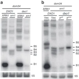
The results in Fig.1and Supplementary Fig. 1 allow us to infer the precise positions of ribosomes on B5 and B4 RNA species in the dom34 mutant backgrounds. Our experiments all converge to the conclusion that the B5 species corresponds to RNAs covered by trisomes (Fig.1g). We also conclude that three ribosomes cover the 71-nt B4 RNA in dom34 mutant cell extracts as this species is resistant to Xrn1 (Supplementary Note 1), its 5′-region is protected from RNase I digestion in vitro (Supplementary Note 2) and a significant proportion remains associated with fractions corresponding to three ribosomes in sucrose gradients (Fig.1e, f). Dxo1 trimming of 3′-NGD fragments in Xrn1-deficient cells. We strongly suspected that the B4 species was the original NGD product, and because B3 and B2 RNAs were exclusively detected in Xrn1-deficient cells, we speculated that these RNAs might be derived from B4 by an alternative 5′–3′ exoribonuclease. We therefore asked whether the 5′–3′ exoribonucleolytic activity of Dxo1, which plays an important role in 5′-end capping quality control22, might explain the presence of the B3 and B2 RNAs. Remarkably, deletion of both XRN1 and DXO1 genes in a dom34 background completely abolished the production of the B3 and B2 RNAs, and only the B4 3′-NGD species remained detectable by northern blot analysis (Fig.2a) or in primer extension assays (Supplementary Fig. 2a). Complementation of the dom34/xrn1/
dxo1 mutant with wild-type Dxo1 restored B3 and B2 RNA production to a significant extent, but a catalytic mutant failed to do so (Fig. 2b). We took advantage of the almost exclusive pre-sence of the B4 3′-NGD RNAs in dom34/xrn1/dxo1 mutant cells to ask how this RNA is protected by ribosomes by adding Xrn1 to cell extracts as described above (Supplementary Fig. 1b). Some of the B4 RNA was Xrn1-resistant (Supplementary Fig. 2b) in accordance with our hypothesis that a portion of this species is protected by three ribosomes in xrn1/dom34 cell extracts (Fig. 1g). Interestingly, the decrease in the amount of B4 RNA was correlated with an almost equivalent increase of a 47-nt species (Supplementary Fig. 2b, c), suggesting that disomes persist on the majority of the 3′-ends of B4 RNAs in dom34/xrn1/dxo1 cells in vivo. We thus propose that two populations of B4 RNAs co-exist in Xrn1-deficient cells in vivo, with one population covered by three ribosomes, especially in dom34 mutants, and the other only covered by two ribosomes, but having a 5′-protuding RNA extremity due to the absence of 5′–3′ exoribonucleases.
Mapping of the primary NGD endonucleolytic cleavage site. The results described above suggest that the principal band detected in the absence of Xrn1 and Dxo1 (B4 RNA) is a specific 3′-product of NGD cleavage in our constructs (Fig.2a). While B4 RNA resistance to Xrn1 in vitro (Supplementary Fig. 1b) could be explained by a third ribosome dwelling after cleavage, we also considered the possibility that its 5′-phosphorylation state could contribute to its stability, since both Xrn1 and Dxo1 require 5′-phosphorylated extremities to degrade RNA22,28. We therefore asked whether the B4 RNA naturally has a monophosphate (5′-P) or a hydroxyl group (5′-OH) at its 5′-end by treating RNA purified from dom34 cell extracts with T4 polynucleotide kinase to see whether this would stimulate attack of B4 by Xrn1 in vitro. Remarkably, the B4 RNA was completely degraded by Xrn1 only after 5′-phosphorylation by T4 kinase in vitro (Fig.3a), demon-strating that the B4 RNA has a 5′-OH extremity in dom34 cells. A portion of the abundant B1 RNA persisted during Xrn1 treatment in kinase buffer (Fig. 3a, and see the “Methods” section), but parallel Xrn1 digestion in optimal buffer confirms that B1 and B5 RNAs were totally digested (i.e., are fully mono-phosphorylated), while the B4 RNA remained resistant (Supplementary Fig. 3a). It has recently been demonstrated that the Hel2 ubiquitin-protein ligase is crucial for the activation of NGD cleavages29. Consistent with our hypothesis that the B4 species corresponds to the 3′ NGD cleavage product, this RNA is no longer produced in the hel2 mutant (Supplementary Fig. 3b).
By definition endonucleolytic cleavage of RNA results in the production of 5′ and 3′-RNA fragments. However, corresponding 5′ and 3′ fragments have never been definitively demonstrated in the case of NGD-targeted mRNAs. We thus searched for the corresponding 5′-NGD fragment for the B4 3′-NGD RNA. To map the 3′-end of 5′-NGD RNAs, total RNA preparations from ski2 and ski2/dom34 mutants were ligated to a pre-adenylated oligonucleotide linker using truncated RNA ligase to perform 3′-RACE (Fig.3b)30,31. The ski2 mutant context was used to limit 3′-trimming of these RNAs in vivo, and RNAs were pre-treated with T4 polynucleotide kinase to modify 3′ phosphates or 2′–3′ cyclic phosphates to 3′-OH to permit RNA ligation31. The major RT-PCR product was of the expected size (66 bp; Fig. 3c and Supplementary Fig. 3c) and verified by sequencing the resulting clones (Fig. 3d). The identification of a matching 5′-NGD
fragment for the B4 3′-NGD RNA, confirms that an endonu-cleolytic event occurred at this precise position. The same procedure performed on RNAs isolated from ski2 mutants where Dom34 was still active yielded the same major PCR product, also verified by sequencing (Supplementary Fig. 3d). Thus, while the
B1 B5 B4 B3 Vec B1 B2 B4 B2 B3 B5 a xrn1 dxo1 Pwt Pmut b dom34 Vec 5S 5S DXO1 xrn1 dxo1 dom34 DXO1 xrn1 XRN1 XRN1 XRN1
Fig. 2 Dxo1 creates the heterogeneity of 3′-NGD RNA fragments in Xrn1-deficient cells. 8% PAGE followed by northern blot analysis using probe prA showing steady-state levels of RNAs in dom34 and other indicated mutant strains. The 5S rRNA served as a loading control. a Impact of DXO1 deletion on B2 and B3 RNA production.b Plasmid expression of wild-type Dxo1 (Pwt) or a Dxo1 catalytic mutant (Pmut) (mutant E260A/D262A)22.
The vector control is plasmid pRS313 (Vec). Source data are provided as a Source Datafile.
dom34 mutation facilitates the detection of NGD fragments by increasing their stability, the cleavage event itself is Dom34-independent.
The fate of 5′-NGD RNAs. We anticipated that following NGD cleavage of mRNA1, ribosomes that had initiated translation on the 5′-NGD fragments would advance to the new 3′-end and the RNA be subjected to Xrn1 trimming, similar to the process that generates B1 and B5 (Fig.4a). Since the B4 3′-NGD RNAs were
cut in the +1 reading frame (Fig. 1g), upstream ribosomes on these 5′-NGD RNAs would be expected to stall with one nucleotide in ribosome A-site (Fig.4a and Supplementary Fig. 4) and as result produce new RNA fragments 47+ 1, 77 + 1 nts, protected by two or three ribosomes, respectively (see Supple-mentary Fig. 4). Indeed, in northern blots using probe prG, which is complementary to the new 3′-ends generated by NGD cleavage, we detected RNA fragments consistent with a 1-nt increase in size compared to those detected by prA on the same membrane (Fig. 4b). We mapped the 5′-ends of these new ribosome
pro-tected fragments by primer extension assays using prG (Fig. 4c and Supplementary Fig. 4). The detection of 48-nt (and 78-nt) cDNAs only in cells containing active Xrn1 (Fig. 4c) strongly suggests that the new NGD endonucleolytic products are covered by two and three ribosomes, respectively. The production of cDNAs of exactly the predicted sizes (48 and 78 nts) is an independent confirmation that the 3′-extremity of the 5′-NGD product corresponds precisely to the proposed NGD endonu-cleolytic cleavage site (Supplementary Fig. 4). Remarkably, the 3′-extremity of the 5′-NGD RNA was detected in the context of active 3′–5′ exonucleases, meaning that ribosomes run on and cover the 3′-extremity before any 3′–5′ attacks can occur. In summary, we propose that the B4 RNA is produced by endo-nucleolytic cleavage within the footprint of the third stalled ribosome and that at least two upstream ribosomes promptly protect the resulting 5′-NGD fragment from degradation by 3′–5′ exoribonucleases (Fig.4d).
The 5′-OH endocleaved product is phosphorylated by Trl1. Despite the fact that a portion of the B4 species was found to be 5′-hydroxylated in dom34 cell extracts (Fig.3a), a major fraction
of the B4 RNA from dom34/xrn1 and dom34/xrn1/dxo1 cell extracts can be degraded by Xrn1 in vitro without prior 5′-phosphorylation (Supplementary Figs. 1b and 2b), suggesting that B4 accumulates as a 5′-phosphorylated species in this mutant background. A well-characterized factor with RNA kinase activity in yeast is the essential tRNA ligase Trl121,32. Splicing of tRNAs is known to generate 5′OH-intron RNAs which require Trl1 kinase activity to permit their degradation by Xrn1. We therefore asked whether the 3′-NGD B4 RNA fragments were substrates of Trl1. Since TRL1 is an essential gene, we used trl1Δ strains expressing pre-spliced intron-less versions of the 10 intron-containing tRNAs that restore viability33,34. If Trl1 were required for B4 degradation, loss of Trl1 function should increase the amount of B4 RNA. Remarkably, in trl1 versus TRL1 cells, we observed a 24-fold accumulation of the B4 RNA species and only a two-24-fold- two-fold-decrease of B1 RNA levels, while the B5 RNA was barely detectable (Fig. 5a, b). We asked whether the accumulated B4 RNA had a hydroxyl group (5′-OH) at its 5′-end. RNAs were analysed by 12% PAGE allowing separation of 5′-hydroxylated from 5′-phosphorylated RNAs21. B4 RNA migrated as two bands in TRL1 cells, whereas only the lower band was detected in trl1Δ cells. This species was fully shifted to the upper band after 5′-phosphorylation of RNA in vitro (Fig. 5c). We also verified that
the B4 RNA was resistant to Xrn1 treatment in vitro. In contrast to B1 and B5 RNAs (Supplementary Fig. 5), B4 was completely degraded by Xrn1 only after 5′-phosphorylation by T4 kinase in vitro (Fig.5d). We conclude that the B4 RNA accumulates as a fully 5′-OH species in the trl1Δ mutant. Thus, Trl1 is the major kinase involved in phosphorylating the B4 RNA following NGD cleavage.
NGD cleavage products derived from mRNAs containing rare codons. We asked whether we could identify endonucleolytic cleavages on another NGD-targeted mRNAs, using what we learned about this process on truncated mRNAs. We chose an mRNA containing four contiguous rare CGA codons, which we call (CGA)4-mRNA, as an NGD target (Fig. 6a and
Supple-mentary Fig. 6a)5. Similar to the truncated mRNAs, ribosomes were shown to stall when decoding rare codons, producing 5′-and 3′-NGD RNAs (Fig. 6a). As previously demonstrated5,35,
dom34 Xrn1 – – – + + + + – PNK 5S B4 B5 B1 5′ 3′-OH Ligation 5′ rApp-linker 5′ 5′ Reverse transcription PCR amplification RNA digestion 5′ 5′-NGD RNA PNK 3′-NGD RNA 50 bp 150 100 200 5′ 3′ mRNA1 DNA sequences obtained
via 3’-RNA ligase RACE
Linker sequence B4 RNA 5′ 3′ 5′-NGD RNA 3′-end 5′ 3′ 5′ 3′ 5.8S a b c d
ski2dom34 ski2 Control Ladder
Fig. 3 Characterization of the endonucleolytic RNA fragments. a Xrn1 digestion of total RNA extracts from dom34 mutant cells in the presence or absence of polynucleotide kinase (PNK) in vitro. 8% PAGE followed by northern blot analysis using probe prA. The 5S rRNA served as a loading control and 5.8S rRNA as a positive control of Xrn1 treatment.b Flow chart illustrating the method used for 3′-RACE as described in ref.31with minor modifications
according to McGlincy and Ingolia30(see the Methods section).c PCR products obtained from 3′-RACE and separated on a 2% agarose gel. Purified DNAs
for sequencing are indicated by an arrowhead. Prior to PCR, cDNAs were produced from total RNA from ski2, ski2/dom34 mutant cells expressing mRNA1. Control is made of total RNA from ski2/dom34 mutant cells without mRNA1 expression. d Sequences obtained after 3′-RACE performed on ski2 and ski2/ dom34 total RNA. 100% of sequenced clones (omitting a residual 5S rRNA-linker amplification detected) have this DNA sequence. 5′-NGD DNA sequence (in green) and linker sequence (in magenta). Below, the site of mRNA1 is shown before and after the cleavage producing the 3’-NGD RNA B4 and the 3′-extremity of the 5′-NGD RNA confirmed by 3′-RACE. Source data are provided as a Source Data file.
we detected 3′-NGD RNAs fragments in dom34 or DOM34 genetic contexts by northern blotting experiments using probe prB (Supplementary Fig. 6b). The precise identification of endonucleolytic cleavages by primer extension experiments is known to be challenging5 probably because, in contrast to truncated mRNAs, the positioning of ribosomes on contiguous rare codons is variable. Wefirst asked whether we could detect 5′-NGD RNAs (Fig.6a) using the same procedure as for NGD-targeted truncated RNAs (Fig.4a, b). By probing the (CGA)4
-mRNA in a large region upstream of the four CGA codons (Supplementary Fig. 6a), we detected RNA bands using a probe annealing 71 nts upstream of the first rare codon (probe prH, Fig. 6b and Supplementary Fig. 6a). Similar to the 5′-NGD
RNAs produced from NGD-targeted mRNA1 (Fig. 4b), RNA detection required a dom34 genetic background (Fig.6b). The profile of the 5′-NGD RNAs resulting from endonucleolytic cleavages of (CGA)4-mRNA was remarkably similar to the B1,
B4 and B5 RNAs from the truncated mRNA1. We then treated these RNAs with Xrn1 and, as anticipated, we observed that the ~71-nt RNA, like the B4 RNA, was Xrn1-resistant, and that ~47-nt and ~77-nt RNAs, like the B1 and B5 RNAs, were Xrn1-sensitive (Fig. 6c). These results strongly suggest that NGD-targeted (CGA)4-mRNAs are a similar source of truncated
RNAs which are, in turn, processed like mRNA1 by the NGD pathway.
The detection of short RNA species by prH probe suggested that endonucleolytic cleavages occurred just downstream, in a region located ~70 nts upstream of the cluster of rare codons (Supplementary Fig. 6a). We thus set out to map the NGD cleavage sites on the (CGA)4-mRNA, using 3′-RACE for the
detection of the 3′-ends of 5′-NGD RNAs in ski2 and ski2/dom34 mutant cells (Supplementary Fig. 6c). We obtained major RT-PCR products of about 45 bp that were purified, cloned and sequenced (Fig. 6d and Supplementary Fig. 6d). The 3′-end
sequences (Fig.6e) formed three clusters, C1, C2 and C3 (Fig.6g), that map to ~71 nts upstream of the second, third and fourth rare codon, respectively, consistent with cleavage within the footprint of the third ribosome as seen for the truncated mRNA1. No 3′-ends were detected within the region covered by two ribosomes, comforting the notion that disomes are not competent for NGD endonuclease activation. Xrn1 arrests mapping to 17–18 nts upstream of the A-site of the twofirst ribosomes positioned with either the second or third CGA codon in the A-site were also detected by primer extension assay (Fig. 6f–h). The strongest
Xrn1 arrests corresponded to those where the lead ribosome contains the third CGA codon in the A-site (Fig.6h), suggesting that the major stall occurs on this codon. Typically, Xrn1 is preferentially blocked 17 nts upstream of the first ribosomal A-site residue26. We speculate that this 1-nt difference reveals distinct conformations of stalled ribosomes on rare codons versus
P A P A P A P A P A P A d a Endocleavage in frame +1 b c producing 3′ NGD B4 RNA 5′ 3′ Xrn1 arrest Probe prG 48 nts 5′-NGD RNAs 3′-NGD RNAs 3′ 5′ (1nt in A site) 5′ 3′ 5′-NGD RNAs Xrn1 5′-NGD RNA (48 nts) 5′ 3′ 5′ 3′ 3′ B4 RNA (71 nts) B4 RNA (71 nts) 17 nts 3′ Probe prG 5′ 3′ Cleavage 71 nt C 78 nts 48 nts T A G 3′ Primer prG 47 nt 77 nt 5S Xrn1 Primer prG Primer prA
ski2dom34xrn1dom34 ski2dom34xrn1dom34 xrn1dom34 dom34
dom34 dom34
Fig. 4 Analysis of the fate of 5′-NGD RNAs. a Schematic model of mRNA1 before and after the endonucleolytic cleavages producing B4 RNAs. 5′-NGD resulting RNAs are shown here covered by two ribosomes and processed by Xrn1 to 48-nt RNAs (see also Supplementary Fig. 4 for 5′-NGD RNAs covered by three ribosomes).b 8% PAGE followed by northern blot analysis using probe prG showing steady-state levels of RNAs in the indicated mutant strains (left panel). Same membrane has been probed with prA as a ladder (right panel), and sizes of B5 (77nt), B4 (71 nt) and B1 (47nt) are indicated. The 5S rRNA served as a loading control.c Primer extension experiments using probe prG to determine the 5’-end of RNAs (see also Supplementary Fig. 5). d Schematic model of ribosome positioning on mRNA1 before and after the unique endonucleolytic cleavage producing B4 RNAs, localized 8 nts upstream of thefirst P-site nt. The position of disomes on the resulting 48-nt 5’-NGD RNA is shown with the distal ribosome having 1 nt in the A site (see also Supplementary Fig. 4). Source data are provided as a Source Datafile.
truncated mRNAs. All these results taken together suggest that the (CGA)4-mRNA and truncated mRNA1 are NGD-targeted in
a highly similar process that results in cleavage within the footprint of the third ribosome, 71 nts upstream of the stall site for the leading ribosome.
Cleavages have been proposed by others to occur in the region covered by disomes using primer extension experiments5,29. We thus analysed an mRNA containing rare codons with an identical ribosome stalling sequence to that previously examined5 (Supplementary Fig. 6e). We demonstrate that primer extension arrests detected in the region covered by disomes are abolished in dxo1/xrn1 mutant cells, suggesting that they are the products of subsequent trimming by these enzymes. In conclusion, our data suggests that stalled disomes on truncated mRNAs or on mRNAs containing short CGA repeats are poorly competent for NGD endonucleolytic cleavages. Endonucleolytic cleavages instead occur upstream of collided disomes, in agreement with other 3′-RACE analyses27. Our data suggests that these cleavagesfirst occur within the mRNA exit tunnel of the third stacked ribosome and those queuing further upstream.
Discussion
In this study, wefirst characterized the 3′-NGD RNA fragments produced near the 3′-end of truncated mRNAs that mimic nat-ural cleaved mRNAs known to be NGD targets. One advantage of studying the 3′-NGD products of truncated mRNAs is that the precise positioning of stalled ribosomes results in 3′-NGD RNA fragments of specific sizes. Indeed, the precise identification of endonucleolytic cleavages is known to be challenging for mRNAs containing rare codons5,29because the positioning of ribosomes on multiple contiguous rare codons is variable. Using a ribozyme
to efficiently generate precise 3′-ends within an open-reading frame, we were able to obtain detailed information about ribo-some positioning on 3′-NGD RNAs, and provide the first precise mapping of the original site of endonucleolytic cleavage on an NGD substrate. Our model suggests that this cleavage occurs 71 nts from the 3′ end of the truncated mRNA, 8 nts upstream of the P-site codon in the third stacked ribosome (Figs.1g and7). This localizes the 5′-extremity of cleaved RNA fragment within the mRNA exit tunnel, 4 nts downstream of the expected nucleotide position of a canonical mRNA that emerges from the ribosome and becomes available for cleavage by RNase I in vitro, classically used in ribosome foot-printing studies. A buried cleavage site is consistent with the idea that the NGD endonuclease might be the ribosome itself. However, we cannot fully exclude the possibility that the stalled ribosome allows access to an external nuclease with a specific conformation to penetrate this far into the mRNA exit tunnel.
Xrn1 treatment of various mutant cell extracts suggested that the predominant ribosome configuration on truncated mRNAs is disomes. Interestingly, the existence of disomes on truncated mRNAs has been previously reported in ribosome-profiling analysis20 and stacking of two or more ribosomes has been proposed as a prerequisite for the activation of the endonu-clease27. The latter observation led to the proposition that ribosome collision triggers NGD cleavage upstream of disomes. Our data suggests that NGD endonucleolytic cleavage detected within the third stalled ribosome, suggests that the first two stalled ribosomes (disome) are not competent for the activation of the endonuclease activity (Fig.7). Our 3′-RACE experiments
did not amplify DNA products corresponding to RNAs matching the predicted sizes of NGD-cleaved RNAs within the second (41 nts) orfirst stalled ribosome (15 nts) (predicted sizes 95 and 125
0 0.2 0.4 0.6 0.8 1 5S B4 B1 B5 trl1/dom34 Xrn1 – + + + + – – – PNK 5.8S 5S c – + – + +PNK P OH TRL1 dom34 Amount relative to 5S
*
5 μg 1.25 μg B1 B4 trl1 TRL1 dom34 + 5S B1 B5 B4*
trl1 TRL1 b a d ΔFig. 5 Endonucleolytically cleaved 5′-OH RNAs are phosphorylated by Trl1. a 8% PAGE followed by northern blot analysis using probe prA. Levels of 3′-NGD RNA fragments in trl1/dom34 cells compared with those from TRL1/dom34 cells. b B1 and B4 RNA quantification relative to 5S rRNA from three independent experiments as shown ina. c 12% PAGE followed by northern blot analysis using probe prA. Treatment using T4 PNK to determine 5’-OH and 5’-P B4 RNA positions in the indicated strains. One-fourth of trl1/dom34 total RNA treated was loaded to limit scan saturation and allow TRL1/dom34 B4 RNA detection. The 5S rRNA served as a loading control.d As in Fig.3a, Xrn1 digestion of total RNA extracts from trl1/dom34 mutant cells in the presence or absence of T4 PNK treatment in vitro. A minor band detected in trl1 is indicated by an asterisk (see also Supplementary Fig. 5 in which this band is detectable in TRL1 cells). Error bars indicate standard deviation (s.d.) calculated from three independent experiments. Source data are provided as a Source Datafile.
nts, respectively), indicating that they do not occur to any sig-nificant level. The major ~65-bp RT-PCR product corresponded perfectly to an RNA cleaved 71nt upstream of the 3′-extremity of mRNA1, suggesting this is the primary site of NGD cleavage. This also suggests that the particular conformation of disomes on
these mRNAs is incompatible with an NGD endonuclease activity cutting upstream of the ribosome P-site. The ability to induce this precise NGD cleavage appears thus to be a normal property of stalled ribosomes, with disomes (and monosomes) being exceptions. P A P A P A P A P A P A C3 5′ 3′ Xrn1 arrest Probe prH 5′-NGD RNAs 3′ 3′ 3′ 3′ 3′ Xrn1
(CGA)4-mRNA (CGA)4-mRNA
3′-NGD RNAs Probe prB 5′ RNA1 DOM34 XRN1 SKI2 Δ Δ Δ Δ + + + + + + + Δ + + Δ + + + Δ + + Δ + + C 47 71 77
*
prH prA nts 5.8S – + SKI2 ski2 – + Xrn1 dom34 47 71 77 nts C2 Xrn1 5′ 5′ Xrn1 Xrn1 Xrn1 Rare codons prJ primer extension Control C T A G Sequences obtainedby 3′-RNA ligase RACE
Linker sequence (38%) 5′ (23%) 5′ (23%) 5′ 5′-NGD RNA 3′ -end (15%) 5′ 50 bp 150 100 200 5′ 3′ Xrn1 3′-ends detected –71 nts C1 C1 C2 Xrn1 Xrn1 5′ –71 nts C3 –71 nts Xrn1
Xrn1 arrests not detected
17 nts (CGA)4 mRNA Rare codons Rare codons a b c g d e f h ski2dom34 ski2 Ladder Control
Fig. 6 Identification of the endonucleolytic cuts on the NGD targeted (CGA)4-mRNA. a Schematic view of the (CGA)4-mRNA. 5′- and 3′-NGD RNAs are
products of NGD. The lightningflash represents the potential endonucleolytic cleavage upstream of the ribosome stall site. Probes prB and prH are indicated. 5′-NDG RNAs are shown processed by Xrn1 as described in Figs.4a, d and Supplementary Fig. 4.b Northern blot analysis using probe prH showing steady-state levels of RNAs in the indicated mutant strains. Same membrane has been probed with prA as a ladder, and sizes of mRNA1 products, such as B5 (77 nt), B4 (71 nt) and B1 (47 nt) are indicated. Only the dom34 lane is shown. See Supplementary Fig. 6a for the sequence probed by prH. The 5S rRNA served as a loading control. Total RNA from WT cells without (CGA)4-mRNA expression served as a control, noted C. A non-specific band is
indicated by an asterisk.c Xrn1 treatment in vitro of total RNA from dom34 or dom34/ski2 mutant cells and northern blot using probe prH. The 5.8S rRNA is a positive control of Xrn1 treatment.d PCR products obtained from 3′-RACE (see also Fig.3c). Prior to PCR, cDNAs were produced from cells expressing (CGA)4-mRNA. Total RNA from cells without (CGA)4-mRNA expression served as a control.e Sequences obtained after 3′-RACE performed in d on
ski2/DOM34 total RNA. Sequence distribution is given in percentage. f Primer extension experiments using probe prJ to determine the 5′-end of RNAs. Xrn1-specific arrests are indicated by arrowheads. g Positioning of 3′-ends detected by 3′-RACE on (CGA)4-mRNA from ski2/DOM34 cells (magenta
arrowhead). Arrowhead sizes are proportional to the relative number of sequences obtained. Three cleavage clusters, C1, C2 and C3 were defined (see Supplementary Fig. 6d). Xrn1 arrests deduced fromf are indicated by black arrowhead with sizes proportional to the intensity of reverse stops observed in f. h Schematic view of the ribosome positioning on (CGA)4-mRNA deduced from Xrn1 arrests combined with the positioning of endonucleolytic cleavages
We considered the possibility that the dom34 mutation may exaggerate the ribosome stalling and allow cleavage beyond what would naturally be observed. As discussed in the section Intro-duction, the analysis of NGD RNA fragments is facilitated by the dom34 mutation and is crucial for RNA stabilization when ana-lysing truncated mRNAs by northern blotting experiments5,20. In the presence of Dom34, and more efficient ribosome dissociation, the exosome would certainly be more actively involved once the first endonucleolytic cleavage event has occurred5,20. Importantly, however, our 3′-RACE experiments confirmed the existence of 5′-NGD products with ends matching the 5′-extremity of the 3′-NGD B4 RNA in cells containing Dom34 (Fig.3c, d). Thus, NGD endonucleolytic cleavage does not occur randomly upstream of the ribosomal stall site and is not an artefact of dom34 genetic context.
These observations were used to map endonucleolytic clea-vages that occur on a second NGD-target mRNA containing rare
codons, also in a DOM34 genetic context. Endonucleolytic clea-vages occurred 71nts upstream of thefirst residue in the leading ribosome A-site, in the region potentially covered by the third ribosome. Therefore, mRNAs containing rare codons are pro-cessed similar to truncated RNAs, although cleavage accuracy is slightly affected, correlating with specific Xrn1 arrests (Fig. 6f) that might be explained by a particular conformation of thefirst stalled ribosome29.
It has been recently reported that Cue2 is the endonuclease that cleaves NGD/NSD targeted RNAs36,37. Its action has been pro-posed to occur in the ribosome A site and therefore deviates significantly from our observations. We do not have a good explanation for this difference. However, previous experiments with the naturally occurring truncated HAC1 mRNA are con-sistent with our mapping of the endonucleolytic cleavage site20. The HAC1 intron is known to be excised by Ire1, but RNA ligation can be incomplete and lead to a truncated but translated
3′ New run of endonucleolytic cut Phosphorylation by Trl1 Xrn1 3′ 3′ 5′OH 3′-NGD RNA Endonucleolytic cut Disome
1nt in A site 5′-NGD RNA 5′ 5′ 3′ B4 RNA B1 RNA Xrn1 5′-p 5′-p 5′-p B3 RNA Dxo1 Dxo1 B4 RNA B4 RNA B2 RNA Dxo1 trimming 5′OH Endonucleolytic cut in the 3rd ribosome Endonucleolytic cut 3′ Disome 5′-p 5′-p 5′-p Xrn1 3′ 3′ 3′ Ribosome dissociation
and RNA phosphorylation by Trl1
3′ B5 RNA Disome Decapping or 3′ B5 RNA 3′ B1 RNA Endonucleolytic cut upstream of the 3rd ribosome m7Gpp m7Gpp Xrn1 Xrn1
Hel2-independent endo-exo attacks
5′OH 3′ 5′OH 5′OH 5′OH 3′ 3′ 3′ 3′ 3′ 3′ 3′ 5′
Fig. 7 Model of No-Go decay pathway involving Trl1 kinase and 5’–3’ exoribonucleases. Top of figure, the third ribosome is represented as competent for NGD endonuclease activation. We propose that the twofirst stalled ribosomes are not properly conformed to trigger the endonucleolytic process. NGD endonuclease cleavage (lightningflash) occurs 8 nts upstream of the first P-site residue, within the mRNA exit tunnel of the ribosome. Upstream ribosomes covering the resulting 5′-NGD fragments can advance and stall on the new 3′-end with 1 nt in the ribosomal A-site. Colliding ribosomes on this new RNA fragment can induce a novel NGD endonuclease activation. On 3′-NGD RNAs, like B4 RNAs, the NGD-competent ribosome dissociates and facilitates access of Trl1 RNA kinase to the 5′-hydroxylated 3′-NGD RNA, but we cannot exclude that the leading ribosome dissociates and upstream ribosomes run to form a new disome with 5′-protruding RNA. Once the RNA is 5′-phosphorylated, the processive 5′–3′ exonucleolytic activity of Xrn1 can degrade and produce B1 RNA. Alternatively, upon Xrn1 inactivation, 5′–3′ exonucleolytic digestion of this RNA by Dxo1 can occur and produce trimmed RNAs, such as B3 and B2 RNAs. Middle offigure, upstream of the 3rd ribosome, ribosomes are also competent for NGD endonuclease activation. Here, the endonucleolytic cleavage occurs in the 4th ribosome and B5 RNAs can derive from such RNAs after phosphorylation and 5′–3′ digestion. Bottom of figure, alternative pathways are proposed: B1 and B5 RNA production could be initiated by decapping or via Hel2-independent uncharacterized endo/exonucleolytic attacks.
mRNA38,39. Green and colleagues showed by ribosome-profiling analysis that ribosomes stall at the 3′-end of the first exon of the HAC1 mRNA, leading to an endonucleolytic cleavage ~70 nt upstream from the 3′-end9. Consistent with this observation, our data suggests that the NGD endonuclease cleaves 71 nt upstream of the 3′-end of the truncated mRNA1 (B4 RNAs, Fig.1g). We also showed that the NGD endonuclease cleaves mRNA1 in the +1 reading frame. As a consequence, upstream ribosomes run forward on the 5′-NGD mRNA and stall with 1 nt in the A-site (Figs. 1g and 7). This is in agreement with ribosome-profiling
analysis reporting the predominance of short RNAs having one 3′-nucleotide in the ribosome A site20,36.
We showed that the Trl1 kinase plays a role in NGD, and we propose that the resistance of the B4 3′-NGD RNA fragments to Xrn1 attacks in vitro and in vivo (Fig. 3a and Supplementary Figs. 1b and 3a) is a direct consequence of the 3rd ribosome pre-venting access to Trl1, with dissociation of this ribosome triggering the 5′-phosphorylation of the B4 RNA (Fig.7). It is also possible that the leading ribosome dissociates, and that upstream ribosomes run forward to form a new disome with a 5′-protruding RNA (Fig.7). Once the RNA is 5′-phosphorylated, the processive 5′–3′
exonucleolytic activity of Xrn1 can degrade to produce the disome protected B1 RNA (Fig. 7). The fact that RNAs longer than B5, which accumulate in the trl1 mutant (Fig.5a), are Xrn1-resistant (Fig.5c and Supplementary Fig. 5) coupled with the absence of the B5 RNA upon Xrn1 inactivation (Fig.1b–d) lead us to propose that
the B5 RNA originates from 5′-OH RNAs cleaved within ribo-somes upstream of the 3rd ribosome, are phosphorylated by Trl1 and are digested by Xrn1 until it bumps into downstream ribo-somes (Fig.7). A portion of the B1 species (Fig. 5a and Supple-mentary Fig. 5a) is detected upon Trl1 inactivation, however. Therefore, 5′-phosphorylation of 5′-hydroxylated B4 RNAs only accounts for a portion of B1 RNA production. In agreement, the hel2 mutation abolishes B4 RNA detection without impacting B1 and B5 RNA levels (Supplementary Fig. 3b). Mutations can thus reveal the existence of alternative pathways. In this regard, we learned that the inactivation of Xrn1 can lead to a Dxo1 trimming and production of B2 and B3 RNAs (Fig. 7). We cannot exclude the possibility that a portion of the B1 and B5 RNAs derive from alternative pathways, such as the canonical 5′–3′ decay pathway (i.e., decapping and Xrn1 5′–3′ digestion)24or an uncharacterized endo and/or exonucleolytic decay pathway (Fig.7). Lower levels of NGD RNAs in a dcp2 mutant (Fig. 1b, c) do not contradict a decapping requirement in some of their production, but the poor growth of dcp2 mutant cells prevents us from drawing further conclusions. The potential existence of alternative pathways does not overshadow the strong impact of the trl1 mutant on NGD RNA production (Fig.5).
We show that the NGD pathway produces RNAs bearing a 5′-hydroxyl group (Figs.3a,5and 7). 5′-hydroxyl and 2′–3′ cyclic
phosphate 3′ extremities are typically generated by metal-independent endoribonucleolytic reactions40. We also show that the Trl1 kinase phosphorylates these 3′-NGD fragments to allow degradation by 5′–3′ exoribonucleases. Thus, in addition to its role in tRNA splicing and in phosphorylation of 5′-hydroxylated exon of the HAC1 mRNA34, Trl1 is an important player in NGD pathway. This study also provides mechanistic insights that will help to go further in the comprehension of mRNA surveillance pathways in connection to NGD.
Methods
Yeast media, plasmids, strains, and oligonucleotides. The media, strains of S. cerevisiae, oligonucleotides, synthetized DNAs and plasmids used in this study are described in the Supplementary Methods and Supplementary Tables 1, 2, 3 and 4, respectively.
Northern blot analysis. 8 OD600nmof exponentially growing cells were collected
by centrifugation. RNA was extracted by adding on cell pellet 500 µl of buffer AE (50 mM Na acetate [pH 5.3], 10 mM EDTA), vortexing, and adding 500 µl of phenol previously equilibrated in buffer AE. After vortexing, the tubes were incubated at 65 °C for 4 min and then frozen in a dry ice–ethanol mix, followed by a 15-min centrifugation at room temperature. A second extraction with 500 µl of phenol–Tris–EDTA–chloroform was performed. RNA from 360 µl of supernatant was precipitated with 40 µl of 3 M Na acetate (pH 5.3) and 2.5 volumes of ethanol and centrifuged at 4 °C for 30 min. The pellet was washed in 80% ethanol, dried at room temperature and resuspended in water. After electrophoresis, RNA was blotted onto a Hybond-N+ membrane (Amersham) and hybridization to end-labelled oligodeox-ynucleotide probes was carried out at 42 °C overnight in Roti-Quick solution (Carl-roth). The Hybond-N+ membrane was washed successively at 42 °C in 5 × SSC–0.1% SDS and twice in 1 × SSC–0.1% SDS. Blots were exposed to PhosphorImager screens, scanned using a Typhoon FLA 9500 (Fuji), and quantified with ImageJ software. Gel electrophoresis for separation of RNA molecules. Total RNA was resolved by 8% TBE-urea polyacrylamide or 1.4% TBE-Agarose gels, and to dissociate 5′-hydroxylated from 5′-phosphorylated RNAs, total RNA was resolved by 12% TBE-urea polyacrylamide gels. All gels were followed by northern blot analysis. In vitro RNA digestion. RNA digestion of 20 OD260nmof cell extracts were
per-formed by using 1 unit of Xrn1 (Biolabs) in NEB buffer 3 at 25 °C during 30 min unless otherwise indicated. NEB Buffer 3 was replaced by T4 PNK buffer (NEB) in kinase assays in the presence or absence of Xrn1 (Figs.3a and5d). For RNase I treatment of cell extracts, 20 OD260nmof extracts (prepared without heparin) were
incubated with 0.5, 1, or 2 µl of RNase I (Invitrogen, 100 units/µl) 30 min at 25 °C. For total RNA treatment, 5 µg of RNA were digested 30 min at 25 °C. All RNase treatments were followed by RNA extraction and northern blot analysis as described above.
Polysome analysis. Yeast cells were grown exponentially to 0.8 OD600at 28 °C and
the 200 ml cell culture was quickly cooled in ice water and then centrifuged and washed in 20 ml of buffer A containing 10 mM Tris pH 7.4, 100 mM NaCl and 30 mM MgCl2, and 0.5 mg/ml heparin. Heparin was only omitted for RNase I
treatment24. Cycloheximide was not used to prevent any drug-induced ribosome
positioning. The cell pellet was resuspended in 0.6 ml of buffer A. A 1.4 g amount of glass beads was added, and cell lysis was performed by bead beating twice for 30 s each time (with intermittent cooling). After lysis, the cell extract was clarified for 5 min at full speed in a microcentrifuge. The equivalent of 20 OD260nmunits of cell
extract (80μg of total RNA) was then layered onto linear 10–50% sucrose density gradients. Sucrose gradients (in 10 mM Tris–HCl [pH 7.4], 70 mM ammonium acetate, 30 mM MgCl2) were prepared in 12 × 89 mm polyallomer tubes (Beckman
Coulter). Polysome profiles were generated by continuous absorbance measurement at 254 nm using the Isco fraction collector. Gradients were collected in 20 fractions and processed for northern blotting as described above.
Primer extension. Radiolabelled primers (primers prA and prE for mRNA1, and primer prJ for (CGA)4-mRNA) were used and Maxima H Minus reverse
tran-scriptase (ThermoFisher) was used to synthesize a single-stranded DNA toward the 5′-end of the RNA. The size of the labelled single-stranded DNA was determined relative to a sequencing ladder (ThermoFisher Sequenase sequencing kit) on 5% TBE-urea polyacrylamide gel. Oligonucleotides were radio-labelled with [γ-32P] ATP with the T4 polynucleotide kinase (NEB).
3′-end RNA mapping. Mapping was performed according to the 3′-RNA ligase-mediated RACE method31with minor modifications: Total RNA preparations were
first 3′-dephosphorylated using T4 PNK for 1 h at 37 °C without ATP and pre-adenylated linker (Universal miRNA cloning linker, NEB) ligation was performed during 4 h at 22 °C in the presence of truncated RNA ligase 2 (NEB)30. Reverse
transcriptase reactions were performed using primer prE complementary to the linker sequence. PCR primer prF specific to mRNA1, or primer prK specific to (CGA)4-mRNA, were used with primer prE in PCR reactions (Supplementary
Figs. 3a and 6c). PCR products were purified, cloned into Zero Blunt TOPO PCR Cloning vector (Invitrogen), transformed and plasmids sequenced.
Reporting Summary. Further information on research design is available in the Nature Research Reporting Summary linked to this Article.
Data availability
A Reporting Summary for this Article is available as a Supplementary Informationfile. The authors declare that all data supporting thefindings of this study are available within the Article and its Supplementary Informationfiles or from the corresponding author upon reasonable request. The source data underlying Figs. 1b–f, 2a, b, 3a, c, 4b, c, 5a–d and 6b–d, f and Supplementary Figs. 1b, d–i, 2a–c, 3a, b, 5 and 6b, e are provided as a Source Datafile in the Supplementary Information and at FigShare with DOI information (0.6084/m9.figshare.11317613).
Received: 8 November 2018; Accepted: 11 December 2019;
References
1. Doma, M. K. & Parker, R. Endonucleolytic cleavage of eukaryotic mRNAs with stalls in translation elongation. Nature 440, 561–564 (2006).
2. Passos, D. O. et al. Analysis of Dom34 and its function in No-Go Decay. Mol. Biol. Cell 20, 3025–3032 (2009).
3. Chen, L. et al. Structure of the Dom34–Hbs1 complex and implications for No-Go Decay. Nat. Struct. Mol. Biol. 17, 1233–1240 (2010).
4. Letzring, D. P., Dean, K. M. & Grayhack, E. J. Control of translation efficiency in yeast by codon–anticodon interactions. RNA 16, 2516–2528 (2010). 5. Tsuboi, T. et al. Dom34:hbs1 plays a general role in quality-control systems by
dissociation of a stalled ribosome at the 3′ end of aberrant mRNA. Mol. Cell 46, 518–529 (2012).
6. Simms, C. L., Hudson, B. H., Mosior, J. W., Rangwala, A. S. & Zaher, H. S. An active role for the ribosome in determining the fate of oxidized mRNA. Cell Rep. 9, 1256–1264 (2014).
7. Sinturel, F. et al. Cytoplasmic control of sense–antisense mRNA pairs. Cell Rep. 12, 1853–1864 (2015).
8. Simms, C. L., Thomas, E. N. & Zaher, H. S. Ribosome-based quality control of mRNA and nascent peptides. RNA 8, e1366 (2017).
9. Guydosh, N. R. & Green, R. Translation of poly(A) tails leads to precise mRNA cleavage. RNA 23, 749–761 (2017).
10. Frischmeyer, P. A. et al. An mRNA surveillance mechanism that eliminates transcripts lacking termination codons. Science 295, 2258–2261 (2002). 11. van Hoof, A., Frischmeyer, P. A., Dietz, H. C. & Parker, R. Exosome-mediated
recognition and degradation of mRNAs lacking a termination codon. Science 295, 2262–2264 (2002).
12. Kuroha, K. et al. Receptor for activated C kinase 1 stimulates nascent polypeptide-dependent translation arrest. EMBO Rep. 11, 956–961 (2010). 13. Brandman, O. et al. A ribosome-bound quality control complex triggers
degradation of nascent peptides and signals translation stress. Cell 151, 1042–1054 (2012).
14. Defenouillere, Q. et al. Cdc48-associated complex bound to 60S particles is required for the clearance of aberrant translation products. Proc. Natl Acad. Sci. USA 110, 5046–5051 (2013).
15. Shen, P. S. et al. Protein synthesis. Rqc2p and 60S ribosomal subunits mediate mRNA-independent elongation of nascent chains. Science 347, 75–78 (2015).
16. Johnson, A. W. & Kolodner, R. D. Synthetic lethality of sep1 (xrn1) ski2 and sep1 (xrn1) ski3 mutants of Saccharomyces cerevisiae is independent of killer virus and suggests a general role for these genes in translation control. Mol. Cell. Biol. 15, 2719–2727 (1995).
17. Shoemaker, C. J., Eyler, D. E. & Green, R. Dom34:Hbs1 promotes subunit dissociation and peptidyl-tRNA drop-off to initiate no-go decay. Science 330, 369–372 (2010).
18. Meaux, S. & Van Hoof, A. Yeast transcripts cleaved by an internal ribozyme provide new insight into the role of the cap and poly(A) tail in translation and mRNA decay. RNA 12, 1323–1337 (2006).
19. Guydosh, N. R., Kimmig, P., Walter, P. & Green, R. Regulated Ire1-dependent mRNA decay requires no-go mRNA degradation to maintain endoplasmic reticulum homeostasis in S. pombe. eLife 6, e29216 (2017).
20. Guydosh, N. R. & Green, R. Dom34 rescues ribosomes in 3′ untranslated regions. Cell 156, 950–962 (2014).
21. Wu, J. & Hopper, A. K. Healing for destruction: tRNA intron degradation in yeast is a two-step cytoplasmic process catalyzed by tRNA ligase Rlg1 and 5 ′-to-3′ exonuclease Xrn1. Genes Dev. 28, 1556–1561 (2014).
22. Chang, J. H. et al. Dxo1 is a new type of eukaryotic enzyme with both decapping and 5′–3′ exoribonuclease activity. Nat. Struct. Mol. Biol. 19, 1011–1017 (2012).
23. Parker, R. RNA degradation in Saccharomyces cerevisae. Genetics 191, 671–702 (2012).
24. Hu, W., Sweet, T. J., Chamnongpol, S., Baker, K. E. & Coller, J. Co-translational mRNA decay in Saccharomyces cerevisiae. Nature 461, 225–229 (2009). 25. Ingolia, N. T., Ghaemmaghami, S., Newman, J. R. & Weissman, J. S.
Genome-wide analysis in vivo of translation with nucleotide resolution using ribosome profiling. Science 324, 218–223 (2009).
26. Pelechano, V., Wei, W. & Steinmetz, L. M. Widespread co-translational RNA decay reveals ribosome dynamics. Cell 161, 1400–1412 (2015).
27. Simms, C. L., Yan, L. L. & Zaher, H. S. Ribosome collision is critical for quality control during No-Go Decay. Mol. Cell 68, 361–373 (2017).
28. Nagarajan, V. K., Jones, C. I., Newbury, S. F. & Green, P. J. XRN 5′->3′ exoribonucleases: structure, mechanisms and functions. Biochim. Biophys. Acta 1829, 590–603 (2013).
29. Ikeuchi, K. et al. Collided ribosomes form a unique structural interface to induce Hel2-driven quality control pathways. EMBO J. 38, e100276 (2019). 30. McGlincy, N. J. & Ingolia, N. T. Transcriptome-wide measurement of
translation by ribosome profiling. Methods 126, 112–129 (2017).
31. Adkar-Purushothama, C. R., Bru, P. & Perreault, J. P. 3’ RNA ligase mediated rapid amplification of cDNA ends for validating viroid induced cleavage at the 3′ extremity of the host mRNA. J. Virol. Methods 250, 29–33 (2017). 32. Phizicky, E. M., Consaul, S. A., Nehrke, K. W. & Abelson, J. Yeast tRNA ligase
mutants are nonviable and accumulate tRNA splicing intermediates. J. Biol. Chem. 267, 4577–4582 (1992).
33. Cherry, P. D., White, L. K., York, K. & Hesselberth, J. R. Genetic bypass of essential RNA repair enzymes in budding yeast. RNA 24, 313–323 (2018). 34. Cherry, P. D., Peach, S. E. & Hesselberth, J. R. Multiple decay events target
HAC1 mRNA during splicing to regulate the unfolded protein response. eLife 8, e42262 (2019).
35. Chen, H. & Xiong, L. The bifunctional abiotic stress signalling regulator and endogenous RNA silencing suppressor FIERY1 is required for lateral root formation. Plant Cell Environ. 33, 2180–2190 (2010).
36. D’Orazio, K. N. et al. The endonuclease Cue2 cleaves mRNAs at stalled ribosomes during No Go Decay. eLife 8, e49117 (2019).
37. Glover, M. L. et al. NONU-1 encodes a conserved endonuclease required for mRNA translation surveillance. Preprint athttps://doi.org/10.1101/674358(2019). 38. Cox, J. S. & Walter, P. A novel mechanism for regulating activity of a
transcription factor that controls the unfolded protein response. Cell 87, 391–404 (1996).
39. Harigaya, Y. & Parker, R. Global analysis of mRNA decay intermediates in Saccharomyces cerevisiae. Proc. Natl Acad. Sci. USA 109, 11764–11769 (2012). 40. Gonzalez, T. N., Sidrauski, C., Dorfler, S. & Walter, P. Mechanism of
non-spliceosomal mRNA splicing in the unfolded protein response pathway. EMBO J. 18, 3119–3132 (1999).
Acknowledgements
This work has been supported by AAP Emergence Sorbonne Université, SU-16-R-EMR-03 and by the“Initiative d’Excellence” program from the French State grant “DYNAMO”, ANR-11-LABX-0011-01. S.C. was a recipient of fellowship from the Ministère pour la Recherche et la Technologie (MNRT). A.N. was supported by a doc-toral grant from“DYNAMO”, ANR-11-LABX-0011-01. We thank Jay Hesselberth for providing trl1 mutant and Jeff Coller for the dcp2Δ mutant. We thank Ciaran Condon, Josette Banroques and Kyle Tanner for technical assistance. We thank Ciaran Condon for comments on the manuscript and constructive discussions. Thefirst two authors are listed in chronological order of contribution.
Author contributions
A.N., S.C., R.S.-F., J.H., C.T. and L.B. designed, performed and analysed data. L.B. wrote the manuscript.
Competing interests
The authors declare no competing interests.
Additional information
Supplementary informationis available for this paper at https://doi.org/10.1038/s41467-019-13991-9.
Correspondenceand requests for materials should be addressed to L.B.
Reprints and permission informationis available athttp://www.nature.com/reprints
Publisher’s note Springer Nature remains neutral with regard to jurisdictional claims in published maps and institutional affiliations.
Open Access This article is licensed under a Creative Commons Attribution 4.0 International License, which permits use, sharing, adaptation, distribution and reproduction in any medium or format, as long as you give appropriate credit to the original author(s) and the source, provide a link to the Creative Commons license, and indicate if changes were made. The images or other third party material in this article are included in the article’s Creative Commons license, unless indicated otherwise in a credit line to the material. If material is not included in the article’s Creative Commons license and your intended use is not permitted by statutory regulation or exceeds the permitted use, you will need to obtain permission directly from the copyright holder. To view a copy of this license, visithttp://creativecommons.org/ licenses/by/4.0/.