Applications of Transient Grating
Spectroscopy to Radiation Materials Science
The MIT Faculty has made this article openly available.
Please share
how this access benefits you. Your story matters.
Citation
Short, Michael P., Cody A. Dennett, Sara E. Ferry, Yang Yang, Vikash
K. Mishra, Jeffrey K. Eliason, Alejandro Vega-Flick, Alexei A. Maznev,
and Keith A. Nelson. “Applications of Transient Grating Spectroscopy
to Radiation Materials Science.” JOM 67, no. 8 (June 25, 2015):
1840–1848.
As Published
http://dx.doi.org/10.1007/s11837-015-1496-3
Publisher
Springer US
Version
Author's final manuscript
Citable link
http://hdl.handle.net/1721.1/106986
Terms of Use
Creative Commons Attribution-Noncommercial-Share Alike
Applications of Transient Grating Spectroscopy to Radiation
Materials Science
Michael P. Short
∗, Cody A. Dennett, Sara E. Ferry, Yang Yang
†, Vikash K. Mishra
‡,
Jeffrey K. Eliason, Alejandro Vega-Flick, Alexei A. Maznev, Keith A. Nelson
§June 3, 2015
Abstract
The ability to study radiation damage in-situ promises to yield great scientific insights into the mechanisms of radiation damage accumulation, di-rectly enabling the rapid innovation and qualification of materials for nuclear applications. This is a chal-lenging task, as the measurement technique must be non-contact, non-destructive, rapid, and still allow for online irradiation without interference. Appli-cable methods of mechanical spectroscopy are sur-veyed, noting their potential usefulness at character-izing radiation-induced microstructural changes in-situ. The laser-based surface acoustic wave (LSAW) technique appears most suited for these studies, due to its non-contact, non-destructive nature, its abil-ity to rapidly probe materials to the depth of ion irradiation, and the large number of deconvolvable components extractable from its signal. Work is pro-posed to separate the individual mechanisms of irra-diation damage using in-situ and ex-situ LSAW anal-ysis, through a suite of single-effect and integrated experiments.
1
Introduction
For over fifty years, nuclear materials researchers have grappled with the scientific and financial diffi-culties of studying radiation response in nuclear sys-tems. In-core instrumentation is notoriously difficult, because it must fit within the confines of nuclear re-actor cores while also withstanding intense radiation fields. More fundamental studies are performed us-∗Corresponding author. Tel: (617) 347-7763, Email:
hereiam@mit.edu
†Department of Nuclear Science and Engineering,
Mas-sachusetts Institute of Technology
‡Department of Mechanical Engineering, University of
Arkansas
§Department of Chemistry, Massachusetts Institute of
Technology
ing spallation sources and ion accelerators, which ir-radiate samples with neutrons and charged particles, respectively [1]. However, the latter studies differ sig-nificantly from those using neutrons. They typically apply fluxes higher than those found in nuclear reac-tor cores by multiple orders of magnitude, resulting in significant dose rate effects on material property response. Furthermore, the damage resulting from charged particles is not necessarily representative of the damage resulting from neutrons at the same flux level, due to neutron-atypical effects such as injected interstitials.
The unit of material radiation exposure used in studies of radiation damage is the displacements per atom (DPA), which was developed to account for varying levels of material damage as radiation type and energy is changed [2]. It replaced an older, sim-pler unit consisting of the total fluence above a cer-tain energy, typically between 0.1 MeV and 1 MeV. The DPA allows for the proper accounting of higher damage cascade complexity and energy-dependence of damage cross sections, and is therefore linked to re-sultant changes in material property changes, which ultimately are the quantities of interest to nuclear materials engineers. Thus, the introduction of the DPA as the unit of radiation damage in materials helped unify formerly disparate studies in radiation materials science.
However, the DPA is still fundamentally an expo-sure parameter, which only accounts for the ballis-tic stage of radiation damage cascades [2]. The link between the number of displaced atoms (DPA) and the resultant, long-term accumulation and evolution of radiation damage in the form of multiscale ma-terial defects has been the subject of a great num-ber of studies, recently summarized in a comprehen-sive tome on nuclear materials [3]. Variations in the dose-property response of the same materials ex-posed to identical DPA have been observed as func-tions of temperature [4], dose rate [5], type of ra-diation [6], duty cycle (beam rastering) of charged
particle beams [7, 8, 9], imposed stresses [10], pro-cessing and resultant microstructure [11], secondary precipitates or impurities [12], and injection or co-generation of gases such as helium during irradiation [13, 14]. These variations demonstrate a clear dif-ference between applied dose and accumulated dam-age for differing material and irradiation conditions. In addition, the actual analysis of radiation damage most often consists of time-consuming TEM analy-sis, in which artifacts due to varying sample thick-ness and preparation can often obscure the details of accumulated irradiation damage. Were a rapid, non-destructive method of quantification of radiation damage to exist, it would allow for faster innovation in the field of nuclear materials, and it would augment the ability of the DPA to describe resultant changes in material properties during and following irradia-tion.
In this study, a new method of quantifying mate-rial property changes during irradiation is proposed, based on in-situ laser-induced surface acoustic wave (LSAW) analysis. This non-contact mechanical spec-troscopy technique is ideally suited for online mea-surement of material property changes during irra-diation in a variety of sample microstructures and environments. Recent studies, focusing either on the quantification of individual effects of irradiation (such as helium co-injection or dislocation density changes) or on the integrated effects of irradiation, are shown to reliably correlate LSAW signal changes to radia-tion damage in DPA. Next, representative studies of the LSAW response of highly pure copper to irradi-ation are presented to illustrate the potential useful-ness of LSAW analysis of radiation damage. Finally, methods describing a possible in-situ method of ra-diation damage studies are proposed. These methods have the potential to vastly accelerate the pace and accuracy of radiation damage experiments, as well as to reveal mechanisms of breakaway phenomena [15] and potential self-organized criticality [16] arising from the self-organization of radiation-induced mate-rial defects.
2
Mechanical Spectroscopy for
Radiation Materials Science
The missing link between understanding the relation-ship between radiation dose and material property changes is a thorough understanding of how radia-tion damage accumulates spatially in real time. Ions in particular affect just a thin layer of material, rang-ing from tens of microns for MeV protons to hundreds of nanometers for similarly energetic heavy ions. An
ideal technique for in-situ analysis of radiation dam-age would therefore:
• be non-destructive • be non-contact
• collect data extremely rapidly
• have radiation resistant probe materials
• have the ability to probe near-surface phenom-ena, particularly to study charged particle irra-diation
• study multiple material properties at once • should function well in extreme environments of
low/high temperature
• function with samples immersed in a vacuum, in high pressure gases, and in corrosive fluids Recent significant progress has been made to carefully design and implement proton irradiation experiments to accurately simulate neutron damage [1]. Even so, in-situ mechanical spectroscopy has the potential to reveal more mechanistic material changes during ir-radiation, further improving the correlation between neutron and charged particle irradiation.
2.1
Potential Methods for Mechanical
Spectroscopy of Radiation
Dam-age
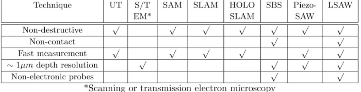
Conventional ultrasonic testing (UT) methods can detect flaws as small as tens of microns, which is too low resolution to detect radiation-induced dam-age modes ranging in size from point defects to voids or precipitates tens of nanometers in size. Piezo-based SAW technologies [17] and acoustic methods such as scanning acoustic microscopy (SAM) [18], scanning laser acoustic microscopy (SLAM) [19], and its 3D cousin holographic SLAM (HOLOSLAM) [20] all require require either a mechanical (solid) or a fluid (oil, water) coupling, precluding the use of sam-ples in extreme environments. Surface Brillouin scat-tering (SBS) measures very fine fluctuations in inci-dent light due to changes in thermal vibration spectra on the atomic level, and therefore requires measure-ment times in the range of hours, meaning that one data point can take longer to acquire than an en-tire irradiation experiment. SBS has, however, been used to measure disorder and amorphization in ion-implanted GaP via acoustic velocity variations [21].
Technique UT S/T SAM SLAM HOLO SBS Piezo- LSAW
EM* SLAM SAW
Non-destructive √ √ √ √ √ √ √
Non-contact √ √
Fast measurement √ √ √ √ √ √
∼ 1µm depth resolution √ √ √ √
Non-electronic probes √ √
*Scanning or transmission electron microscopy
Table 1: Potential mechanical spectroscopy methods for in-situ analysis of radiation damage from ion beams, with their strengths and limitations
Table 1 summarizes the potential mechanical spec-troscopy methods for the analysis of radiation dam-age, along with their matches for the key character-istics of an ideal technique.
SAW analysis has been used to successfully de-termine material properties of interest to radiation materials scientists. Frass and Hess used fiber op-tic coupled lasers to excite SAW waves to study elastic and mechanical properties of thin films [22]. Hurley et al. used SAW spectrometry using sepa-rated pump and probe beams to measure the elastic properties of TiN thin films grown on Si, including thickness and Young’s modulus [23]. Ruiz and Nagy performed SAW dispersion measurements on surface treated metals [24], confirming that SAW spectra can be used to identify the extent of different surface de-formation modes, such as surface roughness, compres-sive residual stress, and cold work. This study points strongly to the possibility that different single effects of radiation damage on microstructure, like those in Ruiz and Nagy’s study, should be separable in a very similar way. Their SAW waves were produced using a piezoelectric element, but this is does not affect the mechanism by which the detection is performed.
A. G. Every reviewed the various methods of measuring near-surface elastic properties of solids and thin films in 2002 [25]. Methods discussed in-clude SBS (mentioned above), laser-generated sur-face acoustic waves (LSAW), and acoustic microscopy (AM). Of these, LSAW is the most suitable method for performing rapid, in-situ, and non-contact deter-mination of the change in elastic properties due to various modes of radiation damage. AM uses me-chanical excitations to induce standing or propagat-ing waves across a surface, but these require contact with the specimen (such as with a piezoelectric ele-ment). The probe itself would be irradiated, or would otherwise interfere with any top-down surface modifi-cation process, such as reactive ion etching of silicon, helium implantation, plasma ion implantation, or ra-diation damage in general.
Every reports that LSAW can be used to measure certain properties of thin films, such as Young’s mod-ulus, Poisson’s ratio, and film thickness [25]. Mea-surement can take place via four methods:
1. Interferometry, whereby the probe laser is split into a reference and a probe beam, and the two are combined in an interferometer to measure surface-normal changes in local specimen height due to the presence of SAWs.
2. Reflectivity changes, since a traveling density wave will affect the reflectivity of the surface. This mode requires only one probe laser beam, and its intensity changes are recorded.
3. Deflection methods, since a Rayleigh wave will curve the surface of the specimen as it travels through (or stays as a standing wave), whereby the angle of deflection is measured using a posi-tion sensitive detector.
4. Diffraction methods, including transient grat-ing (TG) techniques, whereby a laser is used to probe changes in the diffractive properties of a material caused by Rayleigh wave formation. The literature is unusually scant regarding the use of non-contact ultrasonics to quantify radiation dam-age, though much work with contact-based ultra-sonics to quantify and understand irradiation dam-age has been performed. Thompson and Holmes found the Young’s modulus of neutron-irradiated sin-gle crystal copper to increase [26], which the au-thors explain via hindered movement of dislocation line segments due to irradiation-produced defects. This explanation was confirmed in later studies [27]. Tittmann quantified the effect of dislocation density and its interaction with ultrasonic waves [28]. He also reports that Ogi and Hirao used ultrasonic backscat-tering to evaluate thermal aging embrittlement of duplex stainless steels [29], though these were MHz ultrasonic waves, unlike the GHz LSAW waves pro-duced on a surface.
Matlack et al. studied the amount of neutron radi-ation damage present in reactor pressure vessel steels using nonlinear ultrasound [30, 31], by measuring a longitudinal wave non-linearity parameter beta as a function of neutron fluence as an indicator of radia-tion damage. The authors do report that effects from surface roughness and condition, as well as thick-ness variation, affect their results. This work there-fore uses one lumped parameter to describe radia-tion damage, and does not uncover the separate mi-crostructural mechanisms, such as defect clustering, void formation, or dislocation network formation, re-sponsible for its accumulation. It is also not yet clear whether the mechanical response of these materials can be reliably correlated to the non-linearity param-eter alone. Complex radiation effects in materials require a technique which returns suitably complex information, but can also be separated into simpler, single effects.
Additional studies of radiation-induced material property changes using LSAW analysis point to its promising in-situ use during irradiation. Its use in probing the depths of and changes due to ion im-plantation has been investigated in semiconductors [32, 33], ceramics [34], and diamond [32]. LSAW with changing analysis depths has been used to reconstruct depth profiles of Rayleigh wave velocity and elastic constants [35, 36]. LSAW was also used to determine the thickness of amorphized layers of ion-implanted silicon [37], simultaneously acquiring the layer’s den-sity, Young’s modulus, and Poisson’s ratio. Simi-lar changes were investigated in lithium niobate thin films during ion implantation [17], suggesting that LSAW is equally viable as an in-situ technique, with the added advantage of being a non-contact method. However, its use has yet to be demonstrated on nu-clear structural materials, except by Hofmann et al. [38].
2.2
Description of the LSAW Method
An illustration of the LSAW setup used in this study is shown in Figure 1, based on the methods described by Rogers et al. [40]. Here, two laser outputs (1) are sent through a diffraction grating (2), where the +/-1 diffraction orders (3) are made parallel and fo-cused onto the sample (5). The pump beams create a “transient grating,” by inducing parallel regions of thermal expansion with a spatial period determined by the diffraction grating period. The spatially pe-riodic “ripple” pattern at the sample surface is mea-sured by diffraction of the probe light. The diffracted signal beam is co-linear with the reflected reference beam, interfering with it (a process called heterodyne
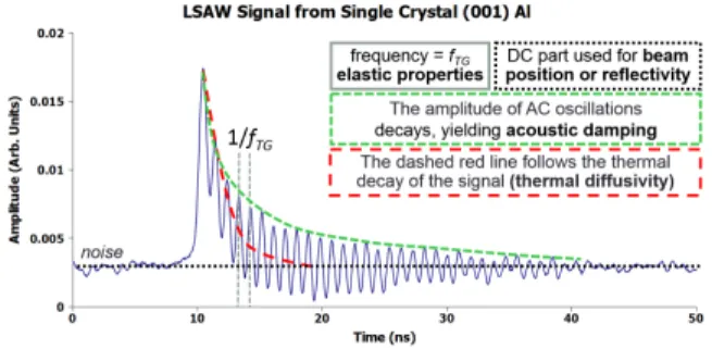
Figure 2: LSAW signal decomposition, showing fre-quency, thermal decay, and acoustic damping on unirradiated, single crystal, (001) aluminum using a 2.75 µm grating spacing
detection [41], which deliberately utilizes co-linear probe and reference beams to amplify the signal). The heterodyne-amplified signal can then be sent si-multaneously to a signal detector (6) to measure its time-dependent intensity, and to a quadrant detector (not done in this study) to provide feedback for any sample motion. This keeps the sample in the plane of the crossed beams and perpendicular to the bisector of the beam paths.
The signal from an LSAW measurement can be decomposed into a number of different components, each revealing different properties about the mate-rial being studied. Figure 2 shows the simplest of these, which can be extracted using data averaged from a single set of conditions. Here, the funda-mental frequency fT G can be easily extracted from a
Fourier transform of the signal, which leads directly to the Rayleigh wave velocity, since SAWs are in-duced with a known wavelength. Subsequent mea-surements of fTG with different phase mask grating
spacings (dP M) can lead to direct measurements of
the speed of sound, as well as properties such as elas-tic constants, the Young’s modulus, the shear mod-ulus, the bulk modmod-ulus, and Poisson’s ratio. The decay constant of the overall curve envelope gives in-sights into the thermal conductivity of the material. A faster decay means a more thermally conductive material, as the DC component of the signal decays faster as the heat from the pump lasers spreads be-tween the thermally excited regions within the analy-sis area. Finally, the decay of the periodic oscillations of frequency fTG on top of the thermal curve
enve-lope yields information about the acoustic damping of SAW waves in the material.
Figure 1: Diagram of the LSAW facility used in this study, adapted from [39]
2.3
Measurement
of
Radiation
Damage-Relevant
Quantities
by SAW Analysis
Three recent studies have been performed using SAWs which directly access quantities of interest to radiation materials scientists. First, Banet et al. [42] studied the acoustic signals of annealed and unan-nealed silicon-on-insulator (SOI) substrates. This single variable study, while preliminary in nature, examined the effect of varying dislocation density (which is known to increase rapidly with irradia-tion to a saturairradia-tion level [43]) on the time-dependent acoustic signals from SAW analysis. Annealed and unannealed substrates have low and high disloca-tion densities, respectively. The data show a time-dependent intensity curve for each case, whereby the signal level in mV directly corresponds to the amount of light entering a photodetector, resulting from the oscillation of a SAW induced by the pump laser. The annealed (low dislocation density) case exhibits very rapid overall curve envelope decay, indicating higher thermal diffusivity assuming the same material den-sity, as well as a relatively low acoustic damping ratio, visible as subsequent oscillations in signal following the initial peak. By contrast, the unannealed (high dislocation density) sample showed lower curve enve-lope decay, indicating lower thermal diffusivity, while oscillations following the initial signal peak all but disappear, indicating high acoustic damping. Stud-ies have shown clear decreases in thermal conduc-tivity as a function of increasing dislocation density in semiconductors [44], and in metals particularly at cryogenic temperatures by dislocation scattering of phonons [45].
Next, Hurley et al. have proposed the use of prop-agating LSAW to study the effects of neutron
ra-diation on material property changes [46]. The re-searchers used an interferometer to generate time-dependent 2D images of SAW wave propagation on copper samples with high texture (anisotropy), pro-duced by rolling, to study one of the simulated ef-fects of neutron irradiation. The results suggest that LSAW methods can be used to study texture changes due to recrystallization during irradiation, by sep-arating the pump and probe beams. The authors also report on using picosecond ultrasonics to mea-sure bulk elastic properties in precisely sectioned FIB samples of candidate materials. Because the LSAW method can be used to determine quantities such as Young’s modulus by varying the LSAW wavelength, it could be used to measure the changes in elas-tic properties during irradiation. Many studies have shown significant changes in Young’s modulus with ir-radiation. Changes have attributed it to effects rang-ing from increases due to increasrang-ing point defect or small cluster concentrations [47] to decreases due to void swelling [48].
Finally, a recent study by Hofmann et al. [38] examined the changes in LSAW propagation due to helium implantation of single crystal tungsten, both pure and with dilute alloying elements of Re or Ta, for fusion reactor first wall applications. Here, a suite of molecular dynamics and density functional theory simulations were compared with x-ray microdiffrac-tion and LSAW measurements to study the changes in void swelling and elastic moduli due to helium im-plantation. Rayleigh wave velocity was used with a known grating spacing to determine the change in Young’s modulus and Poisson’s ratio, assuming sim-ilar densities for the materials. The results show a statistically significant reduction in both Rayleigh wave velocity, Young’s modulus, and shear modulus for helium implanted tungsten, while statistically
sig-Table 2: Proposed single-effect experiments to sepa-rate changes due to individual radiation damage ef-fects on LSAW signal, using single crystal, known-orientation materials
nificant increases in all three are measured and cal-culated for tungsten with an increased self intersti-tial atom (SIA) concentration. These results show that additional quantities relevant to radiation dam-age studies, from the intermediate (SIA concentra-tion) to the end results (Young’s and shear moduli) can be measured using LSAW analysis. However, using the technique in this ex-situ manner still has the same limitations of low sample throughput and lack of time resolution as the ion beam studies in [1]. Further studies by Logan et al. [49] show the appli-cability of measuring LSAW signal changes of both frequency shift and acoustic damping on irradiated Kapton films, suggesting that the technique should be even more widely useful at quantifying radiation damage in a broad class of materials.
3
Proposed Methods of LSAW
Analysis of Radiation Effects
3.1
Using LSAW Ex-Situ to Study
In-dividual Radiation Effects
Deconvolving multiple concurrent radiation effects in real materials will likely be too challenging at the start. Therefore, initial tests which highlight individ-ual effects of radiation damage, such as just voids, re-gions of amorphization, point defect concentrations, or dislocation density, are proposed in Table 2. These single effect studies will be far more informative at the start, as each single effect should have measurable, in-tuitive effects on an LSAW signal, as summarized in Figure 3.
For example, the helium ion microscope can be used to create both regions of amorphization and sin-gle bubbles to simulate voids. Short He-irradiation times using a square grid pattern creates regular regions of amorphization, while longer irradiation creates single helium bubbles inside the
amorphiza-Figure 3: Intuitively expected changes in LSAW sig-nal with representative, single effects of radiation damage
tion regions. Subsequent annealing recrystallizes the amorphized region, leaving the voids intact. Amor-phous materials should carry an acoustic wave more poorly than single crystal materials, so a decrease in the acoustic damping time constant could indicate an increase in amorphization. The voids, if created in regular patterns, may vibrate at their own superlat-tice density, in addition to the material’s underlying Rayleigh wave frequencies.
Cold work by rolling or compression increases the dislocation density of the material to an extent be-fore new grains are formed. These dislocations would scatter acoustic waves and phonons, increasing the thermal decay constant and decreasing the acoustic decay time constant from Figure 2. This effect was already observed during LSAW analysis of annealed vs. unannealed silicon-on-insulator (SOI) substrates [42]. Counting surface etch pits or extraction of a TEM sample can confirm the dislocation density.
Even the effects of vacancy concentration may be measurable using in-situ LSAW on highly cooled specimens, to minimize vacancy migration and re-combination. Dienes proposed that a 1% vacancy concentration should impart a 1% change in all the elastic constants in copper [50], while Nabarro as-serted that the change should be higher (2.3%) for the same vacancy concentration [51]. Later, Ackland reported an expected change of just under 1% in the C44 elastic constant for a simulated copper lattice
with a vacancy concentration of 7.3 · 10−4 [52], at-tainable by quenching near the melting point in some metals, though the author admits a not entirely sat-isfactory explanation of experiments by this method. Recent measurements by the authors of this study on single crystal aluminum found variability between LSAW scans of single crystal aluminum to be repeat-able within 0.2%, suggesting that a high vacancy concentration measured in-situ with LSAW may be achievable during cryogenic irradiation.
Performing future studies on single crystal mate-rials such as tungsten, aluminum, or beryllium may provide more distinctive insights into the mechanisms of radiation damage and their results on LSAW prop-agation, due to less noise resulting from polycrys-tallinity and sample preparation. Realistic limita-tions of equipment come into play for this analysis, however. For example, should one desire to study the LSAW spectral change of diamond, one would need a facility with a frequency resolution of approximately 20 GHz for the Rayleigh wavelengths used in the facil-ity in this study. Significant, order-of-magnitude level steps in equipment cost are incurred past the 2-4GHz level, both for oscilloscopes and for time-dependent light detectors with high enough sensitivity at desired wavelengths.
3.2
Using LSAW In-Situ to Identify
Mechanisms of Radiation Effects
The unique aspects of LSAW as described above make it ideally suited to in-situ radiation studies. Data can be taken at the repetition rate of the pump laser, meaning that hundreds of thousands of signals can be averaged in under two minutes. As long as the medium surrounding the sample is optically transpar-ent to the lasers, the LSAW method can be used. So far, LSAW has been performed on solids in air and to measure thermal properties of transparent fluids, but in theory, the LSAW method can be adapted to study coupled corrosion and radiation damage in real-time of a solid, polished surface immersed in a corrosive, transparent fluid at high-temperature. Most impor-tantly, however, is the ability to take rapid radiation response data, which has the potential to provide new insights into the mechanisms of radiation damage, not just rates, by studying the shapes of the dose-response curves to identify breakaway phenomena.
Figure 4 shows a schematic for an in-situ LSAW irradiation beamline. Here, the ion beam hits the sample, which can be dynamically positioned by x-y-z-tilt stages to maintain the sample surface and angle in the laser focal plane. Irradiations would take place at an angle, so that an LSAW analysis system remains perpendicular to the sample surface, with laser beams entering through an optical window. Proton irradi-ation experiments in extreme environments can be enabled by increasing the proton beam energy, such that it can enter and exit thin, proton-transparent windows (such as Ti-foil), thus separating the experi-mental environment from the vacuum required in the accelerator beamline. A quadrant detector or online analysis of the signal DC amplitude can be used to determine the position of the sample relative to the
Figure 4: Conceptual drawing of an in-situ LSAW beamline, allowing for simultaneous irradiation and LSAW interrogation. Experiments in corrosive fluids or other extreme environments are possible by heat-ing/cooling the sample, changing the fluid within the beamline end station (provided it is optically trans-parent), and the probe beam may be partially di-verted to a Raman spectrometer for simultaneous chemical change information during irradiation.
laser focal spot, allowing dynamic translation of the sample via the stages.
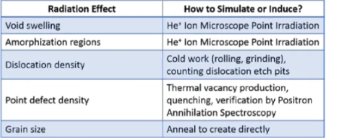
The potential impact that this new method may have on radiation damage studies is partially due to the high throughput of data possible by in-situ LSAW analysis. A study by Garner et al [54] on the relative swelling of ferrite and sorbite grains is shown as an illustrative example in Figure 5. Best fit curves have been drawn to describe the data, though the ferrite data show a smooth exponential relationship, similar to the Kozlov relation [55], while the sorbite grains are drawn with a bi-linear relationship, as proposed by Kalchenko et al. for 304 stainless steel irradiated in EBR-II [56]. This practice has been common in the radiation damage community, though it inadver-tently labels sparse data with mechanistic informa-tion via the curve fit model that may or may not match the mechanism of radiation damage accumu-lation. By contrast, if in-situ LSAW data were to be collected during irradiation, it could fill in the gaps between irradiation data points, providing real mech-anistic information via the shape of the curve during the sudden (or not) onset of void swelling. Radiation damage effects such as these have been proposed as self-organizing into superlattices [57], and have more recently been identified as hallmarks of mesoscale sci-ence [58] due to their potential self-organizing
quali-Figure 5: Data on swelling of ferrite and sor-bite grains, showing bi-linear and exponential fitting curves drawn to explain the data [53]. In-situ LSAW analysis could fill in gaps in this data, helping to ex-plain which, if any, proposed models and mechanisms are most valid. Reprinted with permission from El-sevier.
ties [59].
Polycrystalline, anisotropic materials in particular may be studied to pinpoint the onset of anisotropic void swelling. While void swelling is an isotropic phenomenon, the resultant irradiation creep that re-laxes stresses induced by void swelling will follow the anisotropy of the material, and its microstruc-ture. Such anisotropic swelling, mediated by irra-diation creep, has been theoretically predicted [60] and observed in austenitic [61] and duplex stainless steels [62] bombarded by nickel ions. The anisotropic swelling of polycrystalline materials, due to differing grain orientations, will cause step-height changes to occur, non-linearly increasing surface roughness with irradiation. Therefore, the surface roughness induced by anisotropic irradiation creep from swelling can be used to identify the onset of “breakaway” void swelling, as increased surface roughness will increase heat transfer into the region of heated air just above the sample. These air oscillations have been observed in previous LSAW measurements [41], and may act as an indicator of the onset of the end of void incu-bation.
3.3
Comments on Decreased
Experi-mental Time using LSAW
Nowadays, ex-situ data are collected one at a time, where each experimental data point requires its own sample preparation, irradiation, and destructive anal-ysis. It is for this reason that radiation data across a
wide range of materials are so sparse, because acquir-ing this data is extremely time-consumacquir-ing and expen-sive. By contrast, in-situ data collection using LSAW analysis would eliminate all variability due to original sample condition, sample preparation, changing irra-diation and temperature parameters, and preparation for destructive analysis, by using the same sample for all studies. In-situ LSAW therefore has the poten-tial to turn an entire experimental campaign, which may last months to years, into a 1-2 week single ex-periment with vastly reduced sources of exex-perimental error. LSAW methods are therefore positioned to en-able faster innovation in the field of nuclear materials. Of most interest may be the ability to apply the LSAW technique to the vast body of existing mate-rial irradiation studies already published in the open literature. Simply analyzing the current state of ma-terials mentioned in any of the thousands of papers related to ion irradiation, assuming the samples still exist in good condition, can provide rapid validation of the technique. Then, a low-dimensional envelope could be fit to all the changes in LSAW signal for the material studied, to rapidly gain experience with the technique while minimizing additional irradiation experiments.
4
Conclusions
In-situ LSAW analysis has the potential to greatly increase the pace of innovation in nuclear materi-als science and engineering. Its contact, non-destructive nature, the speed of data acquisition, and its flexibility in operating environments make it ide-ally suited to collecting material property data during irradiation. Open questions regarding ion-specific ef-fects such as beam rastering, mechanisms of radiation defect self-organization and breakaway, and coupled effects of material structure and processing on radia-tion response may be answered using this technique. LSAW analysis of irradiated materials, both in the form of ex-situ experiments on archival samples and in-situ studies on new ones, is poised to rapidly gain experience and validate the technique as a new tool in the field of nuclear materials science and engineering. Next steps include studies to determine the sensitiv-ity of LSAW to experimentally simulated, individual microstructural effects of radiation damage, followed by subsequent multiple-effect experiments to deter-mine their separability in the LSAW signal.
Acknowledgments
This manuscript is based upon work supported by the NSF Graduate Research Fellowship under Grant No. 1122374.
References
[1] G. S. Was, Z. Jiao, E. Getto, K. Sun, A. M. Monterrosa, S. A. Maloy, O. Anderoglu, B. H. Sencer, and M. Hackett. Scr. Mater., 88:33–36, 2014.
[2] G. S. Was. Fundamentals of Radiation Materials Science. Springer-Verlag, 2007.
[3] R. Konings, editor. Comprehensive Nuclear Ma-terials, volume 4. Elsevier, 2012. ISBN 978-0-08-056027-4.
[4] P.M. Rice and S.J. Zinkle. J. Nucl. Mater., 258– 263, Part 2:1414–1419, 1998.
[5] N.I. Budylkin, E.G. Mironova, V.M. Chernov, V.A. Krasnoselov, S.I. Porollo, and F.A. Garner. J. Nucl. Mater., 375(3):359–364, 2008.
[6] G.S. Was and R.S. Averback. 1.07 - radiation damage using ion beams. In R. J. M. Konings, editor, Comprehensive Nuclear Materials, pages 195–221. Elsevier, Oxford, 2012.
[7] J.L. Brimhall, L.A. Charlot, and E.P. Simonen. J. Nucl. Mater., 104:1147–1150, 1981.
[8] N. Ghoniem and G.L. Kulcinski. J. Nucl. Mater., 69–70:816–820, 1978.
[9] A. Hishinuma, N. H. Packan, Eal H. Lee, and L. K. Mansur. J. Nucl. Mater., 122(1–3):260– 265, 1984.
[10] I.A. Portnykh, A.V. Kozlov, V.L. Panchenko, V.M. Chernov, and F.A. Garner. J. Nucl. Mater., 367–370, Part B:925–929, 2007.
[11] T. Leffers, B.N. Singh, S.N. Buckley, and S.A. Manthorpe. J. Nucl. Mater., 118(1):60–67, 1983. [12] L.K. Mansur. Nucl. Technol., 40(1):5–34, 1978. [13] L.K. Mansur, E.H. Lee, P.J. Maziasz, and A.P.
Rowcliffe. J. Nucl. Mater., 141–143, Part 2:633– 646, 1986.
[14] D. B. Bullen, G. L. Kulcinski, and R. A. Dodd. J. Nucl. Mater., 122-123:584–589, 1984.
[15] M. P. Short, D. Gaston, C. R. Stanek, and S. Yip. MRS Bulletin, 39(1):71–77, 2014. [16] P. Bak, C. Tang, and K. Wiesenfeld. 59(4):381–
384, 1987.
[17] S. Joneliunas, L. Pranevichius, and R. Valatka. Nucl. Instr. Meth., 182-183, Part 2:761–767, 1981.
[18] B.T. Khuri-Yakub. Ultrasonics, 31(5):361–372, 1993.
[19] L. W. Kessler and D. E. Yuhas. Scan. Elec. Mi-croscopy, 1:555, 1978.
[20] A.C. Wey and L.W. Kessler. Holographic scan-ning laser acoustic microscopy (holoslam): A new qnde tool. In D. O. Thompson and D. E. Chimenti, editors, Review of Progress in Quan-titative Nondestructive Evaluation, pages 975– 982. Springer US, 1990.
[21] J. Zuk, H. Kiefte, and M. J. Clouter. J. Appl. Phys., 73(10):4951–4954, 1993.
[22] A. Frass and P. Hess. J. Appl. Phys, 90:5090, 2001.
[23] D. C. Hurley, V. K. Tewary, and A. J. Richards. Thin Solid Films, 398-399:326–330, 2001. [24] A. Ruiz and P. B. Nagy. Ultrasonics, 42:665,
2004.
[25] A. G. Every. Meas. Sci. Technol, 13(5):R21, 2002.
[26] D. O. Thompson and D. K. Holmes. J. Appl. Phys., 27(7):713–723, 1956.
[27] H. M. Simpson, A. Sosin, and D. F. Johnson. Phys. Rev. B, 5:1393–1401, 1972.
[28] B. R. Tittmann. MRS Proceedings, 503, 1997. [29] H. Ogi and M. Hirao. Res. In Nondestr. Eval.,
9:171, 1997.
[30] K. Matlack, J. J. Wall, J. Y. Kim, J. Qu, Ja-cobs L. J, and H. W. Viehrig. J. Appl. Phys., 111:054911, 2012.
[31] K.H. Matlack, J.-Y. Kim, J.J. Wall, J. Qu, L.J. Jacobs, and M.A. Sokolov. J. Nucl. Mater., 448(1–3):26–32, 2014.
[32] Y. Nagata, K. Yamanaka, H. Ogiso, S. Nakano, and T. Koda. Nondestr. Test. Eval., 8-9(1-6):1013–1023, 1992.
[33] P. Mutti, Z. Sklar, G. A. D. Briggs, and C. Jeynes. J. Appl. Phys., 77(6):2388–2392, 1995.
[34] P. Hartemann and M. Morizot. Variation of sur-face - acoustic - wave velocity produced by ion implantation. In 1974 Ultrasonics Symposium, pages 307–310, 1974.
[35] C. Glorieux, W. Gao, S. E. Kruger, K. Van de Rostyne, W. Lauriks, and J. Thoen. J. Appl. Phys., 88(7):4394–4400, 2000.
[36] J. Goossens, P. Leclaire, X. Xu, C. Glorieux, L. Martinez, A. Sola, C. Siligardi, V. Cannillo, T. Van der Donck, and J.-P. Celis. J. Appl. Phys., 102(5):053508, 2007.
[37] M. Szabadi, P. Hess, A. J. Kellock, H. Coufal, and J. E. E. Baglin. Phys. Rev. B, 58:8941–8948, 1998.
[38] F. Hofmann, M. R. Gilbert D. Nguyen-Manh and, C. E. Beck, A. A. Maznev J. K. Elia-son and, W. Liu, D. E. J. Armstrong, K. A. Nel-son, and S. L. Dudarev. Acta Materialia, 89:352– 363, 2015.
[39] J. A. Johnson, A. A. Maznev, M. T. Bulsara, E. A. Fitzgerald, T. C. Harman, S. Calawa, C. J. Vineis, G. Turner, and K. A. Nelson. J. Appl. Phys, 111:023503, 2012.
[40] J. A. Rogers, A. A. Maznev, M. J. Banet, and K. A. Nelson. Annu. Rev. Mater. Sci., 30(1):117–157, 2000.
[41] A. A. Maznev, K. A. Nelson, and J.A. Rogers. Opt. Lett., 23(16):1319–1321, 1998.
[42] M. Banet, L.P. Allen, K.A. Nelson, M. Fuchs, J.A. Rogers, A. Akthukal, and A. Maznev. Non-contact acoustic wave metrology of SOI sub-strates. In SOI Conference, 1998. Proceedings., 1998 IEEE International, pages 45–46, October 1998.
[43] N. Igata, Y. Kohno, and J. Nishimura. Disloca-tion behavior in the swelling process of hvem ir-radiated stainless steel. In N. H. Packin and A. S. Kumar, editors, Radiation-induced Changes in Microstructure, 13th International Symposium (part I), page 241. ASTM International, 1987. [44] C. Mion, J. F. Muth, E. A. Preble, and
D. Hanser. Appl. Phys. Lett., 89(9):092123, 2006.
[45] W. Wasserb¨ach. Physica Status Solidi (B), 128(2):453–466, 1985.
[46] D. Hurley, M. Khafizov, J. R. Kennedy, and E. Burgett. Mechanical proper-ties of nuclear fuel surrogates using pi-cosecond laser ultrasonics. Technical Report INL/CON-12-27719 PREPRINT, Idaho National Laboratory (INL), 2013. http://www.inl.gov/technicalpublications/ Documents/5808430.pdf [accessed 26.07.14]. [47] I.M. Neklyudov, V.N. Voyevodin, I.N. Laptev,
and O.O. Parkhomenko. VANT, 90(2):21, 2014. [48] A. V. Kozlov, E. N. Shcherbakov, S. A. Averin, and F. A. Garner. Effects of Radiation on Materials, chapter The Effect of Void Swelling on Electrical Resistance and Elastic Moduli in Austenitic Steels, page 66. ASTM International, 2004. ASTM STP 1447.
[49] R. Logan, A.A. Maznev, K. A. Nelson, and J. Megusar. J. Nucl. Mater., 246(2–3):256–259, 1997.
[50] G. J. Dienes. Phys. Rev., 86(4):666, 1952. [51] F. R. N. Nabarro. Phys. Rev., 86(4):665–666,
1952.
[52] G. J. Ackland. J. Nucl. Mater., 152:53–63, 1988. [53] M. B. Toloczko, F. A. Garner, V. N. Voyevodin, V. V. Bryk, O. V. Borodin, V. V. Melˆanychenko, and A. S. Kalchenko. J. Nucl. Mater., 453(1-3):323–333, October 2014.
[54] F. A. Garner. Void swelling and irradiation creep of ferritic-martensitic alloys at very high dpa lev-els produced by either neutron or self-ion irradi-ation. In 2010 ANS winter meeting, Las Vegas, NV, USA, November 2010. Tech source science and engineering consultants.
[55] A..V. Kozlov and I.A. Portnykh. J. Nucl. Mater., 386–388:147–151, 2009.
[56] A.S. Kalchenko, V.V. Bryk, N.P. Lazarev, V.N. Voyevodin, and F.A. Garner. J. Nucl. Mater., 437(1-3):415–423, 2013.
[57] K. Krishan. Nature, 287(5781):420–421, 1980. [58] S. Yip and M. P. Short. Nat. Mater., 12(9):774–
777, 2013.
[59] M. P. Short and S. Yip. Current Opinion in Solid State and Materials Science, 2015. doi:10.1016/j.cossms.2014.12.005.
[60] F. A. Garner, G. L. Wire, and E. R. Gilbert. Stress effects in ion bombardment experiments. In Radiation Effects and Tritium Technology for Fusion Reactors, CONF-750989, pages I–474, 1976.
[61] W. G. Johnston, J. H. Rosolowski, A. M. Turkalo, and K. D. Challenger. Scr. Mater., 6:999–1006, 1972.
[62] F. A. Garner and D. S. Gelles. J. Nucl. Mater., 159:286–309, 1988.
![Figure 1: Diagram of the LSAW facility used in this study, adapted from [39]](https://thumb-eu.123doks.com/thumbv2/123doknet/14054427.460589/6.918.128.791.110.341/figure-diagram-lsaw-facility-used-study-adapted.webp)

![Figure 5: Data on swelling of ferrite and sor- sor-bite grains, showing bi-linear and exponential fitting curves drawn to explain the data [53]](https://thumb-eu.123doks.com/thumbv2/123doknet/14054427.460589/9.918.120.447.105.357/figure-swelling-ferrite-grains-showing-exponential-fitting-explain.webp)
