REVIEW
Tuberculosis: drug resistance, fitness, and strategies
for global control
Erik C. Böttger&Burkhard Springer
Received: 21 May 2007 / Revised: 22 August 2007 / Accepted: 3 September 2007 / Published online: 7 November 2007 # Springer-Verlag 2007
Abstract Directly observed standardized short-course che-motherapy (DOTS) regimes are an effective treatment for drug susceptible tuberculosis disease. Surprisingly, DOTS has been reported to reduce the transmission of multi-drug resistant tuberculosis, and standardized short-course che-motherapy regimens with first-line agents have been found to be adequate treatments for some patients with drug resistant tuberculosis, including multi-drug resistance. These paradoxical observations and the apparent heteroge-neity in treatment outcome of multi-drug resistant tuber-culosis when using standard regimens may be due in part to limitations of in vitro drug susceptibility testing based on unique but mistakenly used techniques in diagnostic mycobacteriology. Experimental data and mathematical models indicate that the fitness cost conferred by a resistance determinant is the single most important param-eter which dparam-etermines the spread of drug resistance. Chromosomal alterations that result in resistance to first-line antituberculosis agents, e.g. isoniazid, rifampicin, streptomycin, may or may not be associated with a fitness cost. Based on work in experimental models and from observations in clinical drug resistant isolates a picture
emerges in which, among the various resistance mutations that appear with similar rates, those associated with the least fitness cost are selected in the population.
Keywords Tuberculosis . Resistance . Treatment . Prevention . Fitness . Susceptibility testing
Tuberculosis (TB) has been and still is one of the most common infectious causes of death on earth [16]. Treatment of tuberculosis disease faces three problems: (1) interrup-tion of further transmission, (2) curing the acute disease, and (3) preventing relapse (most relapses occur within 6– 12 months after completion of therapy). A number of landmark historical clinical studies [e.g. 9, 30, for review 32] have defined the principles which form the basis for successful drug treatment of tuberculosis disease and have resulted in the current concept of standard short-course chemotherapy (SSC). Today, the most commonly used standard chemotherapy of tuberculosis is a combination therapy consisting of a total of six months of drug treatment. Combination therapy is necessary for successful treatment of the acute infection and for prohibiting resistance to emerge, while a minimum treatment length of 6 months is required to prevent relapse of the disease. The ongoing TB pandemic is a serious threat, in particular for the developing countries, which carry most of its burden. Worldwide, the current situation is characterized by increasing numbers of drug-susceptible tuberculosis disease and by emerging drug resistance [46, 47,50, 54]. Much attention has focused on the burden of multi-drug resistant (MDR) TB [18, 47, 55]. At present, MDR-TB continues to be a significant problem, not only in the developing countries, but also in the Baltic region, parts of the former Soviet Union, and other areas of the world. E. C. Böttger (*)
:
B. SpringerNationales Zentrum für Mykobakterien,
Institut für Medizinische Mikrobiologie, Universität Zürich, Gloriastrasse 32,
8006 Zürich, Switzerland e-mail: boettger@immv.uzh.ch Present address:
B. Springer
Institut für Medizinische Mikrobiologie und Hygiene, Beethovenstrasse 6,
Drug resistance and susceptibility testing in the laboratory
Plasmid-mediated mechanisms of resistance are absent in tuberculous mycobacteria, as acquired drug resistance is exclusively due to chromosomal alterations such as mutations or deletions. These chromosomal alterations affect either the drug target itself or bacterial enzymes activating the prodrug. During the past 15 years, significant knowledge has been gained concerning the mechanisms of mycobacterial drug resistance at the molecular level [for review see36]. Isoniazid is a prodrug that requires activa-tion by the catalase-peroxidase enzyme encoded by the katG gene. Resistance to isoniazid is mainly due to alterations within katG. In contrast, resistance to rifampicin or streptomycin is due to mutational alterations in genes encoding the drug target such as rpoB, rpsL, or rrs.
MDR-TB by definition implies resistance to at least isoniazid and rifampicin, the two cornerstone drugs of standard short-course therapy. A treatment based on isoni-azid and rifampicin can not be expected to cure or substantially improve tuberculosis in patients infected with MDR-TB, nor should ineffective treatment reduce the transmission of multi-drug resistant tuberculosis. However, implementation of the DOTS program should reduce acquisition of drug resistance generated by erratic, unsuper-vised therapy and by an unreliable drug supply. When applied to populations with high rates of existing drug resistance, DOTS has been found to further amplify resistance [33]. In epidemiological terms, treating all patients with standard short-course therapy alone will suppress drug-susceptible strains and select for transmission of drug-resistant strains at a population level. Surprisingly, standard short-course therapy has been found to be an effective cure for 30–50% of patients with MDR-TB [19], and implementation of DOTS has been reported to reduce transmission of MDR-TB [17].
How to explain these counterintuitive observations? Meta-analyses of the impact of drug resistance on treatment outcome and transmission are complicated by the use of different methods and drug concentrations for phenotypic drug susceptibility testing in various countries. For the first-line antituberculosis drugs, there is a correlation between the drug susceptibility testing result in-vitro and the clinical usefulness of the drug. Based on historical data gathered in the 1960s [11, 12], there has been a generally accepted consensus on how laboratory testing of drug susceptibility of Mycobacterium tuberculosis should be performed [23, 42]. The definition of resistance in the mycobacteriology laboratory dates back to 1962:“Resistance is defined as a decrease in sensitivity of sufficient degree to be reasonably certain that the strain concerned is different from a sample of wild strains of human type that have never come into
contact with the drug”. This definition is an epidemiolog-ical one. Already in 1969 the prognostic significance of in vitro determined drug resistance had been found to be limited.“There is evidence that the presence of resistance to a single drug has little or no effect on the outcome of treatment with the three drugs isoniazid, streptomycin, and para-amino salicylic acid. Furthermore, even in the pres-ence of primary resistance to two first-line drugs, a bacteriological response is not infrequently obtained with the three drug regimen” [11].
In the diagnostic laboratory, a single drug concentration, termed “critical concentration”, is primarily used for drug susceptibility testing and to categorize a clinical isolate of M. tuberculosis as susceptible or resistant [23]. This “critical concentration”, however, bears little relationship to the drug concentrations which are present in vivo in the patient (see Table 1), e.g. the serum concentrations for isoniazid and streptomycin are 10- to 20-fold higher compared to the “critical concentration”. This contrasts with common procedures established in antibiotic therapy of infectious diseases which take pharmacokinetic proper-ties into account and where the relationship between phenotypic resistance in vitro and drug concentration in vivo is addressed by the definition of breakpoints. Thus, the resistance phenotype determined in vitro is related to the drug levels which are present in vivo. What is needed in diagnostic mycobacteriology are standardized measures of quantitative drug susceptibility testing. For example, iso-lates categorized as resistant according to the “critical concentration”, should be subjected to determination of minimal inhibitory concentrations.
For streptomycin and isoniazid, a significant fraction of clinical TB isolates categorized as resistant in the diagnostic laboratory exhibits only a low-level resistant phenotype [31,51]. A successful treatment outcome despite a resistant phenotype—as determined by routine drug susceptibility testing—most likely reflects limitations of the procedures used to determine drug susceptibility and indicates that
Table 1 Mycobacterial drug susceptibility testing—the critical con-centration Antimicrobial agent MIC [mg/l] of susceptible M. tuberculosis Concentration [mg/l] in serum Concentration [mg/l] used for testing Low High Isoniazid 0.05–0.2 7 0.1 0.4 Rifampicin 0.5 10 2 – Pyrazinamid 20 45 100 – Ethambutol 1–5 2–5 2.5 7.5 Streptomycin 1 25–50 2 6
low-level drug resistance may not correspond to clinical resistance [6, 15, 37]. The NCCLS subcommittee has incorporated parts of these considerations in its guidelines. “In the case of isoniazid, if an isolate is resistant to the critical concentration of 0.1 μg/ml but susceptible to the higher concentration of 0.4μg/ml, the following comment should be given—the test results indicate low-level resis-tance to isoniazid: some evidence suggests that patients infected with corresponding strains may benefit from continuing therapy with isoniazid” [42]. According to results from systematic quantitative drug susceptibility testings at the National Center for Mycobacteria (IMM, University of Zurich), more than one-third of clinical TB strains categorized as resistant to isoniazid in Switzerland exhibit a low-level resistant phenotype with MIC values less than 1.0 mg/l. In contrast, rifampicin resistance, predominantly corresponds to a high-level drug resistant phenotype with MIC values >50 mg/l (our own unpub-lished data). These data lead to the hypothesis that some strains categorized by in vitro drug susceptibility testing as MDR-TB strains may not correspond to clinical multi-drug resistance, in vivo. Treatment of corresponding infections with a standard short-course regimen is likely to cure the acute disease, thus providing an adequate explanation for the seemingly paradoxical observations that DOTS repre-sents not only an effective treatment for a significant fraction of MDR-TB disease, but also reduces transmission of primary MDR-TB. It remains unclear, however, whether SSC based regimens will effectively prohibit relapse under these conditions.
The consequences of erratic drug susceptibility testing are particular severe in terms of treatment options for apparent MDR or XDR tuberculosis. Isoniazid resistance reported by the diagnostic laboratory may lead to the use of second or even third-line antibiotics—compounds which are compromised by severe toxicity and which almost certainly have inferior activity than isoniazid against low-level INH resistant strains. The thioamide drugs, ethion-amide (ETH) and prothionethion-amide (PTH), are reasonable treatment options for MDR tuberculosis. Isoniazid and the thioamide drugs share InhA as the primary target of action [36, 52]. In contrast to isoniazid, the thioamides do not require activation by KatG. Thus, strains with high-level isoniazid resistance due to mutational KatG alterations typically retain thioamide susceptibility. Newly introduced molecular diagnostic tests (e.g. the GenoType MTBDR assay) offer rapid determination of genotypic resistance, as they allow for the direct detection of the most frequent and relevant rpoB, katG and inhA resistance mutations in smear-positive specimens [40]. These tests may assist in therapeutic decisions in treatment of drug-resistant tuber-culosis, e.g. whether to use isoniazid or the alternative thioamide drugs.
Compared to isoniazid, rifampicin, and streptomycin, drug susceptibility testing for ethambutol is particularly problematic [28]. A number of reasons may account for this, e.g. the bacteriostatic nature of ethambutol and reduced activity of the drug in culture medium. Most important, however, is the small difference between the drug concentration used for in vitro drug susceptibility testing and the natural drug susceptibility of wild-type isolates of M. tuberculosis (see Table 1). Thus, minute changes in drug susceptibility will have a major impact on the interpretation of the in vitro test result, with only a narrow range between MICs of susceptible and MICs of resistant isolates of M. tuberculosis. Despite identification of the emb gene cluster, proposed to encode for a mycobacterial arabinosyl transferase, as a target for ethambutol [2], the role of the emb operon in resistance is unclear [43]. In particular, the association of embB codon 306 mutation with ethambutol resistance in M. tuberculosis remains enigmatic [21,24,34]. This is not the least due to the lack of clear-cut gene replacement experiments in M. tuberculosis using the codon 306 mutant embB allele.
Drug resistance and fitness
A common perception in drug resistance implies that drug resistance has a cost: a drug resistance determinant provides an advantage in the presence of the drug, but in the absence of the drug the resistance determinant is associated with a fitness burden [5, for review1]. In mathematical models, the fitness cost of drug resistance is the primary parameter that determines both the frequency of resis-tance at any given level of antibiotic use and the rate at which that frequency will change with changes in antibiotic use patterns. Experimentally, in the laboratory, it has been demonstrated that compensatory mutations may occur which counteract the fitness burden associated with a primary resistance determinant. These compensa-tory mutations have been suggested to maintain the spread of resistance even in the absence of antibiotics [1]. In case a fitness lowering chromosomal alteration occurs, two possibilities thus exist: (1) the mutant carrying the chromosomal alteration becomes extinct, or (2) the chromosomal alteration is fixed in the population by means of a compensatory evolution.
A different and particularly instructive picture emerged when studying the mechanisms of streptomycin resistance in M. tuberculosis [7]. The results of these studies question the dogma of a resistance-associated fitness cost. These investigations were the first to combine data on the frequency of molecular resistance determinants in clinical isolates in vivo with in vitro experimental data on the genetics and costs associated with a resistance determinant.
The procedure involved three steps: (1) to determine the frequency of resistance mutations in drug resistant M. tuberculosis strains isolated from patients versus the frequency in in vitro selected drug resistant mutants, (2) to introduce resistance determinants by means of genetic techniques into a suitable model to obtain isogenic mutants, and (3) to experimentally determine the fitness cost of a resistant determinant in an in vitro competition growth assay. In vitro, a variety of mutations in either ribosomal protein S12 or the small subunit rRNA result in resistance to streptomycin. However, in vivo a strict correlation was found between the frequency of a given resistance mutation in clinical isolates and its fitness cost as determined in vitro. The no-cost Lys→ Arg alteration at position 42 of rpsL is by far the most frequent streptomycin resistance mutation in clinical isolates (see Table2). An important control was to determine the stochastic probability of the different re-sistance mutation in vitro: no such correlation is found here [8,38]. Thus, while the stochastic probabilities of the different resistance mutations may be similar, there is a significant selection in vivo for those resistance mutations which carry no fitness cost.
Crystallographic data on ribosome-drug complexes have helped to understand the fitness cost associated with the various streptomycin resistance mutations [13]. Most of the streptomycin resistance mutations will affect the fidelity of translation by leading to ribosomal hyper-accuracy. Among the different rpsL mutations conferring resistance to streptomycin, e.g. Lys 42→Arg, Thr or Asn, the replacement of lysine by arginine at aa position 42 is the only mutation known that confers resistance to streptomy-cin without affecting the fidelity of translation [26]. Mutation of lysine to arginine would disrupt hydrogen-bonding contacts to the OH groups of streptomycin (see Fig.1) and thereby reduce the affinity of the ribosome for streptomycin, leading to resistance. Compared to the streptomycin resistance mutations RpsL aa 42 Lys→Thr or Asn, however, the Lys→Arg alteration does not affect
intra-ribosomal contacts of aa 42 to small subunit RNA nucleotides and thus translation remains normal.
These recent experimental results addressing the mo-lecular mechanisms of resistance and fitness cost are cor-roborated by epidemiological observations from some 40 years ago [10]. In his investigations, Canetti studied primary versus acquired drug resistance in M. tuberculosis. Primary resistance is defined as infection with a resistant strain, here a resistant strain is transmitted from patient to patient. Acquired resistance is defined as resistance devel-oping in a patient following infection with a drug susceptible strain. “Primary resistance is not a mere replication of acquired resistance. Other factors, such as altered virulence of the resistant strains, or instability inherent to certain types of resistance, may also be at work in producing the difference. If some of the strains with acquired resistance are incapable (through insufficient virulence) of producing new cases of tuberculosis .... the relative frequency of resistance must necessarily be lower in primary than in acquired resistance” [10].
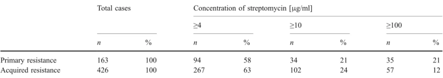
Canetti categorized streptomycin resistance into low-level (≥4 μg/ml), intermediate-low-level (≥10 μg/ml) and high-level (≥100 μg/ml) drug resistance (see Table 3). At this gross level, there was no significant difference between Table 2 Frequency of genotypic alterations in clinicalM. tuberculosis
isolates resistant to streptomycina
Mutations Frequency in clinical isolates Mean relative fitness MIC mg/L rpsL 42 Arg 88% 0.98 >1,000 rrs 523 C 6% 0.94 125 rrs 522 T 3% 0.91 250 Rrs 526 T 2% 0.90 125 rpsL 42 Thr <1% 0.86 >1,000 rpsL 42 Asn <1% 0.85 >1,000
aResistance mutations in rpsL aa position 88 and in rrs positions 501,
912, and 913 are not included; these mutations account for approximately 10% of clinical streptomycin resistant isolates
Fig. 1 Chemical structure of streptomycin, showing interaction of the various groups with specific residues of the Thermus thermophilus small ribosomal subunit; nucleotides correspond to 16S rRNA, Lys 45 (S12) corresponds to ribosomal protein RpsL aa position 42. Figure taken from Ref.13with permission of the publisher
primary and acquired drug resistance with respect to the relative proportions of the different streptomycin resistance levels. In other words, within streptomycin resistant mutants high-level drug resistance mutations must exist which did not impede transmission. For rifampicin, the situation is similar. Different mutations in rpoB result in drug resistance [4, 20, 22, 29]. As with streptomycin, a significant correlation exists between the frequency of a particular mutation in clinical isolates and the fitness cost associated with the mutation (see Table4).
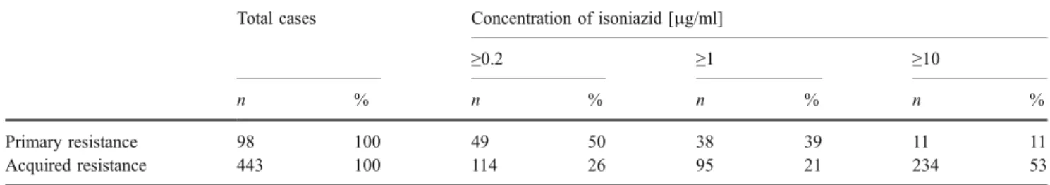
For isoniazid, the situation is different. Compared to acquired resistance, the high-level resistance phenotype is significantly underrepresented in primary resistance (11% versus 53%, see Table5). Apparently, high-level isoniazid resistance is associated with a significant fitness burden which impedes transmission. Multiple chromosomal alter-ations in katG may result in resistance to isoniazid. It was demonstrated some 50 years ago that high-level resistance to isoniazid—as conferred by deletion of katG, i.e. complete loss of katalase-peroxidase activity—is associated with a significant fitness cost [14]. More recently, it was shown that complete loss of KatG activity in clinical isoniazid resistant strains is associated with a secondary mutation resulting in over-expression of the alkyl-hydro-peroxide-reductase AhpC [39]. It was hypothesized that in strains with isoniazid resistance due to deletion of katG, compensatory mutations in ahpC will develop over time, ultimately facilitating transmission and spread of resistant microorganisms. The most frequent isoniazid resistance mutation found in clinical strains, however, is not a nonsense mutation, but a serine to threonine replacement at aa position 315 of KatG [48, 49]. This particular mutation confers an intermediate level of resistance and is not associated with a fitness cost [35]. The 315 Ser→Thr mutation is found in approximately 60% of clinical strains with isoniazid resistance. In the absence of KatG/isoniazid crystal complexes, modelling and computational studies have been used to understand drug–target interaction [3]. These and other studies suggest [25] that a 315 Ser→ Thr mutation in the KatG catalase–peroxidase would alter the binding site for isoniazid but retain the active site properties for proper catalytic function.
Available evidence suggests that within a spectrum of possible mutational resistance alterations each being asso-ciated with a distinct fitness cost, a selection for those resistance mutations with the least resistance-associated cost seems to exist in vivo [4, 8, 29, 35, 38, 49]. This selection is best explained by fluctuating environments, i.e. expansion of mutants experiencing a low fitness cost in the absence of antibiotics during periods in which selection for antibiotic resistance is removed. The rare finding of high cost resistance mutations in clinical isolates can be explained by the stochastic probability of a resistance mutation in a size limited bacterial population and by bottleneck phenomena which take place in transmission. It is under such conditions that compensatory mutations which ameliorate the cost of resistance are likely to occur [38].
A priori, there is no need for compensatory evolution in maintaining persistence and further spread of drug resis-tance, as resistance mutations exist which carry little or no fitness cost at all. As long as for a given drug different resistance mutations exist with only one of these being a no-cost resistance mutation, the stochastic probability of selection for this particular mutation in a given population is much higher than the probability of two mutations occurring either simultaneously or successively with one compensating for disadvantages conferred by the other. The frequent presence of and selection for drug resistance mutations which carry no or only a low fitness cost, also indicates that drug persistence per se can not be expected to restrict transmission of tuberculosis disease.
Table 3 Primary resistance and acquired resistance to streptomycin
Total cases Concentration of streptomycin [μg/ml]
≥4 ≥10 ≥100
n % n % n % n %
Primary resistance 163 100 94 58 34 21 35 21
Acquired resistance 426 100 267 63 102 24 57 12
Table 4 Frequency of rpoB mutations in clinical M. tuberculosis isolates resistant to rifampicina
RpoB mutation Frequency in clinical isolates Mean relative fitness MIC mg/l S 531 L 54% 0.93 >32 H 526 Y 11% 0.82 >32 H 526 D 7% 0.78 >32 S 531 W 4% 0.79 >32 H 526 R 3% 0.56 >32 S 522 L 1% 0.54 16–32 a
Limitations of the DOTS strategy
In 1994, the WHO declared tuberculosis a global emergen-cy and introduced the DOTS strategy for global tuberculo-sis control. However, the plan has produced variable success and despite intensified efforts to diagnose and treat tuberculosis, the rates continue to climb in some regions. Although various experts have argued that the failure of the DOTS strategy to control tuberculosis resulted from failed implementation, poor public health infrastructure, poverty, and the like, recent reports indicate that the problem with the DOTS strategy may be a more principal one [45,53]. For example, tuberculosis rates were found to increase de-spite implementation of DOTS [27]. The DOTS strategy is built around five activities: (1) case detection by sputum smear microscopy among symptomatic patients self-reporting to health services, (2) directly observed therapy using stan-dard short-course regimens, (3) regular supply of medication, (4) governmental commitment to sustained tuberculosis control by providing resources and infrastructural capacity, and (5) a standardized recording and reporting system that allows assessment of individual treatment results and of the tuberculosis control program overall.
The reproductive rate of an epidemic is characterized by three determinants: (1) the duration of infectiousness of an infected patient, (2) the number of contacts between an in-fectious patient and susceptible contacts, and (3) the prob-ability of the infected to become infectious. As a treatment program, DOTS in part targets the duration of infectious-ness. However, DOTS does not address the number of contacts between an infectious patient and susceptible contacts before diagnosis on the basis of passive case-finding [44], nor does it address the probability of the infected to become infectious. It has been estimated that approximately one tenth of those infected develop active disease and half of those with active disease become themselves infectious [41]. Thus, it would be necessary for one tuberculosis case to infect approximately twenty susceptible contacts in order to produce another infectious case and to maintain the same level of infection in the population. Any measure which decreases this number will result in a decline of tuberculosis rates. Two simple calculations may serve to illustrate the limitations of a
mere treatment program based on passive case finding. First, a patient, prior to detection on the basis of passive case finding, infects more than 20 susceptible contacts before transmission is interrupted by treatment. This is a scenario which is quite conceivable under the living conditions present in many of the world’s underdeveloped areas. Second, rather than 10%, a significantly larger fraction of the infected develops active disease, due to either inherent genetic (disease susceptibility and genetic polymorphisms), societal (poverty and starvation), or environmental reasons, e.g. coinfection with HIV. The latter is particularly worrisome due to the widespread nature of the AIDS epidemic and its association with a high risk of developing progressive primary tuberculosis. Under these circumstances, the DOTS program will have significant shortcomings in the global control of TB.
More data are needed to estimate our case finding ability in various settings. Available data indicate that in high burden environments it may be quite low, e.g. in South Africa. In that case, no strategy will reduce incidence, unless case finding ability is enhanced [45]. The current WHO recommendations form an excellent base for tuber-culosis control, but may need to be complemented by a strategy which reflects the biology of the disease by considering determinants which influence the reproductive rate of an epidemic. The DOTS program as a treatment program is ideally combined with a component which actively targets transmission, designed to interrupt the spread of M. tuberculosis in the community. Such a component could be the implementation of contact tracing and active case finding. Can we afford to do it, or rather, can we afford to ignore it?
Acknowledgements We apologize to all colleagues whose work could not be cited due to space limitations. We are indebted to our coworkers and collaborators with whom we had the privilege to share ideas and views. We are thankful to Paul van Helden for thoughtful and critical discussions. We thank A. Makovec for typing the manuscript. Work in the authors laboratories has been supported by the German Research Council, the Bundesministerium für Forschung und Technologie (Germany), the European Commission, the Swiss National Research Foundation, the Bundesamt für Gesundheit (Switzerland), the University of Zurich, and the Niedersächsischer Verein zur Bekämpfung der Tuberkulose (Germany).
Table 5 Primary resistance and acquired resistance to isoniazid
Total cases Concentration of isoniazid [μg/ml]
≥0.2 ≥1 ≥10
n % n % n % n %
Primary resistance 98 100 49 50 38 39 11 11
References
1. Andersson DI, Levin BR (1999) The biological cost of antibiotic resistance. Curr Opin Microbiol 2:489–493
2. Belanger AE, Besra GS, Ford ME, Mikosova K, Belsle JR, Brennan PJ, Inamine JM (1996) The embAB genes of Mycobac-terium avium encode an arabinosyl transferase involved in cell wall arabinan biosynthesis that is the target for the antimycobac-terial drug ethambutol. Proc Natl Acad Sci USA 93:11919–11924 3. Bertrand T, Eady NA, Jones JN, Jesmin, Nagy JM, Jamart-Grégoire B, Raven EL, Brown KA (2004) Crystal structure of Mycobacterium tuberculosis catalase-peroxidase. J Biol Chem 279:38991–38999
4. Billington OJ, McHugh TD, Gillespie SH (1999) Physiological cost of rifampin resistance induced in vitro in Mycobacterium tuberculosis. Antimicrob Agents Chemother 46:1866–1869 5. Bjorkman J, Nagaev I, Berg OG, Hughes D, Andersson DI (2000)
Effects of environment on compensatory mutations to ameliorate costs of antibiotic resistance. Science 287:1479–1482
6. Böttger EC (2001) Drug resistant tuberculosis. Lancet 357:1288– 1289
7. Böttger EC, Pletschette M, Andersson D (2005) Drug resistance and fitness in Mycobacterium tuberculosis infection. J Infect Dis 191:823–824
8. Böttger EC, Springer B, Pletschette M, Sander P (1998) Fitness of antibiotic-resistant microorganisms and compensatory mutations. Nat Med 4:1343–1344
9. British Thoracic Association (1982) A controlled trial of six month chemotherapy in pulmonary tuberculosis: second report results during the 24 months after the end of chemotherapy. Am Rev Respir Dis 126:460–462
10. Canetti G (1965) Present aspects of bacterial resistance in tuberculosis. Am Rev Respir Dis 92:687–703
11. Canetti G, Fox W, Khomenko A, Mahler HT, Menon NK, Mitchison DA, Rist N, Šmelev NA (1969) Advances in techniques of testing mycobacterial drug sensitivity, and the use of sensitivity tests in tuberculosis control programmes. Bull World Health Organ 41:21–43
12. Canetti G, Froman S, Grosset J, Hauduroy P, Langerová M, Mahler HT, Meissner G, Mitchison DH,Šula L (1963) Mycobac-teria: laboratory methods for testing drug sensitivity and resis-tance. Bull World Health Organ 29:565–578
13. Carter AP, Clemons WM, Brodersen DE, Morgan-Warren RJ, Wimberly BT, Ramakrishnan V (2000) Functional insights from the structure of the 30S ribosomal subunit and its interactions with antibiotics. Nature 407:340–348
14. Cohn ML, Kovitz C, Oda U, Middlebrook G (1954) Studies on isoniazid and tubercle bacilli. II. The growth requirements, catalase activities, and pathogenic properties of isoniazid-resistant mutants. Am Rev Tuberc 70:641–664
15. Cynamon MH, Zhang Y, Harpster T, Cheng S, DeStefano MS (1999) High-dose isoniazid therapy for isoniazid-resistant murine Mycobac-terium tuberculosis infection. Antimicrob Agents Chemother 43:2922–2924
16. Corbett EL, Watt CJ, Walker N, Maher D, Williams BG, Raviglione MC, Dye C (2003)The growing burden of tuberculo-sis: global trends and interactions with the HIV epidemic. Arch Intern Med 163:1009–1021
17. DeRiemer K, García-García L, Bobadilla-del-Valle M, Palacios-Martínez M, Palacios-Martínez-Gamboa A, Small PM, Sifuentes-Osornio J, Ponce-de-León A (2005) Does DOTS work in populations with drug-resistant tuberculosis? Lancet 365:1239–1245
18. Dye C, Espinal MA, Watt CJ, Mbiaga C, William BG (2002) Worldwide incidence of multidrug-resistant tuberculosis. J Infect Dis 185:1197–1202
19. Espinal MA, Kim SI, Suarez PG, Kam KM, Khomenko AG, Migliori GB, Baez J, Kochi A, Dye C, Raviglione MC (2000) Standard short-course chemotherapy for drug-resistant tuberculo-sis: treatment outcome in 6 countries. JAMA 283:2537–2545 20. Gagneux S, Long CD, Small PM, Van T, Schoolnik GK, Bohannan
BJM (2006) The competitive cost of antibiotic resistance in Mycobacterium tuberculosis. Science 312:1944–1946
21. Hazbón MH, Bobadilla-del-Valle M, Guerrero MI, Varma-Basil M, Filliol IN, Cavatore M, Colangeli R, Safi H, Billman-Jacobe H, Lavender C, Fyfe J, García-García L, Davidow A, Brimacome M, León CI, Porras T, Bose M, Chaves F, Eisenach KD, Sifuentes-Osornio J, Ponce-de-León A, Cave MD, Alland D (2005) Role of embB codon 306 mutations in Mycobacterium tuberculosis revisited: a novel association with broad drug resistance and IS6110 clustering rather than ethambutol resistance. Antimicrob Agents Chemother 49:3794–3802
22. Huitric E, Werngren J, Juréen P, Hoffner S (2006) Resistance levels and rpoB gene mutations among in vitro-selected rifampin-resistant Mycobacterium tuberculosis mutants. Antimicrob Agents Chemother 50:2860–2862
23. Inderlied CB, Salfinger M (1995) Antimicrobial agents and susceptibility tests: mycobacteria. In: Murray PR, Baron EJ, Pfaller MA, Tenover FC, Yolken RH (eds) Manual of clinical microbiology. American Society for Microbiology, Washington, DC, pp 1385–1404
24. Johnson R, Jordaan AM, Pretorius L, Engelke E, van der Spuy G, Bosman M, van Helden PD, Warren R, Victor TC (2006) Ethambutol resistance testing by mutation detection. Int J Tuberc Lung Dis 10:68–73
25. Kapetanaki SM, Chouchane S, Yu S, Zhao X, Magliozoo RS, Schelvis JP (2005) Mycobacterium tuberculosis KatG (S315T) catalase-peroxidase retains all active site properties for proper catalytic function. Biochemistry 44:243–252
26. Kurland CG, Hughes D, Ehrenberg M (1996) Limitations of translational accuracy. In: Neidhardt FC (ed) Escherichia coli and Salmonella: cellular and molecular biology, 2nd edn. ASM Press, Washington, DC, pp 979–1004
27. Lawn SD, Bekker LG, Middelkoop K, Myer L, Wood R (2006) Impact of HIV infection on the epidemiology of tuberculosis in a peri-urban community in South Africa: the need for age-specific interventions. Clin Infect Dis 42:1040–1047
28. Madison B, Robinson-Dunn B, George I, Gross W, Lipman H, Metchuck B, Sloctsky A, Washabange G, Mazurek G, Riddershof J (2002) Multicenter evaluation of ethambutol susceptibility testing of Mycobacterium tuberculosis by agar proportion and radiometric methods. J Clin Microbiol 40:3976–3979
29. Mariam DH, Mengistu Y, Hoffner SE, Andersson DI (2004) Effect of rpoB mutations conferring rifampin resistance on fitness of Mycobacterium tuberculosis. Antimicrob Agents Chemother 48:1289–1294
30. Medical Research Council Investigation (1950) Treatment of pulmonary tuberculosis with streptomycin and para-amino sali-cylic acid. Br Med J 2:1073–1085
31. Meier A, Sander P, Schaper KJ, Scholz M, Böttger EC (1996) Correlation of molecular resistance mechanism and phenotypic resistance level in streptomycin-resistant M. tuberculosis. Antimicrob Agents Chemother 40:2452–2454
32. Mitchinson DA (1985) Mechanisms of the action of drugs in short-course chemotherapy. Bull Int Union Tuberc 60:36–40 33. Mitnick C, Bayona J, Palacios E, Shin S, Furin J, Alcántara F,
Sánchez E, Sarria M, Becerra M, Smith Fawzi MC, Kapiga S, Neuberg D, Maguire JH, Yong Kim J, Farmer P (2003) Com-munity-based therapy for multidrug-resistant tuberculosis in Lima, Peru. N Engl J Med 348:119–128
34. Mokrousov I, Otten T, Vyshnevskiy B, Narvskaya O (2002) Detection of embB306 mutations in ethambutol-susceptible
clinical isolates of Mycobacterium tuberculosis from Northwest-ern Russia: implications for genotypic resistance testing. J Clin Microbiol 40:3810–3813
35. Pym AS, Saint-Joanis B, Cole ST (2002) Effect of katG mutations on the virulence of Mycobacterium tuberculosis and the implication for transmission in humans. Infect Immun 70: 4955–4960 36. Ramaswamy S, Musser JM (1998) Molecular genetic basis of
antimicrobial agent resistance in Mycobacterium tuberculosis: 1998 update. Tuber Lung Dis 79:3–29
37. Sander P, Böttger EC (1999) Mycobacteria: genetics of resistance and implications for treatment. Chemotherapy 45:95–108 38. Sander P, Springer B, Prammananan T, Sturnfels A, Kappler M,
Pletschette M, Böttger EC (2002) Fitness cost of chromosomal drug resistance-conferring mutations. Antimicrob Agents Chemother 46:1204–1211
39. Sherman DR, Mdluli K, Hickey MJ, Arain TM, Morris SL, Barry CE, Stover CK (1996) Compensatory ahpC gene expression in isoniazid-resistant Mycobacterium tuberculosis. Science 272:1641–1643 40. Somoskovi A, Dormandy J, Mitsani D, Rivenburg J, Salfinger M
(2006) Use of smear-positive samples to assess the PCR-based genotype MTBDR assay for rapid, direct detection of the Mycobacterium tuberculosis complex as well as its resistance to isoniazid and rifampin. J Clin Microbiol 44:4459–4463
41. Styblo K (1980) Recent advances in epidemiological research in tuberculosis. Adv Tuberc Res 20:1–63
42. NCCLS (2003) Susceptibility testing of mycobacteria, nocardiae, and other aerobic actinomycetes. Approved standard NCCLS document M24-A (ISBN-1-56238-500-3). NCCLS, Wayne, Pennsylvania, USA 43. Telenti A, Philipp WJ, Screevatsan S, Bernasconi C, Stockbauer KE, Wieles B, Musser JM, Jacobs WR Jr (1997) The emb operon, a gene cluster of Mycobacterium tuberculosis involved in resistance to ethambutol. Nat Med 3:567–570
44. Uys PW, Warren RM, van Helden PD (2007) A threshold value for the time delay to TB diagnosis. PLoSONE 2(8):e757 45. van Helden PD, Donald PR, Victor TC, Schaaf HS, Hoal EG, Walzl
G, Warren RM (2006) Antimicrobial resistance in tuberculosis: an international perspective. Expert Rev Anti-Infect Ther 4:759–766 46. van Rie A, Warren R, Richardson M, Victor TC, Gie RP, Enarson
DA, Beyers N, van Helden PD (1999) Exogenous reinfection as a
cause of recurrent tuberculosis after curative treatment. N Engl J Med 341:1174–1179
47. van Rie A, Warren R, Richardson M, Gie RP, Enarson DA, Beyers N, van Helden PD (2000) Classification of drug-resistant tuber-culosis in an epidemic area. Lancet 356:22–25
48. van Soolingen D, Borgdorff MW, de Haas PE, Sebek MM, Veen J, Dessens M, Kremer K, van Embden JD (1999) Molecular epidemiology of tuberculosis in the Netherlands: a nationwide study from 1993 through 1997. J Infect Dis 180:726–736 49. van Soolingen D, de Haas PE, van Doorn HR, Kuijper E, Rinder
H, Borgdorff MW (2000) Mutations at amino acid position 315 of the katG gene are associated with high-level resistance to isoniazid, other drug resistance, and successful transmission of Mycobacterium tuberculosis in the Netherlands. J Infect Dis 182:1788–1790
50. Verver S, Warren RM, Beyers N, Richardson M, van der Spuy GD, Borgdorff MW, Enarson DA, Behr MA, van Helden PD (2005) Rate of reinfection tuberculosis after successful treatment is higher than rate of new tuberculosis. Am J Respir Crit Care Med 171:1430–1435
51. Victor TC, Warren R, Butt JL, Jordaan AM, Felix JV, Venter A, Sirgel FA, Schaaf HS, Donald PR, Richardson M, Cynamon MH, van Helden PD (1997) Genome and MIC stability in Mycobac-terium tuberculosis and indications for continuation of use of isoniazid in multidrug-resistant tuberculosis. J Med Microbiol 46:847–857
52. Wang F, Langley R, Gulten G, Dover LG, Besra GS, Jacobs WR Jr, Sacchettini JC (2007) Mechanism of thioamide drug action against tuberculosis and leprosy. J Exp Med 204:73–78
53. Whalen CC (2006) Failure of directly observed treatment for tuberculosis in Africa: a call for new approaches. Clin Infect Dis 42:1048–1050
54. WHO Global Tuberculosis Programme (2002) An expanded DOTS framework for effective tuberculosis control. WHO/CDS/ TB/2002.297. World Health Organization, Geneva
55. WHO/IUATLO Global Project on Anti-Tuberculosis Drug Resis-tance Surveillance (2004) Anti-tuberculosis drug resisResis-tance in the world: third global report. WHO/HTM/TB/2004.343. World Health Organization, Geneva
![Table 1 Mycobacterial drug susceptibility testing—the critical con- con-centration Antimicrobial agent MIC [mg/l] ofsusceptible M](https://thumb-eu.123doks.com/thumbv2/123doknet/14846032.627398/2.892.458.816.886.1071/table-mycobacterial-susceptibility-testing-critical-centration-antimicrobial-ofsusceptible.webp)