HAL Id: hal-01604739
https://hal.archives-ouvertes.fr/hal-01604739
Submitted on 26 May 2020
HAL is a multi-disciplinary open access
archive for the deposit and dissemination of
sci-entific research documents, whether they are
pub-lished or not. The documents may come from
teaching and research institutions in France or
abroad, or from public or private research centers.
L’archive ouverte pluridisciplinaire HAL, est
destinée au dépôt et à la diffusion de documents
scientifiques de niveau recherche, publiés ou non,
émanant des établissements d’enseignement et de
recherche français ou étrangers, des laboratoires
publics ou privés.
Copyright
Protectin D1n-3 DPA and resolvin D5n-3 DPA are
effectors of intestinal protection
Thomas Gobbetti, Jesmond Dalli, Romain A. Colas, Donata Federici Canova,
Marius Aursnes, Delphine Bonnet, Laurent Alric, Nathalie Vergnolle, Céline
Deraison Manuel, Trond V. Hansen, et al.
To cite this version:
Thomas Gobbetti, Jesmond Dalli, Romain A. Colas, Donata Federici Canova, Marius Aursnes, et al..
Protectin D1n-3 DPA and resolvin D5n-3 DPA are effectors of intestinal protection. Proceedings of
the National Academy of Sciences of the United States of America , National Academy of Sciences,
2017, 114 (15), pp.3963-3968. �10.1073/pnas.1617290114�. �hal-01604739�
Protectin D1
n-3 DPA
and resolvin D5
n-3 DPA
are effectors
of intestinal protection
Thomas Gobbettia, Jesmond Dallia,b, Romain A. Colasa,b, Donata Federici Canovaa, Marius Aursnesc, Delphine Bonnetd, Laurent Alricd, Nathalie Vergnollee,f,g,h,i, Celine Deraisone,f,g,h,i, Trond V. Hansenc, Charles N. Serhanb,
and Mauro Perrettia,1
aThe William Harvey Research Institute, Barts and The London School of Medicine, Queen Mary University of London, London EC1M 6BQ, United Kingdom; bCenter for Experimental Therapeutics and Reperfusion Injury, Department of Anesthesiology, Perioperative, and Pain Medicine, Brigham and Women’s
Hospital and Harvard Medical School, Boston MA 02115;cDepartment of Pharmaceutical Chemistry, School of Pharmacy, University of Oslo, N-0316 Oslo,
Norway;dDepartment of Internal Medicine and Digestive Diseases, Pole Digestif, Centre Hospitalier Universitaire (CHU), 31059 Toulouse, France;eInstitut
de Recherche en Santé Digestive (IRSD), Université de Toulouse, 31300 Toulouse, France;fUnit 1220, INSERM, 31300 Toulouse, France;gUnit 1416, Institut
National de la Recherche Agronomique (INRA), 31300 Toulouse, France;hÉcole Nationale Vétérinaire de Toulouse (ENVT), 31300 Toulouse, France;
andiUniversité Paul Sabatier (UPS), 31300 Toulouse, France
Edited by Ruslan Medzhitov, Yale University School of Medicine, New Haven, CT, and approved February 28, 2017 (received for review October 18, 2016)
The resolution of inflammation is an active process orchestrated by specialized proresolving lipid mediators (SPM) that limit the host response within the affected tissue; failure of effective resolution may lead to tissue injury. Because persistence of inflammatory signals is a main feature of chronic inflammatory conditions, in-cluding inflammatory bowel diseases (IBDs), herein we investigate expression and functions of SPM in intestinal inflammation. Targeted liquid chromatography-tandem mass spectrometry-based metabolo-lipidomics was used to identify SPMs from n-3 polyunsaturated fatty acids in human IBD colon biopsies, quantifying a significant up-regulation of the resolvin and protectin pathway compared with normal gut tissue. Systemic treatment with protectin (PD)1n-3 DPA
or resolvin (Rv)D5n-3 DPA protected against colitis and intestinal
ischemia/reperfusion-induced inflammation in mice. Inhibition of 15-lipoxygenase activity reduced PD1n-3 DPAand augmented
intesti-nal inflammation in experimental colitis. Intravital microscopy of mouse mesenteric venules demonstrated that PD1n-3 DPA and
RvD5n-3 DPAdecreased the extent of leukocyte adhesion and
emi-gration following ischemia-reperfusion. These data were translated by assessing human neutrophil–endothelial interactions under flow: PD1n-3 DPA and RvD5n-3 DPA reduced cell adhesion onto
TNF-α–activated human endothelial monolayers. In conclusion, we pro-pose that innovative therapies based on n-3 DPA-derived mediators could be developed to enable antiinflammatory and tissue protec-tive effects in inflammatory pathologies of the gut.
inflammation resolution
|
specialized proresolving mediators|
omega-3 fatty acids|
intravital microscopy|
neutrophilsI
nflammatory bowel diseases (IBDs), the main forms being Crohn’s disease and ulcerative colitis, are chronic inflam-matory conditions of unknown etiology that primarily affect the gastrointestinal tract (1). IBD affects ∼1.5 million Americans,>2 million people in Europe, and several hundred thousands more worldwide (2). Irrespective of its etiopatho-genesis, the chronic nature of IBDs is caused by inflammatory signals (i.e., cytokines, proteases) leading to overt pathology (1). Healthy intestinal mucosa rely on a complex equilibrium: when this is disrupted a robust and persistent inflammatory response develops characterized by mucosal injury, increased epithelial permeability, invasion of bacteria into lamina propria, and marked neutrophil recruitment (1). This paradigm has driven research in the field informing current treatments, including bi-ologics like anti–TNF-α. However, these treatments are immu-nosuppressive (hence plagued by side-effects), expensive, and ineffective in a good proportion of patients (3). The increasing incidence of IBD and the inadequacy of current treatments makes imperative to develop new therapeutic approaches.The ultimate remit of the inflammatory response is to protect the host from exogenous or endogenous dangers: following
removal of the noxious stimuli, the inflammatory process is finely programmed to self-resolve to avoid harmful injury (4, 5). The last step of this physiological multicomponent process is tissue repair, with return to tissue functionality, instructing the adaptive immune response and altogether regaining homeostasis (6, 7). However, if not modulated inflammatory mechanisms induce self-harm, leading to tissue injury, as in chronic diseases including IBDs (8, 9). This beneficial profile of the inflammatory response is ensured with the turning on of resolution programs and the biosynthesis of proresolving mediators that counter-regulate the action of inflammation-initiating mediators (10). Within the effectors of resolution, an important role is emerging for specialized pro-resolving lipid mediators (SPMs): these are families of mediators formed via the stereospecific conversion of essential poly-unsaturated fatty acids (PUFAs) by enzymes, including the lipoxygenases (10). During inflammation, increased local vas-cular permeability leads to edema, which supplies PUFAs from blood to the site of injury, together with elevated numbers of immune cells that carry the biosynthetic enzymes required for the local SPM production (11). These mediators, in turn, in-teract with specific receptors to temper leukocyte reactivity,
Significance
We provide evidence for a functional role of bioactive lipid medi-ators of the docosapentaenoic acid (DPA) metabolome in intestinal inflammation. Supported by changes in DPA-derived mediators in colon biopsies from inflammatory bowel diseases, we studied the pharmacological properties of two mediators. Exogenous adminis-tration of protectin (PD)1n-3 DPAor resolvin (Rv)D5n-3 DPAin mice reduced dextran sulfate sodium-induced colitis though a mecha-nism partly linked to decreased leukocyte–endothelial interaction and reduced granulocyte trafficking, as assessed by intravital microscopy. The translational impact of these data was determined by the ability of PD1n-3 DPAand RvD5n-3 DPA to reduce human neutrophil adhesion onto TNF-α–activated human endothelial monolayers. We propose that n-3 DPA-derived mediators could represent the basis for innovative therapeutic strategies in set-tings of intestinal inflammation.
Author contributions: T.G., J.D., C.N.S., and M.P. designed research; T.G., R.A.C., and D.F.C. performed research; M.A., D.B., L.A., C.D., and T.V.H. contributed new reagents/analytic tools; T.G., J.D., R.A.C., and D.F.C. analyzed data; and T.G., J.D., N.V., C.N.S., and M.P. wrote the paper.
The authors declare no conflict of interest. This article is a PNAS Direct Submission.
Freely available online through the PNAS open access option.
1To whom correspondence should be addressed. Email: m.perretti@qmul.ac.uk.
This article contains supporting information online atwww.pnas.org/lookup/suppl/doi:10. 1073/pnas.1617290114/-/DCSupplemental. IMMUNOLO GY AND INFLAM MATION
dampen inflammatory pain, and promote tissue repair and re-generation (12).
Recently, we reported that n-3 docosapentaenoic acid (n-3 DPA), an intermediary product between eicosapentaenoic acid (EPA) and docosahexaenoic acid (DHA), is converted to novel SPMs by both human and murine leukocytes (13). Production of these novel SPMs was temporally regulated in self-limited in-flammatory exudates. In addition, n-3 DPA-derived 10R,17S-dihydroxydocosa-7Z,11E,13E,15Z,19Z-pentaenoic acid (PD1n-3 DPA)
and 7R,14S-dihydroxydocosa-8E,10E,12Z,16Z,19Z-pentaenoic acid (MaR1n-3 DPA) exerted potent proresolving actions stimulating
hu-man macrophage phagocytosis and efferocytosis (13, 14). Herein we identified the presence of n-3 DPA-derived SPM in human colon and characterized their antiinflammatory and tissue-protective ac-tions of two DPA-derived mediators, denoted as PD1n-3 DPAand
RvD5n-3 DPA
(7S,17S-dihydroxydocosa-8E,10Z,13Z,15E,19Z-pentaenoic acid) in settings of intestinal inflammation.
Results
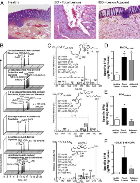
Identification of n-3 DPA Proresolving Mediators in Colon Biopsy from Control and IBD Patients as Well as Mice.Using targeted LC-MS/ MS based lipid mediator (LM) metabololipidomics, we profiled the arachidonic acid (AA), EPA, DHA, and n-3 DPA metab-olomes with human intestinal biopsies from control and IBD pa-tients (seeTable S1for demographics). In the presence of overt inflammation, as typified by marked damage of the mucosal ar-chitecture displayed by biopsies of IBD colon samples (Fig. 1A), levels of leukotriene (LT) B4, prostaglandin (PG) E2, and
thromboxane (TX) B2(further metabolite of the potent
platelet-agonist TXA2) were significantly increased compared with tissue
biopsies from control. In these biopsies we also identified and quantified SPM from all four of the major bioactive metabolomes (Table S2). Of note, we identified mediators from the n-3 DPA bioactive metabolome in both biopsies from control and IBD patients. These mediators were identified in accordance with
Fig. 1. Altered LM-SPM profiles in human colon biopsies from IBD patients. Colon biopsies were obtained from control and IBD patients (seeTable S1for demographic information). (A) H&E staining of human colon biopsies. (Magnification: 20×.) (B and C) SPM profiles were obtained using LC-MS/MS based LM profiling. (B) Multiple reaction monitoring (MRM) chromatograms for identified mediators. (C) Representative MS/MS spectra used for the identification of n-3 DPA resolvin D5 (RvD5n-3 DPA, 7S,17S-dihydroxydocosa–8E,10Z,13Z,15E,19Z-pentaenoic acid), n-3 DPA protectin D1 (PD1n-3 DPA,
10R,17S-dihydroxydocosa-7Z,11E,13E,15Z,19Z-pentaenoic acid), and 15-epi-Lipoxin A4 (5S,6R,15R-trihydroxyeicosa-7E,9E,11Z,13E-tetraenoic acid). Results are representative of n=
30 colon biopsies (Table S2). (D–F) Quantification of RvD5n-3 DPA, PD1n-3 DPAand 10S,17S-diHDHA in the tissue samples. *P< 0.05 vs. healthy tissue.
published criteria, matching retention times and at least six characteristic ions in the MS/MS spectrum, and included n-3 DPA resolvins (RvD1n-3 DPA, RvD2n-3 DPA, RvD5n-3 DPA), protectins
(PD1n-3 DPA, 10S,17S-diHDPA), and maresins (MaR1n-3 DPA)
(Fig. 1B and C andTable S2). Quantification using multiple re-action monitoring demonstrated that RvD5n-3 DPA,PD1n-3 DPA,
and the protectin pathway marker 10S,17S-diHDPA were aug-mented in tissue biopsies from IBD patients compared with those from control (Fig. 1D–F). Taken together, these findings identify SPM, including n-3 DPA-derived SPM in human colonic tissue. In addition, these results indicate an up-regulation of the resolvin and protectin pathways from the n-3 DPA metabolome in IBD.
We next tested if inhibition of endogenous SPM production would impact disease activity. Using targeted LC-MS/MS–based LM metabololipidomics, we profiled the AA, EPA, DHA, and n-3 DPA bioactive metabolomes in colon tissues from naive mice, mice receiving dextran sulfate sodium (DSS), with or without a 15-LOX inhibitor treatment. In colon tissues from naive mice, we identified mediators from all four bioactive metabolomes, in-cluding the n-3 DPA-derived PD1n-3 DPA(Fig. 2 A and B and
Table S3). Administration of DSS leads to up-regulation of several protective pathways, including both DHA and n-3 DPA-derived protectins, as well as the E-series resolvins and the DHA-derived RvD5, suggesting the activation of host-protective mechanisms during ongoing inflammation. Administration of a 15-LOX in-hibitor reduced tissue concentrations of PD1n-3 DPAand increased
levels of several proinflammatory eicosanoids, including PGD2and
PGE2(Table S3). Modulation of SPM profiles was associated with a
decreased colon length, increased macroscopic tissue damage, and neutrophil infiltration (Fig. 2B–D). These results suggest that inhibition of endogenous PD1n-3 DPAleads to a failure to
counter-regulate colonic inflammation, leading to increased tissue damage.
PD1n-3 DPAand RvD5n-3 DPAProtect Mice Against DSS-Induced Colitis.
Given that the enzymatic inhibition of n-3 DPA-derived SPMs is deleterious in colitis and that levels of these mediators are regu-lated in human colon biopsies from IBD patients (Figs. 1 and 2 and
Tables S2andS3), we tested the pharmacological actions of PD1n-3 DPAand RvD5n-3 DPAin DSS-induced colitis (Fig. 3A). Five days
of oral DSS administration to mice induced severe colon in-flammation, evident through significant colon shortening, increased wall thickness, and macroscopic damage score (Fig. 3B). Systemic treatment with PD1n-3 DPA or RvD5n-3 DPA (0.3 μg per mouse
daily) prevented colon length reduction. Treatment with PD1n-3 DPA
significantly reduced colon wall thickness and macroscopic colon damage (Fig. 3B). In DSS-induced intestinal inflammation, tis-sue damage is predominantly mediated by infiltrating neutro-phils (15); thus, we assessed the actions of PD1n-3 DPA and
RvD5n-3 DPAon this parameter by measuring tissue
myeloper-oxidase (MPO) activity (Fig. 3C). Against a marked increase in colonic MPO activity, animals receiving PD1n-3 DPA orRvD5n-3 DPA
displayed reduced MPO values (Fig. 3C). Analyses of cytokines showed a pronounced increase in colonic TNF-α, IL-1β, and IL-6 levels—with a partial decrease in IL-10—in vehicle-treated DSS mice. PD1n-3 DPA, but not RvD5n-3 DPA, significantly
re-duced proinflammatory cytokines levels without correcting IL-10 (Fig. 3D). Both compounds restored the histological architecture of the mucosa, decreasing mucosal ulceration compared with the DSS-treated vehicle group (Fig. 3E).
We then assessed if PD1n-3 DPAwas also protective when given
using a therapeutic treatment. Mice were given DSS for 7 d, followed by 3 d of water. Treatment with either PD1n-3 DPA
(0.3μg/mouse, i.p.) or vehicle commenced at first signs of disease (day 5, beginning of weight loss): PD1n-3 DPA-treated group
displayed significant protection as measured by reduced colon shortening, decreased macroscopic damage, MPO activity, and inflammatory cytokine levels (Fig. 3F).
PD1n-3 DPAand RvD5n–3 DPAPrevent Local and Systemic Inflammation
Following Intestinal Ischemia/Reperfusion.Given the association for intestinal ischemia in the pathogenesis of IBD, we tested if PD1n3 DPAand RvD5n-3 DPAregulated host responses following gut
ischemia (16). Occlusion of the mesenteric artery (ischemia) fol-lowed by reperfusion provoked a robust granulocyte infiltration in the gut with marked histological damage, as characterized by fragments of mucosa detected in the lumen, damage to the villi, and disintegration of the lamina propria (Fig. 4). Postischemic admin-istration of PD1n-3 DPA (most effective at 0.1μg per mouse) and
RvD5n-3 DPA (0.1–1 μg per mouse) significantly reduced
gran-ulocyte infiltration (Fig. 4A and B). Administration of PD1n-3 DPA
preserved structure and length of the villi, whereas RvD5n-3 DPA
was less effective (Fig. 4C). At the dose of 0.1 μg, PD1n-3 DPA
re-duced secondary granulocyte recruitment into the lungs and plasma IL-1β levels (Fig. 4 D and E).
To establish the impact of endogenous n-3 DPA-derived medi-ators in gastrointestinal protection, we assessed the profiles in an-imals with reduced expression of fatty acid elongase 2 (ELOVL2) (17), the enzyme that converts n-3 DPA to DHA (Fig. S1A). Ad-ministration of siRNA to mice reduced ELOVL2 expression in peripheral tissues (Fig. S1B). Mice were then supplemented daily with EPA and subjected to ischemia/reperfusion (I/R) injury. LM profiling identified that plasma levels of n-3 DPA-derived SPM, including RvD5n-3 DPAand PD1n-3 DPA, were significantly increased
(Fig. S1C–EandTable S4). These increases were associated with reduced systemic levels of LTB4and 12S-HHT when compared
with mice given the mock plasmid alone (Fig. S1F). Of note, MPO levels in ELOVL2 knockdown, but not mock siRNA-treated ani-mals supplemented with EPA, were significantly reduced compared with levels found in control mice (Fig. S1G).
PD1n-3 DPAand RvD5n-3 DPARegulate Neutrophil–Endothelial Interactions.
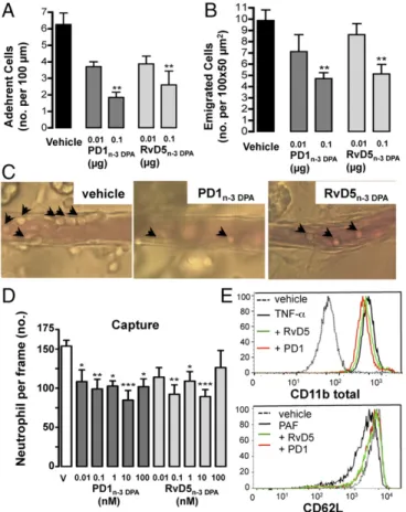
Next, we focused on the process of leukocyte trafficking by using a model of zymosan peritonitis, followed by direct observation of postcapillary venules by intravital microscopy. Administration of PD1n-3 DPA and RvD5n-3 DPA to mice treated with zymosan
markedly reduced leukocyte numbers in the exudates, compared with the vehicle-treated mice. Low doses of RvD5n-3 DPAdecreased
neutrophil counts by >40% (Fig. S2). PD1n-3 DPA was more
ef-fective at the lower dose of 0.1μg.
I/R of the mesenteric microcirculation followed by intravital microscopy allowed monitoring of the processes of leukocyte rolling, adhesion, and emigration (18). The dose of 0.1 μg was selected and administered before reperfusion. Treatment with PD1n-3 DPAor RvD5n-3 DPA decreased the number of adherent
leukocytes onto the postcapillary endothelium (Fig. 5A) but did not affect the intermittent contacts typical of the rolling process
Fig. 2. 15-lipoxygenase inhibition decreased colon PD1n-3 DPAlevels and
increased tissue damage. Mice received PD 146176 (100 mg/kg orally daily). On day 8, tissues were harvested and LM were identified and quantified using LM metabololipidomics. (A) Representative MS/MS spectra used for the iden-tification of PD1n-3 DPA. (B) Colon length, (C) macroscopic damage, and (D)
intestinal MPO activity. Data are means± SEM of six mice per group. *P < 0.05 vs. control.+P< 0.05 vs. DSS vehicle-treated group (V), one-way ANOVA, followed by Dunnett’s post hoc test.
IMMUNOLO
GY
AND
INFLAM
(95± 13% of the response in vehicle-treated animals; not signif-icant). A significant reduction in the extent of leukocytes emi-grated into the subendothelial tissue was also quantified (Fig. 5B). Representative images are in Fig. 5C.
Preincubation of human neutrophils with PD1n-3 DPAor
RvD5n-3 DPA drastically reduced cell capture (Fig. 5D)
com-pounded by an effect on cell adhesion and transmigration onto TNF-α–activated endothelial monolayers (Fig. S3), with activity between 10 pM and 100 nM. No effect was observed on the number of human cell rolling on the endothelium (Fig. S3). These results made us dwell on human neutrophil functions. Incubation of whole blood with TNF-α up-regulated cell surface expression of total CD11b. Addition of PD1n-3 DPAor RvD5n-3 DPAbefore TNF-α led
to a 32± 4% and 28 ± 4% of reduction for total CD11b immu-nostaining (n = 4 donors, P < 0.05) (representative histograms in Fig. 5E). There was some degree of selectivity in these responses because the two mediators did not modulate basal CD62L expres-sion nor TNF-α–induced CD62L shedding (102 ± 6% with respect to appropriate control;n = 4 donors, not significant). This finding is in contrast to what observed with PAF stimulation, with an effect of PD1n-3 DPA or RvD5n-3 DPA on CD62L shedding (∼50 ± 6%
reduction of PAF response for either mediator,n = 4 donors, P < 0.05) (representative histogram in Fig. 5E).
Discussion
In the present study, we report the tissue-protective actions of two n-3 DPA-derived mediators, denoted as PD1n-3 DPA and
RvD5n-3 DPA. These mediators: (i) reduced inflammation following
DSS-induced colitis; (ii) prevented local and systemic inflammation following intestinal I/R; and (iii) regulated neutrophil–endothelial interactions, which may occur downstream modulation of adhesion molecule activation. Of translational relevance, we could identify n-3 DPA SPM in human colon biopsies. These results suggest a relevant profile for these SMPs as an innovative therapeutic ap-proach to control intestinal inflammation like in IBD.
Two decades of research now support the concept that resolu-tion of inflammaresolu-tion is an active process brought about by specific mediators and receptors (12). We studied herein resolution, or its lack, in the persistent and unresolved inflammation typical of IBD (1). Of relevance, the gut is in a state of underlying inflammation and it is plausible that endogenous checkpoints are properly op-erative to avoid progression to chronic conditions (19). Moreover, the relapsing and remitting nature of IBD together with the spontaneous resolution that sometimes is observed (20), suggest existence of contrasting endogenous processes enacted by proin-flammatory and proresolving mediators during the pathogenesis of the disease. SPM are produced by leukocytes, including neutrophils and macrophages, via transcellular biosynthesis and in a cell-autonomous manner (21, 22). Other cell types, including epithe-lial cells (23) and platelets (21), may also contribute to the SPM production because they carry some of the initiating enzymes in the biosynthesis of these molecules. Many of these cell types, including macrophages and epithelial cells, are present in human naïve (healthy) colon biopsies and their numbers are increased in IBD (24). In the present study we found an increase in both proin-flammatory mediators, and a decrease in the proresolving n-3
Fig. 3. PD1n-3 DPAand RvD5n-3 DPAprotect mice against DSS-induced colitis.
(A) Mice had access to drinking water with or without 2.5% DSS for 5 d, then switched to normal water for an additional 3 d before colon collection for analyses (day 8). Animals were treated intraperitoneally with vehicle (100μL PBS 0.01% EtOH) or with 0.3μg PD1n-3 DPAor RvD5n-3 DPAdaily from days 0–5.
Control mice (CTL) received only drinking water. Day 8 analyses: (B) colon length, wall thickness, and macroscopic damage; (C) intestinal MPO activity; (D) cytokines (TNF-α, IL-1β, IL-6, and IL-10) levels as determined in colon ho-mogenates using ELISA. (E) Histological score with representative images of colon sections. (Magnification: 50×.) (F) In another set of experiments, mice received DSS as above, but 0.3μg PD1n-3 DPAwas administered daily from day
5. Day 10 analyses included colon length, macroscopic damage score, MPO activity, and tissue cytokine levels. In all cases, data are means± SEM of six mice per group. *P< 0.05 versus CTL.+P< 0.05,++P< 0.01 vs. DSS
vehicle-treated group, one-way ANOVA, followed by Dunnett’s post hoc test.
Fig. 4. Exogenous administration of PD1n-3 DPAor RvD5n-3 DPAprevents local
and systemic I/R injury. Mice were subjected to intestinal ischemia (30 min) followed by 5-h reperfusion (I/R) or sham operation (S). Animals were given vehicle (100 μL), PD1n-3 DPA, or RvD5n-3 DPA (0.1μg intravenously, unless
otherwise specified) before reperfusion. After 5-h reperfusion, multiple markers were quantified: (A) Gut MPO activity; (B) intestinal histological score on colon sections stained with H&E; (C) representative images of colon sections (magnification: 50×); (D) lung MPO activity; (E) plasma IL-1β levels. Data are mean± SEM of 6–8 mice per group. *P < 0.05, **P < 0.01 vs. sham;+P<
0.05,++P< 0.01 vs. vehicle group, one-way ANOVA, followed by Dunnett’s post hoc test.
PUFA mediators, including PD1n-3 DPA. Of note, levels of these
mediators were within their bioactive concentration (femtomolar to nanomolar) (13, 14), a feature aligned with recent findings where SPM at bioactive concentrations were identified in human tissues including spleen, lymph nodes, plasma (25), and also milk (26). The protective role of endogenous SPM was tested using a 15-lipoxygenase inhibitor that decreased the formation of PD1n-3 DPAand that was
associated with an increase in the production of proinflammatory molecules and colon inflammation.
SPM derived from n-3 PUFA-like DHA and EPA have been linked to protective actions in intestinal inflammation (27). Ad-ministration of resolvin E1, resolvin D2, 17(R)-hydroxy DHA, aspirin-triggered resolvin D1, and Maresin 1 resulted in an im-proved disease activity score, reduced colonic damage, and lower polymorphonuclear infiltration in several models of colitis (27). In
the present study we report that administration of PD1n-3 DPA or
RvD5n-3 DPAwas effective in preventing the hallmark of
intes-tinal inflammation after DSS administration to mice, including an effect on colon length, microscopic damage score, and gran-ulocyte recruitment. These effects could be secondary to the regulation of proinflammatory cytokine production, as in the case of PD1n-3 DPA. The clinical efficacy of anti-TNF therapy in IBD
(3) supports this mechanism. Of interest, both PD1n-3 DPAand
RvD5n-3 DPAdid not regulate colonic levels of IL-10, setting these
mediators apart from LXA4(28) or other proresolving mediators,
like Annexin-A1 (29, 30) andα-melanocyte stimulating hormone (31). To substantiate the physio-pathological impact of this new pathway, it was important to observe that inhibition of the enzyme responsible for conversion of DPA to DHA led to increased tissue levels of n-3 DPA-derived SPM, an effect linked to a reduction in systemic eicosanoid levels during I/R injury, supporting the host protective actions of these molecules.
IBD is characterized by marked infiltration of inflammatory cells and elevated concentration of inflammatory mediators, both within the gut and in the circulation (32). DSS-induced colitis is a useful model to study the early initiating event in IBD, such as the neutrophil recruitment to the intestinal wall and the enhanced production of proinflammatory cytokines that typifies the inflammatory cascade (33, 34). Among the properties of n-3 PUFA-derived mediators (10, 13), PD1n-3 DPAor RvD5n-3 DPA
displayed potent bio-actions that are relevant to the pathophys-iology of IBD, including their ability to regulate neutrophil re-cruitment (vide infra). Each of these mediators also displayed characteristic regulation of inflammatory cytokines whereby PD1n-3 DPAregulated TNF-α, IL- 1β, and IL-6, whereas RvD5n-3 DPA
only partially reduced IL-1β levels. Thus, these findings suggest that these two mediators may activate distinct protective re-sponses potentially through engagement of different receptors.
At 0.1μg (equivalent to ∼90 nmol), PD1n-3 DPAand RvD5n-3 DPA
significantly reduced neutrophil recruitment in zymosan peritonitis and intestinal ischemia reperfusion injury in mice. Because the latter model results in secondary organ injury (35), we could com-plement the pharmacological properties of these two compounds by establishing their ability to confer both intestinal tissue protection and reduce excessive granulocyte recruitment into the lungs, to-gether with decreased level of plasma IL-1β.
The series of molecular and cellular events that lead to neu-trophil recruitment are well characterized (36). Using intravital microscopy of mesenteric venules, we investigated the actions of these n-3 DPA mediators on specific processes within the leuko-cyte recruitment cascade in vivo, finding that both RvD5n-3 DPA
and PD1n-3 DPAspecifically regulated cell adhesion while leaving
cell rolling unaffected. This observation was also replicated with human primary cells, using the flow-chamber system. The potent actions of PD1n-3 DPA and RvD5n-3 DPA on TNF-α–induced
neutrophil recruitment under shear conditions indicate that conversion of n-3 DPA into proresolving mediators can coun-terbalance the actions of proinflammatory mediators. Single-cell analyses with human neutrophils identified at least one mecha-nism by which these mediators could regulate inflammatory responses. Both molecules modulated adhesion molecule ex-pression on the neutrophil cell surface, with the intriguing observation that the specific target (CD11b or CD62L) might be stimulus-dependent.
In conclusion, the present study offers evidence that endoge-nous LMs derived from the n-3 DPA biosynthetic pathway exert beneficial actions on leukocyte reactivity and cytokine pro-duction, regulating the outcome of intestinal inflammation. Thus, PD1n-3 DPAand RvD5n-3 DPAcan be used as guidance for
the development of innovative therapeutic strategies to control the imbalanced inflammatory status typical of IBD and other inflammatory pathologies.
Fig. 5. PD1n-3 DPAand RvD5n-3 DPAreduce neutrophil–endothelial
interac-tions. Mice were subjected to intestinal ischemia (30 min) followed by reper-fusion (90 min) (I/R). Animals were treated intravenously before reperreper-fusion with 0.01 or 0.1μg PD1n-3 DPA, RvD5n-3 DPA, or vehicle (100μL PBS 0.01% EtOH).
Postcapillary venules were imaged and recorded for offline quantitation of white blood cell interaction with the endothelium. (A) Number of adherent cells to the postcapillary venule endothelium and (B) emigrated cells in the subendothelial space. (C) Representative light-microscopy images are shown for vehicle, PD1n-3 DPA, and RvD5n-3 DPA(0.1μg per mice). Black arrows identify
leukocytes adherent to postcapillary venules. (Magnification: 400×.) Data are mean± SEM of six mice per group. **P < 0.01 vs. respective vehicle, one-way ANOVA followed by Dunnett’s post hoc test. (D) Human neutrophils were incubated with vehicle, PD1n-3 DPA, or RvD5n-3 DPA(0.01–100 nM) prior
perfu-sion over TNF-α–stimulated endothelial cell monolayers for 8 min, and the extent of cell capture was quantified. Results are mean± SEM of six distinct cell donors, *P< 0.05, **P < 0.01, ***P < 0.001 vs. vehicle, one-way ANOVA, followed by Dunnett’s post hoc test. (E) Whole blood from healthy volunteers was incubated with vehicle (PBS 0.1% EtOH), PD1n-3 DPA, or RvD5n-3 DPA
(100 nM; indicated as PD1 or RvD5) for 15 min at 37 °C, and then stimulated with TNF-α or PAF. Histograms of CD11b and CD62L expression on human neutrophils are shown (representative from four distinct blood donors).
IMMUNOLO
GY
AND
INFLAM
Materials and Methods
An extended version of materials and methods is presented inSI Materials and Methods. All procedures were performed under the ethical approval both for animal and human cell work. For studies involving mice, all procedures were performed under the United Kingdom Animals (Scientific Procedures) Act, 1986. Human cells were prepared according to an approved protocol (East London & the City Local Research Ethics Committee; no. 06/ Q605/40; P/00/029 ELCHA). The Ethics Committee approved the human research protocol ( Clin-icalTrials.gov Identifier: NCT01990716) to collect biopsies and clinical in-formation. Written informed consent was received from participants before inclusion in the study.
Sample Extraction and LM Metabololipidomics. Human and mouse gastroin-testinal tissues extraction and LM metabololipidomics was conducted as described previously (25).
Induction and Assessment of DSS-Induced Colitis. Mice were provided access ad libitum to water containing 2.5% DSS over a 5-d period. Every other day, the DSS solution was replenished. On day 5, DSS was replaced with normal drinking; on day 8 the animals were killed. Control mice received only drinking water. From days 1–5, RvD5n-3 DPA, PD1n-3 DPA, or vehicle (0.3μg/100 μL in saline
0.01% EtOH) were administered intraperitoneally daily. In other cases, the 15-lipoxygenase inhibitor PD 146176 (Cayman Chemical) was used at 100 mg/kg. For the therapeutic protocol, mice were provided access to water con-taining 2.5% DSS over a 7-d period. On day 7, DSS was replaced with normal drinking water for 3 d. PD1n-3 DPAor vehicle (0.3μg/100 μL in saline 0.01%
EtOH) were administered intraperitoneally daily (from day 5 to day 10). At the end of day 8 or 10, the colon was removed, measured, and examined for the consistency of the stool found within, as well as the gross macroscopic appearance and length, as described previously (37, 38). Samples harvested for histology were analyzed by light microscopy and scored (37, 38). MPO
activity was performed as previously described (39). Intestinal levels of cy-tokines were assayed using ELISA kit. Colonic samples were harvested for metabololipidomics analysis, as previously described (25).
Intestinal I/R Injury. In anesthetized mice, the superior mesenteric artery (SMA) was occluded for 50 min; on reperfusion, PD1n-3 DPA, RvD5n-3 DPA, or
vehicle was given, and tissues collected at 5 h. MPO activity was measured as an index of neutrophil infiltration (39). Tissue sections (5μm) were stained with H&E. Microscopic histological damage score was evaluated blindly (40). Cytokines were assayed by ELISA. Specimens of the jejunum-ileum were also collected for intestinal lipid extraction and LC/MS/MS measurements. In other experiments, mice were transfected with either 40μg mock or ELOVL2 siRNA and given EPA (every other day for 6 d) or vehicle. ELOV2 expression in the liver was determined after 6 d by Western blot analysis. ELOVL2 siRNA or control animals were subjected to 50-min intestinal ischemia followed by a 5-h reperfusion, as described above.
Intravital Microscopy of the Mouse Mesenteric Microcirculation. I/R was per-formed with 30-min ischemia and 90-min reperfusion; intravital microscopy of the mesentery was conducted as described previously (18). One to three postcapillary venules were analyzed for the extent of cell rolling, adhesion, and emigration.
ACKNOWLEDGMENTS. This work was funded by the William Harvey Re-search Foundation (T.G. and M.P.). J.D. received funding from the European Research Council under the European Union’s Horizon 2020 Programme for Research and Innovation (Grant 677542) and a Sir Henry Dale fellowship, jointly funded by the Wellcome Trust and the Royal Society (Grant 107613/Z/ 15/Z). C.N.S. is supported by National Institutes of Health Grant P01GM095467. N.V. is supported by the European Research Council (Grant ERC-2012-StG-20111109).
1. Kaser A, Zeissig S, Blumberg RS (2010) Inflammatory bowel disease. Annu Rev Immunol 28:573–621.
2. Ananthakrishnan AN (2015) Epidemiology and risk factors for IBD. Nat Rev Gastroenterol Hepatol 12:205–217.
3. Neurath MF (2014) Cytokines in inflammatory bowel disease. Nat Rev Immunol 14:329–342. 4. Serhan CN, Savill J (2005) Resolution of inflammation: The beginning programs the
end. Nat Immunol 6:1191–1197.
5. Tabas I, Glass CK (2013) Anti-inflammatory therapy in chronic disease: Challenges and opportunities. Science 339:166–172.
6. Nathan C (2002) Points of control in inflammation. Nature 420:846–852.
7. Kotas ME, Medzhitov R (2015) Homeostasis, inflammation, and disease susceptibility. Cell 160:816–827.
8. Maloy KJ, Powrie F (2011) Intestinal homeostasis and its breakdown in inflammatory bowel disease. Nature 474:298–306.
9. Colgan SP (2015) Neutrophils and inflammatory resolution in the mucosa. Semin Immunol 27:177–183.
10. Serhan CN (2014) Pro-resolving lipid mediators are leads for resolution physiology. Nature 510:92–101.
11. Kasuga K, et al. (2008) Rapid appearance of resolvin precursors in inflammatory ex-udates: Novel mechanisms in resolution. J Immunol 181:8677–8687.
12. Serhan CN, Chiang N, Dalli J (2015) The resolution code of acute inflammation: Novel pro-resolving lipid mediators in resolution. Semin Immunol 27:200–215.
13. Dalli J, Colas RA, Serhan CN (2013) Novel n-3 immunoresolvents: Structures and ac-tions. Sci Rep 3:1940.
14. Aursnes M, et al. (2014) Total synthesis of the lipid mediator PD1n-3 DPA: Configurational assignments and anti-inflammatory and pro-resolving actions. J Nat Prod 77:910–916. 15. Kiesler P, Fuss IJ, Strober W (2015) Experimental models of inflammatory bowel
dis-eases. Cell Mol Gastroenterol Hepatol 1:154–170.
16. Ibrahim CB, Aroniadis OC, Brandt LJ (2010) On the role of ischemia in the patho-genesis of IBD: A review. Inflamm Bowel Dis 16:696–702.
17. Pauter AM, et al. (2014) Elovl2 ablation demonstrates that systemic DHA is endoge-nously produced and is essential for lipid homeostasis in mice. J Lipid Res 55:718–728. 18. Brancaleone V, et al. (2013) A vasculo-protective circuit centered on lipoxin A4 and
aspirin-triggered 15-epi-lipoxin A4 operative in murine microcirculation. Blood 122:608–617. 19. Fiocchi C (2008) What is“physiological” intestinal inflammation and how does it
differ from“pathological” inflammation? Inflamm Bowel Dis 14(Suppl 2):S77–S78. 20. Cosnes J, Gower-Rousseau C, Seksik P, Cortot A (2011) Epidemiology and natural
history of inflammatory bowel diseases. Gastroenterology 140:1785–1794. 21. Abdulnour RE, et al. (2014) Maresin 1 biosynthesis during platelet-neutrophil
inter-actions is organ-protective. Proc Natl Acad Sci USA 111:16526–16531.
22. Dalli J, Serhan CN (2012) Specific lipid mediator signatures of human phagocytes: Mi-croparticles stimulate macrophage efferocytosis and pro-resolving mediators. Blood 120: e60–e72.
23. Clària J, Lee MH, Serhan CN (1996) Aspirin-triggered lipoxins (15-epi-LX) are gener-ated by the human lung adenocarcinoma cell line (A549)-neutrophil interactions and are potent inhibitors of cell proliferation. Mol Med 2:583–596.
24. Magro F, et al.; European Society of Pathology (ESP); European Crohn’s and Colitis Organisation (ECCO) (2013) European consensus on the histopathology of in-flammatory bowel disease. J Crohn’s Colitis 7:827–851.
25. Colas RA, Shinohara M, Dalli J, Chiang N, Serhan CN (2014) Identification and sig-nature profiles for pro-resolving and inflammatory lipid mediators in human tissue. Am J Physiol Cell Physiol 307:C39–C54.
26. Arnardottir H, Orr SK, Dalli J, Serhan CN (2016) Human milk proresolving mediators stimulate resolution of acute inflammation. Mucosal Immunol 9:757–766. 27. Schwanke RC, Marcon R, Bento AF, Calixto JB (2016) EPA- and DHA-derived resolvins’
actions in inflammatory bowel disease. Eur J Pharmacol 785:156–164.
28. Souza DG, et al. (2007) The required role of endogenously produced lipoxin A4 and annexin-1 for the production of IL-10 and inflammatory hyporesponsiveness in mice. J Immunol 179:8533–8543.
29. Babbin BA, et al. (2008) Annexin A1 regulates intestinal mucosal injury, inflammation, and repair. J Immunol 181:5035–5044.
30. Leoni G, et al. (2015) Annexin A1-containing extracellular vesicles and polymeric nanoparticles promote epithelial wound repair. J Clin Invest 125:1215–1227. 31. Maaser C, et al. (2006) Crucial role of the melanocortin receptor MC1R in
experi-mental colitis. Gut 55:1415–1422.
32. Hanauer SB (2006) Inflammatory bowel disease: Epidemiology, pathogenesis, and therapeutic opportunities. Inflamm Bowel Dis 12(Suppl 1):S3–S9.
33. Hyun E, Andrade-Gordon P, Steinhoff M, Beck PL, Vergnolle N (2010) Contribution of bone marrow-derived cells to the pro-inflammatory effects of protease-activated receptor-2 in colitis. Inflamm Res 59:699–709.
34. Hyun E, Andrade-Gordon P, Steinhoff M, Vergnolle N (2008) Protease-activated re-ceptor-2 activation: A major actor in intestinal inflammation. Gut 57:1222–1229. 35. Grommes J, Soehnlein O (2011) Contribution of neutrophils to acute lung injury. Mol
Med 17:293–307.
36. Lindbom L, Werr J (2002) Integrin-dependent neutrophil migration in extravascular tissue. Semin Immunol 14:115–121.
37. Motta JP, et al. (2011) Modifying the protease, antiprotease pattern by elafin over-expression protects mice from colitis. Gastroenterology 140:1272–1282.
38. Motta JP, et al. (2012) Food-grade bacteria expressing elafin protect against in-flammation and restore colon homeostasis. Sci Transl Med 4:158ra144.
39. Gobbetti T, et al. (2012) Serine protease inhibition reduces post-ischemic granulocyte recruitment in mouse intestine. Am J Pathol 180:141–152.
40. Chiu CJ, McArdle AH, Brown R, Scott HJ, Gurd FN (1970) Intestinal mucosal lesion in low-flow states. I. A morphological, hemodynamic, and metabolic reappraisal. Arch Surg 101:478–483.
41. Damazo AS, Yona S, Flower RJ, Perretti M, Oliani SM (2006) Spatial and temporal profiles for anti-inflammatory gene expression in leukocytes during a resolving model of peritonitis. J Immunol 176:4410–4418.
42. Norling LV, Dalli J, Flower RJ, Serhan CN, Perretti M (2012) Resolvin D1 limits polymorphonuclear leukocyte recruitment to inflammatory loci: Receptor-dependent actions. Arterioscler Thromb Vasc Biol 32:1970–1978.