ORIGINAL PAPER
Double incision iso-anatomical ACL reconstruction:
the freedom to place the femoral tunnel within the anatomical
attachment site without exception
Markus P. Arnold&Victoria Duthon&Philippe Neyret&
Michael T. Hirschmann
Received: 29 September 2012 / Accepted: 3 October 2012 / Published online: 24 October 2012 # Springer-Verlag Berlin Heidelberg 2012
Abstract
Aim The present paper describes the rationale behind the surgical technique and the clinical results of the iso-anatomical, single bundle bone patellar-tendon bone anterior cruciate ligament (ACL) reconstruction.
Method Using a second incision on the distal lateral femur an outside-in femoral tunnel is drilled. Guided by a special aiming device it is possible to place the femoral tunnel in the centre of the ACL footprint in every single case.
Conclusion Since every crucial step of the procedure is under visual control, the technique is safe and reliable, which is mirrored by good clinical results.
Introduction
The knee with a torn anterior cruciate ligament (ACL) is an archetypical example of an apparently simple repairable structure. But the more we know, the more we realise the complexity of the ACL. It is abundantly clear that it is a difficult job to bring an injured knee back into its former envelope of function and achieve joint homeostasis [1].
In fact, this might be one of the reasons why the ACL and the questions around its optimal treatment are one of the topics in knee surgery that have led to such an overwhelm-ing number of publications.
As the arthroscopic ACL reconstruction techniques gained popularity in the mid 1980s the technical focus was more on simplicity than on anatomical restoration of the ACL insertion sites. To achieve this technical simplicity, significant compromises in terms of restora-tion of the femoral attachment site had to be made. The so-called single incision ACL reconstruction technique became the most popular. Drilling the femoral tunnel through the primarily established tibial tunnel leads to several anatomical compromises. For example, since the freedom to manoeuvre the guiding pin into the anatom-ical centre of the femoral ACL-attachment site is mark-edly reduced, it is impossible to place the femoral tunnel anatomically low into the centre of the native ACL attachment site [2] (Fig. 1).
Because of these well-known shortcomings of the single incision technique, orthopaedic surgeons strived to find other more anatomical ways to reconstruct the ACL. To date, it is consensus that a lower femoral tunnel placement leads to biomechanical as well as clinical advantages [3–6]. There are two ways to achieve an anatomically placed femoral tunnel:
1. Drilling of the femoral tunnel through the medial ar-throscopy portal (inside-out)
2. Drilling of the femoral tunnel through a small additional lateral incision (outside-in)
At first glance the medial portal technique is appeal-ing. However, avoiding a second incision comes along with two major risks. First, a significant iatrogenic
M. P. Arnold (*)
:
M. T. HirschmannDepartment of Orthopaedic Surgery and Traumatology, Kantonsspital Baselland/Bruderholz,
4101 Bruderholz, Switzerland e-mail: markus.arnold@ksbh.ch
V. Duthon
:
P. NeyretDepartment of Orthopaedic Surgery, Centre Albert Trillat, HCL, Groupement Hospitalier Nord,
69004 Lyon, France
damage to the cartilage of the medial femoral condyle may occur. Second, drilling of the femoral tunnel has to be performed in deep knee flexion, which makes visual control of this crucial step impossible. Based on these facts we not only believe that there is a legitimate place for the almost forgotten, but also a well-proven double-incision ACL-reconstruction technique [7].
The aim of this paper is to present the principles of the double-incision ACL reconstruction technique, its clinical results and discuss the pros and cons of this specific type of ACL reconstruction.
The operation technique
The operation technique has previously been published in detail [8]. Hence, the principles are only briefly described as follows.
Patient positioning
The patient is placed in supine position with the lower leg hanging and the distal femur orientated horizontally. A tourniquet and a leg holder is used, which is placed approximately 10 cm proximal to the patella.
Harvesting and preparation of the graft
An anterolateral skin incision from the patella down to the tibial tuberosity is performed. Then a 10-mm wide central strip of the patellar tendon is prepared including a small bone block from the patella and a longer, wedge shaped bone block from the tibial tuberosity (Fig.2). Both are then armed with strong resorbable sutures. Finally the harvest site is closed.
Arthroscopic part
Femoral tunnel preparation
The intercondylar notch and the tibial and femoral ACL attachment sites are debrided respecting the bony landmarks under arthroscopic view. It is of crucial importance to visu-alize the most posterior attachment area of the ACL at the deep posterior femoral condyle, back at the bone-cartilage transition. In most cases visualization is enhanced when the arthroscope is placed into the medial portal.
Guided by a special outside-in aiming device, a short second skin incision is established. The aiming device is placed in the center of the native ACL attachment resulting in an extra-articular starting point on the distal lateral femoral
Fig. 1 Schematic drawing of the lateral femoral notch wall. a Normal ACL insertion placed deep in the notch with a long high-low extension along the femoral condyle cartilage bor-der. b High tunnel position resulting from transtibial dril-ling. c Low iso-anatomical sin-gle bundle femoral tunnel position in the centre of the femoral ACL insertion. d Dou-ble bundle tunnel positions. [Previously published in Springer publications (KSSTA), reprint permission granted]
metaphysis. The guide wire is then drilled from outside into the joint from proximal to distal. The guide pin position is verified via the medial portal and then overdrilled using a 10 mm cannulated drill bit. The resulting 10 mm tunnel is expanded using a manual mill bit (distal 9 mm, proximal 13 mm) again from outside-in (Fig.3). This step is again checked by placing the arthroscope in the medial portal.
Tibial tunnel preparation
In a more familiar way, the tibial tunnel is established placing a tibial aiming device between the tibial spines far enough backwards. Graft notch impingement is not really an
issue due to the anatomic femoral tunnel placement. The tibial tunnel is then drilled outside-in.
Antegrade graft introduction and graft fixation
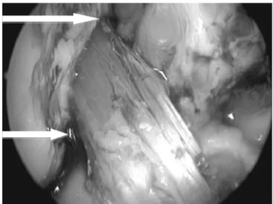
Shuttle sutures are introduced through the femoral tun-nel and pulled out through the tibial tuntun-nel. The graft is then inserted into the femoral tunnel. Here it is possible to determine the rotational position of the bone block in the femoral tunnel. The goal is the tendon’s backside to face anteriorly. Hence, the tendon fibers, which emerge eccentrically from the bone block, are placed as poste-rior as possible mimicking the round shape of the bone cartilage interface (Fig. 4).
Under constant pull distally the bone block is then tapped into the femoral tunnel until a press-fit anchorage is achieved, ideally flush at the femoral aperture. A good indicator for optimal press-fit fixation is the same high-pitched sound we are used to from tapping a non-cemented hip-stem into the proximal femur (Fig. 5). Pre-conditioning, check for isometry and tibial graft fixation with an interference screw complete the procedure.
Clinical results
Reports of this technique, one with an associated lateral extra-articular tenodesis, have been published by the Lyon knee group [9–11]. Two other papers by the same group summarized the clinical results of patients after intra-articular double incision ACL reconstruction.
Bouattour et al. [12] reported a retrospective series of 83 patients (n086 knees) who underwent arthroscopic recon-struction of the ACL using a central one-third patellar ten-don autograft. The mean follow-up was six years postoperatively. The patients were satisfied or very satisfied in 88.5 % of the cases. Forty-two patients (61 %) returned to
Fig. 3 Creation of the cone-shaped femoral tunnel. The 10-mm femoral tunnel is expanded using the manual mill bit allowing for a press-fit fixation of the wedge-shaped bone block in the cone shaped tunnel. [Previously published in Springer publications (EJOS), reprint permission granted]
Fig. 4 The manual mill bit entering the femoral tunnel, creating the outside-in cone-shaped tunnel. It is under constant control with the arthroscope in the medial portal viewing inside-out. [Previously pub-lished in Springer publications (EJOS), reprint permission granted]
a
b
Fig. 2 Schematic drawing of the prepared BPTB-graft. Note the small, tapered bone block A originating from the patella and the wedge shaped bigger bone block B originating from the tibial tuberosity which will be tapped outside-in into the cone shaped femoral tunnel, blocking itself. [Previously published in Springer publications (EJOS), reprint permission granted]
their sports activities and 9/12 high-level athletes returned to sports at an identical level as preoperatively. Final Interna-tional Knee Documentation Committee (IKDC) score is shown in Table 1. They highlighted that good objective outcome was correlated with a more anatomical femoral position and preservation of the meniscus.
Ait Si Selmi et al. [13] also reported a case series of 60 patients who underwent a double-incision BTB ACL recon-struction. The mean follow-up was 16 months. The patients were satisfied or very satisfied in 93.5 % of the cases. Subjective IKDC score was 88.4±15. Gain of subjective score was +13 and 82 % of patients were rated A or B at the final IKDC scoring (Table1).
In conclusion, with regards to these studies the double-incision technique was a safe way to anatomically recon-struct the ACL leading to convincingly good clinical results.
Discussion
This paper intended to describe the technical principles of the double incision iso-anatomic ACL reconstruction tech-nique. In the following the pros and cons of the proposed procedure are discussed.
The only minor disadvantage of the proposed technique is the need for a short second skin incision on the lateral femur. This is however outweighed by a number of technical advantages, which are inherent to this technique.
First, the biomechanically more critical femoral tunnel is primarily drilled. There is no pressure loss through leaking of the saline during this step.
Second, the proposed ACL reconstruction technique facilitates placing the femoral tunnel in the centre of the native ACL attachment, which is lower than usually achieved in transtibial ACL techniques [2]. Furthermore, the created femoral tunnel aperture has a rather circular shape with a defined diameter rather than an ovaloid shape when the drill enters the notch in a tangential direction.
Third, when compared to techniques where the medial portal is used for drilling of the femoral tunnel, there is no risk of damage to the medial femoral condyle. It is an easy and reproducible ACL reconstruction technique. Every step of the surgery is arthroscopically controlled and hence it can be routinely used in a teaching hospital setting.
Fourth, as no fixation device is used on the femoral side (press-fit technique) it is less costly than other described techniques. And fifth, revision surgery is considered to be less difficult. The surgeon is not limited due to the initial tunnel or fixation device in placing the femoral tunnel.
Clinical results
Although there is a tremendous amount of scientific data comparing single with double-incision ACL reconstructions in anatomical and cadaveric studies, only few clinical stud-ies compared the outcome in patients undergoing ACL reconstruction using these two techniques.
In 1999 Howell [14] compared the clinical outcome of the single- and double-incision ACL reconstruction techniques. At two years follow-up in both groups the objective was achieved, it was therefore concluded that there was no clear superiority of either technique. But the subjective IKDC score was significantly lower in the single- than in the two-incision group (P00.008). It could be speculated that the more reliable elimination of the pivot shift phenomenon using an iso-anatomic double-incision ACL-reconstruction might be the main reason for this clinically relevant difference.
Conclusion
The double incision, iso-anatomical ACL reconstruction is a relatively simple, safe, reliable and reproducible technique. Anatomical femoral tunnel placement is fa-cilitated without exception.
Overlooking the results of the three major ACL-reconstruction techniques, one may conclude that the single incision single bundle technique results are bio-mechanically reasonable, the iso-anatomical single bun-dle reconstructions are almost perfect, leaving only
Fig. 5 The ACL graft in place. Note the high-low extension of the graft (between the two white arrows) mimicking the placement of the normal ACL. [Previously published in Springer publications (EJOS), reprint permission granted]
Table 1 Final International Knee Documentation Committee (IKDC) score of two clinical studies on double-incision ACL reconstruction
Study Final IKDC score
A B C D
Bouattour et al. 25 % 50 % 21 % 4 %
marginal room for improvement for the double bundle ACL reconstructions.
Conflict of interest statement No funds were received in support of
this study. No benefits in any form have been or will be received from a commercial party related directly or indirectly to the subject of this manuscript.
References
1. Dye SF (1996) The knee as a biologic transmission with an
envelope of function: a theory. Clin Orthop 325:10–18
2. Arnold MP, Kooloos J, van Kampen A (2001) Single-incision technique misses the anatomical femoral anterior cruciate ligament insertion: a
cadaver study. Knee Surg Sports Traumatol Arthrosc 9(4):194–199
3. Arnold MP, Verdonschot N, van Kampen A (2005) ACL graft can replicate the normal ligament’s tension curve. Knete Surg Sports Traumatol Arthrosc 13(8):625–631
4. Kanaya A, Ochi M, Deie M, Adachi N, Nishimori M, Nakamae A (2009) Intraoperative evaluation of anteroposterior and rotational stabilities in anterior cruciate ligament reconstruction: lower fem-oral tunnel placed single-bundle versus double-bundle
reconstruc-tion. Knee Surg Sports Traumatol Arthrosc 17(8):907–913
5. Loh JC, Fukuda Y, Tsuda E, Steadman RJ, Fu FH, Woo SL (2003) Knee stability and graft function following anterior cruciate
liga-ment reconstruction: comparison between 11 o’clock and 10
o’clock femoral tunnel placement. Arthroscopy 19(3):297–304
6. Scopp JM, Jasper LE, Belkoff SM, Moorman CT 3rd (2004) The effect of oblique femoral tunnel placement on rotational constraint of the knete reconstructed using patellar tendon autografts.
Arthroscopy 20(3):294–299
7. Neyret P, Demey G, Servien E, Lustig S (2012) Traité de chirurgie du genou. Elsevier Masson, Paris, pp 42–64
8. Arnold MP, Biedert RM, Van Loon C, Hirschmann MT (2011) Isoanatomical bone-patellar tendon-bone single-bundle ACL re-construction: the wedge that gives the edge! Eur J Orthop Surg Traumatol Epub 22.3. 2011
9. Dejour H, Dejour D, Ait Si Selmi T (1999) Chronic anterior laxity of the knee treated with free patellar graft and extra-articular lateral plasty: 10-year follow-up of 148 cases. Rev Chir Orthop
Reparatrice Appar Mot 85(8):777–789
10. Dejour H, Walch G, Neyret P, Adeleine P (1988) Results of surgically treated chronic anterior laxities. Apropos of 251 cases reviewed with a minimum follow-up of 3 years. Rev Chir Orthop
74(7):622–636
11. Pernin J, Verdonk P, Si Selmi TA, Massin P, Neyret P (2010) Log-term follow-up of 24.5 years after intra-articular anterior cruciate ligament reconstruction with lateral extra-articular augmentation. Am J Sports Med 38(6):1094–1102
12. Bouattour K, Chatain F, Ait Si Selmi T, Neyret P (2002) Arthroscopic reconstruction of the anterior cruciate ligament. Rev Chir Orthop Reparatrice Appar Mot 88(2):130–138 13. Ait Si Selmi T, Fabié F, Massouh T, Pourcher G, Adeleine P,
Neyret P (2002) Greffe du LCA au tendon rotulien sous arthro-scopie avec ou sans plastie antéro-externe Etude prospective randomisée à propos de 120 cas. Paper presented at the 10èmes Journées Lyonnaises de Chirurgie du Genou, Lyon
14. Howell SM, Deutsch ML (1999) Comparison of endoscopic and two-incision techniques for reconstructing a torn anterior cruciate