HAL Id: inserm-01155015
https://www.hal.inserm.fr/inserm-01155015
Submitted on 26 May 2015HAL is a multi-disciplinary open access archive for the deposit and dissemination of sci-entific research documents, whether they are pub-lished or not. The documents may come from teaching and research institutions in France or abroad, or from public or private research centers.
L’archive ouverte pluridisciplinaire HAL, est destinée au dépôt et à la diffusion de documents scientifiques de niveau recherche, publiés ou non, émanant des établissements d’enseignement et de recherche français ou étrangers, des laboratoires publics ou privés.
Distributed under a Creative Commons Attribution| 4.0 International License
Comparison between 3D Supersonic Shear Wave
Elastography and Magnetic Resonance Elastography: a
preliminary experimental study
Jinlong Yue, Marion Tardieu, Felicia Julea, Linda Chami, Olivier Lucidarme,
Xavier Maître, Claire Pellot-Barakat
To cite this version:
Jinlong Yue, Marion Tardieu, Felicia Julea, Linda Chami, Olivier Lucidarme, et al.. Comparison be-tween 3D Supersonic Shear Wave Elastography and Magnetic Resonance Elastography: a preliminary experimental study. Journées RITS 2015, Mar 2015, Dourdan, France. pp 142-143. �inserm-01155015�
Actes des Journées Recherche en Imagerie et Technologies pour la Santé - RITS 2015 142
Comparison between 3D Supersonic Shear Wave Elastography and Magnetic
Resonance Elastography: a preliminary experimental study
J.L. Yue
1 2 3∗, M. Tardieu
1, F. Julea
1, L. Chami
4, O. Lucidarme
4, X. Maˆıtre
1, C. Pellot-Barakat
2 31 Imagerie par R´esonance Magn´etique M´edicale et Multi-Modalit´es, IR4M, CNRS, Univ Paris-Sud, Orsay, FRANCE.
2 Imagerie Mol´eculaire in vivo, IMIV, UMR 1023 Inserm/CEA/Univ Paris-Sud-ERL 9218 CNRS, CEA/I2BM/SHFJ, Orsay, FRANCE. 3 Laboratoire d’Imagerie Biom´edicale, INSERM, CNRS, Univ Pierre et Marie Curie, Paris, FRANCE.
4 Service de Radiologie, Hˆopital Piti´e-Salp´etri`ere, APHP, Paris, FRANCE ∗ Corresponding author: [email protected]
Abstract - Ultrasound Supersonic Shear Wave raphy (SSWE) as well as Magnetic Resonance Elastog-raphy (MRE) allow accessing the mechanical properties of human tissues. SSWE is usually performed using a 2D probe. 3D SSWE is now available but needs to be validated. We compared 3D SSWE with both 2D SSWE and MRE which is inherently 3D on a breast phantom. We found that 3D SSWE is reproducible and provides e-lasticity estimates comparable to those obtained with the validated 2D SSWE. We also showed that 3D SSWE and MRE exhibit quite different elasticity moduli, but they re-veal similar qualitative trends in the phantom. Although no relationship could be drawn between the two modali-ties, this study provides a first basis for comparison and a guide for potential improvements.
Index Terms - Biomechanics, Magnetic Resonance Imaging, Ultrasound, Elastography
I. INTRODUCTION
Elasticity imaging has emerged recently and is rapidly evolving. Among different approaches, Ultrasound Su-personic Shear Wave Elastography (SSWE) and Magnet-ic Resonance Elastography (MRE) have partMagnet-icular attrac-tive prospecattrac-tives for clinical applications. The two modal-ities are different on principles, experimental conditions and reconstruction methods. It should thus be meaning-ful to compare the two modalities in order to get a better understanding of the mechanical properties of human tis-sues. There has been some research dedicated to the spe-cific comparison of the two modalities in 2D dimension [1]. Since 3D SSWE is now available, a 3D comparison of the two modalities optimizing the registration between the elastograms can be performed. This is the aim of this work. Since 3D SSWE has not yet been deeply investigat-ed, a first study is carried out here to validate the reliability of 3D SSWE.
II. MATERIALS AND METHODS
II.1. Reproducibility of 3D SSWE and comparison with 2D SSWE
A breast phantom containing homogeneous inclusions and background (model 059, CIRS, Norfolk, VA, USA) was imaged by the Aixplorer ultrasound system (Supersonic Imagine, Aix-en-Provence, France). First, ten acquisitions
of the same inclusion were performed by the same operator using a 3D probe (SLV16-5 MHz) while repositioning the probe between each acquisition. Five different inclusions were then imaged using a 2D transducer (SL15-4 MHz) as well as the 3D transducer to validate 3D SSWE. The pixel size was 0.2 × 0.2 mm2. The different slices of the inclu-sions were scanned with the 2D transducer in order to get a better registration with the 3D transducer. An automat-ic thresholding segmentation was performed to extract the mean elasticity Emean and standard deviation SD of each inclusion.
II.2. Comparison between 3D SSWE and MRE A more realistic breast phantom with inclusions embedded in heterogeneous background (model 073, CIRS, Norfolk, VA, USA) was used for the comparison. First, one selected dense inclusion was imaged using the 3D transducer of the Aixplorer. Next, the whole phantom was imaged and me-chanically characterized at 85 Hz on a 1.5T MR Scanner (Achieva, Philips Healthcare, Best, The Netherlands) with a dual flexible coil (Flex-M, Philips Healthcare, Best, The Netherlands). The voxel size was 2 × 2 × 2 mm3. The 3D maps of shear elastic modulus were inferred by inver-sion of the equation of motion [2]. External markers were used to identify the corresponding inclusion in 3D SSWE and MRE. A manual segmentation was performed to ex-tract the Emean and SD values in the selected inclusion.
III. RESULTS
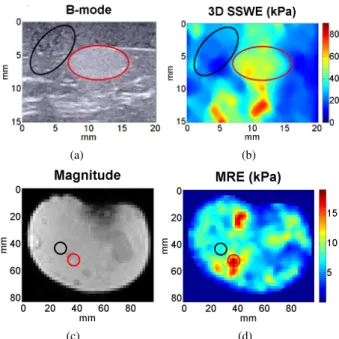
The variability for the Emean values between ten acquisi-tions of 3D SSWE is smaller than 5%. Figure 1(a) repre-sents the five investigated inclusions in the first phantom. Figure 1(b) represents a typical SSW elastogram of inclu-sion 4. Figure 2 shows that 3D SSWE and 2D SSWE mea-surements are comparable. The Emean and SD values ob-tained by the 3D probe tend to be lower than that obob-tained by the 2D probe. Figure 3 represents the B-mode image, the SSW elastogram, the magnitude image, the MR elas-togram of the selected inclusion in the second phantom. The Emean values of the selected inclusion and its back-ground obtained by two modalities are reported in Table 1. Elasticity values of 3D SSWE are more than three times higher than that of MRE, while the ratio between inclusion and background are similar between the two modalities.
143 Actes des Journées Recherche en Imagerie et Technologies pour la Santé - RITS 2015
(a) (b)
Figure 1: (a) Position of the imaged inclusions in the breast phantom (model 059) (b) SSW elastogram of one slice of inclusion 4 obtained by 2D SSWE
Figure 2: Emean and SD values obtained by the 2D (blue) and 3D (red) probes for 5 inclusions
3D SSWE MRE
Inclusion (kPa) 45.4± 8.1 14.3± 1.9 Background (kPa) 24.7± 1.6 6.9± 0.2
Ratio 1.84 2.07
Table 1: Emean obtained by 3D SSWE and MRE IV. DISCUSSION-CONCLUSION
It was proven that 3D SSWE measurements are repro-ducible, and seem to be more precise than 2D ones, as shown by the lower values of SD obtained in homoge-neous regions of interest. The Emean values of 3D SSWE measurements are in average about 10% lower than that of 2D ones, which is opposite to the literature [3]. Many fac-tors could explain this difference. The major explanation could be that the in vivo breast masses measured in the literature exhibited inhomogeneous elastic properties and complex structures, since only one slice of the mass was considered for comparison between 2D and 3D SSWE. It is thus very likely that the registration was incomplete. In our study the inclusions had homogeneous elastic properties and simple structures (spherical inclusions) and we imaged different slices in 2D in order to get a full registration with the 3D probe.
(a) (b)
(c) (d)
Figure 3: (a) B-mode image (b) SSW elastogram (c) Mag-nitude image (d) MR elastogram. The red and black el-lipses represent the segmented inclusion and a region of the background (around 40 mm2)
We found that SSWE provided much higher elasticity values than MRE, while the ratio between inclusion and background was about the same with both modalities. The different excitation wave frequencies seem to be the major element of discrepancy between the two modalities (around 350 Hz for 3D SSWE and 85 Hz for MRE), because the elasticities measured in both modalities are frequency-dependent. To improve the precision of the comparison study, the spatial resolution of MR elastograms will be increased; meanwhile rhehological models will be used to link the elasticity values recorded by both modalities; finally more advanced segmentation methods will be used to better extract the values within the inclusions.
In conclusion, the study demonstrates the reliability of 3D SSWE, and it sustains the need for rheological modeling to properly challenge SSWE and MRE.
REFERENCES
[1] M. Tanter, et al. ”L’´elastographie par ultrasons ou r´esonance magn´etique: de nouveaux outils de diagnos-tic en canc´erologie”, M´edecine Nucl´eaire, 2007, Vol. 31, pp. 132-141.
[2] R. Sinkus, et al. ”Viscoelastic shear properties of in vi-vo breast lesions measured by MR elastography”, Mag-netic Resonance Imaging, 2005, Vol. 23, pp. 159-165. [3] S. H. Lee, et al. ”Differentiation of benign from
malignant solid breast masses: comparison of two-dimensional and three-two-dimensional shear-wave elastog-raphy”, European radiology, 2013, Vol. 23, pp. 1015-1026.