HAL Id: hal-03011376
https://hal.archives-ouvertes.fr/hal-03011376
Submitted on 18 Nov 2020
HAL is a multi-disciplinary open access
archive for the deposit and dissemination of
sci-entific research documents, whether they are
pub-lished or not. The documents may come from
teaching and research institutions in France or
abroad, or from public or private research centers.
L’archive ouverte pluridisciplinaire HAL, est
destinée au dépôt et à la diffusion de documents
scientifiques de niveau recherche, publiés ou non,
émanant des établissements d’enseignement et de
recherche français ou étrangers, des laboratoires
publics ou privés.
Genome-Wide Level
Coline Arnould, Gaëlle Legube
To cite this version:
Coline Arnould, Gaëlle Legube. Analyzing Homologous Recombination at a Genome-Wide Level.
Journal of Molecular Biology, Elsevier, 2021, 432 (3), pp.427-438. �10.1007/978-1-0716-0644-5_29�.
�hal-03011376�
The Secret Life of Chromosome Loops upon
DNA Double-Strand Break
Coline Arnould and Gaëlle Legube
LBCMCP, Centre de Biologie Integrative (CBI), CNRS, Université de Toulouse, UT3, Toulouse, France
Correspondence to
Gaëlle Legube:
[email protected]
https://doi.org/10.1016/j.jmb.2019.07.036
Abstract
DNA double-strand breaks (DSBs) are harmful lesions that severely challenge genomic integrity, and recent
evidence suggests that DSBs occur more frequently on the genome than previously thought. These lesions
activate a complex and multilayered response called the DNA damage response, which allows to coordinate
their repair with the cell cycle progression. While the mechanistic details of repair processes have been
narrowed, thanks to several decades of intense studies, our knowledge of the impact of DSB on chromatin
composition and chromosome architecture is still very sparse. However, the recent development of various
tools to induce DSB at annotated loci, compatible with next-generation sequencing-based approaches, is
opening a new framework to tackle these questions. Here we discuss the influence of initial and DSB-induced
chromatin conformation and the strong potential of 3C-based technologies to decipher the contribution of
chromosome architecture during DSB repair.
© 2019 The Authors. Published by Elsevier Ltd. This is an open access article under the CC BY-NC-ND license
(
http://creativecommons.org/licenses/by-nc-nd/4.0/
).
The DNA Double-Strand Break
Response and Repair
DNA double-strand breaks (DSBs) represent
challenging lesions for cells, as they can lead to
major genome rearrangement such as
transloca-tions, aneuploidy, and deletions/amplifications.
While previously considered to be almost exclusively
induced by environmental agents (radiation,
chemi-cals) with the exception of programmed induction by
endogenous nucleases (during meiosis and
immu-noglobulin loci rearrangement), it is now well
admitted that DSBs are also regularly triggered
during normal cell metabolism. More specifically, the
development of DSB mapping technologies, such as
BLESS, BLISS, DSBCapture, END-seq or BrlTL
[1–6]
(reviewed in Ref.
[7]), has recently revealed
insights on the distribution of endogenous DSBs
across the genome. These genome-wide analyzes
identified transcriptionally active loci and
transcrip-tion start sites (TSS) as particularly prone to
breakage (e.g., Refs.
[3,6], reviewed in Refs.
[7,8]).
These studies also further revealed a compelling
connection between DSB production and
chromo-some architecture, identifying binding sites for CTCF
and cohesin (main genome organizers) as
preferen-tial damage sites due to topoisomerase activity and/
or fork collapse
[5,9
–12]
. At loop anchors,
topoisom-erase II recruitment could further trigger production
of DSB upon transcriptional activation of close-by
genes
[9–11].
Beyond the influence of chromosome loops in
DSB formation, supported by a growing body of
evidence, an emerging concept is also that DSB
formation further modifies chromosome architecture
and organization in the nuclear space. Given the
potential for unscheduled rejoining of two DSBs to
trigger translocations, and the previously reported
bias for translocation to occur in active loci in cancer
cells, it is urgent to understand how DSBs modify
chromosome organization and architecture and how
this impacts genome (in)stability.
0022-2836/© 2019 The Author. Published by Elsevier Ltd. This is an open access article under the CC BY-NC-ND license (http://creativecommons.org/licenses/by-nc-nd/4.0/). Journal of Molecular Biology (2020) 432, 724-736
Various pathways contribute to repair DSB in
eukaryotes, including homologous recombination
(HR) and non-homologous end joining (NHEJ)
(reviewed in Refs.
[13,14]). HR entails processing
of DNA ends in order to generate single strand DNA
(resection) which will invade a homologous copy of
the broken locus, further used as template for DNA
synthesis. NHEJ rather directly rejoins the two DNA
ends. Mechanistically, these two pathways are
profoundly different and likely necessitate different
b
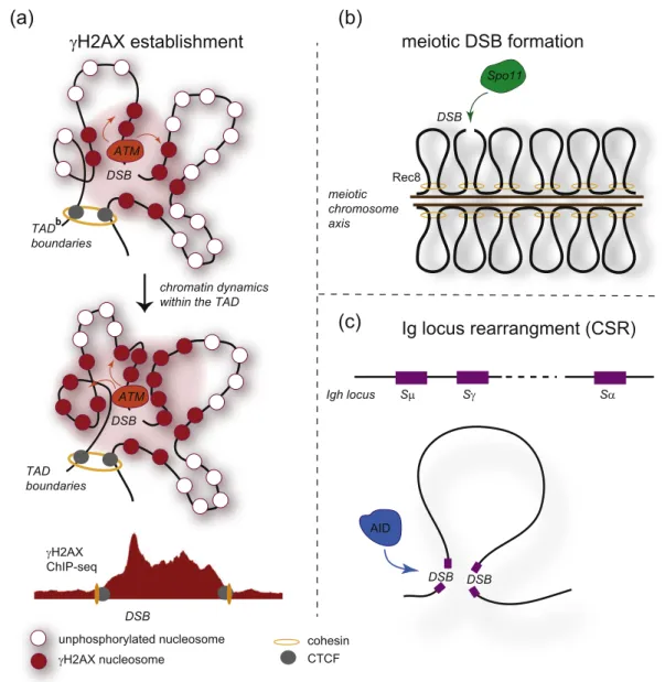
γH2AX establishment
DSB ATM TAD boundaries DSB ATM TAD boundaries chromatin dynamics within the TADDSB γH2AX ChIP-seq meiotic chromosome axis Rec8 DSB Spo11 Sμ Sγ Sα Igh locus
meiotic DSB formation
Ig locus rearrangment (CSR)
DSB DSB AIDunphosphorylated nucleosome cohesin CTCF γH2AX nucleosome
(a)
(b)
(c)
Fig. 1. Contribution of the initial chromatin conformation into
γH2AX establishment and programmed DSB induction and
repair. (A) The initial chromosome conformation may dictate
γH2AX spreading following DSB induction. In this model,
ATM, the main H2AX kinase is locally recruited at the DSB. Once bound, it is able to phosphorylate H2AX containing
nucleosomes brought to its physical proximity, thanks to chromatin dynamics that takes place within the TAD. Sustained
signaling and ATM activation eventually trigger the phosphorylation of H2AX on the entire TAD. In this model,
γH2AX
distribution, as observed by ChIP-seq, should mimic the 3D chromatin conformation. (B) Chromosome conformation is
critical during meiotic breaks formation by Spo11. During prophase, meiotic chromosomes are strongly reorganized with
the formation of DNA loops anchored to a proteinaceous axis. Spo11 generates DSBs within DNA loops, which can further
pair with the homologous chromosome in order to produce crossover and to complete meiosis. The 3D chromatin structure
and the chromosomal axis are required for both DSB production by Spo11 and to ensure the
“homologous bias” (i.e., the
choice of the homologous chromosome rather than the sister chromatid, as a template for HR). (C) Chromosome
conformation is also critical for the rearrangements that occur on immunoglobulin loci, in order to generate immunoglobulin
isotypes (class switch recombination (CSR)) and the antibody repertoire (VDJ recombination). For example, during CSR
(shown here), the long-range physical interactions between switch (S) sequences on the heavy chain locus (Igh) allow two
DSBs to be rejoined.
chromatin composition and properties for proper
execution. At a molecular level, ChIP-seq and
imaging studies have started to determine the
histone modifications landscape assembled at
DSB, as well as their function in DSB repair
(reviewed in Ref.
[15]). However, the conformation
of chromatin around DSBs and the chromosome
organization in damaged nuclei have only recently
started to be investigated.
One of the striking feature of the DNA damage
response is the assembly of microscopically visible
foci in the nucleus, which display massive
phos-phorylation of the H2AX histone variant (γH2AX)
[16], as well as accumulation of repair factors.
Although efforts have been made to understand
the protein content of these foci, their exact
composition and conformation at the DNA level is
still unknown. One of the main reasons for this
scarcity of data stands in the long-lasted inability to
control the position of induced DSB on the genome.
Indeed, except in yeast where the ability of the HO
endonuclease to cleave the MAT locus for mating
type switching was utilized as a tool to investigate
site-specific DSB repair, DSBs have routinely been
induced by exposure to genotoxic (drugs and
radiation) generating damage in a heterogeneous
manner in the cell population and at unknown (but
not necessarily random) positions, which precluded
the use of Chromosome Conformation Capture
(3C)-based methods to investigate chromatin
conforma-tion around DSBs. This has been solved recently,
thanks to the development of several tools to induce
breaks at annotated positions, using restriction
enzymes and homing endonucleases (e.g., AsiSI,
I-PpoI), Zinc Fingers and TALE Nucleases, or the
CRISPR/Cas9 system
[17]. With these novel
exper-imental systems, the molecular characterization of
DNA conformation around DSB and more generally
the folding of damaged chromosomes within the
nucleus are now within reach.
Two main questions should primarily focus our
attention. First, we need to understand how the initial
chromosome conformation and organization in the
nucleus may contribute to ensure proper DSB
signaling and repair. Second, we shall wonder how
this initial chromatin conformation is modified upon
damage to participate in repair events safeguarding
genome integrity.
How Does Initial Chromatin Architecture
Contribute to DSB Signaling and
Re-pair?
The genomic localization of DSBs strongly impacts
their signalization and repair. For instance, clear
evidence suggests that DSB occurring in
heterochro-matin
[18,19], rDNA
[20]
or transcribed loci
[8]
displays
specialized repair pathways. The chromatin
composi-tion of the broken locus (involving for example histone
marks like H3K36me3 for transcription-coupled DSB
repair
[21–23], or bona fide chromatin constituents
such as KAP1 for heterochromatic repair
[24,25]) and
its spatial position within the nucleus (e.g., at the
nuclear periphery
[26]) have been clearly established
as main contributors in determining which pathway
should be used at each genomic location (a decision
known as
“DSB repair pathway choice”)
[27].
Howev-er, at this stage it is not known whethHowev-er, beyond the
sub-nuclear localization of a locus and its chromatin
composition, the chromosome conformation also
plays a role in DSBs signaling and repair.
Neverthe-less, some hints suggest that this is likely the case.
Initial chromatin conformation regulates HR
The fact that chromosome conformation within the
nucleus regulates HR is particularly evident from studies
in yeast, showing that efficiency of sub-telomeric
recombination is strongly affected by telomeres
cluster-ing and anchorage
[28,29]. Beyond telomeres, moving
an HO site at different positions along yeast
chromo-somes revealed a compelling correlation between the
frequency of HR and the proximity with the
homolo-gous locus, observed by 3C before damage induction
[30]. Thus, initial spatial proximity between the broken
locus and a donor sequence is a key feature that
determines the efficiency of HR.
Initial chromatin conformation could regulate
γH2AX spreading
Evidence also suggests that chromosome
archi-tecture might control
γH2AX spreading (reviewed in
Refs.
[31,32]) (Fig. 1). Indeed,
γH2AX mapping by
ChIP-chip around multiple DSBs induced by the
restriction enzyme AsiSI in human cells (the
so-called DIvA cell line for DSB Induced via AsiSI)
revealed that
γH2AX spreads on 1-2 megabases
surrounding DSBs, in a manner that is (i)
reproduc-ible and constrained within boundaries, (ii) not
necessary symmetrical around the break and (iii)
uneven with peaks and valleys, suggesting that the
surrounding epigenomic landscape and/or
chroma-tin architecture may regulate
γH2AX spreading
[33].
A follow-up study uncovered a potential function for
cohesins in regulating
γH2AX distribution and in
insulating transcribed genes encompassed in
γH2AX domains from transcription extinction
[34].
Moreover, by then, comparison of published Hi-C
data generated in undamaged cells
[35]
with
γH2AX
domains boundaries revealed a striking tendency of
γH2AX spreading to stop at topologically associating
domain (TAD) boundaries
[34]. In agreement, the
occupancy of the chromatin looping factor CTCF
was found juxtaposed to
γH2AX foci using
super-resolution light microscopy
[36]. Altogether, these
results raise the exciting hypothesis that once bound
to the DSB, the kinase(s) involved in H2AX
phosphorylation allows
γH2AX spreading by
modi-fying nucleosomes brought into spatial proximity,
thanks to the initial chromatin architecture
surround-ing the DSB (the
“Intra-TAD model”
[31,32]) rather
than by linearly walking along the chromosome. This
hypothesis is further supported in yeast, where
γH2A
spreading was also found to occur in trans (on other
chromosomes) when a DSB was induced close to a
centromere, as a consequence of centromeres
clustering within nuclei
[37].
Initial chromatin conformation regulates
production and repair of programmed DSB
Beyond these evidences that pre-existing
chro-mosome architecture contributes to DSB signaling
and repair, chromosome organization is also likely a
key feature in the repair of programmed DSBs
induced during meiosis and immunoglobulin loci
rearrangements (Fig. 1). During the prophase of
meiosis, chromosomes undergo profound
reorgani-zation which entails progressive condensation, loss
of long-range inter-chromosomal contacts, TADs
dissolution and the formation of arrays of chromatin
loops anchored to a chromosomal axis
[38–43].
Within this context, the topoisomerase-like Spo11
endonuclease induces DSBs in a tightly regulated
manner, which are further processed and
—for some
of them—converted into mature crossovers,
neces-sary to ensure chromosome segregation and
com-pletion of meiosis. Importantly, crossover formation
depends on the
“homologous bias” that consists in
choosing the homologous chromosome as a
tem-plate over the sister chromatid (reviewed in Refs.
[44,45]). Notably, germ cells-specific chromosome
architecture plays a critical role in both determining
DSB
DSB
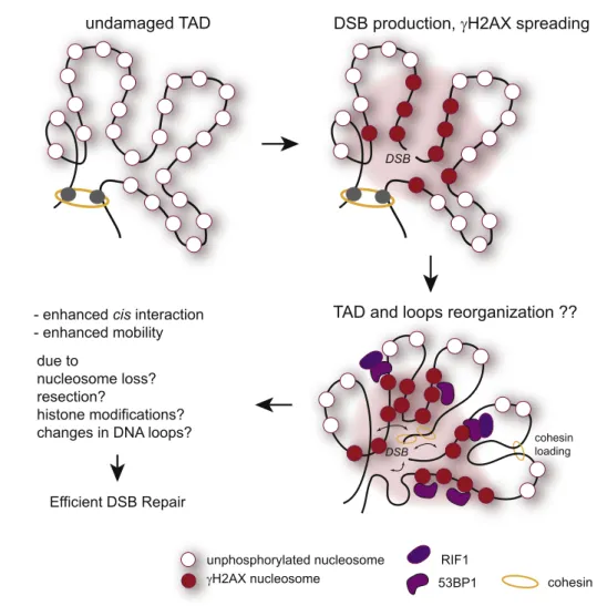
undamaged TAD
DSB production,
γH2AX spreading
TAD and loops reorganization ??
cohesin loading
- enhanced cis interaction
- enhanced mobility
Efficient DSB Repair
due to
nucleosome loss?
resection?
histone modifications?
changes in DNA loops?
RIF1
53BP1 cohesin unphosphorylated nucleosome
γH2AX nucleosome
Fig. 2. DSB-induced modification(s) of the chromosome conformation in cis to the break. Following DSB production and
γH2AX spreading, the 3D conformation of damaged TAD could also be modified, due to the binding of cohesin, CTCF or
repair proteins with potential function in chromatin architecture such as 53BP1 and RIF1. The DSB-induced histones
modifications (including
γH2AX spreading), nucleosome loss or/and generation of single strand DNA (resection) may also
collectively change the dynamics of chromatin within TADs. Altogether, these changes could translate in enhanced
mobility and efficient DSB repair.
the distribution of DSBs along the chromosome and
in the homologous bias. Indeed, in Saccharomyces
cerevisiae, Spo11-dependent DSBs are being
formed within DNA loops in a manner that depends
on multiple axial factors such as the meiosis-specific
cohesin subunit Rec8
[46,47], the Spp1 protein
[48],
Red1
[46], or the Spo11 accessory complex, RRM
(Rec114
–Mei4–Mer2)
[49]. In addition, components
of the structural axis (such as Rec8 or Red1) are also
strongly involved in regulating the homologous bias
[47]
and the axis further acts as a platform for
recombination. This peculiar chromosome
architec-ture that is assembled in meiotic cells hence displays
a prominent role into DSB production and repair.
Programmed DSBs also occur at the
Immunoglob-ulin (Ig) loci to ensure V(D)J recombination for
antibody diversification and class switch
recombina-tion (CSR) to generate different antibody isotypes. On
the Ig heavy-chain locus (Igh), productive CSR results
in a deletion event after recombination between two
Switch (S
H) sequences, located up to 100 kb apart.
Here as well, the initial 3D chromatin conformation
exerts a regulatory role on both break formation and
repair
[50,51]
(reviewed in Ref.
[52]). For instance,
deletion of the CTCF-binding sites encompassed in
the Igh Superanchor (SA), correlates with a decrease
in cohesin-mediated loop extrusion (detected by
“stripes” on Hi-C maps) and reduces CSR
[50].
Similarly, V(D)J recombination, both on Igκ and Igh
loci, strongly relies on long-range chromatin
interac-tions. For instance, deletion of CTCF-binding sites in
the intergenic control region-1 (IGCR1) upstream the
D segments on Igh locus impairs normal V to DJ
recombination and B-cell development in a manner
that coincides with modification of chromosomal loops
[53,54]. Similarly, deletion of a specific
enhancer-CTCF-bound element on the Ig
κ locus perturbs the
antibody repertoire in a manner that also correlates
with the loss of long-range interaction
[55].
It is hence clear from all these studies that the initial
chromosome architecture contributes to DSB signaling,
processing and repair. However, while our knowledge
regarding the role of long-range chromatin interactions
and TADs during repair of programmed DSBs quickly
expands, our understanding of their function into repair
of endogenous DSBs in somatic cells still lags behind
and will necessitate future 3C-based studies using
sequence-specific DSB induction systems.
How Does Chromatin Architecture
Change Post-damage In Cis to DSB, within
γH2AX Domains?
Another important question that needs to be
addressed is the nature of the changes in
chromo-some architecture following damage (Fig. 2). Indeed,
DSB-induced modifications in the size of DNA loops
or the position of TADs boundaries could regulate
chromatin flexibility (stiffness), thereby regulating the
DSB mobility in the nucleus
[56]. This could also help
to
“burry” (i.e., protect) the DSB from its environment,
which therefore may have profound impacts on
translocation biogenesis, partner choice for HR and
more generally on genome integrity. Plenty of
evidence supports that chromatin in cis to DSB
displays a different behavior in terms of mobility,
rigidity and compaction (reviewed in Refs.
[31,57]).
In particular, laser-mediated, localized damage
triggers a rapid chromatin decompaction at the
sites of breaks
[58–60], in agreement with
observa-tions that
γH2AX foci displays decondensed-like
appearance
[61,62], suggesting that indeed a DSB
induces dramatic changes in chromatin
conforma-tion in cis. However, nearly all studies were
performed using imaging, and our current
knowl-edge of DSB-promoted 3D changes at the level of
DNA sequence remains incredibly sparse. The first
experiment using 3C methodology to assess cis
modification of long-range interactions was
per-formed in yeast following induction of a single DSB
within the MAT locus by the HO endonuclease
[63].
Strikingly long-range contacts were dramatically
reduced following DSB in asynchronous cells,
while such a decrease was not observed in
G1-arrested cells
[63], suggesting that end-processing
(which occurs specifically in S/G2) rather than
γH2A
spreading and checkpoint activation (occurring all
throughout the cell cycle) was responsible for
decreased chromosomal contacts. Reduced
chro-mosome interaction frequency was further shown to
depend on Rad51 loading and attributed to the
sequestration of the DSB at the nuclear periphery
[63,64]
(see next section). However, this decrease in
long-range interaction following DSB was not
report-ed in human cells
[65]. Instead, by using Capture
Hi-C in the DIvA system (in which ~ 100 DSBs can be
induced at annotated loci
[21,33]), it was shown that,
in average, the DSB itself engages more long-range
contacts with neighboring sequences encompassed
in
γH2AX domains than before break induction
[65]
(reviewed in Ref.
[66]). Contrary to yeast, DSBs have
not been found to relocate to the nuclear envelope in
mammalian cells, which may account for the
discrepancy between both studies. Notably,
en-hanced interactions between the DSB and DNA
loci embedded in
γH2AX domains would be in
agreement with the increased mobility of DNA ends
reported in multiple studies (reviewed in Refs.
[31,57,67]).
Of note, the resolution achieved by Capture-HiC in
the above-mentioned study
[65]
was insufficient to
draw conclusions regarding the DNA loops
reorga-nization around DSBs. Interestingly, although this
was not directly assessed using Hi-C or 4C
experiment, strong evidence suggests that
chromo-some loops are reorganized in cis to DSB induced by
Spo11 in yeast meiotic cells. Indeed, in a WT strain,
DSB production by Spo11 is negatively
counter-acted in a ~ 100-kb window around an initial
Spo11-created break. Notably, this phenomenon, called
negative interference, depends on the yeast ATM
ortholog Tel1, one of the main kinases activated
during the DNA damage response
[68]. This led to
the proposal that, in cis to an initial break, ATM/Tel1
activation could drive chromatin loops
reorganiza-tion, themselves being required and targeted for
DSB formation (see previous section), hence
con-tributing to negative interference and ensuring a
proper distribution of DSBs along meiotic
chromo-somes
[68,69].
It is likely that the usual suspects shaping
chromosome architecture, that is, the cohesin
complex and CTCF, are involved in such
DSB-induced DNA loops reorganization. Studies in both
yeast and mammals have demonstrated that
cohe-sins and their loaders/regulators are recruited at
DSBs
[34,70
–81]
, and post-translationally modified
following damage (e.g., Refs.
[76,77,82–85]).
Strik-ingly, in yeast, DSB-induced cohesin binding takes
place on a large chromosomal domain surrounding
the break
[71,72], in contrast to mammalian cells,
where it only occurs on few kilobases
[34]. While
cohesin loading has long been involved in sister
chromatid cohesion during HR
[75,83,84,86–90], in
mammalian cells, it is also recruited at DSBs during
G1 phase
[34,74], suggesting that this complex
exerts a function beyond sister chromatid cohesion
at DSB. In agreement, cohesin regulates
transcrip-tional repression of genes immediately in cis to
DSBs
[74], insulates the active genes located farther
within the
γH2AX domain to maintain their
transcrip-tion
[34]
and controls the NHEJ repair pathway
[91].
Similarly, CTCF has also been shown to be recruited
at DSBs
[92
–94]
and to contribute to HR
[92,93].
Beyond these well-known architectural proteins
involved in chromatin looping, DSB repair factors
may also contribute to shape chromatin 3D structure
in cis to DSB. Among these, 53BP1 is an interesting
candidate since it was shown to spread on entire
γH2AX domains
[95]
and to be a critical determinant
of the architecture of the Igh locus, even before
damage formation
[51,96]. In addition, the 53BP1
effector Rif1, which is recruited at DSB to regulate
resection
[97
–99]
, is also of interest, as it was
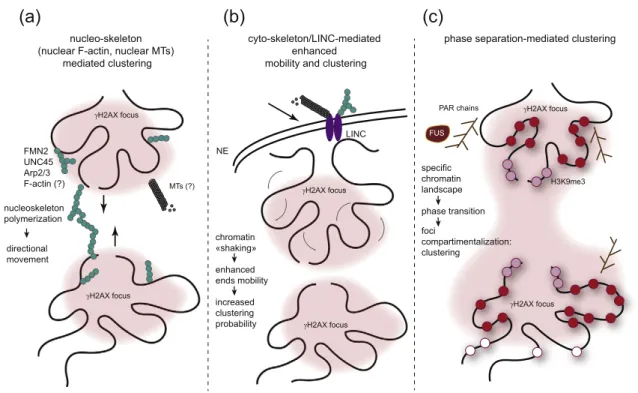
FMN2 UNC45 Arp2/3 F-actin (?) γH2AX focus γH2AX focus MTs (?) γH2AX focus nucleo-skeleton (nuclear F-actin, nuclear MTs)
mediated clustering
phase separation-mediated clustering
γH2AX focus H3K9me3 PAR chains FUS γH2AX focus γH2AX focus cyto-skeleton/LINC-mediated enhanced mobility and clustering
LINC NE chromatin «shaking» enhanced ends mobility increased clustering probability directional movement nucleoskeleton polymerization specific chromatin landscape phase transition foci compartimentalization: clustering
(a)
(b)
(c)
Fig. 3. Changes in chromosome conformation upon damage in trans such as during DSB clustering. Both live cell
imaging and 3C-based methods allowed to demonstrate that multiple DSBs can coalesce together within a single
γH2AX
focus. However, the mechanisms that ensure clustering are unclear and may entail various pathways. (A) The
nucleoskeleton (both polymerized actin and/or microtubules) could allow for DSB mobilization and clustering in a
directional manner. (B) The cytoskeleton could also contribute to clustering thanks to the transmission of forces from
cytoskeleton to chromatin via the LINC complex, embedded in the nuclear envelope. In this context, the forces transmitted
to chromatin may trigger a general increase in chromatin dynamics, increasing the probability of
γH2AX collision/
clustering. (C) Finally, the chromatin landscape established following damage could allow for compartmentalization,
thanks to phase separation.
recently shown by 4C-seq to be a main organizer of
chromatin architecture in unchallenged cells
[100].
Importantly, both factors are essential for productive
CSR
[97,98,101,102], which further highlight their
potential role in organizing the 3D structure of
DSB-surrounding chromatin.
However, despite all these studies, a clear picture
of the DNA conformation within
γH2AX foci is still
awaited. Mapping of architectural proteins using
ChIP-seq as well as determining chromatin
confor-mation by 3C-based approaches around annotated
DSBs will certainly help to better understand the
nature and function of DSB-induced chromosome
loops changes.
How Does Chromatin Architecture
Change Post-damage In Trans to DSB,
within the Nucleus?
In addition to the changes in cis described in the
previous section, damaged chromosomes also
experience more global reorganization within the
nucleus. This is particularly apparent in yeast and
Drosophila, where persistent, heterochromatic and/
or rDNA breaks are relocalized at the nuclear
periphery
[63,64,103,104]
(reviewed in Ref.
[31]).
Similarly in mammals, DSBs induced in rDNA and
α
satellites are extruded at the periphery of the nuclear
sub-compartment (nucleolus and heterochromatic
focus, respectively)
[105
–107]
. Beyond these
large-scale reorganizations, DSBs are also capable of
clustering together (i.e., regrouping in one visible
focus
[65,108
–115]
(reviewed in Refs.
[31,116]).
Using Capture Hi-C to map long-range interactions
following induction of multiple annotated DSBs on
the human genome, it was recently demonstrated
that DSBs can cluster together if they occur in
transcriptionally active, RNA Pol II-bound, loci
[65]
(reviewed in Refs.
[8,15,116]). Of interest, clustering
was mostly observed in G1 cells
[65,108]
and
coincided with delayed repair
[65], suggesting that
it may contribute in
“poising” DSB repair in order to
ensure faithful genetic information recovery
[65,116].
The mechanism(s) at work to ensure DSB
clustering and other DSB mobility events are still
under investigation but may rely on both active/
directional and passive/diffusive movement (Fig. 3).
Cyto and nucleo-skeleton networks
Evidence suggests that DSB end mobility and
DSB clustering are mediated at least in part, thanks
to the cyto-and nucleo-skeleton network (reviewed in
Refs.
[116,117]). Indeed, formin 2, an actin
organiz-er, as well as the Arp2/3 actin branching factor and
the Arp2/3 activator WASP are required for
cluster-ing in human cells
[65,114]
(Fig. 3, left panel).
Moreover, nuclear myosin 1 and actin were recently
reported as mediating damaged-induced
homolo-gous chromosome pairing in G0/G1 cells, in a
manner that depends on the ability of actin to
polymerize (by the use of actin mutants)
[118]. Of
importance, although nuclear actin filaments
(F-actin) have been reported and involved in relocating
heterochromatic DSBs in Drosophila nuclei
[119],
such actin filaments still remain to be observed in
mammalian nuclei. Indeed, DNA damaging agents
do induce nuclear F-actin
[120], but no clear link with
damage sites was reported. More recently, actin was
described to form foci colocalizing with
γH2AX rather
than filaments following damage
[114]. Hence, the
contribution of nuclear F-actin during clustering still
needs further clarification. On another hand, the
microtubule (MTs) network may also contribute to
DSB mobility and clustering. Perturbation of MTs
using drugs impairs DNA ends mobility
[121,122],
although this was not observed in other settings
[115,123]. Moreover, nuclear MTs were observed in
yeast damaged nuclei and proposed to mediate
directional movement
[124]. While DSB-induced
nuclear MTs still need to be identified in other
conditions and organisms, it is nevertheless clear
from many studies that the LINC complex,
embed-ded in the nuclear envelope and connecting the
cytoskeleton (including cytoplasmic MTs) to nuclear
lamina and chromatin, also controls DSB mobility
and clustering
[65,121]
(reviewed in Refs.
[31,116]).
It was therefore proposed that the cytoskeleton may
also contribute to DSB mobility and clustering by
transmitting forces from the cytoplasm to chromatin
through the nuclear envelope and the LINC complex
[121]
(reviewed in Ref.
[116]) (Fig. 3, middle panel).
Contribution of phase separation in
compart-mentalization
It is also tempting to speculate that phase
separation could contribute into DSB clustering, as
reported for heterochromatin foci formation (Fig. 3,
right panel). Indeed, H3K9me3 covered chromatin
tends to phase separate, thanks to the contribution
of heterochromatin protein 1 (HP1)
[125,126]. In this
respect, it is of interest that HP1 is involved in DSB
repair and recruited at DSB
[79,127–132].
More-over, DNA damage foci were found to form
liquid-like compartments in a manner that is seeded by
poly-ADP-ribose polymerase (PARP) activity and
by the formation of poly-ADP-ribose (PAR) chains
[133], as well as by the contribution of the
low-complexity domain RNA binding proteins, such as
FUS
[134]. Notably, a recent study reported that
53BP1 foci display droplet-like behavior, and that
their assembly, fusion and dissociation are phase
separation dependent
[135]. Hence, the chromatin
landscape established in cis to DSB (including
histone modifications but also low-complexity
domain proteins, or other repair proteins
recruit-ment at sites of damage) may contribute to
compartmentalize DSB repair sites through a
phase separation-driven mechanism.
Concluding Remarks
While our knowledge of the nature and function
of chromatin during DSB repair recently greatly
expanded, more studies are now necessary to
understand the nature and function of chromatin
conformation in these processes. Importantly,
modifications of chromosome looping likely display
essential function in safeguarding genome integrity
and driving genome evolution. For instance,
chromosome architecture is strongly linked to the
generation of translocation involving the Igh locus
[136], and DSB clustering is a key player in
translocation biogenesis
[137]. In conclusion, time
has now come to make use of the ever growing,
sequencing-based, methodologies designed to
investigate chromosome architecture at the highest
achievable resolution, to tackle the function of
chromatin conformation and looping in genome
stability.
Acknowledgments
We apologize to our colleagues whose works
could not be included in this review owing to
space limitations. The G.L. laboratory is funded
by grants from the European Research Council
(ERC-2014-CoG 647344), Agence Nationale pour
la Recherche (14-CE10-0002-01 and
ANR-18-CE12-0015-02), the Institut National contre le
Cancer (INCA), and the Ligue Nationale contre le
Cancer (LNCC).
Declaration of Competing Interest
The authors have no competing interest to
declare.
Received 23 May 2019;
Received in revised form 24 July 2019;
Accepted 30 July 2019
Available online 8 August 2019
Keywords:
DNA double-strand breaks repair;
DSB clustering;
γH2AX;
chromatin;
topologically associating domains
Abbreviations used:
DSB, DNA double-strand break; TSS, transcription start
site; HR, homologous recombination; NHEJ,
non-homologous end joining; 3C,
Chromosome Conformation Capture; TAD, topologically
associating domain; CSR, class switch recombination;
MT, microtubule.
References
[1] W.X. Yan, R. Mirzazadeh, S. Garnerone, D. Scott, M.W. Schneider, T. Kallas, et al., BLISS is a versatile and quantitative method for genome-wide profiling of DNA double-strand breaks, Nat. Commun. 8 (2017) 15058,
https://doi.org/10.1038/ncomms15058.
[2] A. Biernacka, Y. Zhu, M. Skrzypczak, R. Forey, B. Pardo, M. Grzelak, et al., i-BLESS is an ultra-sensitive method for detection of DNA double-strand breaks, Commun Biol 1 (2018) 181,https://doi.org/10.1038/s42003-018-0165-9. [3] N. Crosetto, A. Mitra, M.J. Silva, M. Bienko, N. Dojer, Q. Wang,
et al., Nucleotide-resolution DNA double-strand break mapping by next-generation sequencing, Nat. Methods 10 (2013) 361–365,https://doi.org/10.1038/nmeth.2408.
[4] A. Canela, S. Sridharan, N. Sciascia, A. Tubbs, P. Meltzer, B.P. Sleckman, et al., DNA breaks and end resection measured genome-wide by end sequencing, Mol. Cell 63 (2016) 898–911,
https://doi.org/10.1016/j.molcel.2016.06.034.
[5] N. Shastri, Y.-C. Tsai, S. Hile, D. Jordan, B. Powell, J. Chen, et al., Genome-wide identification of structure-forming repeats as principal sites of fork collapse upon ATR inhibition, Mol. Cell 72 (2018) 222–238.e11,https://doi. org/10.1016/j.molcel.2018.08.047.
[6] S.V. Lensing, G. Marsico, R. Hänsel-Hertsch, E.Y. Lam, D. Tannahill, S. Balasubramanian, DSBCapture: in situ cap-ture and sequencing of DNA breaks, Nat. Methods 13 (2016) 855–857,https://doi.org/10.1038/nmeth.3960. [7] N. Puget, K. Miller, G. Legube, Non-canonical DNA/RNA
structures during transcription-coupled double-strand break repair: roadblocks or bona fide repair intermediates? DNA Repair (Amst) 102661 (2019)https://doi.org/10.1016/j. dnarep.2019.102661.
[8] A. Marnef, S. Cohen, G. Legube, Transcription-coupled DNA double-strand break repair: active genes need special care, J. Mol. Biol. 429 (2017) 1277–1288,https://doi.org/10. 1016/j.jmb.2017.03.024.
[9] A. Canela, Y. Maman, S.-Y.N. Huang, G. Wutz, W. Tang, G. Zagnoli-Vieira, et al., Topoisomerase II-induced chromo-some breakage and translocation is determined by chro-mosome architecture and transcriptional activity, Mol. Cell (2019)https://doi.org/10.1016/j.molcel.2019.04.030. [10] A. Canela, Y. Maman, S. Jung, N. Wong, E. Callen, A. Day,
et al., Genome organization drives chromosome fragility, Cell 170 (2017) 507–521.e18,https://doi.org/10.1016/j.cell. 2017.06.034.
[11] H.J. Gothe, B.A.M. Bouwman, E.G. Gusmao, R. Piccinno, G. Petrosino, S. Sayols, et al., Spatial chromosome folding and active transcription drive DNA fragility and formation of oncogenic MLL translocations, Mol. Cell (2019)https://doi. org/10.1016/j.molcel.2019.05.015.
[12] R. Madabhushi, F. Gao, A.R. Pfenning, L. Pan, S. Yamakawa, J. Seo, et al., Activity-induced DNA breaks
govern the expression of neuronal early-response genes, Cell 161 (2015) 1592–1605, https://doi.org/10.1016/j.cell. 2015.05.032.
[13] E. Mladenov, S. Magin, A. Soni, G. Iliakis, DNA double-strand-break repair in higher eukaryotes and its role in genomic instability and cancer: cell cycle and proliferation-dependent regulation, Semin. Cancer Biol. 37-38 (2016) 51–64,https://doi.org/10.1016/j.semcancer.2016.03.003. [14] R. Scully, A. Panday, R. Elango, N.A. Willis, DNA
double-strand break repair-pathway choice in somatic mammalian cells, Nat. Rev. Mol. Cell Biol. (2019)https://doi.org/10.1038/ s41580-019-0152-0.
[15] T. Clouaire, G. Legube, A snapshot on the cis chromatin response to DNA double-strand breaks, Trends Genet. 35 (2019) 330–345,https://doi.org/10.1016/j.tig.2019.02.003. [16] E.P. Rogakou, D.R. Pilch, A.H. Orr, V.S. Ivanova, W.M.
Bonner, DNA double-stranded breaks induce histone H2AX phosphorylation on serine 139, J. Biol. Chem. 273 (1998) 5858–5868,https://doi.org/10.1074/jbc.273.10.5858. [17] V. Mladenova, E. Mladenov, G. Iliakis, Novel biological
approaches for testing the contributions of single dsbs and DSB clusters to the biological effects of high LET radiation, Front. Oncol. 6 (2016) 163,https://doi.org/10.3389/fonc.2016.00163. [18] A. Fortuny, S.E. Polo, The response to DNA damage in
heterochromatin domains, Chromosoma 127 (2018) 291–300,https://doi.org/10.1007/s00412-018-0669-6. [19] C. Lemaître, E. Soutoglou, Double strand break (DSB)
repair in heterochromatin and heterochromatin proteins in DSB repair, DNA Repair (Amst) 19 (2014) 163–168,https:// doi.org/10.1016/j.dnarep.2014.03.015.
[20] M. van Sluis, B. McStay, Nucleolar reorganization in response to rDNA damage, Curr. Opin. Cell Biol. 46 (2017) 81–86,https://doi.org/10.1016/j.ceb.2017.03.004. [21] F. Aymard, B. Bugler, C.K. Schmidt, E. Guillou, P. Caron, S.
Briois, et al., Transcriptionally active chromatin recruits homologous recombination at DNA double-strand breaks, Nat. Struct. Mol. Biol. 21 (2014) 366–374,https://doi.org/10. 1038/nsmb.2796.
[22] S.X. Pfister, S. Ahrabi, L.-P. Zalmas, S. Sarkar, F. Aymard, C.Z. Bachrati, et al., SETD2-dependent histone H3K36 trimethylation is required for homologous recombination repair and genome stability, Cell Rep. 7 (2014) 2006–2018,
https://doi.org/10.1016/j.celrep.2014.05.026.
[23] M. Daugaard, A. Baude, K. Fugger, L.K. Povlsen, H. Beck, C.S. Sørensen, et al., LEDGF (p75) promotes DNA-end resection and homologous recombination, Nat. Struct. Mol. Biol. 19 (2012) 803–810,https://doi.org/10.1038/nsmb.2314. [24] Y. Ziv, D. Bielopolski, Y. Galanty, C. Lukas, Y. Taya, D.C.
Schultz, et al., Chromatin relaxation in response to DNA double-strand breaks is modulated by a novel ATM- and KAP-1 dependent pathway, Nat. Cell Biol. 8 (2006) 870–876,https://doi.org/10.1038/ncb1446.
[25] A.A. Goodarzi, T. Kurka, P.A. Jeggo, KAP-1 phosphorylation regulates CHD3 nucleosome remodeling during the DNA double-strand break response, Nat. Struct. Mol. Biol. 18 (2011) 831–839,https://doi.org/10.1038/nsmb.2077.
[26] C. Lemaître, A. Grabarz, K. Tsouroula, L. Andronov, A. Furst, T. Pankotai, et al., Nuclear position dictates DNA repair pathway choice, Genes Dev. 28 (2014) 2450–2463,
https://doi.org/10.1101/gad.248369.114.
[27] T. Clouaire, G. Legube, DNA double strand break repair pathway choice: a chromatin based decision? Nucleus 6 (2015) 107–113, https://doi.org/10.1080/19491034.2015. 1010946.
[28] A. Batté, C. Brocas, H. Bordelet, A. Hocher, M. Ruault, A. Adjiri, et al., Recombination at subtelomeres is regulated by physical distance, double-strand break resection and chromatin status, EMBO J. 36 (2017) 2609–2625,https:// doi.org/10.15252/embj.201796631.
[29] N. Agmon, B. Liefshitz, C. Zimmer, E. Fabre, M. Kupiec, Effect of nuclear architecture on the efficiency of double-strand break repair, Nat. Cell Biol. 15 (2013) 694–699,
https://doi.org/10.1038/ncb2745.
[30] C.-S. Lee, R.W. Wang, H.-H. Chang, D. Capurso, M.R. Segal, J.E. Haber, Chromosome position determines the success of double-strand break repair, Proc. Natl. Acad. Sci. U. S. A. 113 (2016) E146–E154, https://doi.org/10. 1073/pnas.1523660113.
[31] A. Marnef, G. Legube, Organizing DNA repair in the nucleus: DSBs hit the road, Curr. Opin. Cell Biol. 46 (2017) 1–8,https://doi.org/10.1016/j.ceb.2016.12.003.
[32] F. Aymard, G. Legube, A TAD closer to ATM, Molecular & Cellular Oncology 3 (2016), e1134411.https://doi.org/10. 1080/23723556.2015.1134411.
[33] J.S. Iacovoni, P. Caron, I. Lassadi, E. Nicolas, L. Massip, D. Trouche, et al., High-resolution profiling of gammaH2AX around DNA double strand breaks in the mammalian genome, EMBO J. 29 (2010) 1446–1457, https://doi.org/ 10.1038/emboj.2010.38.
[34] P. Caron, F. Aymard, J.S. Iacovoni, S. Briois, Y. Canitrot, B. Bugler, et al., Cohesin protects genes against γH2AX Induced by DNA double-strand breaks, PLoS Genet. 8 (2012), e1002460. https://doi.org/10.1371/journal.pgen. 1002460.
[35] E. Lieberman-Aiden, N.L. van Berkum, L. Williams, M. Imakaev, T. Ragoczy, A. Telling, et al., Comprehensive mapping of long-range interactions reveals folding princi-ples of the human genome, Science 326 (2009) 289–293,
https://doi.org/10.1126/science.1181369.
[36] F. Natale, A. Rapp, W. Yu, A. Maiser, H. Harz, A. Scholl, et al., Identification of the elementary structural units of the DNA damage response, Nat. Commun. 8 (2017) 15760,
https://doi.org/10.1038/ncomms15760.
[37] C.-S. Lee, K. Lee, G. Legube, J.E. Haber, Dynamics of yeast histone H2A and H2B phosphorylation in response to a double-strand break, Nat. Struct. Mol. Biol. 21 (2014) 103–109,https://doi.org/10.1038/nsmb.2737.
[38] H. Muller, V.F. Scolari, N. Agier, A. Piazza, A. Thierry, G. Mercy, et al., Characterizing meiotic chromosomes’ struc-ture and pairing using a designer sequence optimized for Hi-C, Mol. Syst. Biol. 14 (2018), e8293. https://doi.org/10. 15252/msb.20188293.
[39] J. Dekker, K. Rippe, M. Dekker, N. Kleckner, Capturing chromosome conformation, Science 295 (2002) 1306–1311,https://doi.org/10.1126/science.1067799. [40] L. Patel, R. Kang, S.C. Rosenberg, Y. Qiu, R. Raviram, S.
Chee, et al., Dynamic reorganization of the genome shapes the recombination landscape in meiotic prophase, Nat. Struct. Mol. Biol. 26 (2019) 164–174, https://doi.org/10. 1038/s41594-019-0187-0.
[41] S.A. Schalbetter, G. Fudenberg, J. Baxter, K.S. Pollard, M. J. Neale, Principles of meiotic chromosome assembly, BioRxiv (2018)https://doi.org/10.1101/442038.
[42] Y. Wang, H. Wang, Y. Zhang, Z. Du, W. Si, S. Fan, et al., Reprogramming of meiotic chromatin architecture during spermatogenesis, Mol. Cell 73 (2019) 547–561.e6,https:// doi.org/10.1016/j.molcel.2018.11.019.
[43] K.G. Alavattam, S. Maezawa, A. Sakashita, H. Khoury, A. Barski, N. Kaplan, et al., Attenuated chromatin compart-mentalization in meiosis and its maturation in sperm development, Nat. Struct. Mol. Biol. 26 (2019) 175–184,
https://doi.org/10.1038/s41594-019-0189-y.
[44] J.P. Lao, N. Hunter, Trying to avoid your sister, PLoS Biol. 8 (2010), e1000519. https://doi.org/10.1371/journal.pbio. 1000519.
[45] V. Borde, B. de Massy, Meiosis: early DNA double-strand breaks pave the way for inter-homolog repair, Dev. Cell 32 (2015) 663–664,https://doi.org/10.1016/j.devcel.2015.03.011. [46] Y. Blat, R.U. Protacio, N. Hunter, N. Kleckner, Physical and functional interactions among basic chromosome organiza-tional features govern early steps of meiotic chiasma formation, Cell 111 (2002) 791–802, https://doi.org/10. 1016/S0092-8674(02)01167-4.
[47] K.P. Kim, B.M. Weiner, L. Zhang, A. Jordan, J. Dekker, N. Kleckner, Sister cohesion and structural axis components mediate homolog bias of meiotic recombination, Cell 143 (2010) 924–937,https://doi.org/10.1016/j.cell.2010.11.015. [48] V. Sommermeyer, C. Béneut, E. Chaplais, M.E. Serrentino, V. Borde, Spp1, a member of the Set1 complex, promotes meiotic DSB formation in promoters by tethering histone H3K4 methylation sites to chromosome axes, Mol. Cell 49 (2013) 43–54,https://doi.org/10.1016/j.molcel.2012.11.008. [49] S. Panizza, M.A. Mendoza, M. Berlinger, L. Huang, A. Nicolas, K. Shirahige, et al., Spo11-accessory proteins link double-strand break sites to the chromosome axis in early meiotic recombination, Cell 146 (2011) 372–383,https://doi. org/10.1016/j.cell.2011.07.003.
[50] L. Vian, A. Pękowska, S.S.P. Rao, K.-R. Kieffer-Kwon, S. Jung, L. Baranello, et al., The energetics and physiological impact of cohesin extrusion, Cell 173 (2018) 1165–1178. e20,https://doi.org/10.1016/j.cell.2018.03.072.
[51] P.P. Rocha, R. Raviram, Y. Fu, J. Kim, V.M. Luo, A. Aljoufi, et al., A damage-independent role for 53BP1 that impacts break order and Igh architecture during class switch recombination, Cell Rep. 16 (2016) 48–55,https://doi.org/ 10.1016/j.celrep.2016.05.073.
[52] E.L. Aiden, R. Casellas, Somatic rearrangement in B cells: it's (mostly) nuclear physics, Cell 162 (2015) 708–711,
https://doi.org/10.1016/j.cell.2015.07.034.
[53] C. Guo, H.S. Yoon, A. Franklin, S. Jain, A. Ebert, H.-L. Cheng, et al., CTCF-binding elements mediate control of V (D)J recombination, Nature 477 (2011) 424–430,https://doi. org/10.1038/nature10495.
[54] S. Jain, Z. Ba, Y. Zhang, H.-Q. Dai, F.W. Alt, CTCF-binding elements mediate accessibility of RAG substrates during chromatin scanning, Cell 174 (2018) 102–116.e14,https:// doi.org/10.1016/j.cell.2018.04.035.
[55] E.M. Barajas-Mora, E. Kleiman, J. Xu, N.C. Carrico, H. Lu, E.M. Oltz, et al., A B-cell-specific enhancer orchestrates nuclear architecture to generate a diverse antigen receptor repertoire, Mol. Cell 73 (2019) 48–60.e5,https://doi.org/10. 1016/j.molcel.2018.10.013.
[56] S. Herbert, A. Brion, J.-M. Arbona, M. Lelek, A. Veillet, B. Lelandais, et al., Chromatin stiffening underlies enhanced locus mobility after DNA damage in budding yeast, EMBO J. 36 (2017) 2595–2608, https://doi.org/10.15252/embj. 201695842.
[57] C. Zimmer, E. Fabre, Chromatin mobility upon DNA damage: state of the art and remaining questions, Curr. Genet. 65 (2019) 1–9, https://doi.org/10.1007/s00294-018-0852-6.
[58] M.S. Luijsterburg, I. de Krijger, W.W. Wiegant, R.G. Shah, G. Smeenk, A.J.L. de Groot, et al., PARP1 links CHD2-mediated chromatin expansion and H3.3 deposition to DNA repair by non-homologous end-joining, Mol. Cell 61 (2016) 547–562,https://doi.org/10.1016/j.molcel.2016.01.019. [59] H. Sellou, T. Lebeaupin, C. Chapuis, R. Smith, A. Hegele,
H.R. Singh, et al., The poly(ADP-ribose)-dependent chro-matin remodeler Alc1 induces local chrochro-matin relaxation upon DNA damage, Mol. Biol. Cell 27 (2016) 3791–3799,
https://doi.org/10.1091/mbc.E16-05-0269.
[60] R.C. Burgess, B. Burman, M.J. Kruhlak, T. Misteli, Activation of DNA damage response signaling by con-densed chromatin, Cell Rep. 9 (2014) 1703–1717,https:// doi.org/10.1016/j.celrep.2014.10.060.
[61] M.J. Kruhlak, A. Celeste, G. Dellaire, O. Fernandez-Capetillo, W.G. Müller, J.G. McNally, et al., Changes in chromatin structure and mobility in living cells at sites of DNA double-strand breaks, J. Cell Biol. 172 (2006) 823–834,https://doi.org/10.1083/jcb.200510015.
[62] G. Dellaire, R. Kepkay, D.P. Bazett-Jones, High resolution imaging of changes in the structure and spatial organization of chromatin, gamma-H2A.X and the MRN complex within etoposide-induced DNA repair foci, Cell Cycle 8 (2009) 3750–3769,https://doi.org/10.4161/cc.8.22.10065. [63] P. Oza, S.L. Jaspersen, A. Miele, J. Dekker, C.L. Peterson,
Mechanisms that regulate localization of a DNA double-strand break to the nuclear periphery, Genes Dev. 23 (2009) 912–927,https://doi.org/10.1101/gad.1782209.
[64] S. Nagai, K. Dubrana, M. Tsai-Pflugfelder, M.B. Davidson, T.M. Roberts, G.W. Brown, et al., Functional targeting of DNA damage to a nuclear pore-associated SUMO-dependent ubiquitin ligase, Science 322 (2008) 597–602,
https://doi.org/10.1126/science.1162790.
[65] F. Aymard, M. Aguirrebengoa, E. Guillou, B.M. Javierre, B. Bugler, C. Arnould, et al., Genome-wide mapping of long-range contacts unveils clustering of DNA double-strand breaks at damaged active genes, Nat. Struct. Mol. Biol. 24 (2017) 353–361,https://doi.org/10.1038/nsmb.3387. [66] T. Clouaire, A. Marnef, G. Legube, Taming tricky DSBs:
ATM on duty, DNA Repair (Amst) 56 (2017) 84–91,https:// doi.org/10.1016/j.dnarep.2017.06.010.
[67] M.J. Smith, R. Rothstein, Poetry in motion: increased chromosomal mobility after DNA damage, DNA Repair (Amst) 56 (2017) 102–108,https://doi.org/10.1016/j.dnarep. 2017.06.012.
[68] V. Garcia, S. Gray, R.M. Allison, T.J. Cooper, M.J. Neale, Tel1(ATM)-mediated interference suppresses clustered meiotic double-strand-break formation, Nature 520 (2015) 114–118,https://doi.org/10.1038/nature13993.
[69] T.J. Cooper, V. Garcia, M.J. Neale, Meiotic DSB patterning: a multifaceted process, Cell Cycle 15 (2016) 13–21,https:// doi.org/10.1080/15384101.2015.1093709.
[70] P.R. Potts, M.H. Porteus, H. Yu, Human SMC5/6 complex promotes sister chromatid homologous recombination by recruiting the SMC1/3 cohesin complex to double-strand breaks, EMBO J. 25 (2006) 3377–3388,https://doi.org/10. 1038/sj.emboj.7601218.
[71] E. Unal, A. Arbel-Eden, U. Sattler, R. Shroff, M. Lichten, J.E. Haber, et al., DNA damage response pathway uses histone modification to assemble a double-strand break-specific cohesin domain, Mol. Cell 16 (2004) 991–1002,https://doi. org/10.1016/j.molcel.2004.11.027.
[72] L. Ström, H.B. Lindroos, K. Shirahige, C. Sjögren, Post-replicative recruitment of cohesin to double-strand breaks is
required for DNA repair, Mol. Cell 16 (2004) 1003–1015,
https://doi.org/10.1016/j.molcel.2004.11.026.
[73] C. Bot, A. Pfeiffer, F. Giordano, D.E. Manjeera, N.P. Dantuma, L. Ström, Independent mechanisms recruit the cohesin loader protein NIPBL to sites of DNA damage, J. Cell Sci. 130 (2017) 1134–1146,https://doi.org/10.1242/jcs. 197236.
[74] Meisenberg C, Pinder SI, Hopkins SR, Wooller SK, Benstead-Hume G, Pearl FMG, et al. Repression of transcription at DNA breaks requires cohesin throughout interphase and prevents genome instability. Mol Cell 2019; 73:212–223.e7. doi:10.1016/j.molcel.2018.11.001. [75] L. Ström, C. Karlsson, H.B. Lindroos, S. Wedahl, Y. Katou,
K. Shirahige, et al., Postreplicative formation of cohesion is required for repair and induced by a single DNA break, Science 317 (2007) 242–245, https://doi.org/10.1126/ science.1140649.
[76] S.-T. Kim, B. Xu, M.B. Kastan, Involvement of the cohesin protein, Smc1, in Atm-dependent and independent re-sponses to DNA damage, Genes Dev. 16 (2002) 560–570,https://doi.org/10.1101/gad.970602.
[77] N. Wu, X. Kong, Z. Ji, W. Zeng, P.R. Potts, K. Yokomori, et al., Scc1 sumoylation by Mms21 promotes sister chromatid recombination through counteracting Wapl, Genes Dev. 26 (2012) 1473–1485,https://doi.org/10.1101/ gad.193615.112.
[78] Hellmuth S, Gutiérrez-Caballero C, Llano E, Pendás AM, Stemmann O. Local activation of mammalian separase in interphase promotes double-strand break repair and pre-vents oncogenic transformation. EMBO J 2018;37. doi: 10.15252/embj.201899184.
[79] Y. Oka, K. Suzuki, M. Yamauchi, N. Mitsutake, S. Yamashita, Recruitment of the cohesin loading factor NIPBL to DNA double-strand breaks depends on MDC1, RNF168 and HP1γ in human cells, Biochem. Biophys. Res. Commun. 411 (2011) 762–767, https://doi.org/10.1016/j. bbrc.2011.07.021.
[80] J. Lightfoot, S. Testori, C. Barroso, E. Martinez-Perez, Loading of meiotic cohesin by SCC-2 is required for early processing of DSBs and for the DNA damage checkpoint, Curr. Biol. 21 (2011) 1421–1430,https://doi.org/10.1016/j. cub.2011.07.007.
[81] X. Kong, A.R. Ball, H.X. Pham, W. Zeng, H.-Y. Chen, J.A. Schmiesing, et al., Distinct functions of human cohesin-SA1 and cohesin-SA2 in double-strand break repair, Mol. Cell. Biol. 34 (2014) 685–698, https://doi.org/10.1128/MCB. 01503-13.
[82] P.T. Yazdi, Y. Wang, S. Zhao, N. Patel, E.Y.-H.P. Lee, J. Qin, SMC1 is a downstream effector in the ATM/NBS1 branch of the human S-phase checkpoint, Genes Dev. 16 (2002) 571–582,https://doi.org/10.1101/gad.970702. [83] E. Unal, J.M. Heidinger-Pauli, D. Koshland, DNA
double-strand breaks trigger genome-wide sister-chromatid cohe-sion through Eco1 (Ctf7), Science 317 (2007) 245–248,
https://doi.org/10.1126/science.1140637.
[84] A. McAleenan, V. Cordon-Preciado, A. Clemente-Blanco, I.-C. Liu, N. Sen, J. Leonard, et al., SUMOylation of the α-kleisin subunit of cohesin is required for DNA damage-induced cohesion, Curr. Biol. 22 (2012) 1564–1575,https:// doi.org/10.1016/j.cub.2012.06.045.
[85] J.M. Heidinger-Pauli, E. Unal, D. Koshland, Distinct targets of the Eco1 acetyltransferase modulate cohesion in S phase and in response to DNA damage, Mol. Cell 34 (2009) 311–321,https://doi.org/10.1016/j.molcel.2009.04.008.
[86] N. Takahashi, M. Quimbaya, V. Schubert, T. Lammens, K. Vandepoele, I. Schubert, et al., The MCM-binding protein ETG1 aids sister chromatid cohesion required for post-replicative homologous recombination repair, PLoS Genet. 6 (2010), e1000817. https://doi.org/10.1371/journal.pgen. 1000817.
[87] C. Bauerschmidt, C. Arrichiello, S. Burdak-Rothkamm, M. Woodcock, M.A. Hill, D.L. Stevens, et al., Cohesin promotes the repair of ionizing radiation-induced DNA double-strand breaks in replicated chromatin, Nucleic Acids Res. 38 (2010) 477–487,https://doi.org/10.1093/nar/gkp976. [88] S. Covo, J.W. Westmoreland, D.A. Gordenin, M.A. Resnick,
Cohesin is limiting for the suppression of DNA damage-induced recombination between homologous chromo-somes, PLoS Genet. 6 (2010), e1001006. https://doi.org/ 10.1371/journal.pgen.1001006.
[89] C. Sjögren, K. Nasmyth, Sister chromatid cohesion is required for postreplicative double-strand break repair in Saccharomyces cerevisiae, Curr. Biol. 11 (2001) 991–995,
https://doi.org/10.1016/S0960-9822(01)00271-8.
[90] H. Dodson, C.G. Morrison, Increased sister chromatid cohesion and DNA damage response factor localization at an enzyme-induced DNA double-strand break in vertebrate cells, Nucleic Acids Res. 37 (2009) 6054–6063,https://doi. org/10.1093/nar/gkp684.
[91] C. Gelot, J. Guirouilh-Barbat, B.S. Lopez, The cohesin complex prevents the end-joining of distant DNA double-strand ends in S phase: consequences on genome stability maintenance, Nucleus 7 (2016) 339–345,https://doi.org/10. 1080/19491034.2016.1194159.
[92] F. Lang, X. Li, W. Zheng, Z. Li, D. Lu, G. Chen, et al., CTCF prevents genomic instability by promoting homologous recombination-directed DNA double-strand break repair, Proc. Natl. Acad. Sci. U. S. A. 114 (2017) 10912–10917,
https://doi.org/10.1073/pnas.1704076114.
[93] K. Hilmi, M. Jangal, M. Marques, T. Zhao, A. Saad, C. Zhang, et al., CTCF facilitates DNA double-strand break repair by enhancing homologous recombination repair, Sci. Adv. 3 (2017), e1601898. https://doi.org/10.1126/sciadv. 1601898.
[94] D. Han, Q. Chen, J. Shi, F. Zhang, X. Yu, CTCF participates in DNA damage response via poly(ADP-ribosyl)ation, Sci. Rep. 7 (2017) 43530,https://doi.org/10.1038/srep43530. [95] Clouaire T, Rocher V, Lashgari A, Arnould C,
Aguirreben-goa M, Biernacka A, et al. Comprehensive mapping of histone modifications at DNA double-strand breaks deci-phers repair pathway chromatin signatures. Mol Cell 2018; 72:250–262.e6. doi:10.1016/j.molcel.2018.08.020. [96] S. Feldman, R. Wuerffel, I. Achour, L. Wang, P.B.
Carpenter, A.L. Kenter, 53BP1 contributes to Igh locus chromatin topology during class switch recombination, J. Immunol. 198 (2017) 2434–2444, https://doi.org/10.4049/ jimmunol.1601947.
[97] J.R. Chapman, P. Barral, J.-B. Vannier, V. Borel, M. Steger, A. Tomas-Loba, et al., RIF1 is essential for 53BP1-dependent nonhomologous end joining and suppression of DNA double-strand break resection, Mol. Cell 49 (2013) 858–871,https://doi.org/10.1016/j.molcel.2013.01.002. [98] M. Di Virgilio, E. Callen, A. Yamane, W. Zhang, M. Jankovic,
A.D. Gitlin, et al., Rif1 prevents resection of DNA breaks and promotes immunoglobulin class switching, Science 339 (2013) 711–715,https://doi.org/10.1126/science.1230624. [99] M. Zimmermann, F. Lottersberger, S.B. Buonomo, A. Sfeir,
control 5′ end resection, Science 339 (2013) 700–704,
https://doi.org/10.1126/science.1231573.
[100] R. Foti, S. Gnan, D. Cornacchia, V. Dileep, A. Bulut-Karslioglu, S. Diehl, et al., Nuclear architecture organized by Rif1 underpins the replication-timing program, Mol. Cell 61 (2016) 260–273,
https://doi.org/10.1016/j.molcel.2015.12.001.
[101] J.P. Manis, J.C. Morales, Z. Xia, J.L. Kutok, F.W. Alt, P.B. Carpenter, 53BP1 links DNA damage-response pathways to immunoglobulin heavy chain class-switch recombination, Nat. Immunol. 5 (2004) 481–487,https://doi.org/10.1038/ni1067. [102] A. Bothmer, D.F. Robbiani, M. Di Virgilio, S.F. Bunting, I.A.
Klein, N. Feldhahn, et al., Regulation of DNA end joining, resection, and immunoglobulin class switch recombination by 53BP1, Mol. Cell 42 (2011) 319–329,https://doi.org/10. 1016/j.molcel.2011.03.019.
[103] T. Ryu, B. Spatola, L. Delabaere, K. Bowlin, H. Hopp, R. Kunitake, et al., Heterochromatic breaks move to the nuclear periphery to continue recombinational repair, Nat. Cell Biol. 17 (2015) 1401–1411,https://doi.org/10.1038/ncb3258. [104] C. Horigome, E. Unozawa, T. Ooki, T. Kobayashi, Ribosomal
RNA gene repeats associate with the nuclear pore complex for maintenance after DNA damage, PLoS Genet. 15 (2019), e1008103.https://doi.org/10.1371/journal.pgen.1008103. [105] K. Tsouroula, A. Furst, M. Rogier, V. Heyer, A.
Maglott-Roth, A. Ferrand, et al., Temporal and spatial uncoupling of DNA double strand break repair pathways within mamma-lian heterochromatin, Mol. Cell 63 (2016) 293–305,https:// doi.org/10.1016/j.molcel.2016.06.002.
[106] M. van Sluis, B. McStay, A localized nucleolar DNA damage response facilitates recruitment of the homology-directed repair machinery independent of cell cycle stage, Genes Dev. 29 (2015) 1151–1163,https://doi.org/10.1101/gad.260703.115. [107] S.M. Harding, J.A. Boiarsky, R.A. Greenberg, ATM
depen-dent silencing links nucleolar chromatin reorganization to DNA damage recognition, Cell Rep. 13 (2015) 251–259,
https://doi.org/10.1016/j.celrep.2015.08.085.
[108] J.A. Aten, J. Stap, P.M. Krawczyk, C.H. van Oven, R.A. Hoebe, J. Essers, et al., Dynamics of DNA double-strand breaks revealed by clustering of damaged chromosome domains, Science 303 (2004) 92–95, https://doi.org/10. 1126/science.1088845.
[109] P.M. Krawczyk, J. Stap, C. van Oven, R. Hoebe, J.A. Aten, Clustering of double strand break-containing chromosome domains is not inhibited by inactivation of major repair proteins, Radiat. Prot. Dosim. 122 (2006) 150–153,https:// doi.org/10.1093/rpd/ncl479.
[110] T. Neumaier, J. Swenson, C. Pham, A. Polyzos, A.T. Lo, P. Yang, et al., Evidence for formation of DNA repair centers and dose-response nonlinearity in human cells, Proc. Natl. Acad. Sci. U. S. A. 109 (2012) 443–448,https://doi.org/10. 1073/pnas.1117849108.
[111] N.W. Cho, R.L. Dilley, M.A. Lampson, R.A. Greenberg, Interchromosomal homology searches drive directional ALT telomere movement and synapsis, Cell 159 (2014) 108–121,https://doi.org/10.1016/j.cell.2014.08.030. [112] P. Caron, J. Choudjaye, T. Clouaire, B. Bugler, V. Daburon,
M. Aguirrebengoa, et al., Non-redundant functions of ATM and DNA-PKcs in response to dna double-strand breaks, Cell Rep. 13 (2015) 1598–1609,https://doi.org/10.1016/j. celrep.2015.10.024.
[113] P.M. Krawczyk, T. Borovski, J. Stap, T. Cijsouw, R. ten Cate, J.P. Medema, et al., Chromatin mobility is increased at sites of DNA double-strand breaks, J. Cell Sci. 125 (2012) 2127–2133,https://doi.org/10.1242/jcs.089847.
[114] B.R. Schrank, T. Aparicio, Y. Li, W. Chang, B.T. Chait, G.G. Gundersen, et al., Nuclear ARP2/3 drives DNA break clustering for homology-directed repair, Nature 559 (2018) 61–66,https://doi.org/10.1038/s41586-018-0237-5. [115] D.P. Waterman, F. Zhou, K. Li, C.-S. Lee, M. Tsabar, V.V.
Eapen, et al., Live cell monitoring of double strand breaks in S. cerevisiae, PLoS Genet 15 (2019), e1008001.https://doi. org/10.1371/journal.pgen.1008001.
[116] A. Guénolé, G. Legube, A meeting at risk: unrepaired DSBs go for broke, Nucleus 8 (2017) 589–599,https://doi.org/10. 1080/19491034.2017.1380138.
[117] V. Hurst, K. Shimada, S.M. Gasser, Nuclear actin and actin-binding proteins in DNA repair, Trends Cell Biol. 29 (2019) 462–476,https://doi.org/10.1016/j.tcb.2019.02.010. [118] V.N. Evdokimova, M. Gandhi, A.V. Nikitski, C.J. Bakkenist,
Y.E. Nikiforov, Nuclear myosin/actin-motored contact be-tween homologous chromosomes is initiated by ATM kinase and homology-directed repair proteins at double-strand DNA breaks to suppress chromosome rearrange-ments, Oncotarget 9 (2018) 13612–13622,https://doi.org/ 10.18632/oncotarget.24434.
[119] C.P. Caridi, C. D’Agostino, T. Ryu, G. Zapotoczny, L. Delabaere, X. Li, et al., Nuclear F-actin and myosins drive relocalization of heterochromatic breaks, Nature 559 (2018) 54–60,https://doi.org/10.1038/s41586-018-0242-8. [120] B.J. Belin, T. Lee, R.D. Mullins, DNA damage induces
nuclear actin filament assembly by Formin-2 and Spire-½ that promotes efficient DNA repair [corrected], Elife 4 (2015), e07735.https://doi.org/10.7554/eLife.07735. [121] F. Lottersberger, R.A. Karssemeijer, N. Dimitrova, T. de
Lange, 53BP1 and the LINC complex promote microtubule-dependent DSB mobility and DNA repair, Cell 163 (2015) 880–893,https://doi.org/10.1016/j.cell.2015.09.057. [122] J. Lawrimore, T.M. Barry, R.M. Barry, A.C. York, B.
Friedman, D.M. Cook, et al., Microtubule dynamics drive enhanced chromatin motion and mobilize telomeres in response to DNA damage, Mol. Biol. Cell 28 (2017) 1701–1711,https://doi.org/10.1091/mbc.E16-12-0846. [123] A. Amitai, A. Seeber, S.M. Gasser, D. Holcman,
Visualiza-tion of chromatin decompacVisualiza-tion and break site extrusion as predicted by statistical polymer modeling of single-locus trajectories, Cell Rep. 18 (2017) 1200–1214,https://doi.org/ 10.1016/j.celrep.2017.01.018.
[124] R. Oshidari, J. Strecker, D.K.C. Chung, K.J. Abraham, J.N. Y. Chan, C.J. Damaren, et al., Nuclear microtubule filaments mediate non-linear directional motion of chromatin and promote DNA repair, Nat. Commun. 9 (2018) 2567,
https://doi.org/10.1038/s41467-018-05009-7.
[125] A.G. Larson, D. Elnatan, M.M. Keenen, M.J. Trnka, J.B. Johnston, A.L. Burlingame, et al., Liquid droplet formation by HP1α suggests a role for phase separation in hetero-chromatin, Nature 547 (2017) 236–240,https://doi.org/10. 1038/nature22822.
[126] A.R. Strom, A.V. Emelyanov, M. Mir, D.V. Fyodorov, X. Darzacq, G.H. Karpen, Phase separation drives hetero-chromatin domain formation, Nature 547 (2017) 241–245,
https://doi.org/10.1038/nature22989.
[127] C. Baldeyron, G. Soria, D. Roche, A.J.L. Cook, G. Almouzni, HP1alpha recruitment to DNA damage by p150CAF-1 promotes homologous recombination repair, J. Cell Biol. 193 (2011) 81–95,https://doi.org/10.1083/jcb. 201101030.
[128] Y.-H. Lee, C.-Y. Kuo, J.M. Stark, H.-M. Shih, D.K. Ann, HP1 promotes tumor suppressor BRCA1 functions during the
DNA damage response, Nucleic Acids Res. 41 (2013) 5784–5798,https://doi.org/10.1093/nar/gkt231.
[129] W. Wu, H. Nishikawa, T. Fukuda, V. Vittal, M. Asano, Y. Miyoshi, et al., Interaction of BARD1 and HP1 is required for BRCA1 retention at sites of DNA damage, Cancer Res. 75 (2015) 1311–1321, https://doi.org/10.1158/0008-5472. CAN-14-2796.
[130] M. Alagoz, Y. Katsuki, H. Ogiwara, T. Ogi, A. Shibata, A. Kakarougkas, et al., SETDB1, HP1 and SUV39 promote repositioning of 53BP1 to extend resection during homol-ogous recombination in G2 cells, Nucleic Acids Res. 43 (2015) 7931–7944,https://doi.org/10.1093/nar/gkv722. [131] M.S. Luijsterburg, C. Dinant, H. Lans, J. Stap, E. Wiernasz,
S. Lagerwerf, et al., Heterochromatin protein 1 is recruited to various types of DNA damage, J. Cell Biol. 185 (2009) 577–586,https://doi.org/10.1083/jcb.200810035.
[132] M.K. Ayrapetov, O. Gursoy-Yuzugullu, C. Xu, Y. Xu, B.D. Price, DNA double-strand breaks promote methylation of histone H3 on lysine 9 and transient formation of repressive chromatin, Proc. Natl. Acad. Sci. U. S. A. 111 (2014) 9169–9174,https://doi.org/10.1073/pnas.1403565111. [133] M. Altmeyer, K.J. Neelsen, F. Teloni, I. Pozdnyakova, S.
Pellegrino, M. Grøfte, et al., Liquid demixing of intrinsically
disordered proteins is seeded by poly(ADP-ribose), Nat. Commun. 6 (2015) 8088, https://doi.org/10.1038/ ncomms9088.
[134] Singatulina AS, Hamon L, Sukhanova MV, Desforges B, Joshi V, Bouhss A, et al. PARP-1 activation directs FUS to DNA damage sites to form PARG-reversible compartments enriched in damaged DNA. Cell Rep 2019;27:1809–1821. e5. doi:10.1016/j.celrep.2019.04.031.
[135] Kilic S, Lezaja A, Gatti M, Bianco E, Michelena J, Imhof R, et al. Phase separation of 53BP1 determines liquid-like behavior of DNA repair compartments. EMBO J 2019: e101379. doi:10.15252/embj.2018101379.
[136] Y. Zhang, R.P. McCord, Y.-J. Ho, B.R. Lajoie, D.G. Hildebrand, A.C. Simon, et al., Spatial organization of the mouse genome and its role in recurrent chromosomal translocations, Cell 148 (2012) 908–921,https://doi.org/10. 1016/j.cell.2012.02.002.
[137] H.J. Gothe, V. Minneker, V. Roukos, Dynamics of double-strand breaks: implications for the formation of chromosome translocations, Adv. Exp. Med. Biol. 1044 (2018) 27–38,