Co-targeting among MicroRNAs is Widespread and Enriched in the
Brain
by
Jennifer M. Cherone
B.A. Molecular and Cell Biology
with an emphasis in Genetics, Genomics, and Development University of California, Berkeley (2011)
Submitted to the Department of Biology
in Partial Fulfillment of the Requirements for the Degree of DOCTOR OF PHILOSOPHY
at the
MASSACHUSETTS INSTITUTE OF TECHNOLOGY
February 2019
D 2019 Massachusetts Institute of Technology. All rights reserved.
Signature of Author:
Signature redacted
L Dpartment of Biology November 16, 2018
Certified by:
Signature redacted
-' Christopher B. Burge Professor of Biology Thesis Supervisor MASSAUNSETT NSTITUTE OF TECHNOLOGY
FEB 08 2019
Signature redacted
/ Amy E. Keating Professor of Biology Chair, Biology Graduate Committee Accepted by:MITLibraries
77 Massachusetts Avenue
Cambridge, MA 02139
http://Iibraries.mit.edu/ask
DISCLAIMER NOTICE
Due to the condition of the original material, there are unavoidable flaws in this reproduction. We have made every effort possible to provide you with the best copy available.
Thank you.
The images contained in this document are of the best quality available.
Co-targeting among MicroRNAs is Widespread and Enriched in the
Brain
by
Jennifer M. Cherone
Submitted to the Department of Biology on November 17, 2018 in Partial Fulfillment of the Requirements for the Degree of Doctor of Philosophy in Biology
Abstract
MicroRNAs (miRNAs) play roles in diverse developmental processes and cellular
differentiation. Distinct miRNAs have hundreds to thousands of conserved binding sites in mRNAs, but typically exert only modest repression on a single site. Co-targeting of individual mRNAs by multiple different miRNAs could be commonly used to achieve stronger and more complex patterns of repression. Comparing target sets of different miRNAs, we identified hundreds of pairs of miRNAs that share more mRNA targets than expected (often -2-fold or more) relative to stringent controls. For one co-targeting pair, miR-138 and miR-137, we validated functional overlap in neuronal differentiation. Clustering of the pairing relationships revealed a group of 9 predominantly brain-enriched miRNAs that share many targets. In reporter assays, subsets of these miRNAs together repressed gene expression by 5- to 10-fold or more, sometimes exhibiting cooperative repression. Our results uncover an unexpected pattern in which certain combinations of miRNAs can collaborate to strongly repress particular targets, and suggest important developmental roles.
Thesis Supervisor:
To my grandparents:
Julie and Tony Cherone
&
Betty and Eugene Millard
For always considering me a "smart investment" and having boundless pride.
Acknowledgements
My journey to where I am today - graduating with my PhD from MIT - has been both a difficult and fortunate one. I never would have thought in my wildest dreams growing up that I could achieve this, and I have many people to thank for helping me to get here.
I grew up in a family of five with two sisters. My father was a mechanic and worked long hard
days in the school bus yard. My mother worked various jobs throughout my childhood to help with the family finances. When I was very young she ran a family daycare business, and I can remember our house always being full of other children. When I was in elementary school, she
started working nights as a waitress - I remember missing her profoundly at bedtime. In later
years, she was lucky to get work in the school district to better match the schedule of my sisters and I - first as a lunch lady and then as a school health assistant - always quick to correct people that she was not a nurse. My parents worked hard to make sure they could provide for us and give us as much as they could. They never took vacations or spent money in any frivolous way.
My parents provided an early example to me of how even with very little, hard work and a strong
will can get you very far. I can remember from the time I was in elementary school, my mother would tell me that I could and would go to college, but that I needed to work very hard so that I could get lots of scholarships - getting it in your head that you belong is half the battle.
I was sandwiched in the middle of my sisters Danielle and Rebecca. They will always be two of
my best friends, and they have both supported and encouraged me more than I can say. I fully credit Danielle with teaching me how to write - I can remember the night still when I was
struggling to write an essay for my high school English class, and with Danielle's guidance something clicked. Those principles have guided my writing since and have been crucial to my success. Rebecca is the kind soul of our family. She calls everyone weekly, just because she wants to 'hear your voice.' During the many times in graduate school when I felt too busy and stressed to talk to anyone, Rebecca never let that happen. I am so grateful to have them both in my life.
My grandparents have also been very influential in my life. On my mother's side, my
grandmother Betty passed away when I was a toddler from breast cancer. While I have few memories of her, I can see my mother's undying love for her daily - and this served as an early motivator for me to want to do research. My grandfather Eugene would always give me a great big bear hug when he would see me, and with his prickly, stubbled face brushing my cheek, he would whisper into my ear, "I am so proud of you." I never really knew exactly what he was proud of, but it made me feel very special and loved.. .and like I was capable of making someone proud. It made me want to work harder - to keep making him proud.
On my father's side, my grandmother Julie was a fiery spirit with a fight inside her, not so unlike myself. She was the first in her family to graduate from high school when her mother did not even know how to read or write. I lived with my grandparents in Cambria, CA for the summer between my sophomore and junior years in high school and worked at Hearst Castle. My
grandmother would pick me up from work and make me go out to eat in my State Parks uniform.
I was mortified to be seen in these green high-waisted puckering pants and oversized khaki shirt
to teach me to never be ashamed or embarrassed of myself. My grandfather, Tony, always expressed an interest in my research and would tell me how impressed he was. This was really special and motivating for me.
My partner in life, Peter McCullagh, has supported me in every imaginable way. When I decided
to go to MIT for graduate school, he picked up his life and moved with me. He has always put my dreams and goals above his own, and I am grateful for his eternal belief in me. In my early days of graduate school, I was always stressed and worried about what I didn't know. Peter would stop me and say, 'Of course you don't know everything. You're a student, and you are here to learn.' I didn't always like hearing this because I didn't always believe it, but it became a sort of mantra for me in tough times - I am a student. I am here to learn. In the last couple years
of my PhD when I begin coming into lab seven days a week, Peter took care of me in every way
- he cooked me breakfast and dinner nearly every day, did all of the laundry, cleaned the house. I am not sure I could have done it without his support.
During my time at UC Berkeley, I became an early version of the scientist I am today. In my sophomore year I got two work-study jobs in different labs. In one, I would make stock reagents and change the trash, and in the other, I would split cells for hours, helping to maintain more than 40 cell lines for the Berkeley campus. It is funny to think back to how much I loved just splitting cells all day, but I felt immensely lucky and I am still grateful to Ann Fischer for giving me the job and teaching me the best possible sterile technique.
In my junior year at Berkeley, I begin doing research at with my professor, Fyodor Urnov, at Sangamo Biosciences. I had been so impressed and inspired by his lectures, I wrote to him asking if he had any research opportunities available, not even fully understanding that he was a senior scientist at a biotech company and not at Berkeley. I don't know why he answered my email out of all of the student emails he received and decided to give me a chance at Sangamo, but I am eternally grateful, as my time at Sangamo came to define me more than any other experience. At Sangamo, I worked hard - I stacked all of my classes on Tuesday and Thursday, and I worked M/W/F at Sangamo for about 30 hours a week. Because of this, I learned an
immense amount. At Sangamo, I worked directly under Bryan Zeitler. Bryan taught me the basis of everything I know in the wet lab. Thanks to him, I became a proficient experimentalist, which was key to my success in graduate school. My lab mates are constantly impressed with all of my tips and tricks - and they are all from Bryan. Bryan was also an avid photographer and rock climber, as well as a father, and he demonstrated to me the importance of having a balanced life. Fyodor was by far one of the biggest influencers in my life. He literally taught me how to pipette. On my first day at Sangamo, he sent me home with a 50mL falcon tube, so that I could practice opening and closing it with just my left hand. During my time at Sangamo, I fell in love with research and was inspired by everything that was possible to do with science - and Fyodor's undying passion and enthusiasm was a major driver behind that. Maybe most importantly,
Fyodor made me see value and possibility in myself. He was one of the first people to believe in me and tell me I was talented, and I cannot say how important that was for me. I keep one lesson
from Fyodor close to my heart and have shared it with others in tough times, he told me, "Jennifer, everyone fails - what matters is how you respond to that failure." I am eternally grateful for Fyodor's mentorship.
At MIT, I was very lucky to be able to join Chris Burge's lab. Chris has some sort of magical power to build the most welcoming yet scientifically rigorous lab environment. Coming out of graduate school, I truly believe that the people you are around influence your growth more than anything else, and in Chris's lab, I can say without a doubt I was around the best. My lab mates were friendly and kind and always willing to take the time to help me or to explain something, and they were brilliant - I am so impressed with each and every one of them and their abilities. I
hesitate to list names because I don't want to leave anyone out, but I want to recognize a couple actions that meant so much. Thank you to Peter Sudmant and Danny Dominguez for sitting down with me and helping me figure out how to organize my research into something that could begin to look like a story and a manuscript. Thank you to Maria Alexis for always being willing to talk about statistics or how to code something - you are the MVP in the lab. Thank you to Bridget Begg for transforming our lab into one where everyone talked and conversed much more than before you got there. And thank you Maria and Bridget for being my graduate student support and friends when things were tough. Thank you to Marvin Jens for your enthusiasm about my project and constant excitement - you brought me back up when I felt down. Thank you Ana Fiszbein and Athma Pai for being phenomenal role models of successful women in science. Thank you all for teaching me how to give a good talk - this is a tough skill and one I will have forever.
Chris provided an excellent space for me to experiment and grow as a scientist. I am grateful for the freedom he gave me to try different things while always supporting my endeavors even when things were not working. His guidance on the computational aspects of this work was crucial to the project's success and rigor. Chris fosters a special way to think and do science in the lab. I will forever know what it means to 'follow the data.' I have truly grown so much since I joined the lab, and I am immensely grateful for this.
My committee members, David Sabatini, Phil Sharp, and Myriam Heiman, were an tremendous
help to me throughout my time at MIT. Committee meetings often resulted in pivotal shifts in my project and focus thanks to their insightful thoughts and suggestions. I would also like to thank Frank Slack for joining as my external committee member for my thesis defense.
I am grateful for my friends and classmates who did not let us drift apart even when I isolated
myself in the lab. Gina Mawla, Justin Chen, and Hannah Wirtshafter - I am so glad that we
could share our experience at MIT together and thank you for all of the support over the years. Thank you Hannah for being there for me when I broke all those bones in my face - I will never
forget your kindness. To my very best friends from Berkeley, Jocelynn Pearl and Albert Luong, thank you for all of the visits and support over the years. I am so proud of you both and
everything you have achieved, and I hope that we can continue to grow together. And a huge thankyou to everyone else that I did not have the space to mention.
Table of Contents
A bstract ...
3
A cknow ledgem ents ...
5
Table of C ontents...
8
C hapter 1: Introduction ...
10
Overview ... 11
A brief history and the m echanics of m icroRNAs ... 11
Initial discovery and large-scale identification of miRNA genes...11
Biogenesis ... 13
Target recognition...15
M echanism s of repression... 18
Factors that influence targeting efficacy... 20
m iRNAs working together ... 25
Combinatorial activity by m iRNAs ... 25
Examples of synergism by m iRNAs... 27
m iRNAs in the brain...28
miR- 124 in neuronal differentiation ... 29
Additional roles of neuronal m iRNAs ... 30
References...33
Chapter 2: Cotargeting among microRNAs is widespread, and is highly
enriched in the brain ...
45
Abstract...46
Introduction...47
Results ... 50
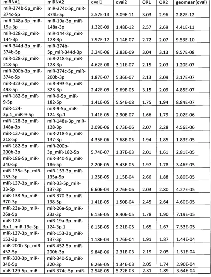
Co-targeting by distinct miRNA pairs is prevalent... 50
The co-targeting miRNAs miR-138 and miR-137 coordinately increase across neuronal differentiation...52
miR-137 can rescue a block in neuronal differentiation caused by loss of mir-138...61
Groups of m iRNAs preferentially share targets with one another... 66
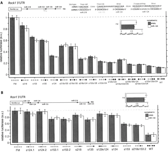
Groups of brain cluster miRNAs can collaborate to exert strong repression ... 67
Co-targeting m iRNA sites with close spacing act cooperatively ... 74
Co-targeting miRNAs direct potent and complex patterns of repression ... 75
Discussion ... 80
Acknowledgem ents ... 83
Author contribution...83
Supplem ental figures ... 84
M ethods ... 89 References...98 C hapter 3: C onclusions ... 103 Im plications ... 104 Future Directions ... 107 References...109
Appendix I: Supplemental Tables to Chapter 2 ... 111
Table I-Si...112
Table I-S2...118
Table I-S3...120
Table I-S4...124
Chapter 1:
Introduction
Overview
microRNAs (miRNAs) are small non-coding RNAs that repress expression of specific target genes through recognition of a sequence complementary to the seed (nucleotides 2 - 8) of
the miRNA. Collectively, the hundreds of confidently annotated miRNAs are predicted to target over 60% of protein coding genes in the mammalian genome (Friedman et al., 2008), yielding a profound amount of regulatory potential en masse. Research over the past two decades has revealed roles for miRNAs in a multitude of diverse cellular processes and their contribution to disease pathogenesis (Bartel, 2018). Here, I will review what we have learned about miRNAs since their initial discovery in 1993 in an effort to connect this to how we think miRNAs function to meaningfully impact gene regulation and the questions that have yet to be answered in the field.
A brief history and overview of microRNAs
initial discovery and large-scale identification of miRNA genes
The first microRNA (miRNA) gene, lin-4, was identified in C. Elegans as an essential regulator of the developmental timing of early larval development. Victor Ambros and
colleagues discovered that lin-4 did not encode a protein but rather two RNA transcripts of approximately 22 and 61 nt in size and proposed that the shorter product may be processed from the from the longer one, later defined as the mature and precursor miRNAs (Lee et al., 1993). These small RNA species contained antisense complementarity to a sequence element found repeated in the 3' untranslated region (UTR) of lin-14 mRNA, which was shown to be necessary for the regulation of lin-14 by lin-4 (Wightman et al., 1993). The true gravity of this finding would come to be understood several years later with the discovery of let-7 in C. Elegans, an
essential regulator of lin-41 in the transition from late larval development to adult cell fates (Reinhart et al., 2000; Slack et al., 2000). And more consequential, let-7 homologs were identified across a range of bilaterian animal species, including vertebrates, ascidian,
hemichordates, and others (Pasquinelli et al., 2000). In the year to follow, small RNA cloning and sequencing experiments in worms, flies, and mammals revealed over a hundred similar short non-coding RNAs, defining a new conserved class of small regulatory RNAs that would be named miRNAs after their small size and unknown function (Lagos-Quintana et al., 2001; Lau et al., 2001; Lee and Ambros, 2001).
The field experienced a rapid expansion in the number and types of studies, defining hundreds of additional miRNA sequences in more than 200 species using both experimental and computational methods (Kozomara and Griffiths-Jones, 2014; Lai et al., 2003; Lim et al., 2003). Sequencing of miRNAs in different mammalian tissues and cell types revealed tissue-specific signatures of miRNA expression (Houbaviy et al., 2003; Lagos-Quintana et al., 2003; 2002; Landgraf et al., 2007), suggesting roles for miRNAs in the definition or maintenance of particular cell types (discussed further in a later section.) High throughput sequencing of small
RNAs paired with stringent criteria for defining a miRNA gene (Lu et al., 2005; Ruby et al.,
2006) puts current estimates of confidently annotated canonical miRNAs at 147 in C. Elegans
(Jan et al., 2011), 164 in Drosophila (Fromm et al., 2015), 475 in mouse (Chiang et al., 2010), and 519 in human (Fromm et al., 2015). These miRNAs can be grouped into seed families with one another on the basis of sharing the same seed sequence and thus likely recognizing and regulating the same set of targets. Of the aforementioned 519 human miRNA genes, 296 fall within 177 seed families conserved among placental mammals, 200 fall within 89 seed families
common ancestor of humans, flies, and nematodes (Bartel, 2018), revealing sets of both deeply conserved and more newly evolved miRNAs.
Biogenesis
miRNAs are encoded in both intergenic regions under the control of their own promoter and within introns of pre-mRNAs, the latter constituting about a quarter of conserved miRNAs and more than half of all poorly conserved mammalian miRNAs (Chiang et al., 2010). These primary miRNA (pri-miRNA) sequences are transcribed by RNA Polymerase II (Pol II) and contain an imperfect hairpin from which the mature miRNA will be processed (Lee et al., 2004). Pri-miRNAs transcribed from intergenic regions are 5' capped and often polyadenylated (Cai et al., 2004), while those contained in introns can be spliced out of the pre-mRNA or transcribed independently but are most commonly co-expressed with the host gene (Baskerville and Bartel,
2005; Rodriguez et al., 2004). Many miRNAs are clustered in the genome and are expressed as a
single polycistronic pri-miRNA from which multiple different miRNAs can be processed, allowing the co-expression of these miRNAs upon transcriptional activation (Lee et al., 2002).
In the canonical pathway, miRNA hairpins are processed out of the pri-miRNA by Microprocessor, a heterotrimeric complex containing one molecule of the Drosha
endoribonuclease and two molecules of its double stranded RNA-binding cofactor DGCR8. Drosha, which contains two RNase III domains, makes two asymmetric cuts approximately 11
bp from the basal ssRNA-dsRNA junction (an interaction which is stabilized and made accurate by the interaction of DGCR8 with the stem and apical junction) producing a -66 nt stem-loop
structure called the precursor miRNA (pre-miRNA), which bears a 5' monophosphate (MP) and a 2 nt 3' overhang (Lee et al., 2003; Nguyen et al., 2015). The pre-miRNA is exported from
nucleus to the cytoplasm by the nuclear export factor, exportin-5, in a RanGTP-dependent manner (Bohnsack et al., 2004; Lund et al., 2004; Yi et al., 2003). Once in the cytoplasm, the pre-miRNA is further processed by Dicer, an endonuclease that makes two cuts to generate the
miRNA duplex with -2 nt 3' overhangs on each end (Zhang et al., 2004). Later studies revealed some sequence specificity beyond the known structural requirements that can enhance the efficiency of processing: GHG motif near the base of the stem (positions 7 -9) with the central
base mismatched (Fang and Bartel, 2015), UG at the 5' base of the stem, UGU at the 5' end of the apical loop, and CNNC 5 nt 3' of the stem (Auyeung et al., 2013). The sequence and structural specificity for miRNA processing has helped to define rules for miRNA gene discovery and can instruct de novo design of miRNA hairpins.
The miRNA duplex is loaded into Argonaute (AGO), which lies at the heart of the RNA-Induced Silencing Complex (RISC), with the help from two chaperone proteins, HSC70/HSP90, that shift AGO to an open conformation permissive of the miRNA duplex, in an ATP-dependent manner (Iwasaki et al., 2010; Kawamata and Tomari, 2010). One of the strands of the miRNA duplex is preferentially selected as the guide strand, which stays in AGO, and the other becomes the passenger strand, which is removed from the complex and degraded. The strand selection is based on the original orientation of duplex loading: AGO favors the strand with the
thermodynamically less stable 5'-terminal pairing as the guide strand (Khvorova et al., 2003; Schwarz et al., 2003), in addition to preference for an AMP or UMP at the 5' end which permits a more optimal fit into the phosphate-binding pocket at the junction of the MID and PIWI domains of AGO (Frank et al., 2010; Suzuki et al., 2015). The miRNA-loaded RISC is equipped and ready to carry out sequence-specific regulation of target genes.
Target recognition
For newly discovered miRNAs, aptly named for their small size but unknown function when first identified, accurate target predictions were key to understanding what functions they might serve in a cell. The complementarity observed between lin-4 or let-7 and sequences in the 3' UTR of each miRNA's respective target, lin-28 or lin-41, provided the first indications as to how miRNAs target specific mRNAs (Lee et al., 1993; Reinhart et al., 2000; Wightman et al.,
1993). The most defining feature for predicting targets of a miRNA is Watson-Crick pairing
through the seed region (nucleotides 2 - 7) at the 5' end of the miRNA (Lewis et al., 2003). This helps to explain why the 5' end of the miRNA is the most conserved region of metazoan miRNA
genes (Lim et al., 2003). Preferential conservation of the canonical target site, defined by uninterrupted pairing to the miRNA seed, vastly improved target predictions (Brennecke et al.,
2005; Krek et al., 2005; Lewis et al., 2005). Bases opposite position I and 8 of the miRNA were
also strongly conserved. While the base opposite position 8 showed a strong bias to provide additional Watson-Crick pairing, the base opposite position 1 had a strong bias for adenosine, regardless of the base in the miRNA (Lewis et al., 2005). The majority of miRNAs actually begin with a U (Lau et al., 2001), but for miRNAs that do not, requiring an A at position I is a better predictor of targeting than Watson-Crick pairing at this position (Baek et al., 2008; Nielsen et al., 2007). This is because the A does not pair with the miRNA but is inserted into a binding pocket of Argonaute which serves to further stabilize the miRNA:target interaction (Schirle et al., 2014). Based on these principles, several canonical target site types were defined; these include: the 8mer, 7mer-m8, 7mer-Al, and 6mer site (sites are listed in order of decreasing preferential conservation and repressive ability). It should be noted that 6mer sites typically
confer regulation barely over that of background, so these sites are generally not considered in most analyses (Agarwal et al., 2015; Grimson et al., 2007; Nielsen et al., 2007).
Supplementary pairing to the 3' end of the miRNA does not strongly increase target site effectiveness (Baek et al., 2008; Grimson et al., 2007). A present but modest effect, possessing higher preferential conservation and conferring greater target repression, is only observed for supplementary pairing centered on miRNA bases 13 to 16 (Grimson et al., 2007), thus the term "3' supplementary pairing" refers to this specific form of pairing to the 3' region. Structural studies support that bases 13 - 16 are special. After target binding is initiated through guide
nucleotides 2 to 5, AGO undergoes a conformational change exposing nucleotides 6 - 8 and 13 -16 (Schirle et al., 2014), thus it is logical that this 3' region would be the most instructive for
target binding after the seed itself. These 3' supplementary sites, containing both 3'
supplementary pairing and full complementarity to the seed, are atypical, ~5% of canonical conserved target sites (Friedman et al., 2008), and have a modest impact on the levels of target repression (Grimson et al., 2007) and target affinity (Salomon et al., 2015; Wee et al., 2012), or if the supplementary pairing does increase affinity, it appears to be specific to only some
miRNAs (Salomon et al., 2015).
3' supplementary pairing can help compensate for a mismatch or bulge in the miRNA
seed - this is called a 3' compensatory site. This site type requires more extensive
complementarity to the 3' region, at least 9 consecutive bases in all experimentally validated examples, and is rarely under selective pressure to be conserved, constituting 1% of
preferentially conserved sites in mammals (Brennecke et al., 2005; Friedman and Burge, 2013; Lewis et al., 2005). However, 3' compensatory sites can have functional reasons for evolving, such as let-7 targeting of lin-41. Lin-41 contains two highly conserved 3' compensatory sites for
let-7, so why conserve these sites so strongly if they are less effective and more difficult to
conserve? The reason lies in that let-7 has several family members in worm, which contain the same seed sequence but have distinct sequence otherwise (Lau et al., 2001; Lim et al., 2003). These paralogs are expressed at earlier stages in development than let-7, and so if these family members were able to target every site in lin-41, they might drive down lin-41 expression too quickly and trigger early differentiation (Abbott et al., 2005; Reinhart et al., 2000). Cleverly, these sites develop imperfect pairing in the seed and extensive pairing to the 3' region to disrupt effective targeting and only restore targeting for specific paralogs with extensive 3'
complementarity (Brennecke et al., 2005; Lewis et al., 2005).
Other forms of non-canonical binding may be more prevalent. High throughput
sequencing of RNAs that crosslinked with AGO revealed high levels of AGO binding with no canonical 6mer target site, making up about 25-50% of all crosslink events (Chi et al., 2012;
2009; Grosswendt et al., 2014; Hafner et al., 2010; Helwak et al., 2013; Loeb et al., 2012). These
studies characterized new non-canonical site types, such as pivot pairing which contains a bulged nt in the seed between position 5 and 6 (Chi et al., 2012), and found evidence that some these sites are quite prevalent, under purifying selection, and repressed but to a lesser extent than canonical motifs (Chi et al., 2012; Grosswendt et al., 2014; Helwak et al., 2013; Loeb et al., 2012). In contrast, other studies have found that these noncanonical AGO-bound mRNAs do not confer detectable levels of repression in experiments where miRNA expression is altered
(Agarwal et al., 2015). McGeary and colleagues, using a new in vitro method to assess relative binding affinities of AGO loaded with different miRNAs (based off of the RNA Bind-n-Seq protocol (Lambert et al., 2014)), identified instances of strong binding to non-canonical sites with affinities that could exceed those of 7mer-m8 and 7mer-A 1 canonical binding in some
instances (McGeary et al.). Many of these non-canonical sites are miRNA-specific, which could explain why these features of non-canonical targeting were not identified with earlier more global searches for features. For miR-124 (which had the largest number of high affinity canonical sites), there were 5 canonical sites higher affinity than the 7mer-m8, and 29 non-canonical sites with higher affinity than the 7mer-Al sites (McGeary et al.). Many of the high affinity non-canonical sites have no seed at all but instead extensive pairing to the 3' region of the miRNA (McGeary et al.). Current models of AGO binding will need to be expanded to explain these new site types; perhaps AGO has more than one binding mode allowing it to accommodate both. Because non-canonical affinities are miRNA-specific, performing AGO-RBNS on additional miRNAs will strengthen future predictions, and in addition, modification of the technique to allow evaluation of miRNA-specific 3' supplementary effects in the presence of a canonical site could provide more accurate prediction of expected repression from individual sites.
Mechanisms of repression
miRNA-mediated repression in animals acts primarily via mRNA destabilization. Upon target recognition and binding by RISC, AGO recruits the adaptor protein TNRC6 (GW182 in
flies and AIN-1/2 in nematodes) (Ding et al., 2005; Rehwinkel et al., 2005) that interacts with the Poly(A)-binding protein (PABPC) in the poly(A) tail and recruits the deadenylase complexes: the PAN2-PAN3 complex and the CCR4-NOT complex (Jonas and Izaurralde, 2015). These
complexes cause shortening of the poly(A) tail, leading to mRNA destabilization through 5' decapping by the Decapping protein 2 (DCP2) and rapid 5'-to-3' decay by the exonuclease XRN1 (Chen and Shyu, 2011).
miRNAs can also act to inhibit translation through the recruitment of DDX6, a helicase that binds the decapping complex, by the CCR4-NOT complex (Chu and Rana, 2006; Jonas and Izaurralde, 2015). DDX6 also interacts with the EIF4E transporter 4E-T, which normally
competes with EIF4G for binding to EIF4E, further enhancing both translational repression and mRNA destabilization (Kamenska et al., 2014; 2016; Nishimura et al., 2015; Ozgur et al., 2015). Kinetic studies of miRNA regulation revealed that translational inhibition occurs very soon after RISC binding to the target transcript, which is followed by deadenylation, decapping, and degradation of the bound mRNA target (Bethune et al., 2012; Djuranovic et al., 2012).
While early characterization focused on the effect of miRNAs on translation, later studies, which compared mRNA levels with protein expression or translational efficiency upon overexpression or depletion of a miRNA, demonstrated that decreases at the level of mRNA could explain the majority (66 -90%) of the steady state repression by mammalian miRNAs
(Baek et al., 2008; Eichhom et al., 2014; Guo et al., 2010; Hendrickson et al., 2009). This result not only had important mechanistic implications for understanding miRNA regulation, but also had practical implications - the effects of a miRNA could be faithfully measured using RNA
sequencing. However, an exception to this result lies in the cells of early embryos, in which translational repression serves as the primary mode of regulation (Bazzini et al., 2012; Subtelny et al., 2014). A notable example, miR-430 targets maternal mRNAs in the early zebrafish embryos, acting first by strongly repressing translation and only triggering decay of target mRNAs at later stages (Bazzini et al., 2012). Poly(A)-tail shortening appears to have very different consequences in the early embryo compared to post-gastrulation cells after zygotic transcription is activated. In early embryonic cells, poly(A)-tail shortening reduces translational efficiency with little effect on mRNA stability, but inversely, poly(A)-tail shortening has strong
destabilizing effects on mRNA with little effect on translational efficiency in cells post-gastrulation (Subtelny et al., 2014).
Alternatively, if a target has sufficiently extensive pairing through the center of the miRNA, AGO2, the only mammalian Argonaute to retain slicing activity (Liu et al., 2004;
Meister et al., 2004), can catalyze cleavage of the target transcript (Hutvaigner and Zamore, 2002; Yekta et al., 2004). In one model, substantial pairing across the target initiates a second site of nucleation at nucleotides 13-16 of the miRNA, eventually releasing the 3' end of the miRNA from its binding pocket and initiating a conformational change in AGO that brings the target in close proximity to the activated DEDH catalytic tetrad core and cleaves the target at the
phosphodiester bond opposite nucleotides 10 and 11 of the miRNA (Bartel, 2018). The 5' and 3' products are then degraded by the exosome and XRN 1 respectively. While this is the major mode of miRNA-mediated repression in plants (Jones-Rhoades et al., 2006), target slicing rarely occurs in mammals and has only been observed in a total of twenty-one mammalian transcripts (Davis et al., 2005; Hansen et al., 2011; Shin et al., 2010; Yekta et al., 2004).
Factors that influence targeting efficacy
Early experiments revealed that miRNA targeting was widespread - hundreds of mammalian genes were repressed following exogenous miRNA overexpression (Lim et al.,
2005). Conversely, inhibiting or ablating an endogenous miRNA also had widespread effects on
the transcriptome (Giraldez et al., 2006; Kriitzfeldt et al., 2005; Rodriguez et al., 2007). While widespread, the effects were quite modest - often repression of any given target is about 20% (Baek et al., 2008; Selbach et al., 2008). These experiments revealed the extensive regulatory effect miRNAs have on the transcriptome, but also urged the question of how to accurately
predict which targets will be regulated, and which targets will be regulated most strongly. There are additional factors beyond the presence of the 7 or 8 nt seed sequence that influence targeting efficacy. Because the study of miRNAs even still today often requires sets of predicted targets to identify direct versus indirect effects of the miRNA, target prediction remains very important.
Conservation generally serves as a good predictor of effective targeting, in addition to being an indicator of biological significance in which a gain in fitness drives the selective maintenance of the target sites (Baek et al., 2008; Brennecke et al., 2005; Krek et al., 2005; Lewis et al., 2005; Neilson et al., 2007). Mammalian miRNAs conserved through vertebrates have an average of more than 300 conserved targets containing 7 or 8mer sites (more than 400 if 6mer sites are included) - overall, more than half of all protein-coding genes are under selective pressure to maintain sites in the 3' UTR for pairing with miRNAs (Friedman et al., 2008). While conservation selects for sites that more likely to be regulated, non-conserved sites can also confer repression (Farh et al., 2005). There are an astounding lOx more poorly conserved 7mer sites than preferentially conserved sites (Farh et al., 2005). Given the huge number of non-conserved sites, one might assume that some fraction of these could exert repressive function and wreak havoc on the regulatory systems, but these non-conserved sites are mostly found in genes expressed primarily in tissues where the cognate miRNA is not expressed (Farh et al., 2005). Thus, sites can accumulate in particular 3' UTR regions (where the gene and miRNA are not co-expressed) without any detrimental effect to the organism, but sites are selectively avoided in 3' UTR regions where the gene and miRNA are both highly expressed because this would
otherwise confer unwanted repression. The "selective avoidance" for sites in highly co-expressed transcripts is so robust that it is possible to accurately predict the expression patterns of miRNAs based on depleted 7 nt motifs in the 3' UTRs of transcripts that are preferentially expressed in
particular tissues (Farh et al., 2005). The most highly expressed genes contain on average about half as many non-conserved 7 nt sites than would be expected by chance (Farh et al., 2005), and provides a reasoning as to why highly expressed "house keeping" genes in animals typically have shorter 3' UTRs in general and compared to orthologs in plants and fungi which do not have extensive 3' UTR targeting - reducing 3' UTR length can be a regulatory strategy to avoid
spurious targeting and repression (Farh et al., 2005; Stark et al., 2005). The combination of conservation and selective avoidance of target sites makes the evolution of almost all
mammalian mRNAs under the influence of miRNAs. For the non-conserved target sites that are not selectively avoided (i.e. mRNAs containing non-conserved sites and the targeting miRNA are co-expressed), some of these may represent newly evolving, species-specific regulation, but many could also lack efficient repressive ability due to non-optimal context features.
Sites are more effective, i.e. drive stronger repression, when A/U-rich sequences flank the seed sequence (Grimson et al., 2007; Neilson et al., 2007). Recent biochemical work from the Bartel Lab also underscores the importance of flanking di-nucleotide (di-nt) content, finding that the two bases immediately upstream and downstream of the seed can alter site affinity more than changing the site type itself (McGeary et al.). The presence of an A or U in these positions enhances affinity, a G reduces affinity, and a C is somewhat neutral. In addition, the 5' flanking di-nt appears to be about 2x more important than the 3' flanking sequence (McGeary et al.), affirming earlier observations that an A/U nt opposite miRNA position 9 gives the strongest repression enhancement (Neilson et al., 2007). With the presence of two 5' flanking A's, a
miR-124 site with a bulge in the seed between nucleotides 5 and 6 is almost just as strong as a perfect 8mer target site (McGeary et al.). These context preferences likely reflect an underlying
site remains in an unpaired state, which is supported by analyses of several independent measures of structure and accessibility (Grimson et al., 2007; McGeary et al.; Robins et al., 2005).
The location of the target site within the transcript also matters. While 7mer and 8mer miRNA seed match sites can be found in the 5' UTR and in the coding sequence (CDS), sites generally only confer repression when located in the 3' UTR. Further, these sites must be at least
15 nt downstream of the stop codon, likely for the same reason that sites in the CDS do not
confer repression - it is hypothesized that the ribosome prevents efficient binding by knocking off proteins in its path (Grimson et al., 2007). Target sites are also generally more effective if they are located near the start or end of the 3' UTR (Grimson et al., 2007), as more complex
folded structures in the center of a long 3' UTR may occlude the site or distance it from interaction with relevant proteins like the PABPC.
Target sites also tend to be more effective if they reside in close proximity to sites for other co-expressed miRNAs (Grimson et al., 2007). This observation is likely due to the fact that target sites can be cooperative (i.e. yield more repression than expected from each site
individually) if they lie within a defined distance from one another, typically 15 to 35 nt between seed starts (Doench and Sharp, 2004; Grimson et al., 2007; Saetrom et al., 2007). Early evidence suggests that TNRC6 (GW182 in flies) may contribute to this observed cooperative effect. TNRC6 is composed of an unstructured N-terminal/Ago-binding domain (ABD) and a C-terminal/silencing domain. In humans, the ABD domain contains more than 30 glycine-tryptophan (GW/WG) repeats, of which three have been shown to mediate an interaction with
AGO (Lazzaretti et al., 2009; Lian et al., 2009; Takimoto et al., 2009; Till et al., 2007). While AGO can only bind a single GW motif, TNRC6 can bind up to three AGO proteins at once
(Elkayam et al., 2017). The presence of multiple guide-primed AGOs and auxiliary target sites in close proximity could result in increased dwell time on the target transcript and help to explain the cooperative effect.
Based on the occurrence of a miRNA target site in the transcriptome, miRNAs will have different target abundances, which has been proposed to alter miRNA activity (Arvey et al.,
2010). The effective target abundance is defined as the number of target sites that must be added
into a cell to achieve half-maximal de-repression of targets (Denzler et al., 2014). miRNAs often have limited expression compared to the many conserved and non-conserved target sites they recognize in hundreds to thousands of mRNAs. One of the most highly expressed miRNAs liver-specific miR-122, which constitutes 72% of the total miRNA in hepatocytes at 135,000 copies per cell, has an even higher effective target abundance (Chang et al., 2004; Denzler et al., 2014;
2016). This means that miRNA levels are rarely saturating and targets will remain responsive to
changes in miRNA expression allowing them to dynamically regulate levels of repression
through their own abundance. The finding that all effective target abundances are quite high also means that the competing endogenous RNA (ceRNA) hypothesis, which posited that fluxes in target abundances from gene expression changes could serve to regulate the activity of a
miRNA, is very unlikely (Salmena et al., 2011). Extending back to our example of miR- 122, 1.5 x 10⁵ sites per cell had to be added to begin to see de-repression of miR- 122 targets (Denzler et al., 2014). This value, and those calculated for additional miRNAs, exceeds the physiological expression of any one gene and is more than the sum of all predicted targets that change in a disease state, thus any changes from competing endogenous miRNAs would be small and likely inconsequential (Denzler et al., 2014; 2016).
The availability of target sites themselves can be regulated, thus turning miRNA
regulation on or off for a particular gene. The usage of different 3'UTR isoforms, alternative last exons (ALEs) or tandem 3' UTRs, can be dynamically regulated and has been shown to lead to the gain or loss of miRNA target sites (Mayr and Bartel, 2009; Miura et al., 2013; Sandberg et al., 2008). For example, highly proliferative cells, like cancer cells, tend to use upstream alterative polyadenylation sites thus shortening the 3' UTR and potentially removing any
miRNA-mediated repression that was encoded in that sequence. RNA editing, such as adenosine-to-inosine editing catalyzed by adenosine deaminases that act on double-stranded RNA (ADAR), could also disrupt or introduce a site for miRNA binding. One study found that 20% of A-to-I editing events were in miRNA seed sites (Borchert et al., 2009; Peng et al., 2012). Lastly, RNA binding proteins (RBPs) can compete for binding and occlude miRNA-loaded RISC binding when an RBP motif is overlapping that of a miRNA site or if the RBP footprint would sterically hinder RISC binding.
miRNAs working together Combinatorial activity by miRNAs
The regulatory logic of miRNAs is incompletely understood - miRNAs often regulate hundreds of targets but confer only a small amount of repression, often ~20%, to each seed match site (Baek et al., 2008; Selbach et al., 2008). However, genes usually contain multiple conserved seed match sites in their 3' UTRs, with an average of 4.2 target sites per targeted 3' UTR (Friedman et al., 2008). While a gene can develop multiple sites to the same miRNA (Mayr et al., 2007), it is much more common to have sites to different miRNA families with only 7% of genes containing one or more conserved site for the same miRNA, and 72% of genes contain
more than one conserved site for any miRNA family (Friedman et al., 2008). The genes most
highly targeted by all miRNAs are enriched for transcriptional regulators and nuclear factors
(Hon and Zhang, 2007), which may hint that miRNAs are more likely to co-target genes in key gene regulatory networks. The repression conferred by two individual sites is multiplicative (also referred to as log-additive), so theoretically, five target sites that individually repress an mRNA to 80% of its prior level could together repress expression to (0.8)5 = -33% of its prior level (Grimson et al., 2007; Neilson et al., 2007). Two sites can confer greater repression together than they can individually if the sites lie within a cooperative distance from one another, most
optimally when there is 13-35 nt between seed starts (Doench and Sharp, 2004; Grimson et al.,
2007; Saetrom et al., 2007). This cooperative effect can yield stronger, more responsive tuning of
gene expression, particularly where miRNAs have differences in temporal expression or varying levels across cell types.
Some early studies selected candidate genes to test for the ability for more than one miRNA to regulate it. However, the results were underwhelming with no more than 2-fold repression for two or three sites in a 3' UTR (Grimson et al., 2007; Krek et al., 2005), and in one case, no repression was observed for three sites until the authors moved the sites artificially closer to one another into a cooperative distance which also would have impacted the entire context they had evolved in (Saetrom et al., 2007). The idea of co-targeting has also been explored on a more global level, but in a limited number of studies. However, it was observed that miRNAs expressed from the same polycistronic clusters, and thus co-expressed, had targets
sets with overlapping genes as well as enrichments for similar pathways (Tsang et al., 2010). This demonstrated that miRNAs expressed in the same fashion have the potential to develop co-targeting relationships, possibly with a stronger and more detectable relationship than pairs of
miRNAs who do not correlate as strongly in their expression patterns. In another study, Obermayer and colleagues found that particular pairs of miRNAs had an enriched number of sites that were co-conserved in a 3' UTR together, suggesting that co-targeting can be a conserved mechanism of regulation (Obermayer and Levine, 2014).
Examples of synergism between miRNAs
Several examples of miRNAs that have synergistic effects or possible co-targeting relationships are known. For some of these, it is has not been investigated where the underlying synergy is coming from - a wider breadth of targets or co-targeting a core set transcripts to drive larger levels of repression - something that may be an important distinction to make as the regulatory logic of miRNAs is investigated. For example, the neuronal miRNAs 9 and
miR-124 can drive the direct conversion of fibroblasts to neurons in combination but not alone (Yoo et al., 2011). In another study, the role of a trio of miRNAs, miR-124, miR-128 and miR-137, in the differentiation of neural stem cells was explored, specifically by knocking down each
miRNA and performing RNA-sequencing to attain a list of semi-validated targets for each miRNA (Santos et al., 2016). Their de-repressed targets sets had a striking amount of overlap leading to the possibility that they synergistically promote differentiation through their large co-targeting network which appears to converge on Spi (Santos et al., 2016). In another instance, Riley and colleagues identified a co-targeting relationship between Epstein-Barr virus (EBV) and
highly expressed human host miRNAs of both viral and human genes involved in cell cycle and
apoptosis (Riley et al., 2012). They validated some of these co-targets with luciferase assays, and for one viral RNA target BHFR1, containing 2 sites for a viral miRNA and one site for miR- 142, they observed about 4-fold repression (Riley et al., 2012).
miRNAs in the brain
miRNA expression profiling has revealed high and specific/enriched expression of many miRNAs in the brain (Landgraf et al., 2007; Sempere et al., 2004) and temporal patterns of expression during development or neuronal differentiation (Krichevsky et al., 2003; Miska et al., 2004; Sempere et al., 2004), suggesting a specific role for many miRNAs in the brain. The
temporal regulation and tissue-specificity of miRNA expression has long suggested that miRNAs may play a role in cellular differentiation and maintenance. In an early experiment,
overexpression of either miR- 124 or miR- 1 in HeLa cells resulted in repression of targets specific to each miRNA but also globally shifted the transcriptome to look more like that of the tissue the transfected miRNA is primarily expressed in, brain and muscle respectively (Lim et al., 2005), demonstrating the role miRNAs can play in shaping the gene expression program of cells.
When Giraldez and colleagues generated maternal zygotic mutants of zebrafish Dicer, ablating the miRNA-processing pathway, they observed gross defects in early brain patterning and morphogenesis, demonstrating that miRNAs are essential for proper brain development (Giraldez et al., 2005). Similar severe phenotypes of defective brain patterning and neuronal differentiation were observed in mammalian systems using conditional knockouts of Dicer or other miRNA biogenesis factors (Choi et al., 2008; Cuellar et al., 2008; Damiani et al., 2008; Davis et al., 2008; Kim et al., 2007). Subsequent studies have revealed prominent roles for miRNAs in many fundamental processes of the brain, including neuronal progenitor expansion, differentiation and migration, in addition to the establishment of synaptic connections and activity-dependent plasticity of these connections (Hu and Li, 2017; Rajman and Schratt, 2017).
miR-124 in neuronal differentiation
Many miRNAs have been shown to play critical roles in neuronal differentiation through diverse cellular mechanisms (Rajman and Schratt, 2017). Here, I will highlight what is known about one of these miRNAs, miR-124, which is one of the most highly expressed, neuron-specific miRNAs (Lagos-Quintana et al., 2002), as well as one of the most well-studied miRNAs. The overexpression of miR- 124 in a variety of systems, including neuronal progenitors, embryonic stems cells, glioma cells and fibroblasts, results in forced neuronal differentiation (Krichevsky et al., 2006; Silber et al., 2008; Xia et al., 2012; Yoo et al., 2011).
One mechanism by which miR-124 drives differentiation is through targeting the small C-terminal phosphatase domain 1 (CTDSP1), which phosphorylates and stabilizes REl silencing transcription factor (REST) (Nesti et al., 2014; Visvanathan et al., 2007). REST binds upstream of neuronal genes and strongly represses their expression, and thus REST must be repressed for neuronal differentiation to occur (Ballas et al., 2005). Interestingly, REST represses the
expression of miR- 124, among other neuronal miRNAs, forming a double-negative feedback loop to strongly drive differentiation in a switch-like fashion (Visvanathan et al., 2007). Because REST represses the expression of many neuronal miRNAs, it has also been proposed that there may be a larger network of miRNAs that target multiple different components of the REST complex to help drive neuronal differentiation and stabilize neuronal gene expression (Wu and Xie, 2006).
miR- 124 also impacts alternative splicing through targeting polypyrimidine-tract-binding protein (PTBP 1), a splicing factor which is expressed in non-neuronal cells and suppresses the inclusion of neuron-specific exons (Makeyev et al., 2007). PTBP1 negatively regulates the
expression of the neuron-enriched homolog, PTBP2, by causing exon skipping and producing a nonsense-mediated decay (NMD) isoform (Makeyev et al., 2007). As PTBP1 is repressed and PTBP2 is activated, the cells transition to a neuronal splicing program. The importance of this regulatory connection is highlighted by the finding that knocking down PTBP 1 alone can drive the conversion of fibroblasts to neurons (Xue et al., 2013).
miR- 124 regulates the switch to neuron-specific ATP-dependent chromatin remodeling complexes, nBAF, by down-regulating the BAF53a subunit found in non-neural and neural progenitor cells, and allowing replacement by the homologous neuron-specific BAF53b (Yoo et al., 2009). BAF complexes regulate nucleosome mobility and chromosome accessibility, and nBAF is essential for many post-mitotic functions including dendritic development (Wu et al.,
2007). There is also strong evidence that both strands of miR-9 repress BAF53a (Yoo et al., 2009).
The Notch pathway controls neural progenitor cell maintenance and inhibits neuronal differentiation (Louvi and Artavanis-Tsakonas, 2006). When the Notch ligand Jaggedi (Jagi) binds the Notch transmembrane receptor, neural stem cell self-renewal is promoted and neuronal
differentiation is blocked (Imayoshi and Kageyama, 2011). miR-124 targets JagI to turn off Notch signaling and allow differentiation to proceed (Cheng et al., 2009; Liu et al., 2011).
Additional roles of neuronal miRNAs
Following neuronal cell specification, miRNAs have also been shown to play an important role in the migration of neurons through targets like doublecourtin, Foxp2, and N-cadherin (Clovis et al., 2012; Gaughwin et al., 2011; Rago et al., 2014). As neurons migrate they polarize, often having to convert from a multipolar to bipolar morphology after they reach their
final location, which once they reach, they begin to grow axons and dendrites - miRNAs are implicated in regulating each level of this (Rajman and Schratt, 2017). Axon guidance through the growth cone is dependent on local translation, and multiple miRNAs have been implicated in the regulation of this process (Rajman and Schratt, 2017). Lastly, synapse development and maintenance depends on the presence of particular miRNAs in the dendritic spine for control of local translation (Rajman and Schratt, 2017). For example, miR-134 negatively regulates
dendritic spine size by inhibiting the local synthesis of Limkl, which promotes actin
polymerization and thus growth of spines (Schratt et al., 2006). Both miR-138 and miR-137 have also been implicated in the regulation of dendritic spines (Siegel et al., 2009; Strazisar et al., 2015).
miRNAs are highly expressed in the CNS and show a striking amount subcellular specificity (Schratt, 2009). Several studies have profiled the axonal miRNA population in comparison to that of the cell body and found an abundant population of axonally expressed miRNAs, as well as an enriched set of miRNAs in the axon implying a selective functional role for certain miRNAs within the axon and at the synapse (Natera-Naranjo et al., 2010; Sasaki et al., 2013). For some of these miRNAs, specific roles have been demonstrated in these distal compartments, for example, mir-9 has been shown to negatively regulate axon length and increase axon branching by locally regulating the expression of its target microtubule-associated protein lb (Mapib), which functions in stabilizing axonal microtubules (Dajas-Bailador et al., 2012). These findings point toward miRNAs having important roles in distinct cells, cellular regions, and cellular processes within the nervous system.
Adenosine-to-inosine editing may have a special role in the brain because ADAR activity is enriched in the brain compared to other tissues. This type of editing has been observed to
occur in pri-miRNAs to block processing by Dicer (Kawahara et al., 2007a) or alter the miRNA seed sequence (Kawahara et al., 2007b), as well as altering the sequence of miRNA target sites (Borchert et al., 2009).
REFERENCES
Abbott, A.L., Alvarez-Saavedra, E., Miska, E.A., Lau, N.C., Bartel, D.P., Horvitz, H.R., and Ambros, V. (2005). The let-7 MicroRNA family members mir-48, mir-84, and mir-241 function together to regulate developmental timing in Caenorhabditis elegans. Developmental Cell 9, 403-414.
Agarwal, V., Bell, G.W., Nam, J.-W., and Bartel, D.P. (2015). Predicting effective microRNA target sites in mammalian mRNAs. Elife 4, 1-3 8.
Arvey, A., Larsson, E., Sander, C., Leslie, C.S., and Marks, D.S. (2010). Target mRNA abundance dilutes microRNA and siRNA activity. Mol. Syst. Biol. 6, 363.
Auyeung, V.C., Ulitsky, I., McGeary, S.E., and Bartel, D.P. (2013). Beyond secondary structure: primary-sequence determinants license pri-miRNA hairpins for processing. Cell 152, 844-858.
Baek, D., Villen, J., Shin, C., Camargo, F.D., Gygi, S.P., and Bartel, D.P. (2008). The impact of microRNAs on protein output. Nature 455, 64-71.
Ballas, N., Grunseich, C., Lu, D.D., Speh, J.C., and Mandel, G. (2005). REST and its
corepressors mediate plasticity of neuronal gene chromatin throughout neurogenesis. Cell 121,
645-657.
Bartel, D.P. (2018). Metazoan MicroRNAs. Cell 173, 20-51.
Baskerville, S., and Bartel, D.P. (2005). Microarray profiling of microRNAs reveals frequent coexpression with neighboring miRNAs and host genes. Rna 11, 241-247.
Bazzini, A.A., Lee, M.T., and Giraldez, A.J. (2012). Ribosome profiling shows that miR-430 reduces translation before causing mRNA decay in zebrafish. Science 336, 233-237.
Bethune, J., Artus-Revel, C.G., and Filipowicz, W. (2012). scientific report. EMBO Reports 13, 716-723.
Bohnsack, M.T., Czaplinski, K., and Gorlich, D. (2004). Exportin 5 is a RanGTP-dependent dsRNA-binding protein that mediates nuclear export of pre-miRNAs. Rna 10, 185-191.
Borchert, G.M., Gilmore, B.L., Spengler, R.M., Xing, Y., Lanier, W., Bhattacharya, D., and Davidson, B.L. (2009). Adenosine deamination in human transcripts generates novel microRNA binding sites. Human Molecular Genetics 18, 4801-4807.
Brennecke, J., Stark, A., Russell, R.B., and Cohen, S.M. (2005). Principles of microRNA-target recognition. Plos Biol 3, e85.
Cai, X., Hagedom, C.H., and Cullen, B.R. (2004). Human microRNAs are processed from capped, polyadenylated transcripts that can also function as mRNAs. Rna 10, 1957-1966.
Moloshok, T., Bort, R., et al. (2004). miR- 122, a mammalian liver-specific microRNA, is processed from her mRNA and may downregulate the high affinity cationic amino acid transporter CAT-1. RNA Biology 1, 106-113.
Chen, C.-Y.A., and Shyu, A.-B. (2011). Mechanisms of deadenylation-dependent decay. WIREs
RNA 2, 167-183.
Cheng, L.-C., Pastrana, E., Tavazoie, M., and Doetsch, F. (2009). miR-124 regulates adult neurogenesis in the subventricular zone stem cell niche. Nat Neurosci 12, 399-408.
Chi, S.W., Hannon, G.J., and Darnell, R.B. (2012). An alternative mode of microRNA target recognition. Nat. Struct. Mol. Biol. 19, 321-327.
Chi, S.W., Zang, J.B., Mele, A., and Darnell, R.B. (2009). Argonaute HITS-CLIP decodes microRNA-mRNA interaction maps. Nature 460, 479-486.
Chiang, H.R., Schoenfeld, L.W., Ruby, J.G., Auyeung, V.C., Spies, N., Baek, D., Johnston, W.K., Russ, C., Luo, S., Babiarz, J.E., et al. (2010). Mammalian microRNAs: experimental evaluation of novel and previously annotated genes. Genes & Development 24, 992-1009. Choi, P.S., Zakhary, L., Choi, W.-Y., Caron, S., Alvarez-Saavedra, E., Miska, E.A., McManus,
M., Harfe, B., Giraldez, A.J., Horvitz, R.H., et al. (2008). Members of the miRNA-200 Family
Regulate Olfactory Neurogenesis. 57, 41-55.
Chu, C.-Y., and Rana, T.M. (2006). Translation repression in human cells by microRNA-induced gene silencing requires RCK/p54. Plos Biol 4, e210.
Clovis, Y.M., Enard, W., Marinaro, F., Huttner, W.B., and De Pietri Tonelli, D. (2012).
Convergent repression of Foxp2 3'UTR by miR-9 and miR-132 in embryonic mouse neocortex: implications for radial migration of neurons. Development 139, 3332-3342.
Cuellar, T.L., Davis, T.H., Nelson, P.T., Loeb, G.B., Harfe, B.D., Ullian, E., and McManus, M.T. (2008). Dicer loss in striatal neurons produces behavioral and neuroanatomical phenotypes in the absence of neurodegeneration. Proc. Natl. Acad. Sci. U.S.a. 105, 5614-5619.
Dajas-Bailador, F., Bonev, B., Garcez, P., Stanley, P., Guillemot, F., and Papalopulu, N. (2012). microRNA-9 regulates axon extension and branching by targeting Map lb in mouse cortical neurons. Nat Neurosci 15, 697-699.
Damiani, D., Alexander, J.J., O'Rourke, J.R., McManus, M., Jadhav, A.P., Cepko, C.L., Hauswirth, W.W., Harfe, B.D., and Strettoi, E. (2008). Dicer inactivation leads to progressive functional and structural degeneration of the mouse retina. Journal of Neuroscience 28,
4878-4887.
Davis, E., Caiment, F., Tordoir, X., Cavaille, J., Ferguson-Smith, A., Cockett, N., Georges, M., and Charlier, C. (2005). RNAi-mediated allelic trans-interaction at the imprinted Rtll/Peg 11 locus. Curr. Biol. 15, 743-749.