HAL Id: tel-01166034
https://tel.archives-ouvertes.fr/tel-01166034
Submitted on 22 Jun 2015
HAL is a multi-disciplinary open access
archive for the deposit and dissemination of sci-entific research documents, whether they are pub-lished or not. The documents may come from teaching and research institutions in France or abroad, or from public or private research centers.
L’archive ouverte pluridisciplinaire HAL, est destinée au dépôt et à la diffusion de documents scientifiques de niveau recherche, publiés ou non, émanant des établissements d’enseignement et de recherche français ou étrangers, des laboratoires publics ou privés.
Evaluation atraumatique de modèles murins de maladies
musculaires génétiques
Aurea Beatriz Martins Bach
To cite this version:
Aurea Beatriz Martins Bach. Evaluation atraumatique de modèles murins de maladies musculaires génétiques. Medical Physics [physics.med-ph]. Université Paris Sud - Paris XI; Universidade de São Paulo (Brésil), 2015. English. �NNT : 2015PA112064�. �tel-01166034�
UNIVERSITE PARIS-SUD
ÉCOLE DOCTORALE :
STITS
Laboratoire de Resonance Magnetique Nucléaire DISCIPLINE PHYSIQUE
THÈSE DE DOCTORAT
préparée en co-tutelle avec l'Universidade de São Paulo
soutenue le 12 / 05 /2015
par
Aurea Beatriz MARTINS BACH
Non-invasive evaluation of murine models for
genetic muscle diseases
Directeurs de thèse : Pierre G. CARLIER Directeur de Recherche – NMR Laboratory AIM-CEA Mariz VAINZOF Professor – Universidade de São Paulo
Composition du jury :
Président du jury : Edson AMARO JR Professor – Universidade de São Paulo
Rapporteurs : Jeanine J. PROMPERS Professor – Eindhoven University of Technology Luciene COVOLAN Professor – Universidade Federal de São Paulo
Examinateurs : Edson AMARO JR Professor – Universidade de São Paulo Emmanuel DURAND Professor – Université Paris Sud
Aurea Beatriz MARTINS BACH
Non-invasive evaluation of murine models for
genetic muscle diseases
Universidade de São Paulo · Université Paris-Sud
São Paulo
2015
Aurea Beatriz MARTINS BACH
Non-invasive evaluation of murine models for
genetic muscle diseases
Avaliação não invasiva de modelos murinos para doenças musculares
genéticas
Evaluation atraumatique de modèles murins de maladies musculaires
génétiques
Tese apresentada ao Instituto de Biociências
da Universidade de São Paulo, para a obtenção
de Título de Doutor em Ciências, Programa:
Ciências Biológicas (Biologia Genética) –
Área de Concentração: Biologia (Genética).
Doutorado
realizado
em
co-tutela
internacional com Université Paris-Sud,
França.
Orientadores: Mariz Vainzof
Pierre Carlier
São Paulo
2015
Ficha Catalográfica
Martins-Bach, Aurea Beatriz
Non-invasive evaluation of murine
models for genetic muscle diseases
Número de páginas 176
Tese (Doutorado) - Instituto de
Biociências da Universidade de São Paulo.
Departamento de Genética e Biologia
Evolutiva, em co-tutela internacional com
Université Paris-Sud, França.
1. NMR 2. Muscle dystrophy 3.
Congenital myopahty 4. Murine models
I. Universidade de São Paulo. Instituto de
Biociências. Departamento de Genética e
Biologia Evolutiva.
Comissão Julgadora
:
Dr. Edson AMARO JR.
Presidente da Banca Examinadora Dra. Jeanine PROMPERS
Revisora da tese Dr(a). Mariz Vainzof Orientadora
Dr. Emmanuel Durand Avaliador
Dra. Luciene COVOLAN Revisora da tese
Dr. Pierre CARLIER Orientador
Acknowledgements
I would like to express my sincere appreciation and gratitude to my supervisors in both sides of the Atlantic Ocean, Dr. Mariz Vainzof and Dr. Pierre Carlier. Thank you both for the scientific guidance, for all the confidence you deposited in me, for the patience, knowledge, enthusiasm, exigency, and for the examples of posture face to life and science I could learn with you.
A special thanks to Béatrice Matot for the all time support: her friendship, patience, endeavor, criticisms, suggestions and happiness are invaluable. I must also thank to Claire Wary, for the immense knowledge and countless lessons you transmitted, with your solid example of professional and person.
I would like to thank those who opened their labs for collaboration: Dr. Marc Bitoun, Dr. Alberto Tannús, Dr. Edson Amaro and Dr. Alberto Ribeiro, thank you for all the scientific and technical support you made available. Thanks also to those from these laboratories, who were always so receptive and helpful, in special: Jackeline Malheiros, Waldir Caldeira, Khallil Chaim and Maria Concepcion Otaduy.
My sincere thanks to Claire Latroche and Gregory Jouvion, for the collaboration and results shared, but also for the patience, character and incomparable good mood.
In both laboratories, I could experience friendly and cheerfull conviviality. I thank to all my colleagues in Brazil and France, for all the help, the stimulating discussions, and for all the fun we have had in the last years. A special thanks to Antonio Ribeiro Fernandes, for the dedicated help; to Noura Azzabou, Ericky Caldas, Paula Onofre-Oliveira, André dos Santos, Camila Almeida, Renata Ishiba, Poliana Martins, Luciane Capelo and Simone Ferreira for the scientific support, discussions and friendship; and to Marta Canovas and Leticia Nogueira Feitosa for the meticulous work and all your help.
My sincere acknowledgements to all those who were present and guided me in the non-conventional administrative issues we faced in this joint-supervision thesis. In special, I would like to thank Julie Vandenheede, Laurence Stephen and Erika Harumi Takamoto, for the limitless patience and for the sympathy always present.
Thanks to the Human Genome Research Center and the Institute of Myology for providing all the support needed for doing this thesis. My acknowledgements to CNPq, CAPES, COFECUB and FAPESP for the financial support.
I must express all my gratitude to my family and friends, for the incentive, the patience with my absence and all the love.
Finally, my deepest thanks to Eduardo, my partner for all moments, always there to support me and to make me grow up. Thank you for sharing with me this and all the achievements and challenges we have faced together.
Summary
ABSTRACT ... 11 RESUMO ... 13 RESUME ... 15 GENERAL INTRODUCTION ... 18 LIST OF PUBLICATIONS ... 24CHAPTER 1. BIBLIOGRAPHIC REVIEW: NON-INVASIVE STUDY OF GENETIC MUSCLE DISORDERS ... 27
MUSCULAR DYSTROPHIES ... 27
Vascular alterations in Duchenne muscle dystrophy ... 30
CONGENITAL MYOPATHIES ... 33
ANIMAL MODELS FOR GENETIC MUSCLE DISEASES ... 35
Animal models for Muscular Dystrophies ... 35
Animal models for congenital myopathies ... 38
NON-INVASIVE EVALUATION OF GENETIC MUSCLE DISEASES ... 39
NMR IN THE STUDY OF ANIMAL MODELS FOR GENETIC MUSCLE DISEASES ... 41
CHAPTER 2. QUANTITATIVE T2 COMBINED WITH TEXTURE ANALYSIS OF NUCLEAR MAGNETIC RESONANCE IMAGES IDENTIFY DIFFERENT DEGREES OF MUSCLE INVOLVEMENT IN THREE MOUSE MODELS OF MUSCLE DYSTROPHY: MDX, LARGEMYD AND MDX/LARGEMYD ... 44
ABSTRACT ... 45
INTRODUCTION ... 46
MATERIALS AND METHODS ... 49
Ethics Statement ... 49
Animals ... 49
Magnetic Resonance Imaging acquisition and analysis ... 50
Histological analysis ... 51
Statistic analysis... 52
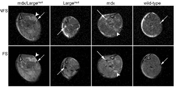
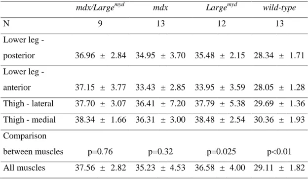
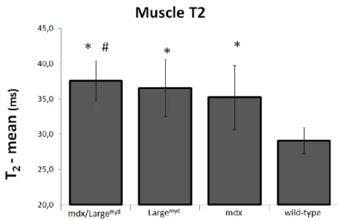
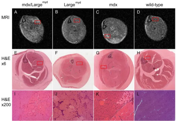
RESULTS ... 53
Muscle T2 ... 54
Muscle texture analysis ... 56
Histological analysis ... 57
DISCUSSION ... 60
CONCLUSIONS ... 65
SUPPORTING INFORMATION ... 66
S2.1. T2 calculation from two images at different echo times ... 66
S2.2. Features selected for Texture Analysis ... 67
COMPLEMENTS TO THE MANUSCRIPT ... 68
Validation of the 2 points T2 measurements with a multiecho sequence ... 68
Post-mortem changes in the T2 values ... 69
CHAPTER 3. STRUCTURAL AND FUNCTIONAL ALTERATIONS OF SKELETAL MUSCLE MICROVASCULAR NETWORK IN DYSTROPHIN-DEFICIENT MDX MICE... 72
ABSTRACT ... 73
INTRODUCTION ... 74
MATERIALS AND METHODS ... 76
Microvascular network organisation in three dimensions ... 76
Histology ... 77
Morphometric analysis ... 77
Nuclear Magnetic Resonance analysis ... 78
Statistics ... 79
RESULTS ... 80
Normal microvascular network organisation in young-adult Flk1GFP/+::mdx mouse. ... 80
Muscle blood perfusion is modified in young-adult mdx mice. ... 84
Muscle bioenergetics in young-adult mice (Table 3.1) ... 86
Alteration of microvascular network organisation in old Flk1GFP/+::mdx mouse ... 88
Alteration of muscle perfusion in old mdx mice ... 92
Muscle bioenergetics in 12 month-old mice (Table 3.2) ... 93
DISCUSSION ... 94
CONCLUSION ... 98
SUPPLEMENTARY INFORMATION ... 99
S3.1. Nuclear Magnetic Resonance analysis ... 99
CHAPTER 4. NON-INVASIVE NMR STUDY OF THE MOUSE MODEL FOR CENTRONUCLEAR MYOPATHY WITH MUTATION IN THE DYNAMIN-2 GENE ...103
INTRODUCTION ... 103
MATERIALS AND METHODS ... 105
Animals ... 105
Nuclear Magnetic Resonance (NMR) ... 105
Data analysis ... 106 Histological analysis ... 108 Statistical analysis ... 108 RESULTS ... 108 Morphometrical evaluation ... 108 T1 measurements ... 110 T2 measurements ... 111 Histological analysis ... 113 DISCUSSION ... 115
CHAPTER 5. PILOT FUNCTIONAL AND METABOLIC EVALUATION OF THE KI-DNM2R465W MICE ...119
INTRODUCTION ... 119
PILOT STUDY 1.EXERCISE AS THE PARADIGM OF MUSCLE STRESS IN KI-DNM2R465W MICE ... 120
Materials and methods ... 120
Results ... 123
Discussion ... 129
PILOT STUDY 2.PROLONGED ISCHEMIA AS THE PARADIGM OF MUSCLE STRESS ... 130
Materials and Methods ... 130
Results ... 131
Discusssion ... 134
PILOT STUDY 3.REGENERATION IN THE DNM2 MICE:T1,T2 AND FUNCTIONAL ANALYSIS AFTER INJURY ... 135
Materials and Methods ... 135
Results ... 136
Discussion ... 140
CHAPTER 6. EVALUATION OF THE POTENTIAL USE OF MICRO-COMPUTED TOMOGRAPHY IN THE STUDY OF MUSCLES FROM MURINE MODELS FOR MUSCLE DYSTROPHIES ...142
INTRODUCTION ... 142
MATERIALS AND METHODS ... 143
Animals ... 143
Micro-CT ... 145
Data Analysis ... 145
Statistical Analysis ... 146
RESULTS ... 146
Phenotypical characterization of the dystrophic muscle with micro-CT ... 146
Evaluation of injured muscle with micro-CT ... 147
DISCUSSION ... 148
GENERAL CONCLUSIONS AND PERSPECTIVES ...152
11
Abstract
Non-invasive evaluation of murine models for genetic muscle diseases
Novel therapeutic approaches are being introduced for genetic muscle diseases such as muscle dystrophies and congenital myopathies, all of them having remained without cure so far. These recent developments have motivated a renewed and augmented interest in non-invasive methods for muscle characterization and monitoring, particularly during and after therapeutic intervention. In this context, animal models are essential to better understand the disease mechanisms and to test new therapies. Recently, significant advances in the non-invasive evaluation of mouse models for genetic muscle diseases have been achieved. Nevertheless, there were still several mouse strains not characterized non-invasively, and it was necessary to develop sensitive methods to identify subtle alterations in the murine affected muscle. The purpose of this thesis was to apply non-invasive techniques in the study of murine models for genetic muscle diseases with variable phenotypes. Three mouse models for muscle dystrophy (mdx, Largemyd, mdx/Largemyd) and one mouse model for congenital myopathy (KI-Dnm2R465W) were studied with Nuclear Magnetic Resonance (NMR) methods. Two dystrophic strains (Largemyd, mdx/Largemyd) and normal mice after injury were studied through micro-Computed Tomography (micro-CT). On NMR, all affected mouse strains presented increased muscle T2, which could be related to variable features in the histological evaluation, including necrosis and inflammation, but also to clusters of fibers under regeneration or with altered cytoarchitecture. The combination of NMR and texture analyses allowed the unambiguous differential identification of all the dystrophic strains, although it was not feasible when comparing the muscle T2 measurements only. Mdx mice showed functional and morphological alterations of vascular network. In the KI-Dnm2R465W mice, a pilot study revealed tendencies of functional impairment. Finally, micro-CT images were unable to detect differences in muscle´s content in dystrophic mice. Altogether, these results not only increased the number of murine models for genetic muscle diseases non-invasively characterized, it also demonstrated some degree of
12 specificity of the imaging anomalies, as revealed by texture analysis. It also showed that non-invasive NMR methods can be sensitive enough to identify subtle alterations in murine muscle phenotype, even in early stages. This thesis was developed under an international joint supervision between France and Brazil, and comprised an important transfer of technology, with the first non-invasive studies of murine muscles performed in Brazil.
13
Resumo
Avaliação não invasiva de modelos murinos para doenças musculares genéticas
Novas abordagens terapêuticas vêm sendo introduzidas para doenças musculares genéticas como distrofias musculares e miopatias congênitas, distúrbios que permanecem sem cura até o momento. Estes recentes avanços motivaram um interesse renovado e crescente por métodos não invasivos para a caracterização e monitoramento do músculo afetado, particularmente durante e após intervenções terapêuticas. Neste contexto, modelos animais são essenciais para uma melhor compreensão dos mecanismos das doenças e para testar novas terapias. Recentemente, avanços significativos na avaliação não invasiva de modelos murinos para doenças musculares genéticas foram alcançados. Entretanto, diversas linhagens de camundongos ainda não foram caracterizadas de maneira não invasiva, e ainda é necessário o desenvolvimento de métodos sensíveis para a identificação precoce de alterações sutis no músculo de camundongos afetados. A proposta desta tese é aplicar técnicas não invasivas inovadoras no estudo do músculo de modelos murinos para doenças musculares genéticas com fenótipos variados. Três modelos murinos para distrofias musculares (mdx, Largemyd, mdx/Largemyd) e um modelo murino para miopatia congênita (KI-Dnm2R465W) foram estudados com métodos de Ressonância Magnética Nuclear (RMN). Duas linhagens distróficas (Largemyd, mdx/Largemyd) e camundongos normais após injúria foram estudados através de micro-Tomografia Computadorizada (micro-CT). Em RMN, todas as linhagens de camundongos afetados apresentaram aumento de T2 muscular, o que foi relacionado a diversas anomalias na análise histológica, como necrose e inflamação, mas também a conjuntos de fibras em regeneração ou a fibras com citoarquitetura alterada. A combinação de RMN com análise de textura permitiu a identificação não ambígua de todas as linhagens distróficas, sendo que apenas a comparação dos valores de T2 muscular não permitiu esta diferenciação. Camundongos mdx mostraram alterações funcionais e morfológicas na rede vascular do músculo. Estudo piloto em camundongos KI-Dnm2R465W revelou tendências de comprometimento da função
14 muscular. Por fim, imagens de micro-CT não permitiram a detecção de diferenças na composição muscular em camundongos distróficos. Este conjunto de resultados não apenas enriquece o painel de modelos murinos para doenças musculares genéticas caracterizados de maneira não invasiva, mas também demonstra um certo grau de especificidade nas anomalias observadas nas imagens, como revelado pela análise de textura. Estes resultados também mostraram que métodos não invasivos de RMN podem ser suficientemente sensíveis para identificar alterações sutis no fenótipo muscular murino, mesmo em estágios precoces. Esta tese foi desenvolvida sob acordo de co-tutela internacional entre a França e o Brasil, e compreendeu uma importante transferência de conhecimento, com os primeiros estudos não invasivos de músculo murino realizados no Brasil.
15
Résumé
Evaluation atraumatique de modèles murins de maladies musculaires génétiques De nouvelles options thérapeutiques sont en cours d'introduction pour les maladies musculaires génétiques telles que les dystrophies musculaires et les myopathies congénitales, maladies jusque là sans traitement causal. Ces développements récents ont suscité un intérêt renouvelé et croissant pour les méthodes atraumatiques en vue de caractériser et de suivre les muscles atteints, en particulier pendant et après une intervention thérapeutique. Dans ce contexte, les modèles animaux sont essentiels pour mieux comprendre les mécanismes des maladies et pour tester des nouvelles thérapies. Récemment, il y a eu des avancées significatives dans l'évaluation atraumatique de modèles murins de maladies musculaires génétiques. Néanmoins, nombre de lignées de souris n'ont pas encore été caractérisées de façon atraumatique et il reste à mettre au point des méthodes plus sensibles pour identifier précocément des altérations subtiles dans le muscle des souris malades. L'objectif de cette thèse est d'appliquer des techniques atraumatiques innovantes à l'étude du muscle de modèles murins de maladies musculaires génétiques avec des phénotypes variés. Trois lignées de souris modèles de dystrophies musculaires (mdx, Largemyd et mdx/Largemyd) et une lignée de souris modèle de la myopathie congénitale (KI-Dnm2R465W) ont été étudiées par des méthodes de Résonance Magnétique Nucléaire (RMN). Deux lignées dystrophiques (Largemyd et mdx/Largemyd) plus des souris normaux après une blessure ont été étudiées par micro-tomographie (micro-CT). En RMN, toutes les souches de souris affectées ont présenté un T2 musculaire augmenté, en relation avec une gamme d'anomalies histologiques, y comprises nécrose et inflammation, mais aussi des groupes de fibres en régeneration ou des fibres avec altérations de l'architecture. Avec la combinaison de la RMN et de l'analyse de la texture, il a été possible d'identifier sans ambiguïté toutes les lignées dystrophiques, alors que la seule mesure du T2 ne permettait pas de les différencier. Les souris mdx ont présenté des altérations fonctionnelles et morphologiques du reseau vasculaire musculaire. Pour les souris KI-Dnm2R465W, des études préliminaires ont révélé une tendance à développer des altérations fonctionnelles musculaires.
16 Finalement, les images de micro-CT n'ont pas pu détecter des différences du contenu musculaire dans les souris dystrophiques. L'ensemble des résultats non seulement enrichit le panel de modèles murins de maladies génétiques musculaires caractérisés de manière atraumatique, il révèle également un certain degré de spécificité des anomalies dans l'imagerie, comme l'a montré l'analyse de texture. Les résultats démontrent aussi que des méthodes de RMN non-invasives peuvent être assez sensibles pour identifier des altérations subtiles dans le phénotype musculaire murin, même à des stades précoces. Cette thèse a été développée dans le cadre d'une co-tutelle internationale entre la France et le Brésil, et elle a comporté un important transfert de compétence, qui a permis de réaliser les premières explorations atraumatiques du muscle murin effectuées au Brésil.
17
18
General Introduction
Muscles are contractile organs essential for life, being responsible for functions such as breathing, locomotion (skeletal muscles), blood circulation (cardiac/smooth muscle) and food propulsion in the digestive system (smooth muscle). Only skeletal muscles are under voluntary control, and attached to bones they compose the locomotor system.
Muscle dysfunctions can be caused by influences like injury, cancer and infections, but can also be originated by genetic defects. Among the genetic disorders affecting primarily skeletal muscles, there are the muscle dystrophies and the congenital myopathies. Muscle dystrophies are caused by alterations in proteins present at the sarcolemma, the sarcoplasm, the basal lamina and even at the nucleus of muscle cells. Muscle dystrophy patients have progressive loss of muscle cells, with consequent weakness. The histopathological pattern includes variation in fiber size, necrosis, infiltration by inflammatory cells, and substitution by connective and adipose tissue (Emery, 2002). Congenital myopathies are caused by alterations in proteins from the contractile filaments, the sarcolemma or the sarcoplasm. Patients with congenital myopathies present weakness generally at birth, and disease progress is less accentuated than in muscle dystrophies. Histopathological analysis of myopathic muscles shows structural alterations such as the presence of rods, cores, or altered positioning of nucleus and other organelles (Nance et al., 2012). There is no cure for genetic muscle diseases, but several therapeutic protocols are in development, from pharmacological to cellular and genetic approaches.
Animal models for genetic muscle disorders play a crucial role in studies aiming to understand genetic, clinical and histopathological processes related to the disease, and are necessary also to test therapeutic protocols. Several animals with molecular, clinical or histological alterations similar to those observed in patients have been identified in nature or generated in laboratory. There are models for muscular diseases from different species, like dogs, cats, hamsters, fishes and pigs, but the murine models are the most commonly studied (Vainzof et al., 2008).
19 Clinical evaluation, muscle proteins dosage in blood, electromyography, genetic studies and muscle biopsy are the most frequent procedures in diagnostic and follow up of muscle diseases. Imaging methods, including computed tomography (CT), ultrasound, radioisotopes based methods (scintigraphy, PET and SPECT) and nuclear magnetic resonance (NMR), are gaining space on the muscle diseases field. Among them, NMR has optimum qualities for soft tissue evaluation, allowing the tri-dimensional visualization of deep structures without involving the use of ionizing radiation. CT images can also be used for the evaluation of patients with muscle diseases, but with less contrast for soft tissues than NMR, and with the use of ionizing radiation (Mercuri et al., 2007).
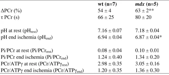
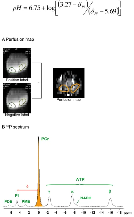
NMR allows image acquisition with different contrasts: T1, T2 and proton density weighted images can give different information about the same anatomical region. Apart of the morphological information, NMR allows metabolic and functional measurements. NMR spectroscopy gives information about the chemical composition of the tissue. 1H and 31P are the most common nuclei for in vivo spectroscopy studies, but different atomic nuclei can be evaluated (19P, 13C, 23Na, 15N, etc). 31P spectroscopy allows the evaluation of tissue pH and of phosphate molecules, being particularly interesting to study the energetic metabolism. 1H spectroscopy can give information about metabolites such as lactate, creatine and lipids. Finally, functional NMR can give information about muscle blood perfusion and oxygen consumption, for example.
In the literature, NMR has been used to evaluate dystrophic patients' muscles (Finanger et al., 2012; Mercuri et al., 2007; Poliachik et al., 2012), heart (Verhaert et al., 2011) and brain (Rae et al., 1998; Razek et al., 2009), with morphological, functional and metabolic analysis. Patients with non dystrophic congenital myopathies have been also evaluated by NMR (Jungbluth et al., 2008; Quijano-Roy et al., 2011), in studies specially focused on morphological alterations such as muscle atrophy and fat replacement. The study of different genetic muscle diseases by CT and NMR images allowed the identification of unique patterns of muscle involvement depending on the primary mutation (Wattjes et al., 2010).
NMR studies from animal models for neuromuscular diseases face the challenge of reducing scale: it is necessary to increase the spatial resolution,
20 especially when considering mice, the most frequent animal for modeling genetic diseases. Despite of it, there is already a considerable amount of NMR data of animal models for neuromuscular disorders. The mdx mouse, model of the most frequent muscle dystrophy, Duchenne Muscular Dystrophy (DMD), has been exhaustively studied by NMR. Muscle morphology and metabolism were evaluated in mdx mouse by in vivo, ex vivo and in vitro MR imaging and spectroscopy (Amthor et al., 2004; Dunn and Zaim-Wadghiri, 1999; Dunn et al., 1991; Griffin et al., 2001; Heier et al., 2014; Mathur et al., 2011; McIntosh et al., 1998a, 1998b; Pratt et al., 2013; Straub et al., 2000; Walter et al., 2005).
In a less extensive amount, different mouse models for muscle dystrophies were studied by NMR (Cole et al., 2002; Pacak et al., 2007; Schmidt et al., 2009; Tardif-de Géry et al., 2000; Walter et al., 2005), as the golden retriever muscular dystrophy dog (Claire et al., 2012; Fan et al., 2014; Thibaud et al., 2007, 2012; Yokota et al., 2009). In addition to the variable NMR methods capable to identify changes in the affected muscles, the use of mathematical tools for image texture analysis is gaining space, since it can identify subtle differences in the pattern of distribution of muscle lesions (Mahmoud-Ghoneim et al., 2006; Nketiah et al., 2014; Pratt et al., 2013; Škoch et al., 2004; Wang et al., 2013). A deeper bibliographic review of genetic muscle disorders and non-invasive muscle evaluation, for both human patients and animal models, is presented in Chapter 1.
Even with an increasing amount of noninvasive data showing differences between affected and normal mice, the use of NMR as an outcome measure depends on the reliability of the technique to track also subtle differences in the muscles. In this context, the Chapter 2 of this thesis aims to evaluate the potential of combining NMR and texture analysis in the comparison of dystrophic muscles under different gene mutations. Three mouse models for muscle dystrophies, covering a wide range of phenotypes, were evaluated by NMR T2 relaxometry: the mdx mouse, with a mild phenotype; the Largemyd mouse, model of the congenital muscular dystrophy 1D, with altered glycosylation of the protein alpha-dystroglycan; and the recently generated double mutant mdx/Largemyd mouse, with both mutations and a very severe phenotype (Martins et al., 2013).
21 It is known that the dystrophic muscle presents microvascular alteration both in patients (Leinonen et al., 1979), dogs (Nguyen et al., 2005) and mice (Bagher et al., 2011; Burch et al., 1981; Palladino et al., 2013). How these microvascular alterations affect the muscle metabolism and function in vivo is yet unexplored. The Chapter 3 of this thesis aims to evaluate in vivo the possible functional and metabolic effects of the microvascular alterations in the mdx mouse muscle. A multimodal approach was used to allow simultaneous measures of muscle perfusion and 31P spectroscopy for the energetic metabolism inference after hypoxic stress. Hypothetical functional or metabolic alterations would take place before muscle degeneration and substitution by connective and adipose tissues, being possible early markers of the disease, and possibly helping to elucidate the mechanisms of the disease.
While dystrophic muscles are characterized by tissue alterations, including inflammation, necrosis, and infiltration by adipose and by connective tissue, congenital myopathies are characterized by structural and functional alterations inside the muscle fibers, which are followed by metabolic and/or functional impairment. The versatility of NMR measures can be helpful in the observation of the impact of these alterations in myopathic muscles.
Animal models for congenital myopathies have been identified in dogs (Beggs et al., 2010; Pelé et al., 2005) and engineered in mice (Buj-Bello et al., 2002; Durieux et al., 2010; Ottenheijm et al., 2013; Pierson et al., 2012; Ravenscroft et al., 2011). Gineste and collaborators published in 2013 three papers characterizing different murine models for nemaline myopathy with noninvasive methods, including NMR (Gineste et al., 2013a, 2013b, 2013c). To our knowledge, these are the only NMR descriptions of myopathic alterations in murine models of congenital myopathies. The Chapter 4 of this thesis aims to describe with in vivo NMR the KI-Dnm2R465W mouse, model of autosomal dominant centronuclear myopathy. This murine model presents the most frequent mutation in the dynamin-2 gene in centronuclear myopathy patients. Its phenotype includes muscle weakness, atrophy, and the presence of histological features observed in patients, like the concentration of oxidative activity at the center of the muscle fibers (Durieux et al., 2010). Nevertheless, the high percentage of centronucleated fibers observed in patients is not present on the mouse model. In these mice, it is not observed infiltration by connective or adipose tissue in the
22 muscle, nor signals of inflammation or necrosis. In the search of additive information about the muscle commitment in this disease, in vivo morphological and relaxometry measures were acquired in this mouse model at two different ages, 3 and 6 months. Additionally, Chapter 5 describes pilot functional and metabolic studies with this mouse model, for the phenotypical characterization and for an initial evaluation of the regeneration process after acute injury. In vivo multiparametric functional NMR, with measurements of muscle blood perfusion, oxygen consumption and energetic metabolism, were carried out.
CT images from patients with muscle diseases can also reveal morphological alterations such as muscle atrophy and fat infiltration (Nakayama et al., 2013a). The muscular X-ray attenuation is reduced in patients with muscle dystrophies (Rickards et al., 1982; Swash et al., 1995), while it is not affected by bed rest (Rittweger et al., 2013), indicating that alterations in the muscle content, like fibrotic or adipose infiltration, would affect its radiodensity. In the recent years, with the development of micro-CT equipments with micrometric resolution, the number of in vivo micro-CT studies with small animals has increased. Nevertheless, the great majority of them are focused on skeletal alterations (Gray et al., 2012; Manske et al., 2012), including studies on dystrophic mice bones (Novotny et al., 2011, 2012). In the literature, there are essays involving micro-ct studies of mouse heart (Detombe et al., 2012), lungs (Lederlin et al., 2012; Paik et al., 2014), and even muscles (Manske et al., 2010; Weber et al., 2012). Nevertheless, these micro-CT muscle studies are focused on muscle volume changes. The possible radiodensity alterations on dystrophic muscle from mouse models are still unknown. In this context, the major objective of Chapter 6 of this thesis is the evaluation of the potential use of X-ray micro tomography in the study of dystrophic muscle. The severe dystrophic mouse model mdx/Largemyd and the parental lineage Largemyd were selected for this essay due to the progressive muscle degeneration and the intense infiltration of the muscle by connective tissue.
23
24
List of Publications
Publications in international journals
Latroche C, Matot B, Martins-Bach A, Briand D, Chazaud B, Wary C, Carlier P, Chrétien F, Jouvion G. Structural and functional alterations of skeletal muscle microvascular network in dystrophin-deficient mdx mice. The American Journal of Pathology. In press (2015).
Martins-Bach AB, Malheiros J, Matot B, Martins PCM, Almeida CF, Caldeira W, Ribeiro AF, Loureiro de Sousa P, Azzabou N, Tannús A, Carlier PG, Vainzof M (2015). Quantitative T2 combined with Texture Analysis of Nuclear Magnetic Resonance Images identify different degrees of muscle involvement in three mouse models of muscle dystrophy: mdx, Largemyd and mdx/Largemyd. Plos One; 24; 10(2):e0117835.
Zatz M, Vieira NM, Zucconi E, Pelatti M, Gomes J, Vainzof M, Martins-Bach A, Garcia Otaduy MC, Bento dos Santos G, Amaro Jr. E, Landini V, Gomes T (2015). Case Report: A normal life without muscle dystrophin. Neuromuscular Disorders; 25(5):371-4.
Martins PCM, Ayub-Guerrieri D, Martins-Bach AB, Onofre-Oliveira P, Malheiros JM, Tannus A, de Sousa PL, Carlier PG, Vainzof M (2013). Dmdmdx/Largemyd: a new mouse model of neuromuscular diseases useful for studying physiopathological mechanisms and testing therapies. Dis Model Mech; 6(5):1167-74.
Martins-Bach AB, Bloise AC., Vainzof M, Rabbani SR (2012). Metabolic profile of dystrophic <i>mdx</i> mouse muscles analyzed with in vitro magnetic resonance spectroscopy (MRS).Magnetic resonance imaging,30(8), 1167-1176.
25
Written communications in international congresses
M. Vainzof, C.F. Almeida, R. Ishiba, A. Martins-Bach, A.L.F. Santos, L. Nogueira (2014). 2871T. Using electroporation as a model of degeneration/regeneration to investigate the regenerative potential in neuromuscular disorders (NMD). 64 th Annual Meeting of the American Society of Human Genetics, 2014, San Diego, CA, United States.
Martins-Bach AB, Malheiros J, Melo Machado PC, Almeida CF, Matot B, de Sousa PL, Tannús A, Carlier PG, Vainzof M (2013). P.1.18 NMR imaging comparison of dystrophic mouse models: mdx, Large, mdx/Large. 18th International Congress of the World Muscle Society, 2013, Asilomar, CA, United States. Neuromuscular Disorders,23(9), 747.
Vainzof M, Calyjur P, Otaduy MCG, Almeida CF, Martins-Bach A, Carlier RY, Gurgel-Giannetti J, Amaro E, Carlier PG (2013). P.4.5 Muscle NMR imaging in the rare E650K mutation in the DNM2 gene in a centronuclear myopathy patient. 17th International Congress of the World Muscle Society, 2013, Perth, Western Australia. Neuromuscular Disorders, 23(9), 761.
Matot B, Jouvion G, Martins-Bach AB, Wary C, Carlier PG (2012). D.P. 16 Metabolic and hemodynamic alterations in the mdx skeletal muscle revisited using multi-parametric functional NMR. 17th International Congress of the World Muscle Society, 2013, Perth, Western Australia. Neuromuscular Disorders,22(9), 822-823.
Martins-Bach AB, Bloise AC, Rabbani SR, Vainzof M (2011). P.1.45 Application of NMR spectroscopy in the study of mdx mouse. 16th International Congress of the World Muscle Society, 2011, Algarve, Portugal. Neuromuscular Disorders, 21(9), 655.
26
Chapter 1
Bibliographic Review: Non-invasive
study of genetic muscle disorders
27
Chapter 1. Bibliographic Review: Non-invasive study of
genetic muscle disorders
Muscular Dystrophies
Muscular dystrophies are an extensive group of progressive genetic diseases where skeletal muscles are primarily affected. Mutations in genes coding for sarcomeric, sarcolemmal, sarcoplasmatic/cytosolic, nuclear or extracellular matrix proteins can cause muscular dystrophies. The absence or altered function of one of these proteins is responsible for a cascade of events that ends in muscle fibers degeneration and necrosis, with substitution of muscle by connective and adipose tissue. Histological analysis of dystrophic muscle reveals fibrosis, muscle fiber size variation, internalization of the nuclei, infiltration by inflammatory and adipose cells, necrosis and muscle fiber degeneration (Dubowitz et al., 2013; Shieh, 2013). Patients present progressive weakness, starting at different ages depending on the disease. More than 30 different forms of muscular dystrophy have been described, being divided in three major groups: dystrophinopathies; limb girdle muscular dystrophy (LGMD) and congenital muscular dystrophies (CMD) (Dalkilic and Kunkel, 2003).
Dystrophinopathies are characterized by mutations in the dystrophin gene, located at Xp21 (Hoffman et al., 1987). There are two forms of dystrophinopathies, Duchenne muscular dystrophy (DMD) and Becker muscular dystrophy (BMD). DMD is the most common muscular dystrophy by incidence, affecting up to 1:3000 boys at birth. In DMD, the gene mutation leads to the absence of the protein dystrophin in muscle and to a severe phenotype. In BMD, the dystrophin protein is present but in less quantity and with abnormal function, leading to a milder phenotype (Emery, 2002). In dystrophinopathies, the skeletal muscle is the major impacted tissue, but additional alterations can be present: cardiomyopathy is observed in DMD patients and heterozygous females (Mirabella et al., 1993; Nigro et al., 1990), slower gastric and intestinal flow are present in DMD patients (Borrelli et al., 2005), and more than 30% of DMD patients have cognitive impairment in a variable degree (Bresolin et al., 1994).
28 Dystrophin is normally expressed not only in skeletal muscle cells, but also in cardiac muscle, central nervous system and smooth muscle cells (Love et al., 1993). In skeletal muscle, the protein dystrophin is located under the sarcolemma, in association with glycosylated proteins, composing the dystrophin-glycoprotein complex (DGC) (Ervasti and Campbell, 1991, 1993). The DGC is responsible for the link between the actin cytoskeleton and the extracellular matrix, acting also on cellular and membrane signaling pathways (Rando, 2001a) and on calcium homeostasis regulation (Gumerson and Michele, 2011). Apart of dystrophin, DGC is composed by the dystroglycan and sarcoglycan complexes, dystrobrevin, syntrophins and sarcospan, which interact with other intra and extracellular proteins (Figure 1.1). Dystrophin interacts by its N-terminal portion with actin filaments in the cytoplasm, while the C-terminal portion interacts with syntrobrevin in the cytoplasm and β-dystroglycan (β-DG) in the sarcolemma. β-DG by its turn is linked to the peripheral membrane protein α-dystroglycan (α-DG), which is responsible for the connection of the DGC with the extracellular matrix protein α-2 laminin. This link occurs through sugar chains in the glycosylated extension of α-DG, with high affinity to Laminin G (LG)-like domains present in various extracellular matrix proteins, such as laminins, perlecan and agrin in muscle, and neurexin in brain (Barresi and Campbell, 2006; Ervasti and Campbell, 1993; Straub and Campbell, 1997).
29
Figure 1.1: Schematic representation of the dystrophin-glycoprotein complex (DGC), showing its components: dystrophin, dystroglycan complex, sarcoglycan complex, α-dystrobrevin, syntrophins and sarcospan. The extracellular (EC) matrix protein laminin-2 binds to the complex through α-dystroglycan, while F-actin, syncoilin and filamin-2 are intracellular (IC) ligands. Calmodulin, Grb2 and nNOS are also associated to the DGC as signaling molecules. The post-traductional modifications are shown: glycosylation in dystroglycans and sarcoglycans, and phosphorylation sites (P) in dystrophin, α-dystrobrevin, β-dystroglycan, syntrophins, α- and γ-sarcoglycans (Rando, 2001a).
Mutations in genes coding for DGC proteins or proteins involved in posttranslational modifications of the DGC components can lead to different forms of muscular dystrophies. Among these, mutations in genes coding for the sarcoglycans are related to LGMD, while alterations in the glycosylation of the protein α-DG are related to LGMD and CMD (Dalkilic and Kunkel, 2003).
30 LGMDs are the more heterogeneous subgroup of muscular dystrophies, including more than 20 different forms. In LGMD, muscles from pelvic and shoulder girdle are those initially affected. The phenotype is highly variable, from severe forms with early onset and rapid progression, to milder presentations, with first symptoms appearing after the 4th decade of life and with slow progression. LGMDs are classified according to the pattern of inheritance (autosomal dominant or recessive) and the causative genes. Mutations in genes related to LGMD can also cause different diseases, such as myofibrilar myopathy, Miyoshi myopathy and CMD (Mitsuhashi and Kang, 2012).
Patients affected by the third subgroup of muscle dystrophies, CMD, present weakness and hypotonia at birth or in the first months of life. Progressive dystrophic alterations are observed in histological analysis, and patients often show involvement in the brain and other organs. The list of genes associated with CMDs is still expanding, but the great majority of them are related to the interaction of the DCG with the extracellular matrix (Emery, 2002; Mercuri and Muntoni, 2012). This interaction is mediated by the α-DG linkage to the extracellular matrix protein laminin. Posttranslational modifications in these two proteins, specifically glycosylation, are essential to maintain and regulate this link. Mutations in the gene coding for dystroglycans are very rare, but alterations in genes coding for proteins that participate in the glycosylation of α-DG, such as POMT1, POMGnT1, FKTN, FKRP and LARGE, are related to different forms of LGMD and CMD (Moore and Winder, 2012; Muntoni et al., 2008).
Vascular alterations in Duchenne muscle dystrophy
DMD is the most studied muscle dystrophy, due to the high incidence and severity. The current hypothesis for the pathophysiology of DMD is that the absence of dystrophin fragilizes the sarcoplasm, which would suffer from micro-lesions due to mechanically induced damage from muscle contractions. Sarcoplasm rupture alters calcium homeostasis, which would result in cell death. Satellite cells would try to regenerate the muscle, but after several rounds of degeneration and regeneration, the
31 satellite cells pool would be exhausted and muscle degeneration would predominate. Processes such as inflammation, fibrosis and impaired vascular adaptation would take place secondarily, even worsening the disease manifestation (Deconinck and Dan, 2007).
Recently, the so-called ―two-hit‖ hypothesis for the disease mechanism in DMD has gained space. According to this hypothesis, dystrophin absence cause not only increased susceptibility to mechanical stress, but also vascular and metabolic alterations that when combined lead to the dystrophic phenotype (Ennen et al., 2013; Rando, 2001b).
Despite of the general lack of dystrophin in the muscle of DMD patients, in initial stages of the disease muscle lesions present in small and random clusters of degenerating fibers, usually surrounded by normal muscle fibers (Engel, 1967; Miike, 1983). This pattern of focal lesions is observed also in healthy muscle when submitted to infarction (Hathaway et al., 1970; Mendell et al., 1971). The hypothesis that muscle vascular insufficiency could be related to the disease manifestation has prompted several studies. In the 70 and 80 decades, optical and electronical microscopy of muscle blood vessels of DMD patients identified only very few nonspecific anomalies. DMD muscle was described as presenting replication of the capillaries’ basement membrane, degenerating and regenerating capillary cells, and platelet aggregation in small blood vessels (Fidziańska et al., 1986; Koehler, 1977; Miike et al., 1987). Nevertheless, no alterations in muscle blood flow at rest could be observed in DMD patients (Bradley et al., 1975; Gudrun et al., 1975; Leinonen et al., 1979).
Even if the recent studies are notably focused on muscle fibers and satellite cells as the major players in muscular dystrophies, it is accepted that alterations in the vascular system may play an important role in the disease manifestation. Skeletal muscle is highly vascularized, and endothelial cells are essential to muscle regeneration. Dystrophin is expressed also in smooth muscle cells, including endothelial cells, but it is absent in DMD patients and mdx mice (Miyatake et al., 1989). The dystrophin absence in endothelial cells reduces its ability to react to shear stress due to blood flow (Loufrani et al., 2001). Since this is a mechanical stimulus for angiogenesis (Ichioka et al., 1997), the dystrophin absence could impair the formation of new vessels in dystrophic muscle (Ennen et al., 2013).
32 Apart of morphological alterations in the vascular network, dystrophin absence may impact on the vascular function as well. Dystrophin protein links to neuronal NO-synthase (n-NOS) in the sarcolemma of normal muscle cells. The enzyme n-NOS is responsible for the muscular production of the local vasodilator nitric oxide (NO), which acts on relaxing smooth muscle in response to increased metabolic demands (Ennen et al., 2013; Kobzik et al., 1994). Apart from vasodilation function, NO has been related to myofiber differentiation, modulation of the contractile force and regulation of exercise-induced glucose uptake in health muscles (Lee et al., 1994; Roberts et al., 1997). In the absence of dystrophin, n-NOS is present in a reduced amount and is not properly anchored to the sarcolemma (Brenman et al., 1995; Crosbie et al., 2002). The reduced and misplaced n-NOS in the dystrophin absent muscle is considered to increase the damage in the disease (Deconinck and Dan, 2007; Rando, 2001b; Wehling et al., 2001). Indeed, DMD and BMD patients have altered vasoconstriction regulation under exercise, which results in functional ischemia (Sander et al., 2000; Thomas, 2013).
The fibrosis intensely present in muscles from DMD patients has also been related to alterations in blood vessels, due to increased myofiber-capillary distances, which can affect muscle function and gas exchanges (Desguerre et al., 2009). It is known that endothelial cells secrete several soluble factors, which act on the muscle regeneration process, and that satellite cells are preferentially present around vessels. An increased distance between capillaries and myofibers may have an impact on muscle regeneration (Arsic et al., 2004; Christov et al., 2007).
In the recent years, studies aiming to develop new therapeutic approaches for DMD with focus on the vascular network have gained space. It is believed that the attenuation of abnormal microcirculation would reduce muscle damage, increase tissue perfusion and reduce the cardiac workload (Ennen et al., 2013). Increasing the NO available in dystrophic muscle has been efficient to ameliorate the dystrophic phenotype in mdx mice, by reducing the post-contraction damage, reducing inflammation, and preserving the number and function of satellite cells (Asai et al., 2007; Brunelli et al., 2007; Voisin et al., 2005). Modulation of vascular endothelial growth factor (VEGF) and its receptor (VEGFR) have also shown benefits in mdx mice (Messina et al., 2007). These results indicate that increasing the microvascular
33 network in muscle could improve the microvascular control and lead to clinical benefits in the dystrophic muscle (Ennen et al., 2013; Shimizu-Motohashi and Asakura, 2014).
Congenital myopathies
Congenital myopathies are a group of genetic muscle diseases with non-dystrophic characteristics in the histological analysis. Nevertheless, clinical presentation in congenital myopathies can be very similar to what is observed in muscular dystrophy patients. Generally, congenital myopathies are characterized by the presence of morphological alterations in the muscle fiber, such as the presence of regions with protein accumulation (rods), regions with altered organelles’ distribution (cores), and altered positioning of the nucleus. Despite of these observations in histological analysis, dystrophic characteristics such as inflammation, degeneration, necrosis, extensive fibrosis and regeneration are not observed in general. Creatine kinase level in blood serum is normal or slightly increased in congenital myopathies, while it is highly increased in muscular dystrophies. Additionally, congenital myopathies are not progressive or have very slow progression, in opposition to the progressive pattern of muscle dystrophies (Sewry et al., 2008; Tubridy et al., 2001).
The three major categories of congenital myopathies are nemaline myopathies, core myopathies and centronuclear myopathies. Nemaline myopathies are characterized by the presence of sarcoplasmic or intranuclear rod structures formed by Z-disk derived material. Mutations in the genes alpha-actin (ACTA1), nebulin (NEB), alpha and beta tropomyosin (TPM3 and TPM2 respectively), troponin T1 (TNNT1), cofilin (CLF2), and a protein of the BTB/Kelch family (KBTBD13) can be related to nemaline myopathies. Patients show proximal weakness, respiratory insufficiency and facial weakness. The symptoms can be present at birth or have childhood- and even adult-onset. In general, patients with intranuclear rods have a more severe phenotype than those with sarcoplasmatic rods (Jungbluth et al., 2008; Nance et al., 2012; Romero and Clarke, 2012).
34 Core myopathies are characterized by the presence of areas without any oxidative or glycolytic enzimatic activity, reflecting the absence of mitochondria in that region. One or more core structures can be present in the muscle fiber, with variable size and distributions. Cores can be characterized as central cores or minicores. Central cores run over all length of muscle fibers, while minicores are smaller areas of disorganized myofibrilar material that are wider than longer in longitudinal histological sections. Based on the histological characteristics, patients are classified as having central core disease or multiminicore disease. Mutations in the ryanodine receptor I (RYR1) or in the selenoprotein N1 (SEPN1) genes can be related to core myopathies. Patients present with proximal muscle weakness, congenital or with early onset, possibly affecting facial muscles. Even if clinical symptoms are present at early age, histological features may not be present, which brings difficulties to the diagnosis (Jungbluth et al., 2008; Nance et al., 2012).
Centronuclear myopathies are characterized by the presence of muscle fibres with internalized or centralized nuclei, in opposition to the peripheral positioning observed in normal muscle fibers. Histopathological characteristics can include abnormal NADH-TR staining, but do not involve extensive muscle degeneration and regeneration. Centronuclear myopathies can be related to mutations in the gene coding for the protein myotubularin (MTM1), which leads to the X-linked recessive myotubular myopathy; mutations in the gene coding for amphiphysin 2 (BIN1), leading to autosomal recessive centronuclear myopathy; mutations in the gene coding for dyanmin-2 (DNM2), with autosomal dominant heritage; or to mutations in the RYR1, hJUMPY and MTMR14 genes in sporadic cases (Bitoun et al., 2005, 2007; Jungbluth et al., 2008; Tosch et al., 2006; Wilmshurst et al., 2010). A mutation in the PTPLA gene was related to centronuclear myopathy in labrador dogs, but until now, there is no report of mutations in this gene causing centronuclear myopathy in humans (Pelé et al., 2005). Clinically, centronuclear myopathies are highly variably regarding the age of onset and pattern of weakness, ranging from severe congenital forms to adult-onset forms with slow progression and mild phenotype (Nance et al., 2012).
35
Animal Models for Genetic Muscle Diseases
There is no established cure for muscular dystrophies and congenital myopathies, which prompted the search for innovative therapeutic protocols, including cellular and genetic therapies. In this context, animal models for these genetic muscle diseases have an essential role in the elucidation of disease pathomechanisms and in the development of new therapeutic strategies. Several animal models for genetic muscle diseases have been described in the literature, including natural and genetically engineered models. These animals mimic the genetic, molecular and/or clinical aspects of the disease, providing information about the pathogenesis of these disorders and allowing tests for therapeutic strategies (Vainzof et al., 2008).
Animal models for Muscular Dystrophies
The Dmdmdx mouse (hereafter called simply mdx) is the most frequently studied mouse model for DMD. This mouse has a stop codon in exon 23 of the murine dystrophin gene, which leads to total absence of this protein in muscle, as observed in DMD patients (Bulfield et al., 1984; Sicinski et al., 1989). The absence of the protein dystrophin leads to an associated reduction of other DGC proteins (Ohlendieck and Campbell, 1991), with consequent progressive muscle deterioration and weakness (Pastoret and Sebille, 1995). Histological analysis shows dystrophic changes such as variation in the caliber of muscle fibers, presence of muscle fibers with centralized nuclei, clusters of degenerating and regenerating fibers, infiltration by inflammatory cells and by connective tissue (Bulfield et al., 1984). As observed in human patients, different skeletal muscles are not identically affected. Diaphragm presents accentuated and earlier dystrophic characteristics, while masseter muscle is partially spared and limb muscles like gastrocnemius have an intermediary phenotype (Muller et al., 2001; Stedman et al., 1991). Nevertheless, differently from human patients, the mdx mouse can continuously regenerate its muscles and has a mild
36 phenotype, which makes the analysis of functional benefits in therapeutic protocols very difficult (Dangain and Vrbova, 1984; Dubowitz, 2004; Tanabe et al., 1986).
Dog models for dystrophinopathies in general have the phenotype closer to that observed in human patients. Spontaneous mutations in the dystrophin gene were observed in Golden Retriever (Valentine et al., 1992), Rotweiller (Winand et al., 1994) and German Shorthaired Pointer dogs (Schatzberg et al., 1999). The Golden Retriever Muscular Dystrophy dog (GRMD) is the more frequently studied canine model to DMD. GRMD dogs have a mutation in the dystrophin gene, leading to absence of the protein in muscle. Histopathological evaluation shows progressive dystrophic alterations in skeletal and cardiac muscles, and the phenotype is very severe. GRMD dogs mimic the molecular, histopathological and phenotypical aspects of DMD, being frequently used in the last step of therapeutic trials before tests in human patients (Shelton and Engvall, 2005). Recently, the GRMD mutation has been transferred to Beagle dogs, a smaller canine strain widely used in research (Shimatsu et al., 2003).
The animal models for LGMD include mutations in the respective genes, with special attention to the sarcoglycans genes. Absence of -sarcoglycan was observed as the primary molecular defect in BIO14.6 hamsters. This hamster has a spontaneous mutation in the gene coding for the protein -sarcoglycan, leading to its absence in muscle (Straub et al., 1998). Mouse models have been generated with null mutation in each of the four sarcoglycans genes (Araishi et al., 1999; Duclos et al., 1998; Hack et al., 1998). Animal models of sarcoglycanopathies present progressive muscular dystrophy with variable degree of severity, in some cases including cardiomyopathy. In all these models, there is a secondary reduction in the expression of the other components of the sarcoglycans/sarcospan complex, and loss of membrane integrity (Allamand and Campbell, 2000).
There are interesting mouse models of congenital muscular dystrophies, with alterations in the link between the DGC complex and the extracellular matrix. Among them, dy/dy and dy2J/dy2J mice were identified in Jackson Laboratories (http://www.jax.org/), presenting total and partial deficiency of the protein α-2 laminin, muscular dystrophy and dysmyelination of the peripheral nervous system. These mouse lineages model merosin-deficient congenital muscular dystrophy
37 (CMD1A), but dy2J/dy2J mice have a milder phenotype than the dy/dy lineage (Vainzof et al., 2008). Two α-2 laminin knock-out strains have been generated, with complete absence of this protein: dy3K and dyW. These mouse strains show severe phenotype, similar to that observed in the dy/dy mouse (Kuang et al., 1998; Miyagoe et al., 1997).
The Largemyd mouse is the murine model for CMD-1D. It has a mutation in the Large gene, which codes for the glycosyltransferase LARGE protein. The mutation leads to a reduced glycosylation of α-DG, and a consequent reduction in its binding to α-2 laminin in the extracellular matrix (Moore and Winder, 2012). Patients with CMD-1D have a very severe phenotype, with muscular and cognitive impairment (Muntoni et al., 2008). Largemyd mice have also a severe phenotype, with accentuated muscle degeneration and substitution by connective tissue. Mice have a shortened lifespan, reduced size and neurological impairment (Grewal and Hewitt, 2002).
Double mutant mice with the mdx background have been created in the attempt to approach the severe phenotype observed in DMD patients, such as the double knockout mdx:utrn-/-, with absence of both dystrophin and utrophin. It has been proposed that the protein utrophin, a homolog of dystrophin, could partially compensate for the absence of this protein in the mdx mice, being responsible for its mild phenotype. Double mutant mice with both utrophin and dystrophin absence have a much more severe muscular and cardiac phenotype than mdx mice, closer to that observed in DMD patients (Deconinck et al., 1997; Grady et al., 1997).
Recently, the double mutant Dmdmdx/Largemyd mouse (hereafter-called mdx/Largemyd) has been generated in our laboratory by crossbreeding of mdx and Largemyd lineages. Homozygous mice for both mutations present a very severe phenotype, worse than both parental lineages. Lifespan is reduced and the degree of muscle degeneration and infiltration by connective tissue is increased when compared to the parental lineages. These mice have a partial deficiency of the enzyme LARGE, and a complete absence of the protein dystrophin. It has special interest in testing cellular or genetic therapeutic protocols, since it allows the evaluation of the dystrophin expression as a marker of therapeutic success (Martins et al., 2013).
38 Animal models for congenital myopathies
There are several mouse models for nemaline myopathy, including mouse models with mutation in the α-actin gene Acta1 (Ravenscroft et al., 2011), the nebulin gene (Ottenheijm et al., 2013; Yamamoto et al., 2013), the muscular troponin T gene (Wei et al., 2014) and the cofilin2 gene (Gurniak et al., 2014). All these mouse models have phenotype very similar to what is observed in human patients. Mouse models for core myopathies include the Sepn1 knockout mouse, which do not present weakness and histopathological features as observed in humans (Rederstorff et al., 2011), and the knock-in models with mutation in Ryr1, which model the clinical and histological alterations observed in patients (Boncompagni et al., 2009; Zvaritch et al., 2009).
The myotubular myopathy murine models include the Mtm1 knock-out (Buj-Bello et al., 2002) and the Mtm1 p.R69C knock-in mice (Pierson et al., 2012), which model the myotubular myopathy with similar clinical and histological features as observed in patients. Fugier and collaborators have developed a mouse with altered splicing in the Bin1 gene and muscle weakness, possibly modeling the autosomal recessive form of centronuclear myopathy (Fugier et al., 2011). Finally, the most frequent mutation in patients with centronuclear myopathy related to dynamin-2 has been inserted in the knock-in KI-Dnm2R465W mouse. Heterozygous mice have only a small proportion of centronucleated fibers, in opposition to what is observed in humans. Muscle atrophy and force reduction are progressive and start at 2 months of age (Durieux et al., 2010).
Dog models for congenital myopathies include the Labrador retriever with mutation in the PTPLA gene (Pelé et al., 2005), the Labrador retriever with mutation in the MTM1 gene (Beggs et al., 2010) and dogs with canine Inherited Myopathy of Great Danes (IMGD), with mutation in the BIN1 gene (Böhm et al., 2013). Similar to the dog models for muscular dystrophies, canine models for congenital myopathies have phenotype similar as the observed in human patients. Mutations in the RYR1 gene have been described in pigs (Fujii et al., 1991), dogs (Roberts et al., 2001) and horses (Aleman et al., 2004), but the major clinical manifestation observed in these animals is the malignant hypertermia phenotype instead of core myopathy. These
39 animal models have spontaneous mutations, in opposition to the engineered mouse models.
Recently, Childers and collaborators could correct the muscle pathology and prolong the lifespan in murine and canine models for myotubular myopathy with mutation in the MTM1 gene. Treatment was efficient when used both in initial and late stages of the disease, raising hope to patients and researchers in this field (Childers et al., 2014). Noninvasive methods to follow the possible benefits of these new therapeutic strategies in clinical trials are highly desirable. In this context, NMR studies in patients and animal models for muscle diseases have been developed, in a first moment to better describe the muscle involvement in these disorders, but with potential use as an outcome measure in clinical trials.
Non-invasive evaluation of genetic muscle diseases
The diagnostic of genetic muscle diseases is based on clinical evaluation, quantification of muscle proteins in serum, electromyography, muscle biopsy and molecular analysis. Due to the large spectrum of genes related to muscle disorders, genetic testing can be a difficult task, especially while next generation sequencing is not widely used. In this way, imaging methods such as Computed Tomography (CT) and Magnetic Resonance Imaging (MRI) can allow the identification of the pattern of muscle involvement, orienting the genetic testing and helping in the differential diagnosis. Muscle MRI and CT from patients with genetic muscle disorders allowed the identification of the pattern of muscle involvement in these diseases, with strong correlation with genotype. The pattern of affected and spared skeletal muscles varies among muscular dystrophies, congenital myopathies and also between other muscle pathologies, such as inflammatory myopathies (Lamminen, 1990; Mercuri et al., 2007; Quijano-Roy et al., 2011, 2012; Wattjes et al., 2010). Early and correct diagnosis can affect the management of muscular dystrophy and congenital myopathy patients, since some of these diseases may involve cardiac and pulmonary complications that require early intervention (Shieh, 2013).
40 Besides of its importance in helping diagnosis, nuclear magnetic resonance (NMR) imaging (MRI), spectroscopy (MRS) and functional analysis (fMRI) have been used to study muscle morphology, metabolism and function in genetic muscle diseases. The non-invasive character of NMR, the use of non-ionizing radiation, the good contrast between fat and muscle tissue, and the possibility of studying muscle metabolism and function makes NMR a very interesting tool in the follow up of the natural history and of the effects of therapeutic trials in muscle disorders.
It has been shown that muscle MRI correlates with clinical evaluation in DMD patients (Liu et al., 1993), but it allows additionally the identification of muscle impairment before the evidence of clinical signs, like in mild BMD patients and DMD female carriers (Tasca et al., 2012a, 2012b). MRI methods that can identify fat infiltration in muscles, presence of edema related to inflammation or necrosis, and that quantify fat and water content in muscles have been extensively applied in the study of DMD patients (Fischmann et al., 2012, 2013; Gaeta et al., 2012; Leroy-Willig et al., 1997; Marden et al., 2005; Pichiecchio et al., 2002; Wren et al., 2008). Quantitative NMR (T1 and T2 measurements) also correlates with clinical parameters and with qualitative evaluation of the fat content in muscle MRI from DMD patients, raising the interest on its use in disease and therapy monitoring (Arpan et al., 2013; Huang et al., 1994; Kim et al., 2010; Mavrogeni et al., 2009; Willcocks et al., 2014).
When applied to different muscle dystrophies, NMR still gives interesting results. It has been shown that muscle and brain NMR correlate with clinical and genetics in myotonic dystrophy (Bachmann et al., 1996) and dysferlinopathies (Paradas et al., 2010). Oculopharyngeal muscular dystrophy patients, who present a very mild phenotype with slow progression, present MRI alterations that correlate with the disease progression, and that can be detected in a more sensitive way with MRI than with clinical evaluation (Fischmann et al., 2012).
Metabolic and functional NMR evaluation of dystrophic muscle have shown impaired metabolism in DMD patients, with changes in pH and altered metabolite ratios in 31P (Newman et al., 1982; Torriani et al., 2012) and 1H MRS (Hsieh et al., 2009). DMD patients also showed higher intracellular Na+ levels in 23Na MRS (Weber et al., 2011). 31P MRS alterations were also observed in milder dystrophies at rest or in exercise protocols, such as in BMD (Lodi et al., 1999; Tosetti et al., 2011)