HAL Id: tel-01498459
https://tel.archives-ouvertes.fr/tel-01498459
Submitted on 30 Mar 2017HAL is a multi-disciplinary open access archive for the deposit and dissemination of sci-entific research documents, whether they are pub-lished or not. The documents may come from teaching and research institutions in France or abroad, or from public or private research centers.
L’archive ouverte pluridisciplinaire HAL, est destinée au dépôt et à la diffusion de documents scientifiques de niveau recherche, publiés ou non, émanant des établissements d’enseignement et de recherche français ou étrangers, des laboratoires publics ou privés.
Mass Spectrometry Study of G-Quadruplex Nucleic
Acids : folding Pathways and Ligand Binding Modes
Adrien Marchand
To cite this version:
Adrien Marchand. Mass Spectrometry Study of G-Quadruplex Nucleic Acids : folding Pathways and Ligand Binding Modes. Other [q-bio.OT]. Université de Bordeaux, 2016. English. �NNT : 2016BORD0196�. �tel-01498459�
THÈSE PRÉSENTÉE
POUR OBTENIR LE GRADE DE
DOCTEUR DE
L’UNIVERSITÉ DE BORDEAUX
ÉCOLE DOCTORALE DES SCIENCES DE LA VIE ET DU VIVANT
SPÉCIALITÉ INTERFACE CHIMIE-BIOLOGIE
Par Adrien MARCHAND
Mass Spectrometry Study of G-Quadruplex Nucleic Acids:
Folding Pathways and Ligand Binding Modes
Sous la direction de : Dr. Valérie GABELICA
Soutenue le 29 Novembre 2016
Membres du jury :
Pr. SCHMITTER Jean-Marie Université de Bordeaux Président
Pr. DEFRANCQ Eric Université Joseph Fourier Grenoble Rapporteur
Pr. AFONSO Carlos Université de Rouen Rapporteur
Pr. BIRKEDAL Victoria Aarhus University, Denmark Examinateur
Pr. LIMBACH Patrick University of Cincinnati, USA Examinateur
R
EMERCIEMENTS
/A
CKNOWLEDGEMENTS
Pour commencer, je tiens à remercier les Pr. Eric Defrancq et Pr. Carlos Afonso d’avoir accepté d’être rapporteurs de ce travail. I also wish to thank both Prof. Victoria Birkedal and Prof. Patrick Limbach for having accepted to examine this thesis. Je remercie également Marie-Paule Teulade-Fichou, non seulement pour faire partie de mon jury, mais aussi pour notre collaboration qui dure maintenant depuis plus de quatre ans. Merci pour le support, les discussions (et aussi les ligands !).
Cela semble évident mais je souhaite aussi remercier Valérie et Fred pour d’innombrables raisons. Tout d’abord, merci de m’avoir fait sortir de ma Belgique natale… Le belge est de nature casanière. Je dois l’avouer, le départ n’a pas été simple d’un point de vue personnel mais il m’a permis de découvrir de nouveaux horizons, de nouvelles perspectives, de découvrir de nouvelles choses. Mais cela n’est pas tout ! Je voudrais aussi vous remercier pour le coté scientifique et la formation que j’ai pu acquérir ici durant ma thèse. La biophysique, la chimie-physique, l’instrumentation, la modélisation moléculaire, la gestion d’étudiants, la création d’un laboratoire (eh oui nous n’étions que trois dans l’équipe au départ !)… Bref, je vais essayer d’utiliser à bien toutes ces compétences.
Merci aussi à Jean-Louis pour les discussions animées lors des “G4-meetings” qui m’ont permis d’aller plus loin dans mon raisonnement. Je pense que cela a contribué aux différents travaux présentés ci-après.
Je souhaite remercier également les membres (présents et anciens) de notre équipe la « BioPhyMS Team ». Merci à Clémence et Stefano de prendre la relève ! Vous allez très bien vous en sortir j’en suis sûr ! Merci à Max pour ses discussions... intéressantes... Merci à Josephine, ma voisine de bureau pendant tout ce temps. Merci à Nina, nouvelle voisine de bureau. Courage pour le laser ! Merci à Sandrine pour les discussions autour du tuning de l’instrument. Merci à Solenne de nous faciliter la vie avec ses scripts ! Merci à Paul. Deux remerciments particuliers à Eric Largy et Valentina D’Atri, deux anciens. Je ne sais pas si c’est la nostalgie qui parle mais... Il était bien ce bon vieux temps où vous êtiez là ! J’espère qu’on va se recroiser vite.
Je tiens à remercier les étudiants (sous-fifres) qui ont réalisé des expériences liées aux projets auxquels j’ai participé. A very special thank to Mike, I really appreciated working with you. I am sure you will go far! Thank you again for the excellent weekend in CA. Thank you also Dominika, the hardest worker I have ever met. Great job with the copper complexes.
Ce travail n’aurait pas pu se réaliser sans l’aide précieuse de plusieurs collègues de l’IECB, notamment de l’équipe Mergny. Merci à Nassima pour les discussions autour des ligands. Merci à Oscar, Samir, Gilmar, Anne, Carmelo.
Merci aussi aux amis. Merci Mathilde, William, Lionel et Aurore pour les bons moments passés ensemble, les cinés, les restos, les soirées…
Enfin, je souhaite remercier ma famille pour son support. Vous avez cru en moi, m’avez supporté et permis de finaliser ce travail. Merci à Emy d’avoir relu ce travail. Merci à Al, Paul, Clems.
Finalement, je ne pouvais pas finir sans remercier Amandine. Elle m’a permis de prendre du recul et de me changer les idées, de ne pas me noyer complètement sous la masse de travail. Parfois, il faut prendre du recul pour mieux avancer. Elle m’apporte un soutien au quotidien et je la remercie pour ça.
T
ABLE OF CONTENT
A
BBREVIATIONS... 5
I.
A
PERÇU EN FRANÇAIS... 7
I.
S
UMMARY/O
UTLINE/O
BJECTIVES... 13
II.
I
NTRODUCTION... 17
II.1.
N
UCLEIC ACIDS... 17
II.1.1 TRIDIMENSIONAL STRUCTURES ... 19
II.2.
G-
QUADRUPLEXES... 21
II.2.1 WHAT IS A G-QUADRUPLEX? ... 21
II.2.2 WHERE ARE G-QUADRUPLEXES FOUND IN BIOLOGICAL SYSTEMS? ... 26
II.2.3 WHY IS IT IMPORTANT TO STUDY G-QUADRUPLEXES? ... 29
II.2.4 HOW TO STABILIZE G-QUADRUPLEXES? ... 32
II.2.5 WHICH IS THE TARGET? ... 35
II.2.6 HOW DO LIGANDS BIND TO G-QUADRUPLEXES? ... 39
II.2.7 HOW TO STUDY G-QUADRUPLEX FOLDING AND LIGAND BINDING MODES? ... 46
II.3.
M
ASS SPECTROMETRY... 53
II.3.1 NATIVE MASS SPECTROMETRY ... 53
II.3.2 ION MOBILITY SPECTROMETRY (IMS) ... 56
II.3.3 MASS SPECTROMETRY AS TOOL TO STUDY SOLUTION PHASE REACTIONS ... 58
III.
E
XPERIMENTAL APPROACHES... 63
III.1.
M
ASS SPECTROMETRY... 63
III.1.1 SAMPLE PREPARATION ... 63
III.1.2 WHICH MASS SPECTROMETER SHOULD BE USED? ... 64
III.1.3 MS INSTRUMENTAL SETTINGS ... 69
III.1.4 ION MOBILITY SPECTROMETRY (IMS) ... 71 1
III.1.5 DATA TREATMENT ... 73
III.2.
C
IRCULAR DICHROISM... 81
III.3.
T
HERMAL DENATURATION EXPERIMENTS... 85
IV.
N
ATIVEMS
FROM POTASSIUM SOLUTIONS... 87
IV.1.
I
NTRODUCTION... 87
IV.2.
A
RTICLE1 ... 91
IV.3.
A
DDITIONAL WORK... 101
IV.3.1 ADDITIONAL STRUCTURAL AND QUANTITATIVE INFORMATION FROM MS ... 101
IV.3.2 HIGHER SALT CONCENTRATIONS ... 103
IV.3.3 OTHER CATIONS THAN K+ ... 105
IV.4.
C
ONCLUSIONS... 107
V.
P
OTASSIUM BINDING TOG-
QUADRUPLEXES... 109
V.1.
I
NTRODUCTION... 109
V.2.
A
RTICLE2 ... 111
V.3.
A
DDITIONAL WORK... 127
V.3.1. K+ BINDING TO SYNTHETIC SEQUENCES ... 127
V.4.
C
ONCLUSIONS... 129
V.5.
P
ERSPECTIVES... 131
VI.
G-
QUADRUPLEX LIGAND BINDING MODES... 133
VI.1.
I
NTRODUCTION... 133
VI.2.
A
RTICLE3 ... 135
VI.3.
A
RTICLE4 ... 141
VI.4.
A
DDITIONAL WORK... 149
VI.4.1 COVALENT LIGANDS ... 149
VI.5.
C
ONCLUSIONS... 153
VI.6.
P
ERSPECTIVES... 155
VII.
G
ENERAL DISCUSSION AND CONCLUSION... 157
VII.1.
S
UMMARY OF THE RESULTS... 157
VII.2.
D
ISCUSSION ABOUT THEMS
RESULTS... 159
VII.2.1 MASS SPECTROMETRY FOR BIOPHYSICAL STUDIES ... 159
VII.2.2 THE IMPORTANCE OF THE BUFFER FOR MS OF CATION-BINDING BIOMOLECULES ... 160
VII.2.3 SOURCES OF ERROR IN THE TREATMENT OF THE MS DATA... 161
VII.3.
D
ISCUSSION ABOUT THEG-
QUADRUPLEX RESULTS... 163
VII.3.1 THE 1-K+ ANTIPARALLEL G-QUADRUPLEXES ... 163
VII.3.2 DO LIGANDS BIND TO G-QUADRUPLEXES BY INDUCED FIT OR CONFORMATIONAL SELECTION? . 164 VII.3.3 LIGAND BINDING MODES: STRATEGIES FOR FUTURE WORK ... 165
VII.4.
O
UTLOOK... 167
VIII.
P
H.D.
THESIS PUBLICATIONS... 169
IX.
R
EFERENCES... 171
X.
A
NNEXES... 197
A
BBREVIATIONS
A Adenine/Adenosine
C Cytosine/Cytidine
CCS Collision cross section CD Circular dichroism
CX-MS Cross linking mass spectrometry DNA Deoxyribonucleic acid
(n)ESI (nano) Electrospray ionization
G Guanine/Guanosine
G4 G-quadruplex(es)
HDX Hydrogen deuterium exchange IMS Ion mobility spectrometry ITC Isothermal calorimetry KCl Potassium chloride
L Ligand
m/z Mass-to-charge ratio
MS Mass spectrometry
NA Nucleic acid(s) NH4OAc Ammonium acetate
NMR Nuclear magnetic resonance spectroscopy PDB ID Protein Data Bank identification number
Q Quadrupole
RNA Ribonucleic acid
SPR Surface plasmon resonance
T Thymine/Thymidine
Tm Melting temperature
TMA Trimethylammonium
TMAA Trimethylammonium acetate TOF Time of flight
ttpy Tolylterpyridine
U Uracil/Uridine
UV Ultraviolet
I. A
PERÇU EN FRANÇAIS
IntroductionEn plus de contenir l’information génétique, l’ADN et l’ARN peuvent être impliqués dans la régulation de processus biologiques. Une structure proposée comme régulateur est le G-quadruplex. Les G-quadruplexes sont des structures non-canoniques d’acides nucléiques formées par des séquences riches en guanines. Un G-quartet est formé par quatre guanines interagissant entre elles via des liaisons hydrogènes. L’empilement de deux (ou plus) de ces G-quartets forme le G-quadruplex, qui est stabilisé par ailleurs par des cations, comme le potassium, positionnés entre des G-quartets adjacents.
Les G-quadruplexes sont trouvés dans des régions clés du génome. En effet, des études bio-informatiques ont montré que des séquences riches en guanines se trouvent dans des promoteurs de gènes et aux télomères. Elles sont présentes dans le génome humain, chez les plantes, chez la souris, dans le génome de virus… Ces structures peuvent être formées durant la réplication de l’ADN ou hors du noyau, sous forme d’ARN. Certains aptamères (des oligonucléotides synthétiques capables de se lier avec haute affinité et sélectivité à leur cible) peuvent aussi former des G-quadruplexes.
L’une des stratégies proposées pour combattre le cancer consiste à stabiliser les G-quadruplexes formés dans les promoteurs de gènes et dans les régions télomériques. En effet, réguler l’expression d’oncogènes (gènes responsables du cancer) est une manière élégante de combattre le cancer car cela implique d’agir directement à la source en supprimant la production d’une protéine. De plus, empêcher la télomérase de se lier aux télomères est une stratégie envisagée pour réduire la prolifération des cellules cancéreuses. Dans les deux cas (dans les promoteurs de gènes et aux télomères), le G-quadruplex agirait en tant que répresseur, c’est-à-dire que sa formation empêcherait soit la liaison de la machinerie de transcription soit la liaison de la télomérase. C’est pourquoi, il est souhaitable de vouloir stabiliser la forme G-quadruplexe par rapport à la forme duplexe. Pour ce faire, de nombreuses équipes synthétisent des ligands spécifiques et affins afin de reconnaitre et de
lier la structure G-quadruplexe. Ces ligands peuvent potentiellement être utilisés comme médicaments.
Objectifs
Le design de ligands de G-quadruplexes n’est cependant pas simple. En effet, il existe beaucoup de topologies différentes de G-quadruplexes dépendant de la molécularité du complexe, de l’orientation des brins dans le cœur du G-quadruplexe, de l’empilement des guanines, des types de boucles… Ce polymorphisme est illustré par la séquence télomérique humaine, pour laquelle au moins 10 structures différentes ont été obtenues expérimentalement.
Pour reconnaitre les G-quadruplexes, le design des ligands implique habituellement de larges plans aromatiques qui sont optimisés pour se lier par des interactions π-π sur les G-quartets extérieurs. Cependant, si ce type d’interaction était le seul mode de liaison, les ligands auraient des affinités similaires pour tous les G-quadruplexes. Or ce n’est pas le cas. D’autres modes de liaison sont en théorie possibles, comme l’intercalation entre des G-quartets, les liaisons avec les sillons ou les boucles. Cependant, aucune structure présentant uniquement un de ces modes de liaison n’a été rapportée.
Les objectifs de cette thèse consistent à déterminer quelles sont les complexes présents en solution, sans et avec ligands, et leurs quantités respectives.
Il existe beaucoup de techniques biophysiques permettant d’étudier les G-quadruplexes, leur repliement et la liaison de ligands. Cependant, ces techniques présentent un de ces deux désavantages : soit la technique ne permet pas d’obtenir d’information sur la structure du G-quadruplex, soit elle ne permet pas de quantifier chacune des structures présentes.
Afin de répondre à ces deux questions (structure et quantité), nous avons utilisé la spectrométrie de masse de type native. En effet, la spectrométrie de masse permet d’obtenir des informations sur la stœchiométrie de complexes non-covalents et de les quantifier. La spectrométrie de mobilité ionique couplée à la spectrométrie de masse apporte des informations sur la structure des complexes.
Résultats
Le noyau des cellules contient une haute concentration en ions potassium qui n’est pas directement compatible avec la spectrométrie de masse. Dans la littérature, des spectres de masse de G-quadruplexes sont typiquement obtenus à partir de solutions d’acétate d’ammonium. Cependant, il est important de replier les G-quadruplexes dans des solutions contenant un cation de nature biologiquement pertinente car ce cation va influencer la topologie de repliement des G-quadruplexes. Pour cette raison, nous avons développé une méthode de préparation d’échantillons nous permettant d’obtenir des spectres de masse à partir de solutions contenant du potassium. Dans notre méthode de préparation, la force ionique est fixée à l’aide d’acétate de triméthylammonium (TMAA). A cette solution est alors ajouté jusqu’à 1 mM de KCl, ce qui permet le repliement des G-quadruplexes. Tout en étant compatible avec la spectrométrie de masse, cette préparation permet aux G-quadruplexes d’être repliés selon des topologies similaires à celles des structures obtenues par résonance magnétique nucléaire (RMN), habituellement à 100 mM de KCl.
Grâce au développement d’un traitement mathématique des adduits non-spécifiques (adapté d’une méthode décrite par Klassen), nous avons pu déterminer les stœchiométries spécifiques en termes de nombre de cations liés, et les quantifier. Nous avons calculé des constantes d’équilibre de liaison du potassium aux G-quadruplexes télomériques humains et avons trouvé que des complexes ne contenant qu’un seul K+ au lieu de 2 (et donc ne possédant
probablement que 2 G-quartets) étaient formés à basse concentration en KCl pour les séquences les plus courtes, ne contenant que le cœur d((GGGTTA)3GGG). Plus des bases sont
ajoutées aux extrémités de cette séquence, plus la liaison des deux ions potassium est coopérative et plus le complexe à 1-K+ devient minoritaire. En combinant les données de
spectrométrie de masse et celles obtenues par dichroïsme circulaire (CD), nous avons obtenu les spectres CD déconvolués de chacune des espèces (à 0-, 1- et 2-K+). Les complexes à 0- et
2-K+ présentent respectivement des signatures CD typiques de structures dépliées et de
G-quadruplexes hybrides. Le complexe à 1-K+ est quant à lui un G-quadruplexe antiparallèle.
Ces résultats peuvent être étendus à d’autres séquences, comme nous l’avons démontré pour les séquences synthétiques 222 et 222T, d((GGGTT)3GGG) et d(T(GGGTT)3GGGT),
respectivement.
Afin d’en étudier le mécanisme de repliement, la liaison des ions potassium aux G-quadruplexes a également été suivie au cours du temps. Nous avons trouvé que six ensembles cinétiques étaient nécessaires pour obtenir rendre compte des observations : un ensemble ne contenant pas de K+, deux ensembles en contenant un seul et trois ensembles
contenant 2-K+. Le meilleur modèle cinétique implique la présence d’un chemin
correspondant à des espèces « mal repliées », incluant notamment un G-quadruplex antiparallèle avec un seul K+ et donc deux G-quartets. La spectrométrie de mobilité ionique
(IMS) couplée à la spectrométrie de masse a confirmé la présence d’au moins trois espèces à 2-K+. Ce mécanisme de repliement, obtenu pour les séquences télomériques humaines, est
généralisable à d’autres systèmes. Nous avons en effet démontré qu’une séquence d’un promoteur (c-myc) se replie aussi selon le même mécanisme. Ces résultats suggèrent que le chemin de repliement impliquant un piège cinétique formé par des G-quadruplexes antiparallèles est un principe général du repliement des G-quadruplexes.
Finalement, nous avons étudié la liaison de ligands à des séquences formant des G-quadruplexes. Premièrement, à travers un criblage sur plus de 20 séquences d’ADN différentes, nous avons montré que deux molécules Cu-ttpy se lient avec une haute affinité et coopérativité aux G-quadruplexes formés par la séquence télomérique humaine : le complexe 2:1 (L:DNA) est majoritaire et le complexe 1:1 est quasiment inexistant. Cette coopérativité inégalée est attribuée à des changements de conformation de la séquence télomérique humaine. Grâce au dichroïsme circulaire (CD), nous avons montré que les structures des G-quadruplexes sont affectées et, avec la liaison des deux ligands Cu-ttpy, les G-G-quadruplexes deviennent de topologies antiparallèles. La stœchiométrie en termes de nombre de K+ est
aussi affectée : les ligands se lient à des structures à 2-K+.
Deuxièmement, nous avons étudié la liaison de certains ligands parmi des plus affins et sélectifs pour les G-quadruplexes (Phen-DC3, 360A et PDS). Ces ligands se lient avec une haute affinité sur ces séquences, en éjectant un potassium, formant un complexe 1:1:1 (DNA:L:K+).
Le complexe est antiparallèle et présente les mêmes caractéristiques que celui observé soit à l’équilibre à basse concentration en KCl, soit à temps court après l’addition de KCl. Ces découvertes peuvent avoir des implications dans le domaine du design de ligands. En effet, une potentielle manière d’obtenir des ligands spécifiques d’une séquence formant des G-quadruplexes pourrait être de cibler des intermédiaires de repliement.
En conclusion, nos résultats démontrent l’importance de caractériser finement la stœchiométrie des complexes par spectrométrie de masse afin de déterminer et d’interpréter correctement les constantes d’équilibre et de caractériser les structures. Dans plusieurs cas, nous avons observé que des changements structuraux étaient accompagnés d’un changement du nombre d’ions potassium liés. La spectrométrie de masse de type native est une méthode de choix pour caractériser ces complexes car elle permet de déterminer et quantifier différentes espèces en solution.
I. S
UMMARY
/O
UTLINE
/O
BJECTIVES
IntroductionIn addition to the storage of genetic information, nucleic acids are involved in the regulation of biological processes. G-quadruplexes, non-canonical structures formed by G-rich nucleic acids, are proposed as biological regulators. These structures are formed by G-quartets, macrocyclic planes formed by four guanines interacting via hydrogen bonds. Two or more G-quartets stacked on top of each other form the G-quadruplex, furthermore stabilized by cations, such as potassium, positioned in between adjacent G-quartets.
G-quadruplexes are found in key regions of the genome. Bioinformatics studies showed that G-rich sequences are found in gene promoters or at the telomeres. These sequences are present in the human genome but also in mice, in viruses… These structures can be formed during DNA replication or in RNA outside the nucleus. Some aptamers (synthetic oligonucleotides able to bind with high affinity and selectivity to their target) can also form G-quadruplexes.
Stabilizing G-quadruplexes in gene promoters and in telomeric regions is a proposed strategy to fight cancer. Indeed, downregulation of oncogene expression is one emerging elegant way to fight cancer because it would act directly at the source, suppressing the expression of a protein. In addition, inhibiting telomerase binding to telomeric DNA may be a strategy to reduce the proliferation of cancer cells. In both cases (oncogene promoters and telomeres), the G-quadruplexes are repressors: their formation would inhibit the binding of the transcription machinery or of the telomerase. Therefore, several groups synthesize highly affine and specific ligands, potential drugs, to stabilize the G-quadruplex over the duplex. Objectives
The design of G-quadruplex ligands is not so simple. Indeed, G-quadruplexes may adopt different topologies, depending on the complex molecularity, strand orientation in the G-quadruplex core, the stacking of guanines, the type of loops… This polymorphism is illustrated by the human telomeric sequence for which at least 10 structures were reported.
To target G-quadruplexes, ligands are usually designed to have large aromatic moieties, to π-π stack on external G-quartets. However, if this was the only binding mode, all ligands would have similar affinities for all G-quadruplexes. Other binding modes are theoretically possible such as intercalation, binding in the grooves or to the loops… Nevertheless, no structure presenting binding modes different from π-π stacking is reported in the literature so far. The thesis objectives consist in determining which complexes are present in solution, with and without ligands, and their respective quantities.
Many biophysical techniques can be used to study G-quadruplex folding and the binding of ligands. However, either the technique cannot bring information about G-quadruplex structures, or it does not allow the quantification of each of these structures. To answer these two questions (structure and quantity), we used native mass spectrometry. Indeed, mass spectrometry allows to obtain the stoichiometry of the complexes and quantify them. Ion mobility coupled to mass spectrometry brings information about the structure.
Results
The first problem was related to the analysis of nucleic acids and cation complexes. In fact, the potassium cation is the relevant cation present in the nucleus but is not directly mass spectrometry-compatible. In the literature, mass spectra of G-quadruplexes are typically obtained from ammonium acetate solutions. However, it is important that G-quadruplexes are folded in solutions containing a cation of relevant nature because the cation’s nature can influence the topology. For this reason, we developed a sample preparation method allowing us to obtain mass spectra from potassium containing solutions. In the developed method, ionic strength is fixed using trimethylammonium acetate (TMAA) doped with up to 1 mM K+
required to fold the G-quadruplexes. This mass spectrometry-compatible sample preparation allows the folding of G-quadruplexes with topologies similar to those obtained in the literature by nuclear magnetic resonance (NMR), usually in 100 mM KCl.
Thanks to a mathematical treatment (adapted from a method described by Klassen) which was designed to take into account the nonspecific adducts, we determined the specific cation stoichiometries and quantified them. We determined equilibrium constants for the binding of the two potassium cations to human telomeric sequences. We found that at low KCl
concentration some complexes are formed with only one K+ instead of the expected two. We
deduced that these complexes only contain two G-quartets. The shortest human telomeric sequences, containing the d((GGGTTA)3GGG) core, are more prone to form this 1-K+ complex.
The binding of the two K+ cations to longer sequences is more cooperative and the 1-K+ species
become minor. By combining the results obtained by mass spectrometry and circular dichroism (CD), we deconvoluted the CD spectra of each of the 0, 1- and 2-K+ species. The 0
and 2-K+ spectra are typical of unfolded and hybrid G-quadruplexes, respectively. The
2-quartet 1-K+ complex is typical of antiparallel G-quadruplex. These results were also obtained
for the synthetic sequences 222 et 222T, d((GGGTT)3GGG) et d(T(GGGTT)3GGGT).
To study the folding pathway of G-quadruplexes, K+ binding was also monitored as a function
of time. We found that six kinetic ensembles of species were needed to fit the data: one species with no K+, two with 1-K+ and three with 2-K+. The best models presented a 1-K+
antiparallel G-quadruplex that acts as a kinetic trap. Ion mobility spectrometry, coupled to mass spectrometry confirmed the presence of at least three species with 2-K+. Interestingly,
this branched folding pathway, obtained for the human telomeric sequences, could be generalized to other sequences. Indeed, we showed that c-myc promoter also fold according to the same mechanism. These results suggest that the folding of antiparallel 1-K+ structures
as kinetic traps is a general tenet of G-quadruplex folding.
Finally, we investigated the binding of ligands to G-quadruplex forming sequences. First, through a screening of over 20 DNA sequences, we showed that two Cu-ttpy ligands bind with high affinity and cooperativity to the human telomeric G-quadruplexes. The 2:1 (L:DNA) complex is a major species whereas the 1:1 complex is almost inexistent. This extreme cooperativity is attributed to conformational changes in the human telomeric structure. Thanks to circular dichroism, we showed that the conformations of human telomeric G-quadruplexes are affected: when two ligands are bound, the G-quadruplex topologies are antiparallel. The K+ stoichiometry, measured by mass spectrometry, is also affected: the
ligands bind to 2-K+ structures.
Second, we studied the binding of some of the most affine and selective G-quadruplex ligands (Phen-DC3, 360A and PDS). These ligands bind with high affinity to the human telomeric G-quadruplexes with the ejection of a K+, forming a 1:1:1 (DNA:L:K+) complex. According to
circular dichroism, this complex is antiparallel and presents the same characteristics as the complex formed at low KCl concentration or at short time scale after KCl addition. Because they provide information on the binding mode, these results are important for ligand design. Indeed, a possible way to obtain ligands specific for one G-quadruplex sequence could be to target folding intermediates.
In conclusion, these results demonstrate the importance of characterizing the stoichiometry of complexes in order to properly determine equilibrium constants and characterize the structures of complexes. In several cases, we found that structural changes were accompanied by a change in the number of K+ cation bound. Native mass spectrometry is a
key technique to characterize these complexes. It allows the determination and quantification of different species formed in solution.
II. I
NTRODUCTION
II.1.
N
UCLEIC ACIDS
DNA is the hereditary material that serves as the storage of all the genetic information needed to make a living being. The genetic information is transmitted through generations. Living cells use nucleic acids to synthetize the proteins they need. Additionally, nucleic acids are implied in other roles, such as regulation and catalysis of chemical reactions in the cell metabolism. Nucleic acids are present inside the nucleus of eukaryotic cells as deoxyribonucleic acids (DNA) but also in the cytoplasm as ribonucleic acids (RNA). Nucleic acids are biopolymers made of four types of subunits, the nucleotides.1 A nucleotide is made of a phosphate group, a sugar
(that can be, either a ribose for RNA or a deoxyribose for DNA), and a base (Figure 1A). Among the bases, the natural and most abundant ones are also classified in two categories (Figure 1B): the purines, made of two aromatic rings (G, guanine and A, adenine) and the pyrimidines, made of one aromatic ring (C, cytosine and T, thymine, for DNA. In RNA, the thymine is replaced by a U, uracil).
Usually a DNA or RNA sequence is named using the one letter code of its sequence, going from the 5’ to the 3’ end. A small d or r at the beginning of the sequence is used to indicate the sugar nature, deoxyribose or ribose, respectively. The 5’ carbon of the sugar on the ith
nucleotide binds to the 3’ carbon of the (i-1)th nucleotide through the phosphate group to form
the phosphodiester linkage (Figure 1C). The nucleotides are therefore connected covalently in a chain through the sugars and phosphates. The alternating sugar-phosphate-sugar-phosphate moieties form the backbone.
Depending on their sequences and experimental conditions (solvent, ionic strength, presence/absence of salt,…) DNA and RNA can adopt different types of structures ranging from the unfolded single stranded form to the tetrastranded G-quadruplex, discussed in the next paragraphs.
Figure 1. Nucleic acid bases. (A) Definition of a nucleotide. (B) Natural DNA and RNA bases. (C) A nucleic acid strand. The bases are color-coded through all this work: G is gold, C pink, A blue, T and U are green.
II.1.1
T
RIDIMENSIONAL STRUCTURESB-form duplex. The most common structure of DNA is the B-helix, first described by J. Watson and F. Crick in 1953.2 It is a double helix that consists in two antiparallel polynucleotides chains
held together by hydrogen bonding between the bases of the strands: G with C and A with T. Furthermore, the paired bases stack one upon another, stabilizing the structure. The helix is right-handed and ten base pairs are needed to make a complete turn of the helix. It possesses a major and a minor groove and the negatively charged phosphates point towards the surface of the structure.
A-form duplex. In some conditions, complementary nucleic acid sequences can adopt other structures such as the A-helix (represented in Figure 2). One major difference comes from the placement of the base pairs within the duplex axis. In the B-DNA, the base-pairs are centered on the main axis while in the A-form they are displaced from the center to be closer to the major groove. This A-form is preferentially adopted by RNA, or by DNA in a dehydrated medium.
Z-form duplex. A third type of helix can also be formed when the sequence has alternating purine-pyrimidine bases. This leads to the formation of a Z-form helix that is left-handed.
Figure 2. The B-, A- and Z- double helices. Left, the B-form structure from PDB ID 424D.3 Middle, the
A-form structure from PDB ID 406D.4 Right, the Z-form structure from PDB ID 2DCG.5
Duplexes are not the only structure that DNA or RNA adopt. Other structures such as the triplex,6 the i-motif7 or the guanine-quadruplex (G-quadruplex) can be formed.
Triplex. A triple helix can be formed by the interaction of a third single strand inside the major groove of a double helix. Two strands form a double helix thanks to the Watson-Crick base pairs and the third strand interacts via hydrogen bonds between its bases and the Hoogsteen face of the bases of the duplex part (Figure 3).
i-motifs. Four strands interacting together two by two via C+-C base pairs (two cytosines
sharing a proton) can form the i-motif structure. They are stable in acidic conditions.
Figure 3. The triplex structure. Left, the triplex structure from the PDB ID 1BWG8 and the hydrogen
network formed for a T-A-T triplet when a triplex structure is folded. Right, the structure of i-motif from PDB ID 1YBR9 and the representation of a C+-C base pair formed in i-motifs.
II.2.
G-
QUADRUPLEXES
II.2.1
WHAT
IS AG-
QUADRUPLEX?
Figure 4. (A) A G-quartet, (B) the stacking of G-quartets gives a G-quadruplex and (C) top and lateral view of the parallel tetramolecular [d(TG4T)]4 G-quadruplex obtained by X-ray crystallography (PDB ID
352D).10
Guanine-rich DNA or RNA strands may form G-quadruplex structures. Bang was the first to observe G-quadruplex formation: guanylic acid in solution forms a gel.11 However, it took half
a century of research to discover its tetra-stranded structure.12 G-quadruplexes are made of
two or more stacked G-quartets, which are hydrogen-bonded macrocycles formed by four guanines (Figure 4A). The H on the N1 and NH2 on the C2 of one guanine are involved in
hydrogen bonds, respectively, with the O6 and N7 of the neighboring guanine. Eight hydrogen bonds per G-quartet are formed. Those hydrogen bonds are called Hoogsteen hydrogen bonds because the Hoogsteen face of one guanine is involved in the hydrogen network.13 Four
G-tracts (runs of G) are needed to form a G-quadruplex that is furthermore stabilized by cations, coordinated between the O6 of guanines.13 For example on Figure 4B, the G-tracts
are made of four guanines, leading to a four G-quartets G-quadruplex with three cations in-between. The interactions stabilizing G-quadruplexes are electrostatic (hydrogen bonds and
dipole-ion interactions), π-π stacking and hydrophobic (water molecules are released upon folding).14
G-
QUADRUPLEX FORMALISMThere are many ways to form G-quadruplexes, depending on the number of strands, their orientation in the G-quadruplex core, the way the loops connect the G-tracts, etc.… Those characteristics describe the topology of a G-quadruplex, and all of them are interconnected. Because many G-quadruplex topologies are possible, and to simplify their description, Webba da Silva has described a recommended geometric formalism and associated nomenclature,15
which is discussed hereafter.
Molecularity. The first characteristic to consider is the molecularity of the complex. Most of the G-quadruplexes studied here were intramolecular, made of one single DNA or ARN strand. However, they can also be made of two or four strands, called respectively bi- and tetramolecular. Although only unimolecular G-quadruplexes may be relevant in vivo, bi- and tetramolecular G-quadruplexes can be formed in vitro.
Figure 5. (A) Unimolecular, (B) bimolecular and (C) tetramolecular G-quadruplexes.
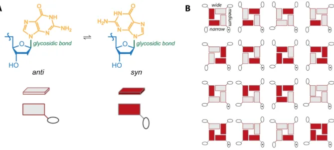
Glycosidic torsion angle. The free rotation of the bases around the glycosidic bond leads to two guanosine conformers: if the base and the sugar are on the same side of the glycosidic bound, the conformation is syn (Figure 6, color-coded in red); if they are on opposite sides of the bond, the conformation is anti (color-coded in light grey). Because each guanine has two possible configurations, there are 16 (4²) different types of G-quartets, all of them represented in Figure 6B. Depending on the secondary structure adopted, the glycosidic bond is locked in one or the other conformation, resulting in the grooves sites of variable size: wide, medium or narrow.
Figure 6. (A) anti/syn configuration of the glycosidic bond: grey = anti and red = syn; this color-code is adopted through all this work. (B) All 16 possible arrangements of the G-quartets, fixing the position of the bottom right sugar (doted sugar). Illustration adapted from Webba da Silva.15
Loop types. The next point is the type of the loops connecting the G-tracts. In a tetramolecular G-quadruplex, such as [TG4T]4 (Figure 4C), all G-tracts are independent and no loop is needed
to connect the strands. However, when the G-quadruplex is bi- or unimolecular, loops are needed. Three types of loops exist: a lateral loop connecting two antiparallel adjacent strands; a propeller loop connecting two parallel adjacent strands; and a diagonal loop connecting antiparallel opposite strands (Figure 7).
Figure 7. (A) Lateral, (B) propeller and (C) diagonal loops.
Strand orientations. The strand orientation is defined by the orientation of the G-tracts involved in the G-quartets following the backbone from the 5’ to the 3’ end. In the literature, this is the most commonly used way to characterize the topology of a G-quadruplex. If all strands point in the same direction, the G-quadruplex is parallel and belongs to group-I. If
three strands point in one direction and the last one in the opposite, the G-quadruplex is hybrid and belongs to group-II. If the strands have opposite direction two by two, the G-quadruplex is antiparallel and belongs to group-III.
Figure 8. (A) Parallel, (B) hybrid, (C) and (D) antiparallel G-quadruplexes.
All those characteristics are interconnected, for example, changing one strand orientation implies changing the loop type, and the glycosidic torsion angle… For those reasons, the interconversion between different structures are far from being simple mechanistically. Those numerous characteristics contribute to the high polymorphism of G-quadruplexes. For example, there are in theory 26 possible unimolecular G-quadruplexes topologies of three loops with three G-quartets.15
Additional sequence/structure-specific features, such as base pairs between bases in the loops or bulges in the G-tract, also contribute to this polymorphism as illustrated with a few structures in Figure 9.
Figure 9. Illustration of the polymorphism of G-quadruplexes. Structures obtained by NMR or X-ray crystallography of different G-quadruplexes. The PDB ID is indicated below the structure: antiparallel 2-quartet G-quadruplex, 1RDE (d(GGTTGGTGTGGTTGG)),16 parallel 3-quartet quadruplex with one
G-tract made of 2 guanines, 2A5P (d(TGAGGGTGGIGAGGGTGGGGAAGG), I stands for inosine),17 parallel
3-quartet G-quadruplex with one long loop, 2LPW (d(AAGGGTGGGTGTAAGTGTGGGTGGGT)),18 and
bimolecular antiparallel G-quadruplex with mini-hairpins in the loops, 2KAZ (2x d(GGGACGTAGTGGG)).19
II.2.2
WHERE
AREG-
QUADRUPLEXES FOUND IN BIOLOGICAL SYSTEMS?
Structural and biophysical studies evidenced that many G-rich sequences can fold into this non-classical type of structures. Initially, it was not known whether the G-quadruplex was a biologically relevant structure. There is now more and more evidence that G-quadruplexes are relevant DNA and RNA secondary structures in vivo.20,21 One of the most convincing
evidence was brought by Biffi et al,22 who developed a G-quadruplex structure-specific
antibody that was used to visualize quantitatively DNA G-quadruplexes in human cells. The quest for G-quadruplex localization in the genome was started by bioinformatics studies. Two works screened for G-quadruplex motifs in the human genome.23,24 The first definition
used to find putative quadruplex forming sequences was the following. Intramolecular G-quadruplexes follow the generalized sequence GaXnGbXoGcXpGd where Ga-d represent the
guanine tracts and Xn-p represent the loops regions that can be made of any nucleotide
combination (G included). The number of guanines in the G-tracts was restricted to 3–5 and the loops were between 1 and 7 nucleotides. This definition led to more than 300 000 putative occurrences. However, the limitation to seven nucleotides maximum in the loop regions is probably too stringent, given that several structures were recently found to have longer loops (examples are shown in Figure 9, PDB ID: 2KAZ, 2LPW).18,19,25 More generally, if the external
loops are short, a long middle loop can be tolerated and could even stabilize the structure if they form secondary structures (such as base pairs26,27). More recently, Phan showed that
G-quadruplexes can be made of 4n–1 guanines: one G-tract can be one guanine shorter, leading to a G-quadruplex made of n–1 G-quartets and a triplet of guanines.28 For these reasons,
Mergny developed a new algorithm to determine G-richness and skewness of regions of the genome.29 They found a much higher number of G4 forming sequences in the human genome
(around 1.5 million). In agreement, experimentally, using a high-resolution sequencing approach, Balasubramanian identified more than 700 000 sequences forming G4 structures.30
In human, yeast and bacterial genomes, G-quadruplex forming sequences are over-represented in certain regions such as the promoters and the telomeres.23,24,29,31,32 Several
indirect evidences for the presence of G-quadruplexes structures come from the DNA binding proteins in bacteria.33 Some proteins are able to induce the folding of G-quadruplexes34 and
others to unwind them.35 The evolutionary conservation of the localization and composition 26
of G4 motifs suggests that G4 have a role in the cells,36,37 even though the exact role of those
structure is not yet fully understood. Some of their potential roles are presented in the next few pages.
Gene promoters. Regions close to the translation start site (TSS) in the promoters of genes (within 1000 nucleotides upstream of the TSS) are enriched in G-quadruplex-forming sequences.23,29,32 For example, a very well-studied G-quadruplex is present in the promoter of
the transcription factor MYC in mammalians.
Telomeres. By definition, the telomere is the sequence at the end of the chromosomes and the proteins that protect it.38 The primary role of the telomere is to protect the chromosome
against degradation or end-to-end fusion. They are made of a double stranded region and an extended 3’ single stranded overhang. Usually, the telomeric 3’ end sequence consists in the repetition of a G-rich motif, for example, in vertebrates, (TTAGGG)n,39 but other G-rich
sequences have been identified for bacteria, yeast or plants.40–42 Several studies have shown
that this G-rich sequence is able to form G-quadruplexes in vivo.22,30,43 In human cultured cells,
a small fluorescent probe44 and an antibody22 suggested the presence of G-quadruplexes in
telomeric regions in vivo. However, researchers are still uncertain about their presence as their folding may be induced by those probes.
During DNA Replication. During its replication, the double stranded DNA is separated into two strands by helicases (proteins responsible for unwinding the DNA structures). First, a preformed G-quadruplex could slow down the helicase unwinding.45 Secondly, during
replication, the leading strand is continuously duplicated and is therefore continuously reforming a duplex. However, the lagging strand’s replication is discontinuous and some portions are free to form secondary structures, such as G-quadruplexes, which could therefore act as transcriptional regulators.38 G-quadruplexes could also be formed in the
double stranded mother-part and prevent or slow down the unwinding by the helicases. A high degree of mutations within the genome of a cell lineage can be caused by most of the human helicases that are able to unwind the G-quadruplexes in vitro.45–47
Outside the nucleus. G-rich RNA sequences are also able to fold into G-quadruplexes, suggesting that RNA G-quadruplexes may have a translation regulation role.48–50 Recently,
fluorescent G-quadruplex probes have been used in living cells and in isolated mitochondria. 27
In this work, the authors suggest their presence also in mitochondria.51 Other potential roles
of the G-quadruplexes in the genome are linked with the epigenetic regulation of gene expression, origin of replication, meiosis, recombination, etc.…38
Viruses. G-quadruplex-forming sequences are also found in viruses. Recently, a G-rich sequence has been identified in the promoter of HIV-1 and the formation in vitro of several G-quadruplexes has been confirmed by NMR spectroscopy.26,52 Another work suggests that
G-quadruplexes can play a role on hepatitis C virus replication.53
G-quadruplexes as therapeutic agents. G-quadruplexes can also be used as therapeutic agents as aptamers, synthetic oligonucleotides able to bind with high affinity and selectivity to their target. Indeed, some aptamers are able to fold into a G-quadruplex structure.16 Short
G-quadruplexes have been evaluated as potential anti-HIV candidates because they bind to HIV-related proteins.54–56 Some G-quadruplexes are also tuned with chemical modifications to
improve their binding affinities towards their target.57
II.2.3
WHY
IS IT IMPORTANT TO STUDYG-
QUADRUPLEXES?
A
LTERING GENE EXPRESSIONBecause many genes have G-quadruplex-forming sequences in their promoter, the transcription could theoretically be influenced by G-quadruplex stability. Surprisingly, there is a discrepancy within gene promoters: promoters of oncogenes (genes that have the potential to cause cancer) are even more enriched in G-quadruplex forming sequences58 whereas
promoters of tumor repressor genes are poorer in those motifs.59 Displacing the equilibrium
towards the formation of the G-quadruplex is a proposed strategy to fight cancer cells (Figure 10).
Stabilizing G-quadruplexes in or near the promoter regions may have positive or negative aspects. If present in the template strand, the G-quadruplex could affect the binding of the polymerase and downregulate the gene expression which would be positive in case of an oncogene. However, G-quadruplexes in human cells tend to be more often found on the other strand.60 They would then have a negative effect as they could facilitate the transcription.
However, they may serve as recruiters to stimulate the transcription… or as blockers by recruiting gene repressors…61 Their exact role is actually still under intense investigation.
Figure 10. Schematic representation of a G-quadruplex-forming sequence in the promoter of a gene. The formation of the G-quadruplex prevent the gene expression.
Some results suggest that the formation of the G-quadruplex would downregulate the gene expression of MYC,62 a transcription factor in mammalians which is associated with cell
proliferation and is overexpressed in 80% of human cancers.63 Displacing the equilibrium
would therefore be positive for the organism.
Downregulation of oncogenes is one emerging elegant way to fight cancer because it would act directly at the source, suppressing the expression of a protein.
P
REVENT TELOMERASE BINDINGAt each cell cycle, the very few end-bases of the chromosome cannot replicate. This leads to telomere attrition, a decreasing of the length of the telomere with cell replication. Once the telomere is too short, the cell dies normally, and this process is called cell senescence.
Telomerase, a ribonucleoprotein responsible for the regeneration of the telomere, is inactive in normal cells but upregulated in 85-90% of malignant cells. This enzyme gives the cancer cells the ability to divide indefinitely.64 Immortality and limitless replicative potential are two
of the hallmarks of a cancer cell.65
Figure 11. Schematic representation of the telomere overhang. The formation of the G-quadruplex prevent the binding of the telomerase and elongation of the telomere. Figure adapted from Wang et
al.66
The substrate for this enzyme is the single stranded DNA overhang. If that overhang is folded, the telomerase cannot bind anymore, which prevents telomere elongation (Figure 11).67 One
research objective is therefore to stabilize the G-quadruplexes formed at the telomeres in 30
order to provide obstacles for the binding of the telomerase, limiting the elongation of the telomere. Inhibiting the telomerase binding may indeed be a strategy to reduce the proliferation of cancer cells.68,69
In addition, because the telomeres are implicated in cell divisions, they may have a role in cell ageing. Indeed, telomere attrition correlates with age-related pathologies.70
II.2.4
HOW
TO STABILIZEG-
QUADRUPLEXES?
Due to their presence in key regions of the genome, it has been suggested that they have the ability to regulate biological events. To affect those biological events using the G-quadruplex structure, a strategy consists in influencing the quadruplex stability. Stabilizing a G-quadruplex means displacing the equilibrium represented in Figure 12 towards the folding of the G-quadruplex.
Figure 12. Factors influencing the G-quadruplex folding equilibrium.
Note that in the genome, DNA G-quadruplex formation competes with duplex (not represented in Figure 12). To the contrary, RNA does not have this constrain and the formation of G-quadruplexes is therefore even more likely to occur. Many parameters have been described to stabilize (or destabilize) G-quadruplexes. The main ones are briefly described hereafter.
Cations. The cation may displace more or less the equilibrium towards the formation of the G-quadruplex depending on its concentration, ionic radius and hydration energy (desolvation and binding of the cation is part of the equilibrium).71 The general trend for
quadruplex-stabilizing cations is the following: Sr2+ > Ba2+ > K+ > Ca2+ > Na+, NH
4+, Rb+ > Mg2+ > Li+ > Cs+.72
The nature of the cation also influences the preferred topology of the G-quadruplex as discussed later. In cells, mostly K+ and Na+ are present and their concentrations are regulated
thanks to the Na+/K+ pump. In the nucleus, potassium is more concentrated than sodium73
and K+ is therefore the most biologically relevant cation for in vitro assays.
Ionic strength. To overcome the electrostatic repulsion between the negative charges of the backbone, a high ionic strength is needed.74 Increasing the ionic strength is, however, not a
convenient strategy to act on the equilibrium in vivo.
Crowding or dehydration agents. Crowding agents such as polyethylene glycol have been used to mimic cell conditions. These agents were shown to be a major determinant of the G-quadruplex stability.75–77 In a previous work, we also showed that organic co-solvents such as
ethanol induced the formation of G-quadruplexes.78 The commonly accepted reason is that
water molecules are released upon folding of the G-quadruplex. Therefore, adding dehydration agents displaces the equilibrium towards the folding of G-quadruplexes.
Temperature. The Gibbs free energy (𝛥𝛥𝐺𝐺1) of the reaction depends on the temperature: lower
temperatures favor the folding of G-quadruplexes. Again, it is not possible to decrease the cell temperature of a living being. This parameter is however used to determine the “G-quadruplex stability”, indicated by the melting temperature (temperature at which 50% of the G-quadruplexes in solution are folded). If the melting temperature is increased/decreased in certain conditions, it means the G-quadruplex is stabilized/destabilized by those conditions. Measuring the melting temperature in different conditions is a widely used method to study G-quadruplex stabilizing/destabilizing conditions. This particular point will be discussed in more details in the next sections.
Ligands. Because none of the above-mentioned parameters can be used to influence G-quadruplexes formation in vivo, researchers had to find a different way to affect the equilibrium. This can be achieved using small molecules (also called ligands) to stabilize the quadruplex structures. Genome-wide approaches (not targeting one specific gene) using G-quadruplex ligands have been conducted to evidence the potential use of G-G-quadruplexes as transcriptional regulators.62,79
The equilibrium in Figure 12 becomes more complicated when a ligand binds to the different structures. The equilibrium in Figure 13 represents some of the equilibria that affect the concentrations of the different species in solution. If the goal is to displace the equilibria towards the G-quadruplex formation, it is therefore important to synthetize ligands affine and selective for G-quadruplexes over the duplex or unfolded species. A review of selected ligands is presented in a specific section (II.2.6), later.
Figure 13. Schematic representation of the solution equilibria at stake when a ligand bind to a G-quadruplex-forming sequence. The green balls are cations and the black disk is a ligand.
II.2.5
WHICH
IS THE TARGET?
Targeting G-quadruplexes with ligands seems to be a promising route to fight cancer for the reasons presented above. However, once injected inside the cell, a ligand could bind to all G-quadruplexes present in the genome and therefore could alter many different processes. Additionally, quadruplex ligands could also theoretically induce the folding of G-quadruplexes in unwanted regions, which may promote unwanted biological effect.
Different G-rich sequences may form G-quadruplexes of different topologies, which could in principle be targeted by different ligands. However, it is not so simple because the same sequence can also fold into different types of G-quadruplexes, ensuing uncertainties about which G-quadruplex structure should be targeted.
The first step to design affine and selective ligands for the G-quadruplex structure is to know the target’s structure. Because most of this work is focused on the human telomeric sequences, the main published human telomeric structures are presented in this section.
P
OLYMORPHISM OF THE HUMAN TELOMERIC SEQUENCESThe human telomeric sequence made of d(TTAGGG) repeats is one of the most studied biologically relevant G-quadruplex forming sequences. Over the years, several structures have come out and this sequence therefore appears to be extremely polymorphic.80
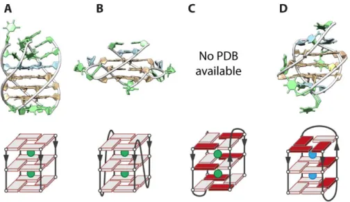
Monomolecular G-quadruplexes are formed by one sequence of four d(TTAGGG) repeats. Bimolecular G-quadruplexes are formed either by two sequences of two repeats or by one sequence of one repeat and one sequence of three repeats. Tetramolecular human telomeric G-quadruplexes are formed by four strands, each made of one d(TTAGGG) repeat. Therefore several G-quadruplexes with different molecularities are published in the literature.
Tetra- and bi-molecular human telomeric G-quadruplexes. Historically, the first topology obtained for a human telomeric sequence is a tetramolecular parallel G-quadruplex.81 Wang
and Patel published, a year later, the first G-quadruplex structure of a derivative of the Tetrahymena telomeric sequence (d(TTGGGGT)).82 Tetramolecular G-quadruplexes do not
have loop constraints and, systematically, the adopted structure is parallel with all glycosidic angles in anti-conformation, as also observed for the human telomeric G-quadruplex
[d(TTAGGGT)]4 (Figure 14A).83 A bimolecular human telomeric G-quadruplex structure was
also obtained in K+ conditions by X-ray crystallography.84 The G-quadruplex is parallel with two
propeller loops (Figure 14B). In solution, Phan showed that this structure is in equilibrium with an antiparallel structure (Figure 14C).85 Those structures have grooves of different sizes. In the
parallel form, all grooves are of medium size whereas in the antiparallel one, two grooves are wide and two are narrow.
Another particular structure is made of a three-repeat sequence combining with a one-repeat sequence, forming a hybrid G-quadurplex (Figure 14D).86 Those bimolecular G-quadruplexes
may be relevant, for example, in the case of the t-loop formation.
Figure 14. Structures of tetra- and bimolecular G-quadruplexes formed by the human telomeric sequence obtained in K+ (green balls in the schematic view) or Na+ (blue balls in the schematic view)
solutions from NMR or X-ray. (A) Parallel tetrameric G-quadruplex [d(TTAGGGT)]4, PDB ID: 1NP9.83 (B)
Parallel bimolecular G-quadruplex [d(TAGGGT)2]2, PDB ID: 1K8P.84 (C) Antiparallel G-quadruplex
[d(TAGGGT)2]2.85 (D) Asymmetric bimolecular G-quadruplex observed for d(GGGTTA)2GGGT +
d(TAGGGT), PDB ID: 2AQY.86
Intramolecular human telomeric G-quadruplexes. However, more relevant structures may be intramolecular, made of four G-tracts (Figure 15). Over the past 15 years, at least six different folds have been published. The final topology depends on many parameters: the nature of the monovalent cation, the concentrations, the ionic strength, the pH, the presence of crowding agents and the exact sequence. Indeed, there are 16 different ways of delimiting the d(TTAGGG)n sequence, all containing at least the 21 bases central core d(GGGTTA)3GGG
and a maximum of 4 G-tracts.
Figure 15. Structures of the intramolecular G-quadruplexes formed by the human telomeric sequences obtained in K+ (green balls in the schematic view) or Na+ (blue balls in the schematic view) solutions
from NMR or X-ray. (A) Parallel G-quadruplex formed by 22AG, PDB ID: 1KF1.84 (B) Antiparallel
G-quadruplex formed by 22AG, PDB ID: 143D.87 (C) Hybrid-1 G-quadruplex formed by 24TTG, PDB ID:
2GKU.88 (D) Hybrid-2 structure formed by 26TTA, PDB ID: 2JPZ.89 (E) 2-quartet antiparallel G-quadruplex
formed by 22GT, PDB ID: 2KF8.90 (F) 2-quartet antiparallel G-quadruplex formed by the modified
sequence 22CTA, PDB ID: 2KM3.91
By X-ray diffraction of crystals obtained from K+ solutions, Parkinson et al. obtained a parallel
structure made of three G-quartets for 22AG, (d(A(GGGTTA)3GGG) (Figure 15A).84 However,
studies in diluted solutions suggest that the crystal packing may bias the resulting structure,92
even though parallel G-quadruplexes could be relevant in molecular crowded conditions (and therefore in the nucleus).75,76,93 The NMR of 22AG in diluted solution containing 100 mM Na+
cations indicated that the formed G-quadruplex is antiparallel (Figure 15B).87 In potassium
solutions by NMR spectroscopy, the 22AG structure has not been elucidated but several structures for closely related sequences were obtained. The mutated sequence 24TTG (d(TT(GGGTTA)3GGGA) folds into a hybrid-1 structure88 as well as 23TAG, (d(TA(GGGTTA)3GGG)
(PDB ID: 2JSM)94 and 25TAG, (d(TA(GGGTTA)3GGGTT) (PDB ID: 2JSL) (Figure 15C).94 Another
hybrid type structure, the hybrid-2 has been published for the 26TTA sequence, (d(TTA(GGGTTA)3GGGTT) (Figure 15D).89 The difference between hybrid-1 and -2 comes from
the order of the loops: hybrid-1 loops are respectively 5’-propeller-lateral-lateral-3’ and the hybrid-2 loops are 5’-lateral-lateral-propeller-3’. A third structure, called hybrid-3, is a 2-quartet antiparallel G-quadruplex with a triplet of guanines (Figure 15E). About 70% of 22GT, (d((GGGTTA)3GGGT) (PDB ID: 2KF8), fold into this conformation90 and 23AG,
(d(A(GGGTTA)3GGGT), is also able to adopt this topology.95 Finally, the modified 22CTA
sequence (d(A(GGGCTA)3GGG, the TTA loops are replaced by CTA loops) also form a 2-quartet
antiparallel structure91 additionally stabilized by a G:C:G:C platform.
In the cellular context, there are uncertainties about which structures are really present. An NMR study suggest the coexistence of the 2-quartet (hybrid-3) and the hybrid-2 type quadruplexes for sequences with more than four repeats that form multiple G-quadruplexes.96 Similarly, interactions between two adjacent G-quadruplexes in longer
sequences have been proposed and could bring additional stability to the G-quadruplex structures formed in the telomeric regions and influence the topology.97
II.2.6
HOW
DO LIGANDS BIND TOG-
QUADRUPLEXES?
A good G-quadruplex ligand has to present a good affinity and a good selectivity versus other secondary structures. In this section, we will discuss the types of ligand scaffolds synthetized and tested in the literature and their potential binding modes.
Theoretically, there are four different potential binding sites on a G-quadruplex structure: the ligand could interact with the bases in the loops and bind via loop recognition, it could intercalate between the G-quartets, bind in the grooves (electrostatic interactions) or stack on external quartets (π-π stacking) (Figure 16). The rational design mainly targets external G-quartets because it is the common feature to all G-quadruplexes. Indeed, over the years, different molecular scaffolds have appeared in the literature but one common point consists in large polyaromatic moieties, prone to π-π stack.98,99 Several structures of ligands stacked
on external G-quartets of different G-quadruplexes were obtained by NMR spectroscopy and X-ray crystallography,100–106 therefore, the usually accepted binding mode for those molecules
is π-π stacking.13 Note that the structure of a peptide interacting with the two external
G-quartets of a parallel G-quadruplex has been published, this also supports that those binding sites are interesting G-quadruplex recognition motifs.107
Figure 16. Potential interacting sites on a G-quadruplex structure.
However, other binding sites such as groove binding, loop recognition or intercalation may be as important targets but only a few structures presenting such binding modes are published
in the literature. In theory, taking into consideration particular features (loops, grooves…), that are topology-dependent, may theoretically increase the selectivity for G-quadruplexes over other structures and eventually lead to inter-G-quadruplex selectivity. Only one complex implying groove binding was obtained108,109 and some attempts have been made to target the
loops110 but those binding modes are as of today not the most common ones.
The most common G-quadruplex ligands usually have a very large aromatic plane, larger than those of traditional duplex intercalators.111 This leads to increased selectivity for the
G-quadruplex compared to the unfolded and/or duplex forms. Additionally, ligands have been improved to bind with higher affinity to the G-quadruplex structure via electrostatic interactions. Indeed, most of the ligands have positively charged groups that can interact with the negative nucleic acids backbone. However, this interaction could also bring nonspecific binding and therefore decrease the selectivity for the G-quadruplex.
In the next sections, we will briefly describe a non-exhaustive list of reported G-quadruplex ligands. They are classified into four categories according to their general molecular shape and will discuss three of each category: porphyrins and ligands with large aromatic planes, metallo-complexes, bisquinoline derivatives and macrocycles.
For a more complete list of G-quadruplex ligands, a database was published112 and several
reviews discuss some of the most recent ligands.98,99,113
P
ORPHYRINSThe aromatic core of porphyrins makes their conception as G-quadruplex ligands almost straightforward. Derivatization of the core with chains of various nature, including or not positive charges, was a common way to improve the affinity of those molecules.114
The tetramethylpyridinium porphyrin, TMPyP4, one of the most studied G-quadruplex ligand, increases the melting temperature of some G-quadruplexes by values as high as 17 °C115 and
inhibits telomerase.116 Hurley also showed that TMPyP4 was able to regulate the expression
of the c-myc oncogene.117 In addition, a surprising structure with one of the two binding
ligands interacting with an external loop has been obtained.118 However, this could come from
a nonspecific binding. Indeed, a series of studies evidenced TMPyP4’s lack of selectivity.115,119–
121 It was even shown that this ligand unfolds very stable RNA G-quadruplexes, displacing the
equilibrium towards the unfolded species.122
Figure 17. Examples of porphyrin G-quadruplex ligands.
The development of Mn(III)-porphyrins by Pratviel’s group improved the selectivity for G-quadruplexes. Surface plasmon resonance (SPR) evaluation of the dissociation constants indicated sub-micromolar constants for the G-quadruplex, compared to values higher than tens of micromolar for duplexes.123 This increased selectivity comes supposedly from the
steric hindrances of the side chains.124
Those two previously described porphyrins bare positive charges that probably increase the interaction between the ligands and the DNA. A negatively charged porphyrin, N-methyl-mesoporphyrin IX (NMM), possesses two negative charges at physiological pH. Its affinity is affected by the presence of those charges but it presents a stronger selectivity for G-quadruplexes versus duplexes.125 It was also shown that NMM has a preferential binding to
parallel G-quadruplexes whose external G-quartets are particularly exposed. The exalted fluorescence when bound to a quadruplex and its selectivity toward parallel quadruplexes permitted its use as a strand orientation reporter for parallel G-quadruplexes.126
M
ETALLO-
COMPLEXESFigure 18. Examples of metal complexes G-quadruplex ligands.
Some of the porphyrins can complex a metal cation, changing the distribution of the charges on the ligand and potentially influencing the binding affinity.127 Researchers also developed
metal complexes to target G-quadruplexes because they are easy to synthetize and could lock an organic moiety in a specific conformation. The ideal metallo-complex binds the G-quadruplex with its metal positioned over the cation channel of the G-G-quadruplex and its organic moiety(ies) stacked on the quartet.128 The Ni(II)-salophen complex was designed this
way and stabilizes the G-quadruplex form by up to 33 °C, which made this ligand one of the best temperature stabilizers.128 It is also a telomerase inhibitor.129 Further optimization is
possible by modifying the chemical groups on the central phenyl moiety and the metal nature.130,131
Terpyridines in interaction with various metal cations have been employed as G-quadruplex ligands.132 The platinum- and palladium-tolylterpyridine (Pt- and Pd-ttpy complexes) were
analyzed by mass spectrometry and were shown to bind covalently to the DNA.133
Teulade-Fichou showed that the Pt-ttpy ligand is covalently bound to the adenines located in the loop regions of the human telomeric G-quadruplex.134 Another study revealed the importance of
the metal geometry of those ligands: to obtain the best selectivity over duplexes, the trigonal bipyramidal shape obtained for the Copper-tolyterpyridine, Cu-ttpy, ligand is the best because it impedes duplex intercalation.132 Derivatization strategies around the terpyridine moiety has
been explored.135,136 This ligand complex with a G-quadruplex has also been used, for
completely different purposes, as a metalloenzyme for Diels-Alder reactions catalysis.136
Other types of metallo-complexes have been employed to target DNA G-quadruplexes such as ruthenium and di-ruthenium complexes.137,138 The rationale was the use of a large aromatic
plane in between the metal cores that bare the charges. The dinuclear {Ru(phen)2}2-tpphz binds