HAL Id: hal-02268713
https://hal.archives-ouvertes.fr/hal-02268713
Submitted on 21 Aug 2019
HAL is a multi-disciplinary open access
archive for the deposit and dissemination of
sci-entific research documents, whether they are
pub-lished or not. The documents may come from
teaching and research institutions in France or
abroad, or from public or private research centers.
L’archive ouverte pluridisciplinaire HAL, est
destinée au dépôt et à la diffusion de documents
scientifiques de niveau recherche, publiés ou non,
émanant des établissements d’enseignement et de
recherche français ou étrangers, des laboratoires
publics ou privés.
Determination of MC-based predictive models for
personalized and fast kV-CBCT organ dose estimation
H. Chesneau, M. Vangvichith, Eric Barat, Caroline Lafond, Delphine
Lazaro-Ponthus
To cite this version:
H. Chesneau, M. Vangvichith, Eric Barat, Caroline Lafond, Delphine Lazaro-Ponthus. Determination
of MC-based predictive models for personalized and fast kV-CBCT organ dose estimation. ESTRO 36,
May 2017, Vienna, Austria. Radiotherapy and Oncology, 123 (Supplement 1), S151; poster PV-0287,
2017, ESTRO 36, May 5-9, 2017, Vienna, Austria. �10.1016/S0167-8140(17)30730-2�. �hal-02268713�
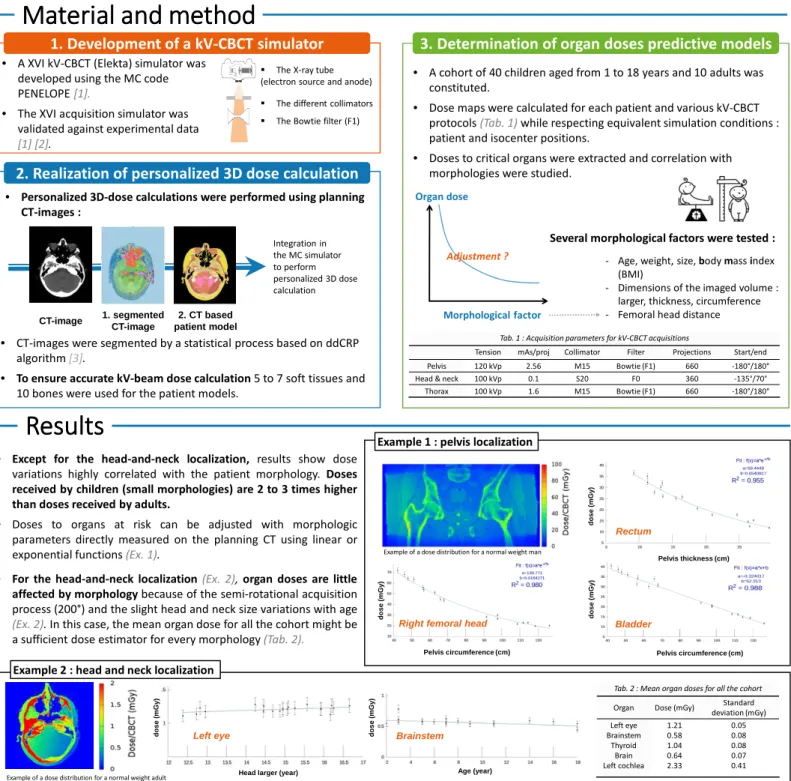
• Except for the head-and-neck localization, results show dose variations highly correlated with the patient morphology. Doses received by children (small morphologies) are 2 to 3 times higher than doses received by adults.
• Doses to organs at risk can be adjusted with morphologic
parameters directly measured on the planning CT using linear or exponential functions(Ex. 1).
• For the head-and-neck localization(Ex. 2), organ doses are little affected by morphology because of the semi-rotational acquisition process (200°) and the slight head and neck size variations with age
(Ex. 2). In this case, the mean organ dose for all the cohort might be a sufficient dose estimator for every morphology(Tab. 2).
Determination of MC-based predictive models for personalized and
fast kV-CBCT organ dose estimation
H. Chesneau 1, M. Vangvichith1, E. Barat 1, C. Lafond2,3, D. Lazaro-Ponthus1
1 - Commissariat à l'Energie Atomique -LIST, Département de physique, Gif-sur-Yvette, France. 2 - Centre Eugène Marquis, Rennes, France
3 - LTSI, Image and Signal Treatment Laboratory, INSERM U 1099, University of Rennes I, France.
Introduction and objective
Introduction and objective
Introduction and objective
Introduction and objective
Material and method
Material and method
Material and method
Material and method
Results
Results
Results
Results
Conclusion
Conclusion
Conclusion
Conclusion
The intensive use of CBCT during radiotherapy treatments induces additional radiations doses delivered on large volumes. If those doses are often neglected, the medical community is starting to ask for an accurate dosimetric tool dedicated to kV-CBCT.
The aim of the present study is to develop a Monte Carlo-based organ doses calculator suitable for clinical environment. To ensure fast dose estimation, the possibility to determine predictive models based on patient morphology was studied.
Tab. 1 : Acquisition parameters for kV-CBCT acquisitions
Tension mAs/proj Collimator Filter Projections Start/end Pelvis 120 kVp 2.56 M15 Bowtie (F1) 660 -180°/180° Head & neck 100 kVp 0.1 S20 F0 360 -135°/70° Thorax 100 kVp 1.6 M15 Bowtie (F1) 660 -180°/180°
This preliminary cohort study demonstrates the possibility to determine mathematical predictive models for personalized and fast kV-CBCT organ dose estimation. Future works will include an extension of the patient cohort to increased the accuracy of the mathematical adjustments and a similar study for the OBI kV-CBCT (Varian). Those perspectives are a part of the French National Research Agency (ANR) project AID-IGRT.
[1] H. Chesneau et al. "Monte Carlo simulation for imaging dose estimation: application to the Elekta XVI kV-CBCT.“ ESTRO annual meeting 34, Barcelona 2015. [2] H. Chesneau et al. "Comprehensive validation of a Monte Carlo kV-CBCT model using OSL and spectral measurements.“ ESTRO annual meeting 35, Torino 2016.
[3] S. Ghosh et al. “Spatial distance dependent Chinese restaurant processes for image segmentation.” Advances in Neural Information Processing Systems Proceedings Books, 2011. Morphological factor
Organ dose
Adjustment ? - Age, weight, size, body mass index
(BMI)
- Dimensions of the imaged volume : larger, thickness, circumference - Femoral head distance
Several morphological factors were tested :
• A cohort of 40 children aged from 1 to 18 years and 10 adults was constituted.
• Dose maps were calculated for each patient and various kV-CBCT protocols(Tab. 1) while respecting equivalent simulation conditions : patient and isocenter positions.
• Doses to critical organs were extracted and correlation with morphologies were studied.
3. Determination of organ doses predictive models
• CT-images were segmented by a statistical process based on ddCRP algorithm [3].
• To ensure accurate kV-beam dose calculation 5 to 7 soft tissues and 10 bones were used for the patient models.
• A XVI kV-CBCT (Elekta) simulator was developed using the MC code PENELOPE [1].
• The XVI acquisition simulator was validated against experimental data
[1] [2]. CT-image 1. segmented CT-image 2. CT based patient model Integration in the MC simulator to perform personalized 3D dose calculation
The X-ray tube (electron source and anode)
The different collimators The Bowtie filter (F1)
• Personalized 3D-dose calculations were performed using planning CT-images :
1. Development of a kV-CBCT simulator
2. Realization of personalized 3D dose calculation
• Limitation : the impact of isocenter position variations was not study in this work.
Pelvis circumference (cm) d o s e ( m G y ) Bladder Right femoral head
Pelvis circumference (cm) d o s e ( m G y )
Example of a dose distribution for a normal weight man
Pelvis thickness (cm) d o s e ( m G y ) Rectum
Example 1 : pelvis localization
Example of a dose distribution for a normal weight adult
Brainstem Age (year) d o s e ( m G y ) d o s e ( m G y ) Left eye
Head larger (year) Example 2 : head and neck localization
Tab. 2 : Mean organ doses for all the cohort
Organ Dose (mGy) Standard deviation (mGy) Left eye Brainstem Thyroid Brain Left cochlea 1.21 0.58 1.04 0.64 2.33 0.05 0.08 0.08 0.07 0.41