HAL Id: hal-02064991
https://hal.sorbonne-universite.fr/hal-02064991
Submitted on 12 Mar 2019
HAL is a multi-disciplinary open access
archive for the deposit and dissemination of sci-entific research documents, whether they are pub-lished or not. The documents may come from teaching and research institutions in France or abroad, or from public or private research centers.
L’archive ouverte pluridisciplinaire HAL, est destinée au dépôt et à la diffusion de documents scientifiques de niveau recherche, publiés ou non, émanant des établissements d’enseignement et de recherche français ou étrangers, des laboratoires publics ou privés.
mutations and clinical aspects
Johanna Palmio, Sarah Léonard-Louis, Sabrina Sacconi, Marco Savarese, Sini
Penttilä, Anna-Lena Semmler, Wolfram Kress, Tahseen Mozaffar, Tim Lai,
Tanya Stojkovic, et al.
To cite this version:
Johanna Palmio, Sarah Léonard-Louis, Sabrina Sacconi, Marco Savarese, Sini Penttilä, et al.. Expand-ing the importance of HMERF titinopathy: new mutations and clinical aspects. Journal of Neurology, Springer Verlag, 2019, 266 (3), pp.680-690. �10.1007/s00415-019-09187-2�. �hal-02064991�
https://doi.org/10.1007/s00415-019-09187-2
ORIGINAL COMMUNICATION
Expanding the importance of HMERF titinopathy: new mutations
and clinical aspects
Johanna Palmio1 · Sarah Leonard‑Louis2 · Sabrina Sacconi3 · Marco Savarese4 · Sini Penttilä1 ·
Anna‑Lena Semmler5,6 · Wolfram Kress7 · Tahseen Mozaffar8 · Tim Lai8 · Tanya Stojkovic9 · Andres Berardo10 · Ricardo Reisin10 · Shahram Attarian11 · Andoni Urtizberea12 · Ana Maria Cobo12 · Lorenzo Maggi13 ·
Sergei Kurbatov14,15 · Sergei Nikitin15 · José C. Milisenda16 · Farzad Fatehi17 · Monika Raimondi18 · Fernando Silveira19 · Peter Hackman4 · Kristl G. Claeys20,21 · Bjarne Udd1,4,22
Received: 22 October 2018 / Revised: 3 January 2019 / Accepted: 3 January 2019 / Published online: 21 January 2019 © The Author(s) 2019
Abstract
Objective Hereditary myopathy with early respiratory failure (HMERF) is caused by titin A-band mutations in exon 344 and considered quite rare. Respiratory insufficiency is an early symptom. A collection of families and patients with muscle disease suggestive of HMERF was clinically and genetically studied.
Methods Altogether 12 new families with 19 affected patients and diverse nationalities were studied. Most of the patients were investigated using targeted next-generation sequencing; Sanger sequencing was applied in some of the patients and available family members. Histological data and muscle MRI findings were evaluated.
Results Three families had several family members studied while the rest were single patients. Most patients had distal and proximal muscle weakness together with respiratory insufficiency. Five heterozygous TTN A-band mutations were identi-fied of which two were novel. Also with the novel mutations the muscle pathology and imaging findings were compatible with the previous reports of HMERF.
Conclusions Our collection of 12 new families expands mutational spectrum with two new mutations identified. HMERF is not that rare and can be found worldwide, but maybe underdiagnosed. Diagnostic process seems to be complex as this study shows with mostly single patients without clear dominant family history.
Keywords Hereditary myopathy · Respiratory failure · Titin · Titinopathy, mutations
Introduction
Hereditary myopathy with early respiratory failure (HMERF, OMIM #603689) is characterized by proximal and/or dis-tal muscle weakness, and early and severe diaphragmatic insufficiency [1–5]. In HMERF, respiratory failure can be a presenting symptom in an ambulant adult patient, which is not a common feature in other genetic myopathies [5–7]. Typical MRI pattern has been reported with fatty degenera-tion of semitendinosus and obturator muscles and anterolat-eral compartment of lower legs early in the disease course [4, 5, 8, 9, 10]. Muscle histopathology shows cytoplasmic bodies usually in subsarcolemmal necklace-like formation,
occasional rimmed vacuoles and myofibrillar disorganiza-tion responsible for Z-disc alteradisorganiza-tions [11].
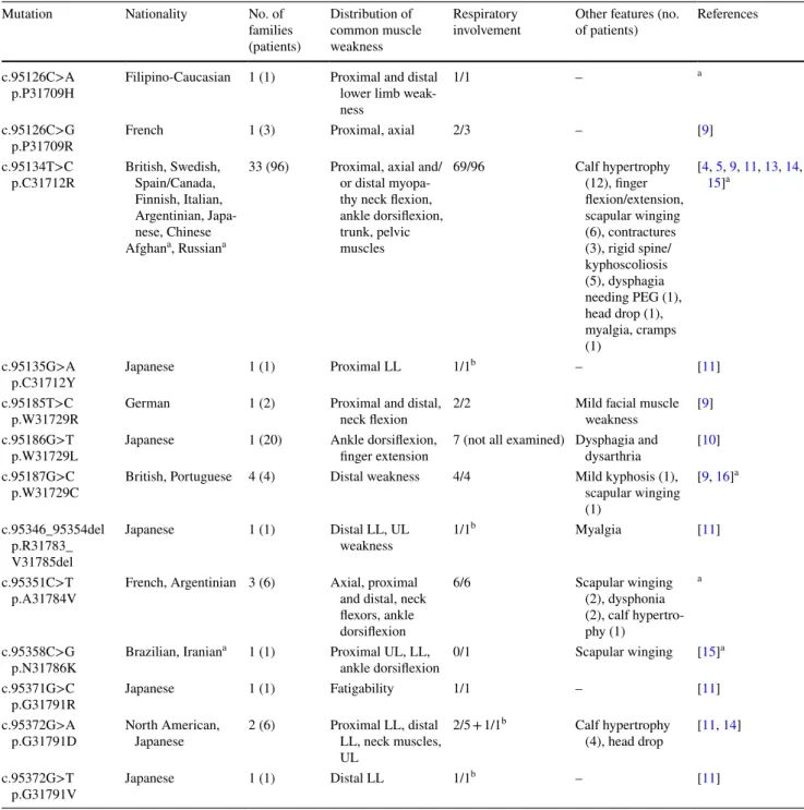
Titin gene mutations in exon 344 encoding the fibronec-tin-3 (FN3) domain in the A-band region of titin are associ-ated with HMERF [4, 5]. The reported mutations mainly show an autosomal dominant inheritance pattern, and are usually private mutations except for the most frequently identified mutation in HMERF patients, c.95134T>C p.C31712R, found in more than 20 families in Europe and Asia (Table 1) [11–14].
We describe here clinical features, pulmonary function tests, histopathological and muscle MRI findings of 19 HMERF patients from 12 families and diverse ethnic ori-gins. Five heterozygous TTN A-band mutations were identi-fied of which two are previously unreported.
* Johanna Palmio johanna.palmio@uta.fi
Methods
Patients
The patients belonged to 12 unrelated families (Fig. 1): one Filipino/Caucasian (A), one Afghan (B), two Italian
(C, D), one Spanish (E) patient, one Russian family (F), two Portuguese patients (G, H), two French families (I, J), one Argentinian (K), one Iranian (L) patient. Family I was from the East of France, with two sisters and her Ger-man cousin affected, while family J was from the South of France with two members affected. In the Russian family F father and daughter were examined and similarly affected.
Table 1 Dominant mutations in TTN A-band-exon 344 with clinical findings
PEG percutaneous endoscopic gastrostomy tube feeding, LL lower limbs, UL upper limbs
a In the present study
b Asymptomatic, found on pulmonary function tests
Mutation Nationality No. of
families (patients) Distribution of common muscle weakness Respiratory
involvement Other features (no. of patients) References
c.95126C>A
p.P31709H Filipino-Caucasian 1 (1) Proximal and distal lower limb
weak-ness
1/1 – a
c.95126C>G
p.P31709R French 1 (3) Proximal, axial 2/3 – [9]
c.95134T>C
p.C31712R British, Swedish, Spain/Canada,
Finnish, Italian, Argentinian, Japa-nese, Chinese
Afghana, Russiana
33 (96) Proximal, axial and/
or distal myopa-thy neck flexion, ankle dorsiflexion, trunk, pelvic muscles 69/96 Calf hypertrophy (12), finger flexion/extension, scapular winging (6), contractures (3), rigid spine/ kyphoscoliosis (5), dysphagia needing PEG (1), head drop (1), myalgia, cramps (1) [4, 5, 9, 11, 13, 14, 15]a c.95135G>A
p.C31712Y Japanese 1 (1) Proximal LL 1/1
b – [11]
c.95185T>C
p.W31729R German 1 (2) Proximal and distal, neck flexion 2/2 Mild facial muscle weakness [9]
c.95186G>T
p.W31729L Japanese 1 (20) Ankle dorsiflexion, finger extension 7 (not all examined) Dysphagia and dysarthria [10]
c.95187G>C
p.W31729C British, Portuguese 4 (4) Distal weakness 4/4 Mild kyphosis (1), scapular winging
(1) [9, 16]a c.95346_95354del p.R31783_ V31785del Japanese 1 (1) Distal LL, UL weakness 1/1 b Myalgia [11] c.95351C>T
p.A31784V French, Argentinian 3 (6) Axial, proximal and distal, neck
flexors, ankle dorsiflexion 6/6 Scapular winging (2), dysphonia (2), calf hypertro-phy (1) a c.95358C>G p.N31786K Brazilian, Iranian a 1 (1) Proximal UL, LL,
ankle dorsiflexion 0/1 Scapular winging [15]
a
c.95371G>C
p.G31791R Japanese 1 (1) Fatigability 1/1 – [11]
c.95372G>A
p.G31791D North American, Japanese 2 (6) Proximal LL, distal LL, neck muscles,
UL 2/5 + 1/1b Calf hypertrophy (4), head drop [11, 14] c.95372G>T p.G31791V Japanese 1 (1) Distal LL 1/1 b – [11]
Fig . 1 P edig ree of t he f amilies. DN A w as collected fr om individuals mar ked wit h an as ter isk*. F illed symbols ar
e affected and open symbols unaffected f
amil y members. Gr ey symbols ar e f am -ily members t hat ar e possibl y affected
There were no other family members diagnosed with spe-cific muscle disease in the rest of the families, although, some patients had relatives with a history of muscle weak-ness and/or respiratory problems (Fig. 1/Table 2). Those family members were already deceased or otherwise not available for this study.
All patients had been clinically examined by the treating neurologist, and data on nerve conduction studies and nee-dle electromyography (EMG), creatine kinase (CK) levels in serum and muscle MRI or CT of the lower limbs were also col-lected. Spirometry test results were available in nine patients. An echocardiogram was performed in six patients. The diag-nosis of HMERF was based on clinical symptoms of respira-tory insufficiency with muscle weakness and the presence of cytoplasmic bodies in muscle biopsy, and/or on a typical pat-tern of muscle involvement on muscle imaging as described previously, i.e., obturator externus, semitendinosus and ante-rolateral muscles in the distal leg [4, 5, 8, 9, 11].
Muscle samples from the patients were snap frozen, and 8–10 µm sections were cut and examined using standard his-tochemical stainings. Samples were also immunostained for different myogenic antigens including myosin heavy chain isoforms (fetal, neonatal, slow and fast MyHC, MHC class I).
Genetic studies
Genomic DNA was extracted from blood by standard methods. Direct Sanger sequencing of the titin exon 344 was performed at Emory Genetics Laboratory (http://genet icsla b.emory .edu/) in patient B and Tampere Neuromuscular Research Center, Finland in patient G. Targeted next-generation sequencing (NGS) was performed as previously described [17] in patients A, C, D, and H–L. Version 2 of the MYOcap gene panel was used that is targeted to the exons of 236 genes including all known genes for muscular dystrophy or myopathy at the time. In Patient E, an NGS panel targeted to the exons of 119 genes known to cause muscular dystrophy or myopathy was per-formed at the genetic service of Hospital de la Santa Creu i Sant Pau, Barcelona, Spain. Targeted sequencing was ordered for the proband of Family F from a commercial laboratory (Genomed Ltd., Moscow, the list of targeted genes: http://price .genom ed.ru/?testi d=826).
TTN variants are described according to the coding DNA
reference sequence (NG_011618.3 or LRG_391), covering transcript variant-IC (NM_001267550.1).
Results
Molecular genetic findings
Novel mutations
A novel A-band mutation c.95126C>A p.P31709H was identified in the proband of family A (III-3). The proband’s mother and two brothers were similarly affected with res-piratory insufficiency and muscle weakness but already deceased before the study. Her affected half-sister needed mechanical ventilation at age 40.
Two French families I and J, as well as one Argen-tinian patient K were identified with a novel mutation c.95351C>T p.A31784V. In family I three affected patients were studied. The proband’s affected father (I-1) in family J was already deceased and not available for the study. However, the son (III-1) was genetically studied as he has mild rigid spine although no muscle involvement. He did not carry the titin mutation. The proband (II-5) and her sister (II-6) also had joint contractures and rigid spine that have probably another genetic background.
The previously unknown variants are not listed in gnomAD (v2.1), but they are listed in ClinVar as vari-ants of uncertain significance (ClinVar ID is 283245 for c.95126C>A p.P31709H and 497143 for c.95351C>T p.A31784V). Moreover, the variant p.P31709H is also reported in dbSNP (rs869320739).
Common mutation
Single patients B–E were found to harbor the most fre-quently identified TTN A-band mutation, c.95134T>C p.C31712R [11, 12]. No other family members of these patients were available for genetic studies. In addition, the proband of family F with several affected members carried the same mutation.
Re‑occurring mutations
Patients G and H were identified with c.95187G>C p.W31729C previously reported in two single patients [9,
16]. In patient L the mutation c.95358C>G p.N31786K, which has also been found in one British patient, was identi-fied [15]. DNA samples were available in five healthy family members of patient L (mother, four siblings), but none of them carried the mutation. The patient and family members were also haplotyped, and the mutation was found to be de novo in the patient as the same haplotype but not the muta-tion was identified in healthy family members.
Table 2 Clinical and genetic data of HMERF patients
TTN A-band-exon 344 c.95126C>A, p.P31709H (novel mutation) Filipino–Caucasian patient A
Patient Sex/age Age at onset First symptoms Muscle weakness
find-ings at examination Respiratory symptoms CK/IU/L Biopsy
A: III-3 F/57 31 Proximal and distal
lower limb weakness Neck flexors, hip flex-ors, ankle
dorsiflex-ors, abductor digiti minimi
FVC 43%, NIV Normal RV, myofibrillar
aggre-gates TTN A-band-exon 344 c.95134T>C, p.C31712R (common mutation)
Afghan patient B
B: II-1 F/30 22 Difficulty climbing
stairs Bilateral left > right calf hypertrophy
Proximal and distal LL and UL, left worse than right Waddling and
step-page gait
FVC 30%, nocturnal
NIV 1.5 × UNL CBs
Italian patient C
C: II-1 M/58 40 Distal weakness
toe-walking Prox UL, prox and dist LL Yes 3 × UNL Necrotic fibers, fibrosis
Italian patient D
D: II-1 M/56 47 Myalgia, steppage gait Severe neck flexors,
severe distal UL and LL, mild-to-mod-erate proximal LL, mild proximal UL
Mild 2–3 × UNL CBs, RV
Spanish patient E
E: II-1 F/59 50 Distal weakness,
respiratory failure Proximal and distal weakness, finger
extension
FVC 30%, NIV Normal Myopathy
Russian family F
F: I-2 F/78a 58 Distal weakness Steppage gait, mild
proximal LL and finger extension
N/A N/A N/A
F: II-1 M/41a 38 Distal weakness Mild tibialis anterior N/A N/A N/A
F: II-3 M/70a 47 Steppage gait Severe distal LL
(tibialis anterior), mild UL (finger extension)
N/A N/A N/A
F:III-2 M/59 30 Ankle dorsiflexion
weakness Severe distal LL (tibialis anterior
1/5), asymmetric UL (finger extension 2/5, 3/5), mild neck flexors and proximal LL
FVC 49% 1.5 × UNL N/A
F:IV-1 F/30 30 N/A Tibialis anterior (4/5),
finger extension (4/5)
N/A N/A N/A
TTN A-band-exon 344 c.95187G>C p.W31729C (re-occurring mutation) Portuguese patient G
G: II-1 M/71 55 Steppage gait Distal LL, mild
kyphosis, scapular winging
Severe, invasive
Clinical findings
Detailed clinical data are presented in Table 2. The mean age at symptom onset was 42 years (range 22–58 years). The main presenting symptoms were related to lower limb proximal or distal weakness in all patients, and to respiratory failure at onset in only two patients.
Clinical characteristics of patient A with the novel TTN mutation c.95126C>A p.P31709H
A 52-year-old female presented with slowly progres-sive proximo-distal myopathy starting at age 31 years. She had respiratory insufficiency and a need for a non-invasive ventilation (NIV). Muscle biopsy at age 45 years
Table 2 (continued)
TTN A-band-exon 344 c.95126C>A, p.P31709H (novel mutation) Filipino–Caucasian patient A
Patient Sex/age Age at onset First symptoms Muscle weakness
find-ings at examination Respiratory symptoms CK/IU/L Biopsy
Portuguese patient H
H: II-1 M/62 55 Left foot drop Severe distal LL, mild
proximal LL and UL (4)
VC 49% nocturnal
NIV N/A Myopathy
TTN A-band-exon 344 c.95351C>T p.A31784V (novel mutation) French family I
I:III-1 F/70 55 Dyspnea Pelvic girdle, neck
flexors, ankle dorsi-flexion, dysphonia
FVC 30%, nocturnal
NIV 1.5 × UNL N/A
I:III-5 F/73 24 Pelvic girdle weakness Hip flexion, neck
flexors abdominal muscles, scapular winging, ankle dorsiflexion, finger extensors FVC 29%, nocturnal
NIV N Myofibrillar aggregates
I:III-6 F/67 54 Respiratory failure Hip flexion, neck
flex-ors, trunk muscles, dysphonia
FVC 42%, nocturnal
NIV N N/A
French family J
J: I-1 M/50a N/A N/A Distal LL Yes N/A CBs
J: II-5 F/58a 44 Distal LL weakness Proximal and distal
LL, proximal UL, axial weakness, scapular winging
Yes, FVC 70% 1.5 × UNL CBs
Argentinian patient K
K: II-1 M/54 40 Dyspnea Deltoid (4/5),
iliop-soas and quadriceps (4/5), tibialis anterior (4−/5, 4/5), toe extension (4−/5, 4/5), unable to walk on heels, steppage gait, calf hypertro-phy
FVC 45%, NIV 2.5 × UNL Normal
TTN A-band-exon 344 c.95358C>G p.N31786K (re-occurring mutation) Iranian patient L
L: II-7 M/42a 26 Difficulty climbing
stairs Generalized mus-cle weakness and
atrophy
Invasive ventilator 2 × UNL CBs
CBs cytoplasmic bodies, CK creatine kinase, F female, FEV1 forced expiratory volume in one second, FVC forced vital capacity, LL lower
limbs, M male, MRC Medical Research Council Scale, N/A not available, NIV non-invasive ventilation support, RV rimmed vacuoles, UL upper limbs, UNL upper normal limit, VC vital capacity, WCB wheelchair bound
showed rimmed vacuoles and subtle lesions compatible with myofibrillar aggregations (Fig. 2a). No cytoplasmic bodies were observed. Muscle imaging could not be per-formed due to patient’s claustrophobia.
Clinical characteristics of families I–K with the novel c.95351C>T p.A31784V mutation
All three patients in family I were followed up for severe respiratory failure which progressed slowly since ado-lescence. One of the patients had been diagnosed with asymptomatic restrictive reduced respiratory capacity after a systematic screening by the school doctor at the age of 12. However, respiratory symptoms, i.e., dyspnea did not become apparent before the age of 50 years in the patient, which resulted in the need for non-invasive nocturnal ventilation. Further, patient II-3 had died at the age of 51 years due to respiratory failure after general anesthesia and was diagnosed with pulmonary embolism at that time. Muscle weakness in the family was pre-dominant at the pelvic girdle, i.e., hip flexion, but also neck flexors and trunk muscles were weak. Ankle dorsi-flexion and finger extension strength was less severely affected. One patient presented with more severe weak-ness including limitation of arm abduction. She needed bilateral help for walking. There was no calf hypertrophy or facial weakness, but dysphonia was noted in patients III-1 and III-6. They all had kyphoscoliosis since ado-lescence. Two patients underwent echocardiography with normal results. Myofibrillar aggregates were present in the muscle biopsy.
The proband (II-5) and her father in Family J showed onset of symptoms in the distal lower limbs. Respiratory insufficiency was also present in both patients. In addition to rigid spine, mandibular and ankle contractures, there were axial weakness and scapular winging in the proband and she needed a stick for walking. Muscle biopsies in both showed cytoplasmic bodies compatible with HMERF findings.
Patient K is a 54-year-old male with severe respira-tory insufficiency, proximal weakness, and no family history. At the age of 40 the first respiratory symptoms and decreased vital capacity (58%) were noted. Muscle symptoms appeared after a few years with proximal upper and lower limb weakness. He was also unable to walk on heels. Respiratory insufficiency progressed to the vital capacity of 45% at age 54 years. EMG was myopathic and serum CK level was elevated, but no abnormal findings were observed in the muscle biopsy (vastus lateralis at age 45 years). Cardiac examination including echocardiogram was also normal. His mother suffered from dyspnea and died suddenly in her sleep at age 70 years.
Patients B–F with the common TTN mutation c.95134T>C p.C31712R
The presenting symptoms of the patients were distal lower leg weakness usually starting with ankle dorsiflexion weak-ness and respiratory insufficiency. The age of onset varied from 20 to 50 years of age. Muscle weakness slowly pro-gressed to encompass both upper and lower limb muscles proximally and distally. Neck flexor and finger extensor weakness were common findings. Respiratory symptoms ranged from asymptomatic or mild to severe with a need for NIV. CK levels were normal or mildly elevated. Mus-cle biopsy findings were available from four families and revealed cytoplasmic bodies and/or unspecific myopathic/ dystrophic changes (Fig. 2b, c).
In retrospect, family history of these patients was positive although none of the relatives had been diagnosed with a specific muscle disease. Patient B had a 41-year-old brother who had similar symptoms since age 32 and used NIV 24 h a day. The mother of patient D who had died at the age of 67 years due to heart attack, had gait difficulties since aged 40 followed by respiratory insufficiency, while patient E had a sister who had muscle weakness and died because of sud-den death around 50 years of age. In addition to the proband and his daughter in Family F, the proband’s father, paternal uncle and grandfather had similar symptoms.
Clinical characteristics of patients G, H, and L with re‑occurring TTN mutations
Patient G, a 71-year-old male patient presented with distal weakness in the lower limbs starting at the age of 55. He presented steppage gait, mild kyphoscoliosis and scapular winging. He also had severe respiratory involvement (since the age of 68) and needed continuous mechanical ventila-tion. No signs of cardiomyopathy were revealed by echocar-diogram. Muscle biopsy showed cytoplasmic bodies. He had no clear family history although his father, who died at age 68 years, had severe muscle weakness, and the father’s sister was wheelchair-bound without a known cause.
Patient H is a 62-year-old male whose symptoms began at age 55 with foot drop, first on the left, then on the right side. He had indications of nocturnal hypoxemia, and res-piratory evaluations showed reduced VC (sitting 48.9%, prone 32.3%) necessitating nocturnal NIV. In addition, mild proximal weakness in the upper and lower limbs was noted. EMG was myopathic, more severe in the anterior leg mus-cles. Muscle histology revealed only unspecific myopathic changes. The proband had ten brothers of which seven were already deceased. Several siblings had gait disturbances and some cognitive deterioration but were not available for evaluation. The father died at 74 years due to cardiac disease and had similar steppage gait but no cognitive impairment.
Patient L is a 40-year-old male with difficulty in climb-ing stairs startclimb-ing aged 26 and no family history. On the first examination, proximal muscle weakness in addition to steppage gait, calf hypertrophy, and macroglossia were present. The weakness was prominent in the shoulder and pelvic girdle, finger extensors and rhomboid muscles; facial muscles were spared. Respiratory failure developed at the age of 30, and at the last examination at age 40 he was bed-ridden and in need of mechanical ventilation. Echocardiog-raphy revealed a mildly decreased ejection fraction of 50% and mild dilated cardiomyopathy. Cytoplasmic bodies were present in his muscle biopsy. The patient died at age 42 as a result of respiratory failure.
Muscle imaging
Muscle MRI was performed in ten patients and CT in two patients. The most typical finding was fatty degenerative changes in semitendinosus and anterolateral muscles of the distal lower leg. In addition, at the pelvic level iliopsoas and gluteal muscles were affected and at the thigh level quadri-ceps and gracilis in some of the patients (Fig. 2d–i).
Discussion
Our study on 12 HMERF families shows that the disease is not that rare as previously understood and can be found worldwide. Together with two novel mutations there are more than ten different TTN A-band mutations identified leading to typical muscle imaging and histology findings [4,
5, 11]. Although the mutations reported here are dominant many of our patients were single cases rendering the diag-nosis of a dominant disease challenging.
Muscle weakness in HMERF together with early res-piratory failure usually involves proximal, distal and trunk muscles as the disease progresses. In contrast to most mus-cular dystrophies the majority of reported HMERF patients either had respiratory insufficiency among the presenting symptoms or developed failure later during the disease
course (Table 1). In our study, the mean age of onset of respiratory symptoms was 50.3 years (range 30–68 years). Three patterns of presenting symptoms can be delineated: (1) distal myopathy especially with ankle dorsiflexion weakness; (2) pelvic girdle weakness, or (3) respiratory insufficiency as a first sign, with the distal presentation being most common in our patients (9/19). Despite vari-able distribution of muscle symptoms, most frequently reported affected muscles were neck flexors, finger exten-sors, ankle dorsiflexors and proximal lower limb muscle weakness (Table 1), which are affected also in many of our patients [4, 5, 9]. Less frequently reported features such as kyphosis, scapular winging, dysphonia or calf hypertrophy [18] were rarely present. Coexisting cardiac symptoms, i.e., arrhythmias were found in HMERF patients with the common c.95134T>C p.C31712R mutation [19]. Only one of our patients had signs of mild dilated cardiomyopathy (patient L), and cardiac abnormalities occurred very rarely also in the previous reports [4, 5, 9, 18].
The hallmarks of the disease, i.e., cytoplasmic bodies in muscle biopsy and typical distribution of muscle involve-ment on imaging, i.e., semitendinosus and anterolateral muscles in the distal leg, were frequent findings also with the novel mutations. Cytoplasmic bodies, rimmed vacuoles and/or myofibrillar aggregates were seen in eight out of 12 patients studied. Thus typical diagnostic changes were not detected in all; however, immunohistochemical stain-ings, e.g., myotilin, desmin or p62, to improve detection of cytoplasmic bodies or myofibrillar aggregates [15] were not consistently used in the patients with consistent diag-nosis. Further, diagnostic findings can be missed in routine examinations as cytoplasmic bodies and rimmed vacuoles can be present only in rare fibers, which apparently is one reason for difficulties to reach a correct diagnosis.
Several dominant mutations in titin A-band have been identified in HMERF patents, c.95134T>C p.C31712R being the most frequent. The common mutation was now diagnosed in patients from the Middle East, South America and Russia. This shows that it is not restricted to Europe, one haplotype or founder mechanism as previ-ously thought [15, 20]. We expand the mutational spec-trum with two novel mutations identified in four families resulting in typical generalized muscle weakness and res-piratory symptoms. In addition to the common mutation, the other reported mutations have been identified in single patients [11, 12]; two of them now found in our patients.
Acknowledgements Maria Ampleeva is thanked for help in bioinfor-matics (Genomed Ltd., Moscow).
Funding J. Palmio was supported by a grant from The Finnish Medical
Foundation, B. Udd was supported by grants from the Finnish Acad-emy, Juselius Foundation and Folkhälsan Research Foundation.
Fig. 2 Histological and muscle imaging findings. Patient a haema-toxylin and eosin staining shows atrophic fibers and rimmed vacuolar pathology. b There are numerous mostly subsarcolemmal cytoplasmic bodies (CBs) present in the biopsy from patient L with Gomori tri-chrome staining but CBs can be present in only occasional fibers as seen in figure c (patient G). d Muscle MRI from Family I (III-4) with the novel mutation shows typical fatty degenerative changes in obtu-ratorius, semitendinosus and anterior lower leg muscles. The same but more severe and diffuse involvement is present in her sister (III-5) (G). The mildest form of involvement is demonstrated in E (patient E) and more advanced fatty degeneration in F (patient B) and H (fam-ily F III-2). CT images in figure I (patient K) show the most typical changes in HMERF marked with arrows
Compliance with Ethical Standards
Conflicts of interest On behalf of all authors, the corresponding author states that there is no conflict of interest.
Ethics approval Systemic collection of clinical data and all genetic studies in Finland were approved by the Ethics committee of Tampere University Hospital, Finland. The participants provided appropriate consent and the study was performed in accordance with the ethical standards laid down in the 1964 Declaration of Helsinki and its later amendments.
OpenAccess This article is distributed under the terms of the
Crea-tive Commons Attribution 4.0 International License (http://creat iveco
mmons .org/licen ses/by/4.0/), which permits unrestricted use, distribu-tion, and reproduction in any medium, provided you give appropriate credit to the original author(s) and the source, provide a link to the Creative Commons license, and indicate if changes were made.
References
1. Chapon F, Viader F, Fardeau M et al (1989) Familial myopathy with “cytoplasmic body” (or “spheroid”) type inclusions, dis-closed by respiratory insufficiency (in French). Rev Neurol (Paris) 145:460–465
2. Edström L, Thornell LE, Albo J, Landin S, Samuelsson M (1990) Myopathy with respiratory failure and typical myofibrillar lesions. J Neurol Sci 96:211–228
3. Chinnery PF, Johnson MA, Walls TJ et al (2001) A novel auto-somal dominant distal myopathy with early respiratory failure: clinico-pathologic characteristics and exclusion of linkage to can-didate genetic loci. Ann Neurol 49:443–452
4. Ohlsson M, Hedberg C, Brådvik B et al (2012) Hereditary myo-pathy with early respiratory failure associated with a mutation in
A-band titin. Brain 135:1682–1694. https ://doi.org/10.1093/brain
/aws10 3
5. Pfeffer G, Elliott HR, Griffin H et al (2012) Titin mutation seg-regates with hereditary myopathy with early respiratory failure.
Brain 135:1695–1713. https ://doi.org/10.1093/brain /aws10 2
6. Pfeffer G, Povitz M, Gibson GJ, Chinnery PF (2015) Diagnosis of muscle diseases presenting with early respiratory failure. J Neurol
262:1101–1114. https ://doi.org/10.1007/s0041 5-014-7526-1
7. Naddaf E, Milone M (2017) Hereditary myopathies with early respiratory insufficiency in adults. Muscle Nerve 56:881–886. https ://doi.org/10.1002/mus.25602
8. Birchall D, von der Hagen M, Bates D, Bushby KM, Chinnery PF (2005) Subclinical semitendinosus and obturator externus
involvement defines an autosomal dominant myopathy with early respiratory failure. Neuromuscul Disord 15:595–600
9. Palmio J, Evilä A, Chapon F et al (2014) Hereditary myopathy with early respiratory failure: occurrence in various
popula-tions. J Neurol Neurosurg Psychiatry 85:345–353. https ://doi.
org/10.1136/jnnp-2013-30496 5
10. Izumi R, Niihori T, Aoki Y et al (2013) Exome sequencing identi-fies a novel TTN mutation in a family with hereditary myopathy
with early respiratory failure. J Hum Genet 58:259–266. https ://
doi.org/10.1038/jhg.2013.9
11. Uruha A, Hayashi YK, Oya Y et al (2015) Necklace cytoplas-mic bodies in hereditary myopathy with early respiratory
fail-ure. J Neurol Neurosurg Psychiatry 86:483–489. https ://doi.
org/10.1136/jnnp-2014-30900 9
12. Savarese M, Sarparanta J, Vihola A, Udd B, Hackman P (2016) Increasing role of titin mutations in neuromuscular disorders. J Neuromuscul Dis 3:293–308
13. Yue D, Gao M, Zhu W et al (2015) New disease allele and de novo mutation indicate mutational vulnerability of titin exon 343 in hereditary myopathy with early respiratory failure. Neuromuscul
Disord 25:172–176. https ://doi.org/10.1016/j.nmd.2014.11.005
14. Toro C, Olivé M, Dalakas MC et al (2013) Exome sequencing identifies titin mutations causing hereditary myopathy with early respiratory failure (HMERF) in families of diverse ethnic origins.
BMC Neurol 13:29. https ://doi.org/10.1186/1471-2377-13-29
15. Pfeffer G, Barresi R, Wilson IJ et al (2014) Titin founder mutation is a common cause of myofibrillar myopathy with early
respira-tory failure. J Neurol Neurosurg Psychiatry 85:331–338. https ://
doi.org/10.1136/jnnp-2012-30472 8
16. Bugiardini E, Morrow JM, Shah S et al (2018) The Diagnostic Value of MRI Pattern Recognition in Distal Myopathies. Front
Neurol 9:456. https ://doi.org/10.3389/fneur .2018.00456
17. Evilä A, Arumilli M, Udd B, Hackman P (2016) Targeted next-generation sequencing assay for detection of mutations in
pri-mary myopathies. Neuromuscul Disord 26:7–15. https ://doi.
org/10.1016/j.nmd.2015.10.003
18. Tasca G, Udd B (2018) Hereditary myopathy with early res-piratory failure (HMERF): still rare, but common enough.
Neuromuscul Disord 28:268–276. https ://doi.org/10.1016/j.
nmd.2017.12.002
19. Steele HE, Harris E, Barresi R et al (2016) Cardiac involvement in hereditary myopathy with early respiratory failure: a cohort study.
Neurology 87:1031–1035. https ://doi.org/10.1212/WNL.00000
00000 00306 4
20. Pfeffer G, Sambuughin N, Olivé M, Tyndel F, Toro C, Goldfarb LG, Chinnery PF (2014) A new disease allele for the p.C30071R mutation in titin causing hereditary myopathy with early
res-piratory failure. Neuromuscul Disord 24:241–244. https ://doi.
org/10.1016/j.nmd.2013.12.001
Affiliations
Johanna Palmio1 · Sarah Leonard‑Louis2 · Sabrina Sacconi3 · Marco Savarese4 · Sini Penttilä1 ·
Anna‑Lena Semmler5,6 · Wolfram Kress7 · Tahseen Mozaffar8 · Tim Lai8 · Tanya Stojkovic9 · Andres Berardo10 · Ricardo Reisin10 · Shahram Attarian11 · Andoni Urtizberea12 · Ana Maria Cobo12 · Lorenzo Maggi13 ·
Sergei Kurbatov14,15 · Sergei Nikitin15 · José C. Milisenda16 · Farzad Fatehi17 · Monika Raimondi18 · Fernando Silveira19 · Peter Hackman4 · Kristl G. Claeys20,21 · Bjarne Udd1,4,22
1 Department of Neurology, Neuromuscular Research Center,
Tampere University Hospital and University of Tampere, 33014 Tampere, Finland
2 Institute of Myology, National Reference Center
for Neuromuscular Disorders, University Hospital of Salpêtrière, UPMC, Paris, France
3 Nice University Hospital, Université Côte d’Azur, Nice,
France
4 Folkhälsan Institute of Genetics and Medicum, Haartman
Institute, University of Helsinki, Helsinki, Finland
5 Department of Neurology, RWTH Aachen University,
Aachen, Germany
6 Institute of Neuropathology, RWTH Aachen University,
Aachen, Germany
7 Institute of Human Genetics, University of Würzburg,
Würzburg, Germany
8 Neurology Department, University of California, Irvine,
Orange, CA, USA
9 Center of Research in Myology, UPMC Univ Paris, INSERM
UMRS, Institut de Myologie, Sorbonne Universités, Paris, France
10 Neuromuscular Unit, British Hospital, Buenos Aires,
Argentina
11 Reference Center for Neuromuscular Disorders and ALS,
CHU La Timone 1338, Marseille, France
12 Centre de Compétences Maladies Neuromusculaires
Hendaye, Hendaye, France
13 Neuroimmunology and Neuromuscular Diseases Unit,
Foundation IRCCS Neurological Institute Carlo Besta, Milan, Italy
14 Regional Medical Diagnostic Centre, Voronezh, Russia
15 Regional Non-governmental Organization «Society
of Neuro-Muscular Diseases Specialists», Moscow, Russia
16 Muscle Research Unit, Internal Medicine Service, Hospital
Clínic de Barcelona and CIBERER, Barcelona, Spain
17 Iranian Center of Neurological Research, Neuroscience
Institute, Tehran University of Medical Sciences, Tehran, Iran
18 Clinica Moncucco, Via Moncucco 10, 6900 Lugano,
Switzerland
19 Hospital São Joao Porto, Porto, Portugal
20 Department of Neurology, University Hospitals Leuven,
Leuven, Belgium
21 Laboratory for Muscle Diseases and Neuropathies,
Department of Neurosciences, KU Leuven, Leuven, Belgium
22 Department of Neurology, Vaasa Central Hospital, Vaasa,