HAL Id: inserm-01761217
https://www.hal.inserm.fr/inserm-01761217
Submitted on 8 Apr 2018HAL is a multi-disciplinary open access archive for the deposit and dissemination of sci-entific research documents, whether they are pub-lished or not. The documents may come from teaching and research institutions in France or abroad, or from public or private research centers.
L’archive ouverte pluridisciplinaire HAL, est destinée au dépôt et à la diffusion de documents scientifiques de niveau recherche, publiés ou non, émanant des établissements d’enseignement et de recherche français ou étrangers, des laboratoires publics ou privés.
biomarkers of selenium status in 1 response to Brazil
nut supplementation (the SU.BRA.NUT study) 2
Janaina L. S. Donadio, Marcelo M. Rogero, Elvira M. Guerra-Shinohara,
Fernando Barbosa Jr, Charles Desmarchelier, Patrick Borel, Alan Sneddon,
John Hesketh, Silvia M. F. Cozzolino
To cite this version:
Janaina L. S. Donadio, Marcelo M. Rogero, Elvira M. Guerra-Shinohara, Fernando Barbosa Jr, Charles Desmarchelier, et al.. Genetic variants in selenoprotein genes modulate biomarkers of selenium status in 1 response to Brazil nut supplementation (the SU.BRA.NUT study) 2. Clinical Nutrition, Elsevier, 2019, 38 (2), pp.539-548. �10.1016/j.clnu.2018.03.011�. �inserm-01761217�
Genetic variants in selenoprotein genes modulate biomarkers of selenium status in
1
response to Brazil nut supplementation (the SU.BRA.NUT study)
2
Janaina L S Donadio1*, Marcelo M Rogero2, Elvira M Guerra-Shinohara3, Fernando
3
Barbosa Jr4, Charles Desmarchelier5, Patrick Borel5, Alan Sneddon6, John Hesketh7, 4
Silvia M F Cozzolino1*
5
1
Department of Food and Experimental Nutrition, Faculty of Pharmaceutical Sciences, 6
University of São Paulo, São Paulo, Brazil; janainadonadio@gmail.com, 7
smfcozzo@usp.br 8
2
Department of Nutrition, Faculty of Public Health, University of São Paulo, São 9
Paulo, Brazil; mmrogero@usp.br 10
3
Department of Clinical and Toxicological Analysis, Faculty of Pharmaceutical 11
Sciences, University of São Paulo, São Paulo, Brazil; emguerra@usp.br 12
4
Department of Clinical, Toxicological and Bromatological Analysis, Faculty of 13
Pharmaceutical Sciences of Ribeirão Preto, University of São Paulo, Ribeirão Preto, 14
Brazil; fbarbosa@fcfrp.usp.br 15
5
C2VN, Aix-Marseille Univ, INRA, INSERM, Marseille, France; 16
charles.desmarchelier@univ-amu.fr, patrick.borel@univ-amu.fr 17
6
The Rowett Institute, University of Aberdeen, Aberdeen, UK; a.sneddon@abdn.ac.uk 18
7
Institute for Cell and Molecular Biosciences, Faculty of Medical Sciences, Newcastle 19
University, Newcastle upon Tyne, UK; j.hesketh@rgu.ac.uk 20
* : Corresponding authors: Silvia M F Cozzolino and Janaina L S Donadio, Department 21
of Food and Experimental Nutrition, Faculty of Pharmaceutical Sciences, University of 22
São Paulo, Av. Prof Lineu Prestes, 580 CEP 05508-900 São Paulo, Brazil. Tel: + 55 11 23
30913625, email: smfcozzo@usp.br and janainadonadio@gmail.com 24
Abstract
25
Background: The beneficial effects of selenium (Se) to human health are exerted by
26
selenoproteins, which can be quantified in blood and used as biomarkers of Se status. 27
Different responses of Se biomarkers after supplementation with selenomethionine and 28
sodium selenite have been observed and some of them could be due to genetic 29
polymorphisms, mainly single nucleotide polymorphisms (SNPs). Brazil nuts are 30
known to be the richest natural source of Se. Objective: Investigate how genetic 31
variations in selenoprotein genes modulate biomarkers of Se status in response to Brazil 32
nut supplementation. Methods: The SU.BRA.NUT study was a four month 33
interventional trial which involved healthy volunteers of both genders, selected in 34
University of Sao Paulo. The supplementation was done with one Brazil nut a day for 8 35
weeks, followed by 8 weeks of washout. Blood samples were collected at 5 time points: 36
baseline, 4 and 8 weeks of supplementation and 4 and 8 weeks of washout for analysis 37
of five biomarkers of Se status – erythrocyte GPx1 (Glutathione Peroxidase 1) activity, 38
plasma GPx3 activity, plasma Se, erythrocyte Se, and plasma selenoprotein P. The gene 39
expression of GPX1, SELENOP, SELENOF and SELENOS was done before and after 8 40
weeks of supplementation. The volunteers were genotyped for SNPs in GPX1 41
(rs1050450, rs3811699 and rs1800699), GPX4 (rs713041), SELENOP (rs3877899 and 42
rs7579), SELENOF (rs5845) and SELENOS (rs34713741). Results: A total of 130 43
volunteers finished the protocol. The concentrations of four biomarkers of Se status 44
increased significantly after 4 and 8 weeks of supplementation, being modulated by 45
gender. In addition, erythrocyte GPx1 activity was associated with rs1050450, rs713041 46
and rs5845. Plasma Se was associated with rs7579 and selenoprotein P with plasma Se 47
at baseline. Nut supplementation significantly increased GPX1 mRNA expression only 48
in subjects with CC genotype at rs1050450. SELENOP mRNA expression was 49
significantly lower in subjects with GG genotype at rs7579 before and after 50
supplementation. Conclusion: Genetic variations in GPX1 and SELENOP genes are 51
associated with different responses of molecular and biochemical biomarkers of Se 52
status after Brazil nut supplementation in healthy Brazilians. The SU.BRA.NUT study 53
was registred at www.clinicaltrials.gov as NCT 03111355. 54
Keywords: Glutathione Peroxidase, SNPs, selenium, polymorphisms, nutrigenetics
55
Abbreviations
56
Se, Selenium; Sec, selenocysteine; SNP, single nucleotide polymorphisms; GPx, 57
glutathione peroxidase enzyme; GPX1, cytosolic glutathione peroxidase gene or 58
erythrocyte glutathione peroxidase gene ; GPX3, plasma glutathione peroxidase gene ; 59
GPX4, phospholipid glutathione peroxidase gene; SELENOP, Selenoprotein P gene; 60
SELENOS, Selenoprotein S gene; SELENOF, Selenoprotein 15 gene; SePP, 61
Selenoprotein P protein; SU.BRA.NUT, Supplementation with Brazil Nuts study. 62
63
Funding source
64
This work was supported by Brazilian grants from São Paulo Research Foundation to 65
JLSD (Fundação de Amparo à Pesquisa do Estado de São Paulo -FAPESP processes: 66
2011/17720-0 and 2015/10146-8). Funding source had no involvement in study design, 67
collection, analysis and interpretation of data from the present research. 68
INTRODUCTION
70
The regular intake of nuts has been proposed to decrease risk for chronic diseases 71
such as cancer, cardiovascular disease and type 2 diabetes 1–4. Brazil nuts (Bertholletia 72
excelsa, family Lecythidaceae) are known to be the richest source of selenium (Se), an 73
essential micronutrient for human health. The main form of Se in Brazil nuts are 74
selenomethionine 5. Se is incorporated as the amino acid selenocysteine (Sec) during 75
translation into 25 selenoproteins encoded by the human genome, many of which show 76
a wide range of functions, including antioxidant defense, redox function, thyroid 77
hormone metabolism, immune function, reproduction and fertility 6,7. 78
Low Se status has been associated with increased risk for several diseases, such 79
as cancer, cardiovascular disease, viral infections, male infertility and inflammatory 80
disorders 7. Potentially, genetic variations could modulate this risk by affecting 81
responses to Se intake 8. Several studies have demonstrated that polymorphisms in
82
genes encoding selenoproteins have functional consequences 9. For instance, the
83
rs1050450 (Pro198Leu) in GPX1 (Glutathione Peroxidase 1) gene was associated with 84
lower erythrocyte GPx1 activity 10,11 and lower plasma Se in humans 12. Although the 85
regulation of selenoproteins expression is mainly during translation, the mRNA 86
expression of selenoproteins, such as SELENOF (Selenoprotein 15), SELENOK and 87
SEPHS1 can be altered by Se status, as shown previously 13. Nevertheless, human
88
studies have failed to demonstrate an association between Se status and selenoprotein 89
transcripts 14–16. Only three studies have observed a positive association between Se 90
supplementation and increased selenoprotein expression in humans (SELENOF, 91
SELENOK, GPX1 and SELENOP) 13,17,18.
92
Glutathione peroxidase 1 (GPx1) activity is sensitive to alterations in Se status in 93
individuals with low to moderate intake 19. The GPX1 gene contains a single nucleotide
polymorphism (SNP) in the coding region, which causes a Proline to Leucine amino 95
acid change at position 198 (rs1050450) 20. This variation has been associated with 96
increased risk for lung, breast, prostate and bladder cancers 11,21–23 and has been found 97
to modulate the response to Se supplementation in healthy subjects 12,24. Carriers of the 98
minor allele T had lower plasma Se at baseline and after one year of supplementation 99
with selenomethionine they had increased urinary Se excretion 12,24. Glutathione 100
peroxidase 4 (GPx4) is the only GPx that can reduce phospholipid hydroperoxides in 101
cell membranes 25. There is a C>T substitution located in the 3’UTR of the GPX4 gene 102
(rs713041) and this variant affects Se incorporation in cell culture models 26 and the 103
response to Se supplementation in healthy adults 27. It was demonstrated that subjects 104
with the TT genotype had lower GPx3 activity after 6 weeks of supplementation with 105
sodium selenite and lower GPx4 activity during the washout period. Also, females with 106
the TT genotype had lower GPx1 and TR1 concentration during the supplementation 107
and the washout period 27. 108
Selenoprotein P (SELENOP) is the major component of blood Se and the key Se 109
transporter in the body 28. Two SNPs with functional consequences are present in this 110
gene; one G>A substitution in the coding region causes an amino acid substitution 111
Alanine to Threonine at position 234 of the protein (rs3877899), and the other G>A 112
substitution is located in the 3’UTR, important for Sec insertion (rs7579). Both SNPs 113
modulate the response to Se supplementation in healthy adults 8. It was demonstrated
114
that carriers of the minor allele A for both SNPs had higher SePP concentrations after 115
supplementation. Also, males with the AA genotype for rs7579 had lower GPx3 activity 116
after supplementation and during the washout period compared with males with the GG 117
genotype 8. Selenoprotein S (SELENOS) is an endoplasmic reticulum (ER)
118
selenoprotein involved in protecting ER from stress caused by misfolded proteins 29. A
C>T substitution located in the promoter region of the gene (rs34713741) has been 120
associated with increased risk for rectal cancer 30. Selenoprotein 15 (SELENOF) is 121
another selenoprotein involved in maintaining ER integrity 31. A SNP in the 3’UTR of
122
this gene, a G>A substitution in position 1125 (rs5845), has been associated with 123
increased risk for rectal cancer 30 and lung cancer 32. 124
Most of the studies investigating the effect of Se supplementation on biomarkers 125
of Se status were conducted using different chemical forms of selenium in different 126
concentrations 8,12,27,33,34. The studies with Brazil nut supplementation were conducted 127
only in specific groups of the population and considered mainly three SNPS in 128
selenoproteins (rs1050450, rs3877899 and rs7579) genes with just three biomarkers 129
evaluated before and after supplementation. No study was conducted in healthy 130
Brazilians using other functional SNPs in selenoproteins, other plasma biomarkers and 131
evaluated the washout period to investigate how the biomarkers return after 132
supplementation withdraw. Therefore, this study was conducted to evaluate if the six 133
functional polymorphisms in selenoprotein genes modulate the response of biomarkers 134
of Se status, on both molecular and biochemical levels, during supplementation with 135
Brazil nuts and the washout period in healthy Brazilians. 136
137
SUBJECT AND METHODS
138
Study population and supplementation protocol
139
The present study involved 130 unrelated healthy volunteers selected at 140
University of Sao Paulo who took part of the Supplementation with Brazil Nuts study 141
(SU.BRA.NUT) described previously 35. Subjects were excluded if they were pregnant,
142
younger than 20y and older than 60y taking multivitamins and mineral supplements, 143
anti-inflammatory drugs, with excessive alcohol consumption, athletes, with chronic 144
diseases such as cancer, diabetes, and cardiovascular disease and obese (BMI > 35). At 145
the beginning of the study (baseline), 20 mL venous blood samples were drawn and the 146
volunteers received. plastic bottles with nuts enough for four weeks. They were oriented 147
to take a daily supplement of one Brazil nut a day with a meal. At the end of four weeks 148
of supplementation, they returned and received another plastic bottle with nuts for the 149
last four weeks. At the end of four (4-week-intervention) and eight weeks (8-week 150
intervention) of supplementation, another 20 mL blood sample was taken, and then two 151
more blood samples were taken after a further four (4-week washout) and eight weeks 152
without intervention (8-week washout) . Volunteers were asked to complete a control 153
calendar and mark with an “x” when they consumed each nut throughout the 154
intervention period. Written informed consent was obtained from all volunteers before 155
blood sampling. The protocol was approved by Faculty of Pharmaceutical Sciences 156
Ethical Committee (CAE: 00961112.3.0000.0067) and was conducted according to the 157
Helsinki Declaration. The SU.BRA.NUT study was registred at clinicaltrials.gov as 158
NCT 03111355. 159
Composition and Se content of Brazil nuts
160
The Se content of a random sample of Brazil nuts representative of the four 161
batches used in the study was analyzed using hydride generation flame atomic 162
absorption spectrometry as described previously 36 and the centesimal composition was
163
done as proposed by the Association of Official Analytical Chemists 37. 164
165
Sample collection
166
Fasting blood samples (20 mL) were drawn by venipuncture into four 5 mL 167
EDTA tubes for quantification of the five biomarkers of Se status. An aliquot of 1.5 mL 168
of whole blood from one EDTA tube was stored into 1,5mL sterile plastic tubes used 169
for DNA extraction and subsequent genotyping, and an aliquot of 500 µL of whole 170
blood from the same EDTA tube was stored into 1,5mL sterile plastic tubes used for 171
RNA extraction and subsequent gene expression. Another 5mL of blood were collected 172
in a tube without anticoagulant to obtain serum for determination of the lipid profile 173
which was described previously 35. The total volume of blood samples collected was
174
25mL. Plasma was separated by centrifugation at 3,000 rpm for 15 min at 4 °C. The 175
erythrocyte pellet was washed three times with 5 mL sterile 9 g/L NaCl solution, slowly 176
mixed by inversion, and centrifuged at 10,000 rpm for 10 min (Eppendorf, C5408) at 4 177
°C, and the supernatant fluid was discarded. Aliquots of whole blood, plasma and 178
erythrocytes were frozen at -80 °C in sterile, demineralized tubes until the analyses 179
were performed. 180
Biomarkers of Se status
181
Plasma Se and erythrocyte Se concentrations were determined by inductively 182
coupled plasma mass spectrometry (ICP-MS, Perkin Elmer DRC II) as described 183
previously 38. Samples were diluted 1:50 into a 15 mL polypropylene tube with a
184
solution containing 0.01% (v/v) Triton X-100, 0.5% (v/v) nitric acid and 10 µg/L of 185
each one of the internal standards. The certified reference material Seronorm Trace 186
Elements Serum (SERO AS, Billingstad, Norway) was used for the quality control 187
assessment. Erythrocyte GPx activity was determined using commercial kit (Randox, 188
Labtest, Minas Gerais, Brazil) according to manufacturer’s instructions. The enzyme 189
activity was evaluated spectrophotometrically at 37 °C at 340 nm using an automated 190
biochemical analyzer Labmax 240 (Labtest, Minas Gerais, Brazil). Hemoglobin (Hb) 191
concentration was also determined spectrophotometrically in order to express 192
erythrocyte GPx activity in U/g Hb. Plasma GPx (GPx3) activity was calculated by the 193
method of Paglia and Valentine (1967), as modified previously 39, using hydrogen
peroxide as a substrate. One unit of GPx3 activity is defined as that which oxidizes 1 195
μmol NADPH/min. SePP concentration was determined in plasma using an in-house 196
SePP ELISA (Enzyme Liked Immunosorbent Assay) assays as described previously 40
197
using a 96-well microplate reader (FLUOstar Omega microplate reader, BMG Labtech, 198
Ortenberg, Germany). Each sample was analyzed in duplicate with aliquots of purified 199
SePP incubated alongside (range 0.25–16 ng/well) acting as the standard curve. A 200
pooled plasma from a previous study conducted at The Rowett Institute was used as a 201
quality control. 202
Genotyping
203
Total genomic DNA was extracted from whole blood using the Purelink 204
Genomic DNA Minikit (Invitrogen, Thermo Scientific, CA, USA) and the final 205
concentration and purity were measured by spectrophotometry at 260 and 280 nm 206
(NanoDrop ND 1000 Thermo Scientific, Wilmington, DE, USA). Genotyping was 207
carried out by real-time PCR using the StepOne Plus Real-Time system with Taqman 208
SNP Genotyping Assays (Applied Biosystems, Thermo Scientific, Foster City, CA, 209
USA). The allelic discrimination was obtained by performing an endpoint read. The 210
SNPs selected were located in GPX1 gene (rs1050450, rs3811699 and rs1800668), 211
GPX4 gene (rs713041), SELENOP gene (rs3877899 and rs7579), SELENOS gene 212
(rs34713741) and SELENOF gene (rs5845). 213
Selenoprotein gene expression
214
Total RNA was extracted from whole blood using the Ribopure Blood Kit 215
(Ambion, Thermo Scientific, Austin, TX, USA) and final concentration was measured 216
in a NanoDrop ND 1000 spectrophotometer (NanoDrop ND 1000, Thermo Scientific, 217
Wilmington, DE, USA). cDNA was synthesized by reverse transcription PCR using the 218
High Capacity Reverse Transcriptase kit (Applied Biosystems, Thermo Scientific, 219
Foster City, CA, USA). Analysis of gene expression was performed by real-time 220
quantitative PCR (qPCR) in the QuantStudio 12K Real Time PCR System using 221
Taqman Gene expression Assays for GPX1, SELENOP, SELENOS and SELENOF 222
(Applied Biosystems, Thermo Scientific, Foster City, CA, USA). GPX4 mRNA 223
expression was not evaluated because the literature states that this protein is ranked high 224
in the hierarchy of selenoprotein expression with no predictable changes in gene 225
expression after supplementation. Glyceraldehyde phosphate dehydrogenase (GAPDH) 226
mRNA expression was used as a reference gene. Relative gene expression was 227
calculated based on the 2-∆∆ Cq method 41. 228
229
Statistical Analysis
230
Continuous variables were tested for normality using the Kolmogorov–Smirnov 231
test. The data were presented as geometric means (CI 95%). Concentrations of the five 232
biomarkers were compared in the different time points using ANOVA repeated 233
measures or Friedman’s test. The percentage of variation for each biomarker was 234
calculated considering the time point immediate before, for example, the percentage of 235
variation for GPx1 activity at 4weeks of nuts was calculated using the formula: 236
(GPx1 activity 4w ) – (GPx1 activity baseline ) ∗ 100 (GPx1 activity baseline )
The variables used for multiple linear regressions in table 5 were done 237
considering only 3 time points: baseline, 8 weeks of nuts and 8 weeks of washout. 238
Therefore, change 1 was referring to the entire supplementation period (baseline until 8 239
weeks of nuts) and change 2 to the washout period (8 weeks of nuts until 8 weeks of 240
washout. For example the variable “Change1_GPx1” was calculated using the formula: 241
(GPx1 activity 8w ) – (GPx1 activity baseline ) ∗ 100 (GPx1 activity baseline )
A genetic dominant model was used to evaluate differences in the presence of 242
the rare allele. In this model, individuals with the rare allele were combined together in 243
one category, leaving the common genotype in another category. Multiple linear 244
regression models were created using the biomarkers at each time of intervention as 245
response variables. Age, body fat percentage, gender, plasma Se, erythrocyte Se and six 246
SNPs were included as predictors. Only six SNPs were used because the three SNPs in 247
GPX1 gene were in linkage disequilibrium with an r = 1 and D’> 0,5. Repeated 248
measures analysis of covariance (ANCOVA) was performed to investigate the effect of 249
the genotypes for SNPs appointed in the linear regression models. The Chi-square test 250
with continuity correction was used to determine whether genotype frequencies 251
followed the Hardy-Weinberg Equilibrium. The haplotype distribution and linkage 252
disequilibrium analysis were done in the software Haploview 4.2. SNPs were 253
considered in linkage disequilibrium when D’> 0,5. Differences were considered 254
significant at P < 0.05. The analyses were performed using the Statistical Package for 255
the Social Sciences software version 17.0 for Windows (SPSS, Chicago, IL, USA) and 256
GraphPad Prism (GraphPad Prism version 5.00 for Windows, GraphPad Software, San 257
Diego, CA, USA). 258
RESULTS
259
A total of 135 adults were enrolled in the study during 2013 and started the 260
supplementation. Of these, 2 females stopped the supplementation complaining of side 261
effects (ex. sickness), leaving 133 subjects that finished the 8 weeks of intervention. Of 262
these 133 volunteers, 2 males withdraw during the first month of washout and one male 263
during the second month, leaving a total of 130 volunteers that finished the entire 264
protocol. 265
266
Characteristics of the volunteers and nut composition
267
The characteristics of the volunteers are summarized in Table 1. Mean age was 268
29.8 ± 9.2 y and mean BMI was 23.3 ± 3.3 kg/m2.There was a 100% adherence to the
269
supplementation confirmed by the control calendar given at baseline, the empty plastic 270
bottles at the end of supplementation and the increase of three times in plasma Se after 8 271
weeks of supplementation with Brazil nuts. The Se content and centesimal composition 272
of Brazil nuts are shown in Table S1. Four different batches were used during the 273
supplementation. The mean ± standard deviation for Se content of these four batches 274
was 100.4 ± 5.3 µg/g. The average weight of the nuts ranged from 3g to 4g, therefore 275
each nut provided from 300 µg of Se to 400 µg of Se, which is approximately six times 276
higher than the RDA (Recommended Dietary Allowances) for adults (55µg/d). 277
278
Effect of Brazil nut supplementation on five biomarkers of Se status
279
The concentrations of the five biomarkers of Se status measured during nut 280
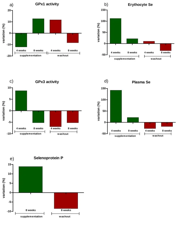
supplementation and washout period are shown in Table 2. GPx1 activity decreased 281
significantly after 4 weeks of supplementation, increased by 8 weeks of 282
supplementation (P < 0.001) and remained high during the first 4 weeks of Brazil nut 283
withdrawal; it finally decreased after 8 weeks washout (P = 0.004). Erythrocyte Se 284
concentrations increased after 4 and 8 weeks intervention and decreased after 8 weeks 285
washout (P < 0.001).There was a significant increase in GPx3 activity after 4 weeks of 286
supplementation (P = 0.004). Similarly, plasma Se concentrations increased 287
significantly after 4 and 8 weeks of supplementation (P < 0.001). During the washout 288
period, there was a sharp decrease in plasma Se compared to 8 weeks of 289
Brazil nut intake (P < 0.001). The concentrations of plasma SePP were also increased 290
after the supplementation and reduced after nut withdrawal (P = 0.001). The percentage 291
of the variation for each biomarker during the supplementation and the washout period 292
is shown in Figure 1. 293
Genotypes and haplotypes in selenoprotein genes
294
Genotype and allele frequencies of SNPs in selenoprotein genes are shown in Table 3. 295
All SNPs were in Hardy-Weinberg Equilibrium. Haplotype analyses showed evidence 296
of linkage disequilibrium for SNPs in GPX1 gene: rs1050450 x rs1800668 (D’= 1.0 and 297
r2= 0.98) and rs1050450 x rs3811699 (D’= 1.0 and r2= 1.0). In fact, in Table 3 the
298
genotypes frequencies of the three SNPs are exactly the same. Only two haplotypes 299
were observed, the common haplotype CCG for rs1050450, 1800668 and rs3811699 300
with a frequency of 74% and the rare haplotype TTA with 25%. Therefore, only 301
rs1050450 was used for further analysis. For the SNPs in the GPX4, SELENOF and 302
SELENOS genes, the frequency of the rare genotypes was between 19% and 8%. No 303
haplotype analysis was performed in those genes because only one SNP was genotyped 304
in each gene.Variables influencing biomarkers of Se status during Brazil nut 305
supplementation and washout period
Multiple linear regression models for the five biomarkers of Se status in each time 307
point of the protocol are shown in Table 4. GPx1 activity was associated with 308
rs1050450 after 4 (P = 0.037) and 8 weeks (P = 0.017) of intervention and with rs5845 309
after 4 weeks (P = 0.003) of intervention with both SNPs reducing GPx1 activity. After 310
8 weeks of washout, GPx1 activity was associated with rs5845 in SELENOF, which was 311
lower in the presence of the rare allele A (P = 0.049) and with rs713041 in GPX4, 312
which increased GPx1 activity in individuals carrying the rare allele T (P = 0.036). The 313
SNP rs713041 in GPX4 gene was associated with Erythrocyte Se concentrations at 314
baseline, reducing its concentrations in the presence of the rare allele T (P = 0.038). 315
Plasma Se concentration was associated with rs7579 in SELENOP (P = 0.034) 316
and rs34713741 in SELENOS (P = 0.038) after 4 weeks of supplementation, which was 317
lower in the presence of the rare allele for both SNPs. Plasma SePP concentrations 318
were positively associated with plasma Se at baseline and GPx3 activity was positively 319
associated with plasma Se after 4 (P = 0.050) and 8 weeks (P = 0.025) of 320
supplementation, as expected due to the same blood compartment location of both 321
biomarkers (Table 4). A complete table of the biomarkers stratified by all functional 322
SNPs can be seen at Supplementary table 2 (Table S2). 323
Erythrocyte GPx1 activity was stratified by rs1050450, as appointed in the 324
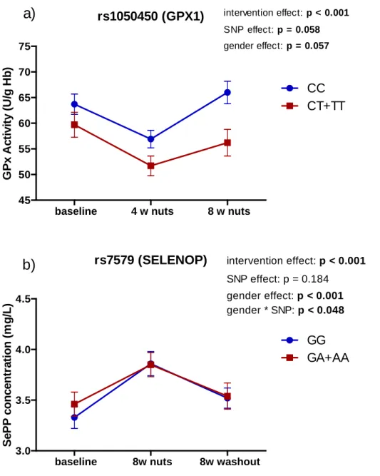
multivariate regression analysis (Figure 2). During the intervention, GPx1 activity was 325
lower in carriers of the rare allele T (CT+TT), almost reaching statistical significance at 326
4 weeks of supplementation (P = 0.057, Figure 2a). SePP concentrations were stratified 327
by rs7579 (Figure 2b). At baseline, SePP concentrations were higher for carriers of the 328
rare allele A, when the interaction with gender was considered (P = 0.048). 329
The percentage of variation on the concentrations of biomarkers of Se status is 330
shown in Table 5. The difference in GPx1 activity comparing baseline and 8 weeks of 331
supplementation (Change1_GPx1) was associated with rs7579 in SELENOP (P = 0.044) 332
and with rs5845 (P < 0.001), in which the presence of the rare allele increased this 333
difference, meaning that the values were higher for carriers of the rare allele A after 8 334
weeks of supplementation. The difference in Erythrocyte Se concentrations during the 335
supplementation (Change1_ Erythrocyte Se) was associated with rs34713741 in 336
SELENOS (P = 0.010), in which the presence of the rare allele T also increased this 337
difference. BMI and gender had a negative effect on the variation in Erythrocyte Se, in 338
which individuals with higher BMI and females had higher values at baseline. The 339
change in GPx3 activity in response to the supplementation (Change1_ GPx3 activity) 340
was negatively associated with rs5845 in SELENOF (P = 0.014), in which carriers of 341
the rare allele A had higher GPx3 activity at baseline. However, interaction with BMI 342
increased the variation (P = 0.011). The only two variables affecting the variation in 343
Plasma Se were gender and BMI, both having a negative effect. The difference in GPx1 344
activity comparing 8 weeks of washout and 8 weeks of supplementation 345
(Change2_GPx1) was negatively associated with gender, in which females had higher 346
GPx1 activity after 8 weeks of supplementation. However, the interaction with 347
rs713041 had the opposite effect: females carriers of the variant allele T had higher 348
GPx1 activity at 8 weeks of washout. Finally, the difference in SePP concentrations 349
comparing 8 weeks of washout and 8 weeks of supplementation (Change2_SePP) was 350
negatively associated with rs5845 in SELENOF gene, in which carriers of the rare allele 351
A had higher SePP concentrations after supplementation (Table 5). 352
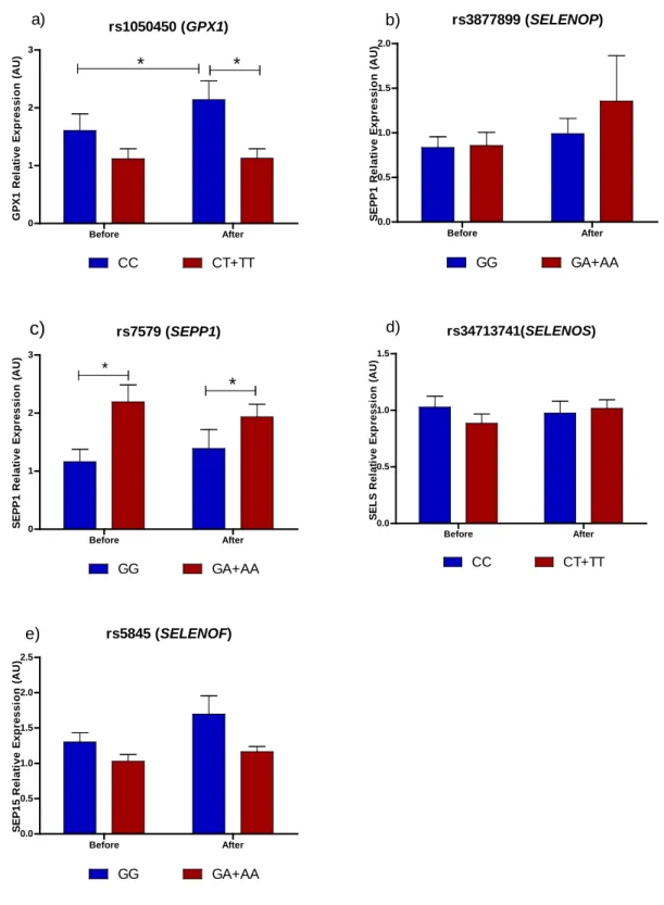
Effect of Brazil nut supplementation on mRNA expression of selenoprotein genes
353
Gene expression of four selenoprotein genes (GPX1, SELENOP, SELENOS and 354
SELENOF) was analyzed on previously genotyped volunteers before and after 8 weeks 355
of Brazil nut supplementation. The results are shown in Figure 3. GPX1 mRNA 356
expression increased after supplementation in individuals with the CC genotype for 357
rs1050450 (P = 0.026) while it did not change for carriers of the T allele (Figure 3a). 358
Consequently, after Brazil nut supplementation, GPX1 mRNA expression was lower in 359
individuals with the CT and TT genotypes compared to the CC group (P < 0.05). 360
SELENOP mRNA expression was higher in carriers of the rare allele A for rs7579 361
either before or after supplementation (Figure 3b, P < 0.05), and nut supplementation 362
did not significantly changed SELENOP mRNA expression whatever the genotype 363
group. No effect of genotypes on SELENOF and SELENOS mRNA expression was 364
observed either before or after the supplementation (Figure 3c and 3d). 365
DISCUSSION
366
The influence of genetic variants on the response to Se supplementation have 367
been proposed before 8,12,27,34. Our results not only confirm these earlier observations, 368
but also extend them by demonstrating that the rs5845 in SELENOF gene modulated 369
erythrocyte GPx1 activity, the variation of GPx1, GPx3 activity after supplementation 370
and the variation on SePP after nut withdraw. This study is the first to report this 371
unexpected association between rs5845 in SELENOF and Se biomarkers after 372
supplementation with Brazil nuts, the first to use SePP concentrations as a biomarker of 373
Se status in Brazilians and the first to measure all five biomarkers during the washout 374
period. Moreover, this study demonstrated that Brazil nut supplementation was effective 375
in increasing mRNA expression of GPX1 and SELENOP and that this effect was 376
modulated by functional polymorphisms on those genes. 377
It was observed a delayed response of the two erythrocyte biomarkers of Se 378
status to Brazil nut supplementation, with values increasing up to the first four weeks of 379
the washout period. This apparently slow response could be explained by erythrocytes 380
having a lifespan of 120 days so that it takes time for changes in selenoprotein synthesis 381
to appear in the mature red cell population 42,43. This is the first study to use plasma 382
SePP concentrations as a biomarker of Se status in healthy Brazilians. The baseline 383
plasma Se concentration of 96.7 µg/L (1.22 µmol/L) would be expected to maximize 384
plasma GPx activity, but not SePP concentration since earlier work has shown that the 385
plasma Se concentration needed to maximize GPx3 activity is about 90 µg/L 44 and to
386
maximize plasma SePP concentration it is approximately 120 µg/L 45. As a result, the
387
studied population could be considered to have a moderate to adequate Se status which 388
was able to respond to Se supplementation with an increase in concentrations of plasma 389
Se and SePP 43,46. We suggest that the threshold for maximize GPx3 activity be
390
reviewed, as our work demonstrated that this population with a baseline plasma Se of 391
90µL/L had a significant increase in GPx3 activity after 4 weeks of supplementation. 392
In our study subjects with the rare allele T for rs1050450 in GPX1 gene had 393
lower GPx1 activity. This observation is consistent with previous studies 10,11,47,48. It is 394
hypothesized that the change of the amino acid Proline to Leucine alters the secondary 395
structure of the protein, which can have profound effects on its activity and stability 21. 396
This was confirmed by in vitro studies where Se supplementation reduced enzyme 397
thermostability for the Leu-variant 49. It was observed that the SNPs rs1050450, 398
rs3811699 and rs1800668 were in linkage disequilibrium. This linkage was also 399
observed in a Japanese study conducted with type 2 diabetic patients 10. One possible 400
explanation for the reduced GPx1 activity is that the presence of these three genetic 401
variations affected the transcriptional process and, as a result, the final enzyme activity 402
was lower. Moreover, in our work, the Brazil nut supplementation was effective in 403
increase GPX1 mRNA expression in whole blood, only in individuals with the CC 404
genotype at rs1050450. Previous studies did not find a positive association of Se 405
supplementation and selenoprotein gene expression 14–16, however, three studies confirm
our results 13,17,18 . Differences may be explained by the lack of the genotype analysis on 407
previous studies, maybe to observe this effect of Se supplementation on mRNA levels 408
of GPX1 is necessary to stratify by genotypes. 409
The presence of rs7579 in the SELENOP gene influenced not only SePP plasma 410
concentrations but also SELENOP mRNA expression. SePP protein concentrations were 411
higher at baseline in carriers of the variant allele A for this 3’UTR SNP. Furthermore, 412
A-carriers had higher SELENOP mRNA levels than GG at baseline and after 413
supplementation. Previous work with humans have not found an association between Se 414
supplementation and SELENOP mRNA expression in white blood cells 14,16. It should
415
be noted that the present work used whole blood for the mRNA expression. Only one 416
study found a positive association by showing that rs7579 influenced SELENOP mRNA 417
expression 17. The SePP protein has two isoforms in plasma, the 50kDa and the 60kDa,
418
that are influenced by the genotype for both SNPs in SELENOP gene. Individuals with 419
the GA genotype for rs3877899 had a lower proportion of the SePP 60kD, with the Sec-420
rich domain 50. This difference in the proportion of SePP isoforms may affect Se 421
availability in plasma for selenoprotein synthesis in different tissues. 422
The 3’UTR region of SELENOF gene has two SNPs which are in the same 423
haplotype. The first one is a located a C>T substitution at position 811 (rs5859) and the 424
other is a G>A substitution at position 1125 (rs5845). The two possible haplotypes are 425
811C/1125G or 811T/1125A 51. Previous work has demonstrated that the SECIS
426
element containing the A variant is less responsive to Se supplementation and may 427
influence the translation of Selenoprotein 15 protein 52. Two biomarkers of Se status 428
were associated with rs5845 in SELENOF gene in our study: erythrocyte Se 429
concentrations and GPx1 activity. We also observed that this SNP modulated the 430
percentage of variation of GPx1 and GPx3 activities after supplementation and the 431
variation in SePP during washout period. To our knowledge, this is the first time that 432
rs5845 is associated with these biomarkers. These were unexpected associations, since 433
the SNP is located in the SELENOF gene and the biological effect was observed in 434
other selenoproteins. However, the biological function of the Selenoprotein 15 is still 435
unknown and maybe the hierarchy of selenoprotein expression may help explain these 436
associations with other selenoproteins. 437
This study has several limitations. The first one is the small sample size, which 438
could have masked significant associations of the genotypes with the biomarkers. The 439
second one is the absence of a control group, however, since the main goal of this 440
before-after interventional trial was to investigate the effect of the genotypes on the 441
response to the dietary intervention, we decided that each person before 442
supplementation would be a better control. The other limitation is the small number of 443
males in the study group which could have biased the gender effect in the statistical 444
modeling. 445
In summary, the results of this study suggest that genetic variants in 446
selenoprotein genes and gender influence the response of plasma and erythrocyte 447
biomarkers of Se status to a daily supplementation with one unit of Brazil nut in healthy 448
adults in Brazil. Furthermore, the genotypes for rs1050450 and rs7579 affected the gene 449
expression of GPX1 and SELENOP. Future nutritional interventions with Brazil nuts 450
should consider the genetic background of the volunteers when evaluating the 451
biomarkers of Se status used in this study. 452
453
Acknowledgments
454
The authors are very grateful to all volunteers who took part in this study. The 455
authors’ responsibilities were as follows – JLSD, MMR, SMFC: designed research; 456
JLSD: conducted research; FB Jr: was responsible for quantification of Plasma and 457
Erythrocyte Selenium; AS: provided essential reagents for Selenoprotein P 458
concentrations and Glutathione Peroxidase 3 activity; JLSD, EMGS, CD, PB: 459
performed data analysis and statistics; JLSD, MMR, JH, PB: wrote the manuscript. All 460
authors read and approved the final version of the manuscript for submission. The 461
authors declare no conflict of interest. 462
References
1. Blomhoff R, Carlsen MH, Andersen LF, Jacobs DR. Health benefits of nuts:
potential role of antioxidants. Br J Nutr. 2006;96(S2):S52.
2. Tapsell LC, Probst YC. Nutrition in the prevention of chronic diseases. World
Rev Nutr Diet. 2008;98:94–105.
3. Donaldson MS. Nutrition and cancer: a review of the evidence for an anti-cancer
diet. Nutr J. 2004;3:19.
4. Grosso G, Yang J, Marventano S, Micek A, Galvano F, Kales SN. Nut
consumption on all-cause, cardiovascular, and cancer mortality risk: a systematic review and meta-analysis of epidemiologic studies. Am J Clin Nutr. 2015 Apr;101(4):783–93.
5. Fairweather-tait SJ, Collings R, Hurst R. Selenium bioavailability : current knowledge and future research. Am J Clin Nutr. 2010;91(2):1484S–91S.
6. Kryukov G V, Castellano S, Novoselov S V, Lobanov A V, Zehtab O, Guigó R,
et al. Characterization of mammalian selenoproteomes. Science. 2003;300(5624):1439–43.
7. Rayman MP. Selenium and human health. Lancet. Elsevier Ltd;
2012;379(9822):1256–68.
8. Méplan C, Crosley LK, Nicol F, Beckett GJ, Howie AF, Hill KE, et al. Genetic
polymorphisms in the human selenoprotein P gene determine the response of selenoprotein markers to selenium supplementation in a gender-specific manner (the SELGEN study). FASEB J. 2007;21(12):3063–74.
9. Hesketh J. Nutrigenomics and selenium: gene expression patterns, physiological
targets, and genetics. Annu Rev Nutr. 2008;28:157–77.
10. Hamanishi T, Furuta H, Kato H, Doi A, Tamai M, Shimomura H, et al.
Functional variants in the glutathione peroxidase-1 (GPx-1) gene are associated with increased intima-media thickness of carotid arteries and risk of macrovascular diseases in Japanese type 2 diabetic patients. Diabetes. 2004;53(9):2455–60.
11. Ravn-Haren G, Olsen A, Tjønneland A, Dragsted LO, Nexø B a., Wallin H, et al.
Associations between GPX1 Pro198Leu polymorphism, erythrocyte GPX activity, alcohol consumption and breast cancer risk in a prospective cohort study. Carcinogenesis. 2006;27(4):820–5.
12. Combs GF, Jackson MI, Watts JC, Johnson LK, Zeng H, Idso J, et al. Differential
responses to selenomethionine supplementation by sex and genotype in healthy adults. Br J Nutr. 2012;107(10):1514–25.
13. Pagmantidis V, Méplan C, Van Schothorst EM, Keijer J, Hesketh JE.
Supplementation of healthy volunteers with nutritionally relevant amounts of selenium increases the expression of lymphocyte protein biosynthesis genes. Am J Clin Nutr. 2008;87(1):181–9.
14. Ravn-Haren G, Bügel S, Krath BN, Hoac T, Stagsted J, Jørgensen K, et al. A short-term intervention trial with selenate, enriched yeast and selenium-enriched milk: effects on oxidative defence regulation. Br J Nutr. 2008;99(4):883–92.
15. Ravn-Haren G, Krath BN, Overvad K, Cold S, Moesgaard S, Larsen EH, et al.
Effect of long-term selenium yeast intervention on activity and gene expression of antioxidant and xenobiotic metabolising enzymes in healthy elderly volunteers from the Danish Prevention of Cancer by Intervention by Selenium (PRECISE) pilot study. Br J Nutr. 2008;99(6):1190–8.
16. Sunde R a, Paterson E, Evenson JK, Barnes KM, Lovegrove J a, Gordon MH.
Longitudinal selenium status in healthy British adults: assessment using biochemical and molecular biomarkers. Br J Nutr. 2008;99 Suppl 3:S37–47.
17. Cardoso BR, Busse AL, Hare DJ, Cominetti C, Horst MA, Mccoll G, et al.
Pro198Leu polymorphism affects the selenium status and GPx activity in response to Brazil nut intake. Food Funct. Royal Society of Chemistry; 2015;9.
18. Jablonska E, Raimondi S, Gromadzinska J, Reszka E, Wieczorek E, Krol MB, et
al. DNA damage and oxidative stress response to selenium yeast in the non-smoking individuals: a short-term supplementation trial with respect to GPX1 and SEPP1 polymorphism. Eur J Nutr. Springer Berlin Heidelberg; 2015;1–16.
19. Ashton K, Hooper L, Harvey LJ, Hurst R, Casgrain A, Fairweather-Tait SJ.
Methods of assessment of selenium status in humans: A systematic review. Am J Clin Nutr. 2009;89(Suppl):2025S–39S.
20. Moscow J a, Schmidt L, Ingram DT, Gnarra J, Johnson B, Cowan KH. Loss of
heterozygosity of the human cytosolic glutathione peroxidase I gene in lung cancer. Carcinogenesis. 1994;15(12):2769–73.
21. Ratnasinghe D, Tangrea J a., Andersen MR, Barrett MJ, Virtamo J, Taylor PR, et
al. Glutathione peroxidase codon 198 polymorphism variant increases lung cancer risk. Cancer Res. 2000;60(22):6381–3.
22. Karunasinghe N, Han DY, Goudie M, Zhu S, Bishop K, Wang A, et al. Prostate
disease risk factors among a New Zealand cohort. J Nutrigenet Nutrigenomics. 2012;5(6):339–51.
23. Zhao H, Liang D, Grossman HB, Wu X. Glutathione peroxidase 1 gene
polymorphism and risk of recurrence in patients with superficial bladder cancer. Urology. 2005;66(4):769–74.
24. Combs GF, Watts JC, Jackson MI, Johnson LK, Zeng H, Scheett AJ, et al.
Determinants of selenium status in healthy adults. Nutr J. 2011;10:75.
25. Bellinger FP, Raman A V, Reeves M a, Berry MJ. Regulation and function of
selenoproteins in human disease. Biochem J. 2009;422(1):11–22.
26. Bermano G, Pagmantidis V, Holloway N, Kadri S, Mowat N a G, Shiel RS, et al.
Evidence that a polymorphism within the 3’UTR of glutathione peroxidase 4 is functional and is associated with susceptibility to colorectal cancer. Genes Nutr. 2007;2(2):225–32.
27. Méplan C, Crosley LK, Nicol F, Horgan GW, Mathers JC, Arthur JR, et al. Functional effects of a common single-nucleotide polymorphism (GPX4c718t) in the glutathione peroxidase 4 gene: interaction with sex. Am J Clin Nutr. 2008;87(4):1019–27.
28. Burk RF, Hill KE. Selenoprotein P-Expression, functions, and roles in mammals.
Biochim Biophys Acta - Gen Subj. Elsevier B.V.; 2009;1790(11):1441–7.
29. Bos SD, Kloppenburg M, Suchiman E, van Beelen E, Slagboom PE, Meulenbelt
I. The role of plasma cytokine levels, CRP and Selenoprotein S gene variation in OA. Osteoarthr Cartil. Elsevier Ltd; 2009;17(5):621–6.
30. Sutherland A, Kim DH, Relton C, Ahn YO, Hesketh J. Polymorphisms in the
selenoprotein S and 15-kDa selenoprotein genes are associated with altered susceptibility to colorectal cancer. Genes Nutr. 2010;5(3):215–23.
31. Ferguson AD, Labunskyy VM, Fomenko DE, Araç D, Chelliah Y, Amezcua C a.,
et al. NMR structures of the selenoproteins Sep15 and SelM reveal redox activity of a new thioredoxin-like family. J Biol Chem. 2006;281(6):3536–43.
32. Jablonska E, Gromadzinska J, Sobala W, Reszka E, Wasowicz W. Lung cancer
risk associated with selenium status is modified in smoking individuals by Sep15 polymorphism. Eur J Nutr. 2008;47(1):47–54.
33. Rayman MP, Searle E, Kelly L, Johnsen S, Bodman-Smith K, Bath SC, et al.
Effect of selenium on markers of risk of pre-eclampsia in UK pregnant women: a randomised, controlled pilot trial. Br J Nutr. 2014;112(1):99–111.
34. Mao J, Vanderlelie JJ, Perkins A V., Redman CWG, Ahmadi KR, Rayman MP.
Genetic polymorphisms that affect selenium status and response to selenium supplementation in United Kingdom pregnant women. Am J Clin Nutr. 2016;103(1):100–6.
35. Donadio JLS, Rogero MM, Guerra‑shinohara EM, Desmarchelier C, Borel P,
Cozzolino SMF. SEPP1 polymorphisms modulate serum glucose and lipid response to Brazil nut supplementation. Eur J Nutr. 2017;
36. Cominetti C, de Bortoli MC, Garrido AB, Cozzolino SMF. Brazilian nut
consumption improves selenium status and glutathione peroxidase activity and reduces atherogenic risk in obese women. Nutr Res. 2012 Jun;32(6):403–7.
37. AOAC Association of Official Analytical Chemists. Official methods of analysis.
15th ed. Washington; 1990.
38. Batista BL, Rodrigues JL, Nunes JA, Souza VC de O, Barbosa F. Exploiting
dynamic reaction cell inductively coupled plasma mass spectrometry (DRC-ICP-MS) for sequential determination of trace elements in blood using a dilute-and-shoot procedure. Anal Chim Acta. 2009 Apr 20;639(1–2):13–8.
39. Brown KM, Pickard K, Nicol F, Beckett GJ, Duthie GG, Arthur JR. Effects of
organic and inorganic selenium supplementation on selenoenzyme activity in blood lymphocytes, granulocytes, platelets and erythrocytes. Clin Sci (Lond). 2000;98(5):593–9.
40. Plonka-Poltorak E, Zagrodzki P, Nicol F, Kryczyk J, Barton H, Westermarck T, et al. Antioxidant agents and physiological responses in adult epileptic patients treated with lamotrigine. Pharmacol Reports. 2013;65(1):99–106.
41. Livak KJ, Schmittgen TD. Analysis of relative gene expression data using
real-time quantitative PCR and the 2(-Delta Delta C(T)) Method. Methods. 2001;25(4):402–8.
42. Robberecht H, Cauwenbergh R Van, Hermans N. Blood selenium levels and
factors influencing concentration values. Trace Elem Electrolytes. 2012;29(3).
43. Thomson CD, Chisholm A, Mclachlan SK, Campbell JM. Brazil nuts : an
effective way to improve selenium status. Am J Clin Nutr. 2008;87:379–84.
44. Duffield AJ, Thomson CD, Hill KE, Williams S. An estimation of selenium
requirements for New Zealanders. Am J Clin Nutr. 1999;70:896–903.
45. Hurst R, Armah CN, Dainty JR, Hart DJ, Teucher B, Goldson AJ, et al.
Establishing optimal selenium status : results of a randomized , double-blind, placebo-controlled trial. Am J Clin Nutr. 2010;91(4):923–31.
46. Da Silva EG, Mataveli LRV, Zezzi Arruda MA. Speciation analysis of selenium
in plankton, Brazil nut and human urine samples by HPLC-ICP-MS. Talanta. 2013;110:53–5.
47. Bastaki M, Huen K, Manzanillo P, Chande N, Chen C, Balmes JR, et al.
Genotype-activity relationship for Mn-superoxide dismutase, glutathione peroxidase 1 and catalase in humans. Pharmacogenet Genomics. 2006;16(4):279–86.
48. Hansen RD, Krath BN, Frederiksen K, Tjønneland A, Overvad K, Roswall N, et
al. GPX1 Pro198Leu polymorphism, erythrocyte GPX activity, interaction with alcohol consumption and smoking, and risk of colorectal cancer. Mutat Res. 2009;664(1–2):13–9.
49. Zhuo P, Goldberg M, Herman L, Lee BS, Hengbing W, Brown RL, et al.
Molecular consequences of genetic variations in the glutathione peroxidase 1 selenoenzyme. Cancer Res. 2009;69(20):8183–90.
50. Méplan C, Nicol F, Burtle BT, Crosley LK, Arthur JR, Mathers JC, et al.
Relative abundance of selenoprotein P isoforms in human plasma depends on genotype, se intake, and cancer status. Antioxid Redox Signal. 2009;11(11):2631–40.
51. Watrowski R, Castillo-Tong DC, Fabjani G, Schuster E, Fischer M, Zeillinger R.
The 811 C/T polymorphism in the 3′ untranslated region of the selenoprotein 15-kDa (Sep15) gene and breast cancer in Caucasian women. Tumor Biol. Springer Netherlands; 2016 Jan 12;37(1):1009–15.
52. Hu YJ, Korotkov K V, Mehta R, Hatfield DL, Rotimi CN, Luke a, et al.
Distribution and functional consequences of nucleotide polymorphisms in the 3’-untranslated region of the human Sep15 gene. Cancer Res. 2001;61(5):2307–10.
TABLE 1
Characteristics of the study volunteers1
Parameters Total (n= 130) Gender, n (%) male 32 (24.6) female 98 (75.4) Age (y), n (%) 20 – 30y 92 (70.8) 31 – 40y 20 (15.4) 41 – 50y 9 (6.9) 51 – 60y 9 (6.9) BMI (kg/m2), n (%) < 25 93 (71.5) 25 – 30 31 (23.8) > 30 6 (4.6) Smoking status, n (%) Never 104 (80.0) ex-smoker 20 (5.4) current 6 (4.6) Physical activity, n (%) yes 94 (72.3) no 36 (27.7)
History of chronic diseases, n (%)
yes 113 (86.9) no 17 (13.1) Race, n (%) caucasian 94 (72.3) black 26 (20.0) asian/indigen 10 (7.7) Plasma Se (µg/L) before supplementation 96.7 ± 29.6 after supplementation 292.8 ± 95.4 1
Numerical variables are presented as mean ± standard deviation (sd). Categorical variables are presented as n (%).
TABLE 2
Concentrations of biomarkers1 of Se status during supplementation with Brazil nuts and
washout period in healthy adults (n = 130)
Biomarker Supplementation period
baseline 4 weeks 8 weeks P value2
GPx 1 activity (U/g Hb) 61.8 (58.8 – 65.1)a 54.4 (52.0 – 57.1)b 61.3 (57.7 – 65.1)a < 0.001 Erythrocyte Se (µg/L) 169.8 (158.8 – 181.4)a 360.2 (342.5 – 378.9)b 438.8 (406.6 – 464.8)c < 0.001 GPx 3 activity (U/L) 528.1 (510.2 – 546.6)a 574.1 (554.4 – 594.5)b 544.4 (523.5 – 566.2)ab 0.004 Plasma Se (µg/L) 90.7 (86.4 – 95.2)a 219.5 (208.4 – 231.2)b 267.0 (252.8 – 282.0)c < 0.001 SePP (mg/L) 3.4 (3.2 – 3.5)a na 3.9 (3.7 – 4.1)b < 0.001
Biomarker Washout period
8 weeks 4 weeks 8 weeks P value
GPx 1 activity (U/g Hb) 61.3 (57.7 – 65.1)a 68.5 (65.3 – 71.9)b 62.8 (58.9 – 67.0)ab 0.005 Erythrocyte Se (µg/L) 438.8 (406.6 – 464.8)a 484.1 (452.7 – 517.5)b 332.1 (307.0 – 359.3)c < 0.001 GPx 3 activity (U/L) 544.4 (523.5 – 566.2)a 507.1 (486.9 – 528.2)ab 480.9 (457.7 – 505.2)b 0.021 Plasma Se (µg/L) 267.0 (252.8 – 282.0)a 195.3 (186.8 – 204.2)b 160.2 (154.3 – 166.2)c < 0.001 SePP (mg/L) 3.9 (3.7 – 4.1)a na3 3.5 (3.4 – 3.7)b 0.001 1
Values are geometric means (CI 95%).
2
ANOVA repeated measures with post hoc Tukey for GPx1 activity; Friedman test with post hoc Dunn for Erythrocyte Se, GPx3 activity and Plasma Se; Wilcoxon test for SePP concentrations. Different letters in the row mean statistical difference in the time points. GPx1 activity and Erythrocyte Se measured in erythrocytes and GPx3 activity, Plasma Se and SePP measured in Plasma. GPx, Glutathione Peroxidase; SePP,
Selenoprotein P.
3
TABLE 3
Genotype and allele frequency of SNPs in selenoprotein genes
GPX1: Glutathione Peroxidase 1, GPX4: Glutathione Peroxidase 4, SELENOP: Selenoprotein P; SELENOF: Selenoprotein 15; SELENOS: Selenoprotein S.
Gene SNP N CC/GG CT/GA TT/AA alleles MAF HWE
(p value) GPX1 rs1050450 130 53.85 40.77 5.38 C > T T: 0.25 0.560 rs3811699 130 53.85 40.77 5.38 C > T T: 0.25 0.560 rs1800668 130 53.85 40.77 5.38 C > T T: 0.25 0.560 GPX4 rs713041 130 38.46 42.31 19.23 C > T T: 0.40 1.914 SELENOP rs3877899 130 53.85 36.15 10.00 G > A T: 0.28 1.428 rs7579 130 38.46 42.31 19.23 G >A T: 0.40 1.914 SELENOS rs34713741 130 53.85 37.69 8.46 C > T T: 0.27 0.332 SELENOF rs5845 130 42.31 46.15 11.54 C > T T: 0.35 0.050
TABLE 4
Multiple Linear Regression models for biomarkers of Se status during daily supplementation with Brazil nuts and washout period in healthy subjects
Dependent Variables
Predictors β coefficient Standard
error
P value GPx1 activity (U/g Hb)
Baseline Erythrocyte Se 0.050 0.019 0.011
4 weeks nuts Erythrocyte Se 0.029 0.012 0.014
rs1050450 (GPX1) -5.385 2.546 0.037 rs5845 (SELENOF) -7.959 2.605 0.003 8 weeks nuts rs1050450 -8.300 3.424 0.017 8 weeks washout rs5845 -8.333 4.189 0.049 rs713041 8.955 4.231 0.036 Erythrocyte Se concentration (µg/L) Baseline Plasma Se 1.570 0.215 < 0.001 rs713041 (GPX4) -26.698 12.752 0.038
4 weeks nuts Plasma Se 0.405 0.150 0.008
8 weeks nuts Plasma Se 0.692 0.158 < 0.001
Plasma Se concentration (µg/L)
4 weeks nuts rs7579 (SELENOP) -0.115 0.054 0.034
rs34713741 (SELENOS) -0.110 0.053 0.038 SePP concentration (mg/dL) Baseline Plasma Se 0.007 0.003 0.015 GPx3 activity (U/L)
4 weeks nuts Plasma Se 0.323 0.165 0.050
8 weeks nuts Plasma Se 0.304 0.134 0.025
1
Multivariate linear regression models were done separately in different time points for each biomarker using SPSS. Only significant p values (< 0.05) are shown in the table. GPx1 activity and Erythrocyte Se measured in erythrocytes and GPx3 activity, Plasma Se and SePP measured in Plasma. GPx, Glutathione Peroxidase; SePP, Selenoprotein P. All the models were adjusted for age, gender and body fat percentage.
TABLE 5
Multiple Linear Regression models for the change1 on biomarkers concentrations during
daily supplementation with Brazil nuts
Dependent Variables Predictors β coefficient Standard
error P value Change1_GPx1 rs7579 7.372 3.624 0.044 rs5845 54.582 11.334 < 0.001 Change1_ Erythrocyte Se rs34713741 262.445 100.909 0.010 BMI -11.849 4.348 0.007 gender -107.745 32.802 0.001 Change1_GPx3 rs5845 -397.353 158.625 0.014 BMI*rs5845 17.412 6.769 0.011
Change1_ Plasma Se gender -57.110 18.159 0.002
BMI -8.429 2.440 0.001
Change2_GPx1 gender -40.057 18.162 0.029
gender*rs713041 26.269 10.728 0.016
Change2_SePP rs5845 -3.110 1.521 0.043
1
: Change 1 = (8 weeks nuts) – (baseline) and Change 2 = (8 weeks washout) – (8 weeks nuts)
All the models were adjusted for age, gender and body fat percentage. GPx, Glutathione Peroxidase; SePP, Selenoprotein P.
GPx1 activity
4 weeks 8 weeks 4 weeks 8 weeks
-20 -10 0 10 20 w ashout supplem entation a) v a ri a ti o n ( % ) Erythocyte Se
4 weeks 8 weeks 4 weeks 8 weeks
-50 0 50 100 150 b)
supplem entation w ashout
v a ri a ti o n ( % ) GPx3 activity
4 weeks 8 weeks 4 weeks 8 weeks
-10 -5 0 5 10 c)
supplem entation w ashout
v a ri a ti o n ( % ) Plasma Se
4 weeks 8 weeks 4 weeks 8 weeks
-50 0 50 100 150 d)
supplem entation w ashout
v a ri a ti o n ( % ) Selenoprotein P 8 weeks 8 weeks -10 -5 0 5 10 15 e)
supplem entation w ashout
v a ri a ti o n ( % )
FIGURE 1. Percentage of variation1 in five biomarkers of Se status during and after daily supplementation with one unit of Brazil nuts in healthy adults
1
The variation was calculated comparing with the time point immediate before: 4 weeks nuts compared to baseline, 8 weeks nuts compared to 4 weeks nuts, 4 weeks washout compared with 8 weeks nuts and 8 weeks washout compared to 4 weeks washout. a) percentage of variation for GPx1 activity, b) percentage of variation for Erythrocyte Se, c) percentage of variation for GPx3 activity, d) percentage of variation for Plasma Se, e) percentage of variation for SePP.
rs1050450 (GPX1)
baseline 4 w nuts 8 w nuts
45 50 55 60 65 70 75 CC CT+TT
a)
intervention effect: p < 0.001 SNP effect: p = 0.058 gender effect: p = 0.057 G P x A ct iv it y ( U /g H b ) rs7579 (SELENOP)baseline 8w nuts 8w washout
3.0 3.5 4.0 4.5 GG GA+AA intervention effect: p < 0.001 SNP effect: p = 0.184 gender effect: p < 0.001 gender * SNP: p < 0.048
b)
S e P P c o n c e n tr a ti o n ( m g /L )FIGURE 2. Biomarkers of Se status stratified by SNPs in GPX1 and SELENOP genes1.
1
Values are geometric means ± standard errors. Two way ANOVA repeated measures adjusted for multiple comparisons with Bonferroni test. a) Erythrocyte GPx1 activity stratified by rs1050450 genotypes, b) Plasma SePP concentration stratified by rs7579 genotypes.
rs1050450 (GPX1) Before After 0 1 2 3 CC CT+TT * * a) G P X 1 R el at ive E x p ressi o n ( A U ) rs3877899 (SELENOP) Before After 0.0 0.5 1.0 1.5 2.0 GG GA+AA b) S E P P 1 R e la ti v e E xp res si o n ( A U ) rs7579 (SEPP1) Before After 0 1 2 3 GG GA+AA c) * * S E P P 1 R e la ti v e E xp res si o n ( A U ) rs34713741(SELENOS) Before After 0.0 0.5 1.0 1.5 CC CT+TT d) S E L S R el at ive E xp ressi o n ( A U ) rs5845 (SELENOF) Before After 0.0 0.5 1.0 1.5 2.0 2.5 GG GA+AA e) S E P 15 R el at ive E xp ressi o n ( A U )
FIGURE 3. Pattern of selenoprotein gene expression in response to Brazil nut
supplementation on previously genotyped volunteers1
1
Gene expression relative to GAPDH a) GPx1 mRNA expression separated by rs1050450 genotypes, b) SELENOP mRNA expression separated by rs3877899 genotypes, c) SELENOP mRNA expression separated by rs7579 genotypes, d) SELENOS mRNA expression separated by rs34713741 genotypes, e) SELENOF mRNA expression separated by rs5845 genotypes.