RESEARCH OUTPUTS / RÉSULTATS DE RECHERCHE
Author(s) - Auteur(s) :
Publication date - Date de publication :
Permanent link - Permalien :
Rights / License - Licence de droit d’auteur :
Bibliothèque Universitaire Moretus Plantin
Institutional Repository - Research Portal
Dépôt Institutionnel - Portail de la Recherche
researchportal.unamur.be
University of Namur
The leptin receptor mutation of the obese Zucker rat causes sciatic nerve
demyelination with a centripetal pattern defect
Gilloteaux, Jacques; Subramanian, Kritika; Solomon, Nadia; Nicaise, Charles
Published in: Ultrastructural Pathology DOI: 10.1080/01913123.2018.1522405 Publication date: 2018 Document Version
Publisher's PDF, also known as Version of record
Link to publication
Citation for pulished version (HARVARD):
Gilloteaux, J, Subramanian, K, Solomon, N & Nicaise, C 2018, 'The leptin receptor mutation of the obese Zucker rat causes sciatic nerve demyelination with a centripetal pattern defect', Ultrastructural Pathology, vol. 42, no. 5, pp. 377-408. https://doi.org/10.1080/01913123.2018.1522405
General rights
Copyright and moral rights for the publications made accessible in the public portal are retained by the authors and/or other copyright owners and it is a condition of accessing publications that users recognise and abide by the legal requirements associated with these rights. • Users may download and print one copy of any publication from the public portal for the purpose of private study or research. • You may not further distribute the material or use it for any profit-making activity or commercial gain
• You may freely distribute the URL identifying the publication in the public portal ? Take down policy
If you believe that this document breaches copyright please contact us providing details, and we will remove access to the work immediately and investigate your claim.
Full Terms & Conditions of access and use can be found at
https://www.tandfonline.com/action/journalInformation?journalCode=iusp20
Ultrastructural Pathology
ISSN: 0191-3123 (Print) 1521-0758 (Online) Journal homepage: https://www.tandfonline.com/loi/iusp20
The leptin receptor mutation of the obese Zucker
rat causes sciatic nerve demyelination with a
centripetal pattern defect
Jacques Gilloteaux, Kritika Subramanian, Nadia Solomon & Charles Nicaise
To cite this article: Jacques Gilloteaux, Kritika Subramanian, Nadia Solomon & Charles Nicaise (2018) The leptin receptor mutation of the obese Zucker rat causes sciatic nerve demyelination with a centripetal pattern defect, Ultrastructural Pathology, 42:5, 377-408, DOI: 10.1080/01913123.2018.1522405
To link to this article: https://doi.org/10.1080/01913123.2018.1522405
© 2018 The Author(s). Published by Taylor & Francis
Published online: 19 Oct 2018.
Submit your article to this journal
Article views: 798
View related articles
BASIC RESEARCH
The leptin receptor mutation of the obese Zucker rat causes sciatic nerve
demyelination with a centripetal pattern defect
Jacques Gilloteauxa,b, Kritika Subramaniana,c, Nadia Solomona, and Charles Nicaiseb
aDepartment of Anatomical Sciences, St George’s University School of Medicine, K.B. Taylor Global Scholar’s Program at Northumbria
University, Newcastle upon Tyne, UK;bUnité de Recherche en Physiologie Moléculaire (URPhyM), Laboratoire de Neurodégénérescence et
Régénération, Département de Médecine, Université de Namur, Namur, Belgium;cDepartment of Clinical and Epidemiological Virology, Rega
Institute of Medical Research, Katholiele Universiteit Leuven, Leuven, Belgium
ABSTRACT
Young male Zucker rats with a leptin receptor mutation are obese, have a non-insulin-dependent diabetes mellitus (NIDDM), and other endocrinopathies. Tibial branches of the sciatic nerve reveal a progressive demyelination that progresses out of the Schwann cells (SCs) where electron-contrast deposits are accumulated while the minor lines or intermembranous SC contacts display exaggerated spacings. Cajal bands contain diversely contrasted vesicles adjacent to the abaxonal myelin layer with blemishes; they appear dispatched centripetally out of many narrow electron densities, regularly spaced around the myelin annulus. These anomalies widen and yield into sectors across the stacked myelin layers. Throughout the worse degradations, the adaxonal membrane remains along the axonal neuro-plasm. This peripheral neuropathy with irresponsive leptin cannot modulate hypothalamic-pituitary-adrenal axis and SC neurosteroids, thus exacerbates NIDDM condition. Additionally, the ultrastructure of the progressive myelin alterations may have unraveled a peculiar, centripetal mode of trafficking maintenance of the peripheral nervous system myelin, while some adhesive glycoproteins remain between myelin layers, somewhat hindering the axon mutilation.
Heading title: Peripheral neuropathy and myelin
ARTICLE HISTORY Received 8 February 2018 Accepted 7 September 2018 KEYWORDS
Leptin receptor; myelin; NIDDM– obesity; sciatic nerve– Schwann cell; Zucker rat
Introduction
In basic neuropathology texts, demyelination could
be acute or chronic. However, the etiology of the
degenerative process related to the nourishing layer
of nerve fiber’s myelin, either involving the central or
the peripheral nervous system (PNS), is complex and
still poorly understood.
1–6In dealing with
periph-eral neuropathies, textbooks bring the topic along
with neuromuscular anomalies.
7The defects are
classified either as (a) axonal neuropathies in which
insults of the axons often consist in degeneration
occurring distally and secondarily to damage the
myelin or (b) as demyelinating neuropathies
char-acterized by Schwann cell (SC) changes wherein
myelin would display abnormal conduction
veloci-ties. This latter type of neural defect is apparently
short-sized and can appear randomly to reduce the
internode myelin sheaths while maintaining the
axonal content. There, changes occurring in the
PNS endoneurium have been seldom investigated.
8–10Recent advances about cooperativity between SC
basal lamina components and axon have revealed
paracrine and juxtacrine interactions with at least
one of the neuregulins.
11,12This report encompasses the fine structure of
scia-tic nerve demyelination injuries in the young male
Zucker rats. A preliminary study of this topic
13followed investigations that have dealt with other
endocrinopathies, such as thyroid gland dysfunctions
(hypothyroidism
14–27and hypercalcemia
14–16),
motricity
27along with a non-insulin-dependent
dia-betes mellitus (NIDDM) or diadia-betes type 2
14–17In
this rat strain, these defects have been linked to a
leptin receptor mutation
28–36, comforted by
pancrea-tic changes.
37–39Additionally, this leptin receptor
defect provokes other hypothalamo-pituitary axis
CONTACTJacques Gilloteaux [email protected] Department of Anatomical Sciences, St George’s University School of Medicine, UNN – School of Health and Life Sciences, Drill Hall 013, Newcastle upon Tyne NE1 8ST, UK; Faculté de Médecine, Université de Namur, Rue de Bruxelles 61, Namur 5000, Belgium
Dedicatedto our colleague Dr Joseph Allan Tucker jr Louise Lenoir Locke, Professor of Pathology, University of Southern Alabama Medical School, Mobile, AL, USA Color versions of one or more of the figures in the article can be found online atwww.tandfonline.com/iusp.
This article has been republished with minor changes. These changes do not impact the academic content of the article. ULTRASTRUCTURAL PATHOLOGY
2018, VOL. 42, NO. 5, 377–408
https://doi.org/10.1080/01913123.2018.1522405
© 2018 The Author(s). Published by Taylor & Francis
This is an Open Access article distributed under the terms of the Creative Commons Attribution-NonCommercial-NoDerivatives License (http://creativecommons.org/licenses/by-nc-nd/4.0/), which permits non-commercial re-use, distribution, and reproduction in any medium, provided the original work is properly cited, and is not altered, transformed, or built upon in any way.
failures.
40–42Because PNS demyelination defects are
often viewed by light microscopy (LM) and not well
illustrated with fine structure, we have aimed to
docu-ment further ultrastructural information on
diabetes-related neuropathy.
Interestingly, the Zucker obese rats bore myelin
anomalies resembling the ones found in
toxicant-induced diabetes in animals
43–52and probably also
those
– not studied by fine structure – found in
unusual human cases of diabetes where leptin
recep-tor was similarly incompetent.
53–69Therefore, the
discussion of our demyelination data includes
dia-betes type 2 considerations along with leptin-linked
endocrine interactions.
Our
micrographic
illustrations
have
been
arranged in a progressive peripheral nerve defects
sequence that could supplement those found in
human diabetes biopsies or those of testing animal
for diabetes and treatments. Additionally, both a
preliminary report presented in Lisbon meeting
70and the analysis of the myelin defects collected
could have unveiled another possible molecular
dynamic mechanism, dealing with the
mainte-nance of the PNS myelin membranes and
compo-nents that could involve a centripetal diffusion out
of either the SCs, marked by an excessive content
in electron-contrasted species.
Materials and methods
The Institutional Animal Care and Use Committee of
the Northeastern Ohio Universities College of
Medicine (now named
‘Northeast Ohio University’),
Rootstown, Ohio, USA have approved the procedures
of animal care, anesthesia, euthanasia, and tissue’s
collection of this study and concomitant ones.
40,41Terminology: Lean Zucker rats have a possible
genotype of the dominant trait Fa homozygous (Fa/
Fa) or heterozygous (Fa/fa), hence called Fa/?, where
the interrogation mark indicates whether fa or Fa
trait is associated with another fa trait (weight)
with-out being unable to verify the corresponding leptin
receptor genotype.
12–18Phenotypically, the obese
Zucker rats possesses both recessive traits (fa/fa)
and consistently showed significant overweight at
matching age.
Five young obese male Zucker rats (fa/fa; 3 month
of age, 398 ± 21.2 g) and five lean littermates (Fa/?)
(201 ± 13.5 g) obtained out of a colony originally
purchased from Charles River Laboratories (Raleigh,
NC) and derived from an original stock
10–13were
housed individually and maintained on a 12 h-light/
12 h-dark cycle (light from 06.00 to 18.00 h). Rats fed
rodent chow (Purina, St Louis, MO) and water ad
libitum. Anesthetized with ether
71rats were perfused
with warm saline (38°C) through aorta for 5 min; then
saline was then replaced by an ad hoc fixative to allow
other studies. The fixative was a mixture of 3%
glutar-aldehyde–paraformaldehyde (1:1) buffered by
phos-phate buffer (pH 7.3–7.4)
72for 30 min in cold
temperature because tissues were primarily used for
immunohistochemistry investigations and one not
necessarily ultrastructure. Excision of sciatic nerve
branches, other organs, and tissues occurred after
brain removal was performed by others
40,41as
exploratory investigations with the aim of potential
other studies on these rodents. At the time, no
quan-titation was planned or performed.
Out of all the lean (Fa/?) and obese (fa/fa)
perfused rat carcasses, several (5–12 mm)
seg-ments excised from the sciatic tibial nerve
branches were not blind-collected; they were
fixed another hour in the same fixative
72, washed
in cacodylate buffer for 30 min (0.1M Na
cacody-late buffer, pH 7.35 and sucrose) and post-fixed
2 h by 2% OsO
4aqueous solution. Samples were
dehydrated, cleared and processed in PolyBed
epoxy resin (Polysciences, Warrington, PA).
One-micrometer thick sections stained with toluidine
blue were examined in an Olympus BX51
photo-microscope (Olympus America, Melville, NY).
Selected areas of LM were ultrathin sectioned,
collected on 50- and 75-mesh hexagonal copper
grids (SPI, West Chester, PA), stained in uranyl
acetate and lead citrate before they were examined
in a Zeiss EM-10 transmission electron
micro-scope (TEM; Carl Zeiss, Thornwood, NY).
Results
Light microscopy
Comparisons between the 1-µm thick sections of
lean (
Figure 1(a,b
)) and obese (
Figures 4(b
) and
5(a,b
)) rat sciatic tibial nerve specimens reveal
that, in both lean and obese nerves, the
popula-tion of large and small myelinated and
unmye-linated fibers can be viewed in all the samples of
nerve branches, including a few single
intramus-cular ones. However, LM aspects poorly resolve
differences at the highest magnifications or by
enlarging the micrographs through
computeri-zed captures. The oblique to longitudinal
sec-tions of the fa/fa sciatic nerve branches and
intramuscular fiber profiles, stained with
tolui-dine blue, displays a myelin layer with peculiar
whorls or sieve-like aspects. Additionally, in the
obese rat nerves, swollen axons and a less dense
internode myelin staining can be found
com-pared with the lean ones. It is only by
Figure 1.(a–c) Lean Fa/? Zucker rat sciatic nerve. One-micrometer thick sections of tibial branches of the sciatic with its epineurium (a,c). Toluidine blue stain. (a): Example of a large branch with tight fascicles contained in the perineurial sheath and two adjacent small branches. The central region contains an obvious vasa nervorum. In (c): A small intramuscular nerve subdivision. Scale in (a) and (b) is 10 µm. (b): TEM pane mounted out of nine micrographs of a further intramuscular branch of (c) demonstrating two small fascicles of five fibers each, surrounded by the epi- and perineurial fibers and an endoneurial loose connective tissue where nerve fibers show their densely contrasted myelin; the most folded ones likely denote their near- or paranodal region. Scale is 5 µm.
Figure 2.(a–b) Lean Fa/? Zucker rat. TEM of cross-sections of isolated intramuscular nerve fibers both surrounded by their endoneurial connective fibers (e). The small part of Schwann cell viewed in intermodal cross-section is the Cajal band, its intranodal myelin and narrow cytoplasm, surrounded by its basal lamina Scale is 1 µm.
ultrastructure
examination
that
differences
between lean or Fa/? nerves can be verified
(
Figures 1(c
),
2(a,b
), and
3(a,b
)), such as the
fine and vacuole-like blemishes revealed along
the myelin of the nerve fibers of obese fa/fa
nerves (
Figures 4(a
) and
5(a
)–
16
).
Figure 3.(a–b) Lean Fa/? Zucker rat. Both TEM views of an internodal segment of myelin from eitherFigure 1(c) or2(aor b) showing its typical basal lamina and the characteristic layering of myelin insulation with 12.5 nm periodicity of the major dense lines spaced by a middle 6.0–6.3 nm minor dense line or intraperiod, corresponding to the external leaflets of the Schwann cell neurilemma. A vasa nervorum endothelium, rich in endo-exocytotic vesicles and its basal lamina is also shown in (a) where the axoplasm contains its neurofibrils and a few neuroreticulum saccules adjacent to adaxonal membrane. (b) is (a) magnified. Scale in (a) is 500 nm and in (b) 12.5 nm is the major dense line periodicity.
Figure 4.(a–b) Obese Zucker rat. (a): LM view of 1 µm-thick cross of a sciatic tibial nerve branch. Toluidine blue stain. Scale is 10 µm. (b): TEM micrograph montage reconstituting a view of (a) section. This pane depicts diverse myelinated fiber damages, in oblique and cross-sections. Some of them were enlarged to further illustrate this study. Note the myelinated and a few Remak nerve fibers accompanied by a loose endoneurial and perineurial supportive tissue. Scale is 1 µm.
Transmission electron microscopy
TEM observations further confirm that no
altera-tions affect the lean rat nerves (
Figures 1(c
),
2(a,b
),
and
3(a,b
)), while all the obese rat myelinated
nerve
samples
display
some
demyelinating
damages and worse are found to be proportional
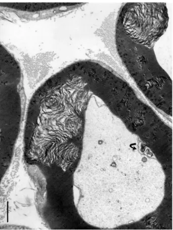
Figure 5.(a–c) (a–b) One-micrometer thick longitudinal to oblique section of a sciatic tibial nerve branch of an obese Zucker rat. Toluidine blue stain. Scales are 10 µm. (a): Example of field of view that shows how difficult is LM resolution to verify whether alterations have occurred but the tiny, poorly stained bulges (*) while nerve fibers’ internodes appear swollen. (b): Tangential views of most myelinated fibers of a fascicle denote changes in myelin with spaced vacuolizations of the insulating myelin that can appear as dark Swiss cheese (upper area). In both (a) and (b) Schmidt-Lantermann areas can be noticed as if oblique gashes in the myelin (both left middle areas). (c): TEM detail of an oblique to longitudinal aspect of one nerve fiber of (b) that resolves the spaced vacuoles and the bulging segments in the myelin to be sectors of focal, demyelination. An endoneurial fibrocyte is adjacent to this nerve fiber section and demonstrates multiple contrasted deposits. Scales equal 10 µm in (a) and (b), in (c) is 1 µm.
to their diameter size (
Figures 4(b
) and
5(b
)–
16
).
In the following paragraphs, descriptions of nerve
fiber injuries of the obese rat nerves are organized
to depict the progression of the damages, from the
smallest to the worst, comforting the neuropathic
changes associated with the NIDDM-associated
leptin receptor defect of this rat strain.
Lean (Fa/?) Zucker rat sciatic nerves
Myelinated fibers.
In these fibers, the
neurolem-nocyte or SC cytoplasm of some small or large
nerve fibers reveals typical indent of the perikaryal
areas (not shown here); it contains typical cell
organelles. The SC nucleus, with its perikaryon,
is in the median internode regions of each
periph-eral nerve fiber, thus the internodal cytoplasm is
most often viewed as a narrow band with the
random TEM sections, named Cajal band. This
‘band’ of the SC contains the abaxonal region
with its outermost myelin layer and sheaths
where, by place, outer mesaxons can be viewed
(
Figures 1(c
) and
2(a,b
)). The axoplasms show
mitochondria, neurotubules, neurofibrils, and a
few adaxonal neuroreticulum cisternae. In the rat
tibial branches, most nerve fibers are typically
myelinated and organized by SCs in concentric
tight layers of neurilemma forming an electron
dense Fermat-like spire enclosing the neurites,
whether found in cross or oblique, or
low-magni-fication sections (
Figure 3(a
)).
Viewed at a higher resolution, the myelin
dis-plays the internodal myelin tightly organized,
with its regularly distanced membranous
archi-tecture and adhesive contacts. There, the typical
major dense lines are viewed as highly
con-trasted thick
‘lines’ which are observed ranging
between 12.5 and 15.5 nm distance periodicity.
There, the thinned SC cytoplasm is spaced by
weakly contrasted intraperiod lines (or minor
lines) that appear with poor contrast, containing
Figure 6.(a–d) TEM of obese rat sciatic nerve. In (a–c): Semi-serial TEM views of perikaryal SC (or parts of Cajal bands containing a mitochondrium, smooth and rough endoplasmic reticulum, polyribosomes and a lipid deposit (dense arrow)) enlarged in insert near (b) showing that no membrane lines the lipid-like inclusion, i.e. not a‘vesicle’. (b): similar area of Cajal band; TEM view as in (a) showing the same lipid deposit. (c): Cajal band perikaryal area of an obese nerve fiber showing a mesaxon area. Long mitochondria cut (m) are shown adjacent to electron dense vesicles or deposits adjacent to a Golgi cistern containing a fibrillar striated content (prepro-collagen?). (d): Small nerve fiber with its SC nucleus and perikaryon with a disorganized mesaxon complex. The curved arrows mark defects in the mesaxon, abaxonal myelin, leaving intact the adaxonal membrane. Scales in (a–d) are all equal to 1 µm.
the adjacent contacting membranes of one
inter-nodal wrapping SC. The periodical distance
between major lines can reach between 20 and
300 nm in width, the widest often located at the
level of Schmidt–Lantermann (S-L) and the
nodal zones (
Figure 3(b
)). In cross-section,
each entire myelin insulating profile shapes like
an annulus, somewhat circular but folded up
near and at the Ranvier’s nodes. The adjacent
to axolemmal with its adaxonal SC cytoplasm, or
so-called Mauthner’s layer, is quasi inexistent
due to the compaction of myelin.
Unmyelinated nerve fibers.
A few unmyelinated
(Remak-like) fibers can be found among the
endo-neurial stroma adjacent to the myelinated ones but
are not illustrated in
Figure 1
, especially when one
has enlarged the small intramuscular sciatic nerve
branches.
The supportive stroma including endoneurium.
This stroma, associated with the basal laminae
produced by the SCs, surrounds every fiber
whether myelinated or unmyelinated. The
endo-neurium reveals its loose endoendo-neurium containing
Figure 7.(a–d) TEM of sciatic nerve fibers of obese Zucker rat with initial myelin damages. (a): Twin dense arrows respectively mark the first and second intraperiod lines spaced with electron dense eposits adjacent to the Cajal band; small arrows and bl: indicate the basal laminae of adjacent nerve fibers. (b): Electron dense deposit enlarges and widen along 9–10th intraperiod line level. (c): Example of small abaxonal cisterns in Cajal band along the outermost myelin sheath. These damages continue as cone-like profiles to reach the adaxonal membrane as discrete to wide intermembranous spaces. (d): example of internodal cross-section with narrow Cajal band where discrete myelin blemishes initiate and a deformed adaxonal membrane, as shown on adjacent fibers. All the scales are equal to 1 µm.
scattered fibroblasts, dispersed bundles of collagen
fibers, in the interstitial, extracellular matrix loose
connective tissue, and few small blood vessels
(
Figure 1(a
)). The perineurium is constituted by
adjacent
fibrocyte-like
cells
providing
nerve
fascicle or even single-nerve fiber external support,
as epineurium subdivisions branch and resolve
into perimysium. This one is a thin fibroblastic
sheath, creating a surrounding channel around
each nerve fiber, as endoneurium with the
Figure 8.Enlarged sector of a fa/fa sciatic nerve fiber showing the Cajal band contains either marbled (arrow) or emptied-like vacuoles facing the abaxonal membrane (arrow) seemingly in contact with the abaxonal myelin, displaying ovoid-shaped alterations rupturing locally the periodicity of the packed myelin annulus. Note along its perimeter and through that annulus the aligned dense component that contrasts as electron dense striped lines into the initial myelin layers that reach deep in it. The basal lamina surrounds the entire SC with its noted extracellular matrix. Scale equals 500 nm.
Figure 9.TEM of obese Zucker rat. A narrow sector of a sciatic nerve with at high magnification showing intraperiod lines or spaces containing densely contrasted elongated hyphen-like buttons in the mid-regions, corresponding to adhesive ‘rivets’ that holds myelin major lines or attachments between the fissuration damages. Scale is 15 nm between two major dense lines.
intercellular basement membrane-like and the
basal lamina of the SCs (
Figures 1(a–c
) and
2(a,
b
)).
Obese rat (fa/fa) sciatic nerves
Myelinated fibers.
Low magnification demonstrates
that all the samples from obese nerves have damaged
nerve fibers shown with LM in a small branch of the
sciatic nerve (
Figures 4(a,b
) and
5(a,b
)). Among the
smallest fibers alterations, some nerve fibers display
defective myelin tight organization in the outer
mesaxon of the SC cytoplasm where altered wrapping
membrane can be seen while the axoplasm content
seems untouched (
Figures 4(b
) and
5(c
)–
13
). Further
away from the perikaryon, internodal SC zones show
other disruptions or anomalies in the outer and inner
mesaxons with adjacent debris to the tight myelin
(
Figure 15
) even though SCs appear to reveal typical
nucleus with perikaryal organelles, clusters of dilated
cisterns of rough and smooth endoplasmic reticulum,
Golgi parts, intermingling polysomes, and
mitochon-dria are recognized (
Figure 6(c,d
)). In
Figure 6(c
), an
example of a lucky field of view displays an elongated
deposit droplet (no limiting membrane) seemingly or
faintly striated but of unclear nature can accompany
other few endoplasmic cisterns where sometimes a
fibril of collagen precursor (pro-collagen?) is viewed;
one could interpret it to be later secreted as part of the
basal lamina (
Figure 6(d
)). At all stages of damage, the
nerve fibers show inner mesaxon changes or
anoma-lies in alignment as well as for the adaxonal lining. The
neuroplasm reveals swelling of the neuroreticulum
but no apparent fibrillar or microtubular changes
(
Figures 4(b
) and
9
–
13
). Near one Golgi cistern,
Figure 10.(a–d): TEM views of obese Zucker rat sciatic nerve fibers cross (a, b, and d) and oblique (c) internode sections demonstrating the spaced vacuolated defects revealed across the width of the myelin annulus sheaths displaying a sort of conical shape, narrow in abaxonal side and enlarged in the adaxonal region. The interval spaces can branch into smaller defects. Either damages favor bulges of the adaxonal membrane and can reveal an axonal content vacuolated. In (d), Cajal band shows several vacuoles as noted inFigure 8. Scales are 1 µm.
adjacent electron dense vesicles (lysosomes?) are
noticed (
Figure 6(d
)). The basal lamina always tightly
surrounds all SCs and does not appear with any
dis-continuities in all TEM views throughout the nerve. In
the endoneurium, collagen eventually shows
errati-cally organized fibers and bundles (
Figure 4(b
)).
Initial damages.
At first, thought to be artifacts,
minute myelin changes appear as if narrow
broad-enings of a few abaxonal and outermost major
dense lines with exaggerate electron dense content
or deposits; these appear made of fine
granular-like aspect caused by tissue processing, revealing
their anionic content (
Figure 7(a–c
)). Noticed in
the first and second major abaxonal lines, these
peculiar deposits appear to also widen the
cyto-plasmic compartment of the SCs and the
inter-membranous spaces separating the intraperiod or
minor dense lines, i.e. extracellular faces of
adher-ing SC’s membranes. The defects correspond with
a disjointing of adhering membranes of the
internodal myelin. Similar SC deposits are noticed
at the mid-level of the myelin (
Figure 7(b
)) made
of unwrapping adjacent myelin sheaths, then
enlarged slits rip them apart in an apparent
cen-tripetal way. Initiated in the Cajal band nurturing
the myelin, the membrane defects appear as tiny
teared sectors that
‘diffuse’ by accretion into each
innermost adjacent layer of myelin minor lines.
There, membrane separation expansions broaden
and disorganize the tight concentric myelin
layer-ing by accumulated contrasted (and, maybe,
poorly contrasted) materials between adjacent,
intervening cytoplasmic tongues thus create rifts
within the myelin annuli. Overall damages create
injuries in the shape of conical pockets pointing
outwardly. Therefore, the morphology of the
damages reinforces the idea of a centripetal
pro-gression toward the adaxonal membrane, ending
brutally as a wide elongated slit at this membrane
or, earlier, within the myelin sheaths. These
altera-tions then appear to branch as sectors with inward
Figure 11.TEM of adjacent obese Zucker rat sciatic nerve fibers with internodal, variable size, enlarged demyelinating sectors and their narrow Cajal bands with pale vacuoles. Defects appear initiated at the abaxonal Cajal band myelin layer with regularly spaced narrow, electron contrasted‘sinks’ enlarged toward the adaxonal layer with obvious onion-like rifts. A curved arrow marks altered adaxonal myelin to be compared with the adjacent fiber where the onion-shape blemish bulges into the axonal space. Scale equals 1 µm.
progress, thus widen the defect zones, as noted
according to the randomness of the examined
nerve fibers sections (
Figures 5(c
),
7(c,d
),
8
, and
9
–
11
).
Demyelination progress.
The myelin degradations
appear to be the worst at the level of the largest
diameter fibers. Again, the defects begin in the narrow
regions of the Cajal bands. There, oblong vesicles,
ranging from 60 to 150 nm in diameter, with marbled
content or similar sized vacuoles can be viewed
adja-cent to or in contact with the outermost sheath of the
abaxonal myelin. Interestingly, most vesicles face the
sites where the initial series of myelin blemishes or
sector fissures occur (
Figures 8
,
9
,
10(a–d
), and 12(a–
f)). Other views of the fissures can also resolve into
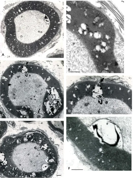
Figure 12.(a–f): TEM aspects of obese Zucker rat sciatic nerve obtained out of thick ultrathin (>500 nm). (a–d): Near entire cross-sections show damages of myelin to appear as‘bubbles’ across several layers of myelin sheaths with higher resolution in (b), (d), (e), and (f). Fusing with each other, myelin degrades throughout with stacked faults or crevices, rupturing sectors with cracks out of internal pockets or wide gashes, leaving intact the adaxonal membrane and the content of the axoplasm. Debris includes waxy, electron dense deposits in the large spaces. Small fatty-like vacuoles in the Cajal bands can be seen in (b)–(f) (arrows). All scales are equal to 1 µm.
further emptied spaces separating myelin layers’
pile-ups. At first, the aligned defect distribution reminds of
widenings of the radial lines in central nervous system
(CNS) as they are regularly spaced along the myelin
annulus profiles (
Figures 5(c
) and
9
–
12(b
)). These
accumulated, piled-up mutilations are sometimes
aligned in the oblique and near longitudinal sections
suggestive of sorts of intraperiod damages propagated
along the outermost layer of the myelin in a periodic
fashion of growing faults toward the adaxonal
mem-brane. The degradation pockets appear as sieve-like
with LM (
Figure 5(b
)) and confirmed with TEM
(
Figure 11(a–f
)). They create inner curved bulges in
transverse sections, also with onion-like aspect limited
by the inner adaxonal membrane, initially viewed as
minor bulges (
Figures 5(c
),
7(d
),
8
,
10(a–d
),
11
, and
13
Figure 13.(a–e): Obese Zucker rat sciatic nerve fibers with aspects of demyelination found as circumferential fissuring of the internodal and paranodal zones as thick onion peeled. Complex whorls with waxy debris are noted in the spaces formed. (a–c): Almost complete splits with adaxonal membrane partly detached from the damaged myelin (curved arrow in (b)). (d–e): complete splits of myelin annulus form quasi two encircling rings out of the intact, single myelin annuli built as spiral (i.e. suggested in (c)). Scales are all equal to 1 µm.Figure 14.(a–c): Pane with paranodal (a–b) to nodal (c) cross-sections of sciatic nerve fibers of an obese Zucker rat with the worse demyelination. (a): Cracks of the myelin annulus with diverse debris and shredded axonal content. (b): Peculiar aspect of paranodal zone with myelin layers retaining points of adhesion (torn in small linkers) creating a peculiar labyrinthine pattern caused by the processing and infoldings of the myelin. (c): Folded node of Ranvier’s region in cross-section with highly contrasted myelin layers with loosen circumferential fissures making the appearance of onion-like aspect and showing a detached adaxonal membrane. In all views, the chaffed SC basal lamina and the axoplasm is reduced into a minute central target-like zone. Scales are 1 µm.
(b
)). The partial or quasi-complete altered myelin now
encompasses dissecting damages through expanded
fissures into vacuolated spaces that progress as
seg-ments with intervening exaggerated spaces along the
circumference of the myelin annulus sections (
Figures
4(b
) and
13(a–e
)). Finally, more fused or coalescent
fissures peel off layers of still adherent sheaths of the
insulating inner layer (
Figures 13(a–e
) and
14(a–c
)).
Figure 15.Near nodal region of an obese Zucker fa/fa rat sciatic nerve fiber. This typical folded region of a myelinated fiber demonstrates apparent myelin adhesion defects not so different that typical node fine structure but appear as large, loosened, open onion-like sectors along with interstices of obliquely-cut tight myelin appears packed, as electron dense stripes. The axoplasm content is vacuolated and the myelin depicts a large star-shaped overall aspect due to partial unwrapping of the myelin layers near the Ranvier node. Scale is 1 µm.
Figure 16.Schematic representation of adipose tissue’s leptin influences with feedbacks on CNS, PNS, and endocrines.
Either erratic or wavy layers reveal the sector’s rough
devastation that eventually completely dissects the
myelin, hacking the entire myelin annulus (
Figure 14
(c
)). The gashes perforate or ruin the entire myelin
layer (
Figure 14(a–c
)).
Altogether, these defects do not usually include
the adaxonal membrane (
Figures 5(c
),
10(d
),
11
,
and
12(a–f
)). These micrographs with important
tearing of the myelin show inward vacuole-like
spaces lined by the adaxonal membrane, leaving
separated the intact neuroplasm and the axonal
content. These myelin tearings feature all sorts of
membranous debris, including some waxy,
elec-tron dense remnants (
Figures 4(b
),
11
,
12(a–f
),
13(a–e
), and
14(a
)). Further, the complex
degrada-tion of the same myelin leaves large adaxonal
spaces and an axonal content compressed to
totally unwrapped myelin in the same area where
typical, undulating tight myelin occurs and
identi-fies the juxta- and paranodal zones (
Figures 4(b
),
13(a–e
),
14(a–c
), and
15
). In the same paranodal
zones, myelin keeps some of its interconnected
membranes leaving remaining ones attached
across the annulus with clear intermembranous,
somewhat punctate junctions. These encompass
SC’s outer membrane leaflet contacts albeit most
of it is fissured by small intraperiod elongated
vacuoles, (
Figures 9
and
14(b,c
)). There, even
though the myelin ravages tear apart the entire
width of its annulus morphology, it remains form
a distorted, multicurved outline where displaced
layers of membranes are still retained together.
Cross-sections
of
those
teased
membranes,
amassed with defects, appear as if they were bales
of wires (
Figure 14(b
)). Following the most
ulti-mate disengagement of the myelin ring in the near
internode and paranode regions, the adaxonal
membrane that has maintained the neurolemma
out of the insulating defects can show breaches
without that of the neuroplasm (
Figures 13(b
)
and
14(a–c
)).
Again, demyelination would likely interfere or
obliterate some of the Zucker nerve conductivity,
as it was suggested by exercise tests.
27It is
inter-esting to view an enlarged small sector out of a
typical myelin damage micrograph to verify that
intraperiod line densities remain as small
interper-iod, elongated contrasted dots, or line-like
densi-ties spaced between the major dense lines unless
they become excessively displaced by some
inter-cellular gaps (
Figure 9
). The gaps can correspond
with intercellular charged components admixed
onto the glycocalyx and rafts. These alterations
change the typical myelin stratification and
stiff-ness thus causing demyelinating defects with
excess in extracellular accumulated repelling
charges contributing to separate them by narrow
to large gaps (
Figures 7(a
) and
9
).
Unmyelinated fibers.
Even though most of the
nerve fibers of the tibial branches of the sciatic nerves
are motor neurons
67, only a few Remak fibers’
membranes examined appear to contain higher
elec-tron-contrasted zones when compared with lean
ones (
Figure 4(b
)). However, more data are needed
to comment on these scarce observations.
Endoneurium and supportive stroma.
Figure 4(b
)
reveals a very loose endoneurium, maybe brought
by some surrounding changes.
Figure 5(c
) also
demonstrates the endoneurium layer, charged
with numerous vacuoles
– including many densely
contrasted ones
– suggests some lipid content
get-ting in close contact with the outer Cajal band,
Figure 17.Diagrammatic representation of a suggested, unex-pected peculiar centripetal diffusion (blue arrows) of myelin compound(s) dispatched by Schwann cells. Axon in black.