HAL Id: tel-01235870
https://tel.archives-ouvertes.fr/tel-01235870
Submitted on 30 Nov 2015HAL is a multi-disciplinary open access archive for the deposit and dissemination of sci-entific research documents, whether they are pub-lished or not. The documents may come from teaching and research institutions in France or abroad, or from public or private research centers.
L’archive ouverte pluridisciplinaire HAL, est destinée au dépôt et à la diffusion de documents scientifiques de niveau recherche, publiés ou non, émanant des établissements d’enseignement et de recherche français ou étrangers, des laboratoires publics ou privés.
Etudes des altérations métaboliques musculaires au
cours de la sclérose latérale amyotrophique : rôle dans le
développement de la pathologie
Lavinia Palamiuc
To cite this version:
Lavinia Palamiuc. Etudes des altérations métaboliques musculaires au cours de la sclérose latérale amyotrophique : rôle dans le développement de la pathologie. Neurobiologie. Université de Strasbourg, 2014. Français. �NNT : 2014STRAJ088�. �tel-01235870�
ÉCOLE DOCTORALE DE LA VIE ET DE LA SANTÉ
U1118 Mécanismes centraux et périphériques de la neurodégénérescence
THÈSE
présentée par :Lavinia PALAMIUC
soutenue le : 10 Septembre 2014
pour obtenir le grade de :
Docteur de l’Université de Strasbourg
Discipline/ Spécialité: Aspect Moléculaire et Cellulaire de la Biologie
Mention Neuroscience
THÈSE dirigée par :
Mme RENÉ Frédérique Dr., Université de Strasbourg
RAPPORTEURS :
M ANDRES Christian Prof., Université de Tours
M CHARBONNIER Frédéric Prof., Université de Paris Descartes
AUTRES MEMBRES DU JURY :
Mme FLORENTZ Catherine Prof., Université de Strasbourg
M GENY Bernard Prof., Université de Strasbourg
M WEYDT Patrick Dr., Université de Ulm
E
TUDES
DES
ALTÉRATIONS
MÉTABOLIQUES
MUSCULAIRES
AU
COURS
DE
LA
SCLÉROSE
LATÉRALE
AMYOTROPHIQUE
:
RÔLE
DANS
LE
DÉVELOPPEMENT
DE
LA
PATHOLOGIE
UNIVERSITÉ DE STRASBOURG
My studentship in the lab of CENTRAL AND PERIPHERAL MECHANISMS OF
NEURODEGENERATION shaped my development, and for that i have to thank all my
colleagues from whom i learned so much over these years. I would like to express my gratitude to Dr. Jean-Philippe LOEFFLER, the director of the lab, who kindly welcomed me in this great team. With care and patience, he ensures a great atmosphere for doing research.
I will forever be indebted to my advisor, Dr. Frédérique RENÉ. It was a great pleasure to work by her side on developing this scientific project. With dedication she passed on to me invaluable skills and knowledge. I would have never achieved my goals without her excellent guidance.
I owe special thanks and appreciation to Dr. Jose-Luis GONZALEZDE AGUILAR, who always had for me great advise and ideas, but also very useful criticism. I want to thank also Dr. Luc DUPUIS who has been for me an example of dynamism and perseverance. To Dr. Caroline ROUAUX and Dr. Alexandre HENRIQUES I am most grateful not only for their
scientific input, but also for their mentorship as young scientists. In this line of great mentors i have to include Dr. Christian GAIDDON who believed in me when i was still a novice with little experience and encouraged and helped me to pursue an academic career. Jose, Caroline, Alex, Luc and Christian gave me invaluable lessons that will help me make my first steps in the tough world of science.
I would also like to thank Prof. Maria Teresa CARRI who kindly welcomed me in her lab at the University of Rome for a short visit and a fruitful collaboration. For this extraordinary „roman“ experience i also have to thank Dr. Cristiana VALLE, a wonderful host.
As a PhD student, i could never thank enough the technical team that was there with me on the battle field. Through troubleshooting, organizing, incredibly useful tips and suggestions, our work at the bench is so much easier thanks to you Annie, Sylvie, Michèle and Marie-Jo. For the efficient logistic organization I want to thank Brigitte. Our lab would simply not function without her prompt assistance!
I want to thank all my colleagues, current and former members of the lab. Thanks to Judith, Yannick, Hus, Florent, Jérôme, Thiebault and Vania i quickly felt as part of the team. Aurélia and Jelena, I found in you not only respected peers but also invaluable friends. I have to thank you for all the support you showed me, especially through these last months that have been particularly difficult. Hajer, Pauline, Laura, Christine, Gina and Steph, you make our lab lively and colorful, not to mention sweet (thanks for all the cookies and cakes). I learned something from each of you and for that I am grateful and and really proud of having been your colleague!
Finally I would like to thank all the members of the jury, who took the time to read and thoroughly evaluate this work.
T
ABLE OF
C
ONTENTS
ABBREVIATIONS 2
INTRODUCTION 6
1 AMYOTROPHIC LATERAL SCLEROSIS – A CLINICALLY AND GENETICALLY DIVERS SYNDROME 7
1.1 Clinical diveristy 7
1.2 Genetic diversity 8
2 PATHOGENESIS: SUPPOSED MECHANISMS AND KEY PLAYERS 14
2.1 Oxidative stress 15
2.2 Protein aggregates 17
2.3 Mitochondrial pathology 20
2.4 Glutamate excitotoxicity 22
3 MULTICELLULAR PATHOLOGY IN ALS 25
3.1 Astrocytes 27
3.2 Microglia 28
3.3 Muscle 29
4 MUSCLE, METABOLISM AND ALS 33
4.1 Muscle structure and physiology 33
4.2 Metabolic regulators of muscle function and fiber type 37
4.3 Muscle metabolism in aging and disease 43
4.4 Metabolic alterations in ALS 46
OBJECTIVES 52
RESULTS I 55
RESULTS II 58
RESULTS III 61
CONCLUSIONS AND PERSPECTIVES 64
SUMMARY IN FRENCH 69
A
BBREVIATIONS
AD Alzheimer s disease
Akt/PKB Akt/protein kinase B
ALS Amyotrophic Lateral Sclerosis
AMP Adenosine monophosphate
AMPA α-amino-3-hydroxy-4-isoxazole propionic acid
AMPK AMP-activated kinase
Atg-1 atrogin 1
ATP Adenosine triphosphate
Bcl2 B-cell lymphoma 2
CaMK calmodulin-dependent protein kinase
CNS Central Nervous System
COX cytochrome oxidase
CPT 1 Carnitine palmitoyltransferase I
CSF cerebrospinal fluid
DRP1 dynamin related protein 1
ERR estrogen related receptor
FA Fatty acid
FALS Familial Amyotrophic Lateral Sclerosis
FAT/CD36 Fatty acid transporter CD36
FG fast glycolytic
FOG fast oxidative glycolytic
FoxO forkhead transcription factor, O box subfamily
FTD frontotemporal dementia
FUS fused in sarcoma
GLT-1/EAAT2 glutamate transporter/excitatory amino-acid transporter 2
GLUT4 glucose transporter 4
HDAC histone de-acetylase
HK Hexokinase
HRE hexanucleotide repeat expansion
Hsc heat shock cognate
Hsp heat shock protein
IGF1 insulin-like growth factor 1
IGFR insulin-like growth factor receptor
iPSC induced pluripotent stem cells
iPSCs induced pluripotent stem cells
MEF 2 Myocyte enhancer factor 2
MND motor neuron disease
mSOD1 mutant Cu/Zn superoxide dismutase
mtDNA mitochondrial DNA
mTOR mammalian target of Rapamycin
MuRF 1 Muscle RING-finger protein 1
MyHC myosin heavy chain
MyLC myosin light chain
NAD+ nicotinamide adenine dinucleotide oxidized
NADH nicotinamide adenine dinucleotide reduced
NMDA N-methyl-D-aspartate
NMJ neuromuscular junction
NRF nuclear respiratory factor
PDH pyruvate dehydrogenase
PDK pyruvate dehydrogenase kinase
PFK1 phosphofructokinase
PGC-1α peroxisome proliferator-activated receptor γ coactivator α
PI3K phosphoinositide 3 kinase
PKB protein kinase B
PLS Primary Lateral Sclerosis
PMA Progressive Muscular Atrophy
PPAR α, γ, β/δ peroxisome proliferator-activated receptor α, γ, β/δ
ROS reactive oxygen species
SALS Sporadic Amyotrophic Lateral Sclerosis
SDH succinate dehydrogenase
Sirt Sirtuin
SO slow oxidative
SOD1 Cu/Zn superoxide dismutase
TDP-43 transactive response DNA-binding protein 43
UCP uncoupling protein
I
NTRODUCTION
1 A
MYOTROPHIC LATERAL SCLEROSIS–
A CLINICALLY AND GENETICALLY DIVERS SYNDROMENeurodegenerative disorders are devastating fatal diseases that continue to challenge the scientific community to find adapted cures, with little breakthroughs so far. This endeavor has a tremendous urgency considering the increasing incidence of these diseases within the current aging population.
1.1
C
LINICAL DIVERISTYIn this category, motor neuron diseases (MND) reached an incidence of 2.16/100,000 people in Europe to date (Logroscino et al., 2010). This group of diseases is characterized by progressive degeneration of upper and/or lower motor neurons accompanied by muscle atrophy. Amyotrophic lateral sclerosis (ALS) is the most common adult MND and presents with degeneration of both upper and lower motor neurons. The site of onset can be either spinal, with symptoms usually appearing first in limbs (65% of all cases) or bulbar affecting first the swallowing muscles (30% of all cases). Denervation and atrophy of respiratory muscles lead to respiratory failure, which is the main cause of death (Kiernan et al., 2011). For the majority of patients age of onset is around 50 years of age and death occurs within 3-5 years from diagnosis. A better prognosis is given for those patients with only upper or lower motor neuron involvement, referred to as primary lateral sclerosis (PLS) and progressive muscular atrophy (PMA) respectively (Hardiman et al., 2011). A small percentage of ALS cases do not fall precisely within these characteristics. Cases of juvenile ALS have been reported, with time of onset before the age of 20. Juvenile ALS has a slow disease progression extending over decades (Ben Hamida et al., 1990), but some aggressive forms have also been reported (Conte et al., 2012). The clinical heterogeneity of ALS is complicated by the association with several other syndromes and
neurodegenerative disorders (Table 1), amongst which frontotemportal dementia (FTD) is the most common (Ling et al., 2013).
1.2
G
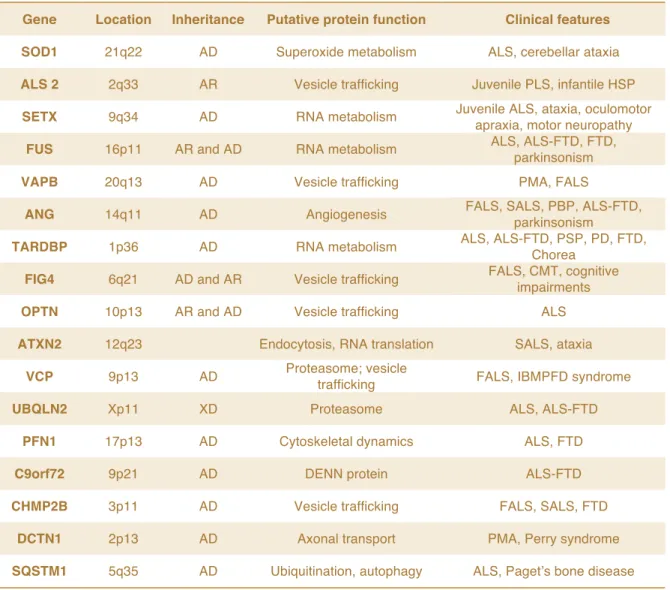
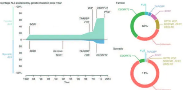
ENETIC DIVERSITYAbout 10% of patients have a familial history (familial ALS or FALS), the remaining 90% being sporadic (sporadic ALS or SALS) with unknown aetiology but clinically indistinguishable from FALS. It is believed however that FALS incidence is underestimated due to flaws in patient history and reduced genetic penetrance of certain mutations. So far more than 20 genes have been implicated in or associated with ALS pathogenesis, most of them discovered within the last 5 years (Table 1). Mutations of all these genes only account for 68% of FALS and 11% of SALS patients (Renton et al., 2014) (Figure 1). These genes have different functions in the cell, which makes the identification of a common pathogenic mechanism, leading to the same disease, laborious. Moreover, the genetic variation introduces some heterogeneity in the clinical characteristics of ALS regarding time of onset, duration of disease and association with other diseases (Table 1). Some mutations can lead to slow developing ALS, juvenile ALS and ALS cases associated with other diseases (FTD, Parkinson, etc). For this reason, more and more often, ALS is defined as a “syndrome” regrouping complex and divers pathophysiological manifestations (Hardiman et al., 2011).
The first mutated gene identified in ALS patients was the Cu/Zn superoxide dismutase 1 (SOD1)(Rosen et al., 1993), an enzyme involved in superoxide scavenging and protection from oxidative stress. This discovery enabled the development of the only animal model for ALS that we have today, which recapitulates most of the clinical and pathological aspects of the human disease including neuronal degeneration and paralysis. Many point mutations (177 according to alsod.org) of the SOD1 gene have been described since 1993, representing 12% of FALS and 1% of SALS cases
Table 1. Main genes involved in ALS pathogenesis
Gene Location Inheritance Putative protein function Clinical features
SOD1 21q22 AD Superoxide metabolism ALS, cerebellar ataxia
ALS 2 2q33 AR Vesicle trafficking Juvenile PLS, infantile HSP
SETX 9q34 AD RNA metabolism Juvenile ALS, ataxia, oculomotor
apraxia, motor neuropathy
FUS 16p11 AR and AD RNA metabolism ALS, ALS-FTD, FTD,
parkinsonism
VAPB 20q13 AD Vesicle trafficking PMA, FALS
ANG 14q11 AD Angiogenesis FALS, SALS, PBP, ALS-FTD,
parkinsonism
TARDBP 1p36 AD RNA metabolism ALS, ALS-FTD, PSP, PD, FTD,
Chorea
FIG4 6q21 AD and AR Vesicle trafficking FALS, CMT, cognitive
impairments
OPTN 10p13 AR and AD Vesicle trafficking ALS
ATXN2 12q23 Endocytosis, RNA translation SALS, ataxia
VCP 9p13 AD Proteasome; vesicle
trafficking FALS, IBMPFD syndrome
UBQLN2 Xp11 XD Proteasome ALS, ALS-FTD
PFN1 17p13 AD Cytoskeletal dynamics ALS, FTD
C9orf72 9p21 AD DENN protein ALS-FTD
CHMP2B 3p11 AD Vesicle trafficking FALS, SALS, FTD
DCTN1 2p13 AD Axonal transport PMA, Perry syndrome
SQSTM1 5q35 AD Ubiquitination, autophagy ALS, Paget’s bone disease
Figure 1.1. ALS-related genes (from Renton et al., 2014). More than 20 genes have been implicated in or
associated with ALS pathogenesis, most of them discovered within the last 5 years. Mutations of all these genes only account for 68% of FALS and 11% of SALS patients. Some of the most important are represented here with the percentage of ALS patients that present with these mutations in FALS and SALS.
(Renton et al., 2014). Using the SOD1 transgenic animals several putative pathogenic mechanisms that may participate to the progressive degeneration of motor neurons have been described. Among them, oxidative stress, protein aggregates, mitochondrial defects, and glutamate excitotoxicity are the most documented. However after 20 years of research, we still do not have the one critical pathogenic mechanism for ALS. The problematic of SOD1-related pathogenesis and the controversies around it will be detailed in the next section.
In 2006, the seminal discovery that Transactive Response DNA-binding protein (TDP-43) is the major component of the ubiquitinated protein inclusions in degenerating motor neurons opened a new path towards understanding ALS pathogenic mechanisms (Neumann et al., 2006). TDP-43 is part of a family of RNA binding proteins, involved in RNA splicing and transport (Buratti et al., 2001; Wang et al., 2004). Subsequently, TDP-43 mutations have also been described, but only in 4% of FALS cases and approximately 1% of SALS (Renton et al., 2014). TDP-43 binds to a myriad of mRNA targets and for this reason the pathogenicity of its mutation is little understood. Recent evidence seems to indicate that mutation of TDP 43 impairs its function of anterograde transport of essential mRNA species from soma to distal neuronal compartments, including the nerve terminals at the neuromuscular junction (NMJ) (Alami et al., 2014). Alterations of RNA metabolism are now thought to be a key player in ALS pathogenesis, as well as other neurodegenerative diseases. In support of this new paradigm, mutations of other genes involved in RNA processing have been identified in ALS patients. Fused in sarcoma (Fus) mutations are localized in the gene’s RNA-binding region and represent 4% of FALS and 1% of SALS (Renton et al., 2014). Hexanucleotide repeat expansion (HRE) in C9ORF72 gene represents today the majority of patients, with incidence in 40% of FALS and 7% of SALS cases. Interestingly, all of these mutations are common feature for both ALS and FTD, one of the most common syndromes associated with ALS.
Developing new models for these newly identified mutations has been a difficult challenge. Some TDP-43 rodent lines were described so far. Transgenic lines expressing human mutant TDP-43 showed an extremely short life span (between 14 and 49 days), motor neuron degeneration and astrogliosis associated with a severe motor phenotype and weight loss (Stallings et al., 2010). These pathological characteristics underline the specificity of TDP-43 toxicity for the motor system and are congruent with an early-onset severe ALS. However, transgenic animals expressing human WT TDP-43 presented a similar phenotype. In view of this observation it was argued that human TDP-43, WT or mutant, is toxic in a mouse background. Nevertheless overexpressing mouse TDP-43 in cortex, hippocampus and striatum induced neuronal pathology and led to a neurological phenotype remembering FTD (Tsai et al., 2010). Equally partial ubiquitous loss of TDP-43 led to progressive loss of neurons (specifically layer V and ventral horn motor neurons), motor dysfunction, paralysis and death (Yang et al., 2014).
ALS-linked Fus mutants affect the ability of mutant FUS to bind RNA and cause mislocalization in the cytoplasm (Daigle et al., 2013). Such mutations in mouse models induce a so-called fusopathy characterized by motor axon degeneration, severe motor phenotype and death (Shelkovnikova et al., 2013). In a zebra fish (Danio rerio) model both mutant FUS and FUS knockdown induced neuromuscular junction abnormalities (Armstrong and Drapeau, 2013). But, like for TDP-43, overexpressing WT human FUS in mice induced an aggressive phenotype with degeneration of spinal cord motor neurons, denervation associated with muscle atrophy and death at 12 weeks of age (Mitchell et al., 2013).
Both loss and gain of function mechanisms have been proposed for C9ORF72. The lack of animal or cellular models expressing mutant C9ORF72 has been a serious drawback in understanding the C9ORF72 pathogenicity. The HREs pose specific technical problems due to their instability, but some advances have been made using the induced pluripotent stem cell (iPSCs) technology (Almeida et al., 2013). Recent reports clearly attest for a gain of
toxic function. Using iPSCs from ALS patients, it was shown that C9ORF72 expression is not particularly modified, and inactivating it rescues the associated phenotype in motor neurons (Sareen et al., 2013). HREs in the C9ORF72 sequence induce the formation of RNA foci, such as G-quadruplexes and RNA-DNA hybrids (Haeusler et al., 2014). This leads to abortive transcription, sequestration of specific HRE-binding proteins and nucleolar stress.
Epidemiological studies have also showed that the incidence of disease and the particularity of the mutations can differ from one geographical area to the other, from one ethnic group to the other, suggesting an important role for environmental factors (Andersen & Al-Chalabi, 2011; Sabatelli et al., 2013). One of the most cited environmental risk factor is physical activity. An active sports-oriented life style has been associated with ALS patients (Scarmeas et al., 2002) and increased incidence amongst professional athletes has also been reported (Chiò et al., 2005). Other factors include diet (Nelson et al., 2000; Okamoto et al., 2007) and toxic substances (e.g. heavy metals, pesticides).
This genetic, clinical and epidemiological diversity of ALS undoubtedly hampered all efforts to find therapeutic targets and design efficient therapeutic approaches. Moreover, the insidious pathology and the limited diagnostic tools lead to a late diagnosis, when therapeutic interventions may no longer be effective. Therefore future lines of research have to concentrate on finding early diagnostic markers on one hand. On the other hand, a big challenge is the development of new animal models that will reflect this genetic diversity and therefore contribute to a better understanding of the complex pathogenicity in ALS.
2 P
ATHOGENESIS:
SUPPOSED MECHANISMS AND KEY PLAYERSSeveral mechanisms are suspected to intervene in the pathogenesis of ALS. A multitude of pathologic alterations have been described and they appear intricately linked at different levels, making it quite difficult to discern between cause and consequence. For this reason their respective participation to the pathogenesis of ALS remains little understood and for most, a matter of controversy.
Since the first mutated gene identified in ALS patients was the SOD1 gene, one of the first causes investigated was the oxidative stress related to the supposed SOD1 loss of function. However, compelling arguments quickly dismissed this possibility. An anecdotal experiment showed that human mutated SOD1 rescues SOD1 knockout Saccharomices cerevisiae just as effectively as the human WT SOD1, to the point where the two strains are phenotypically undistinguishable (Rabizadeh et al., 1995). Moreover, knockout mouse models for SOD1 do not present ALS-like symptoms (Reaume et al., 1996), and the phenotype of transgenic SOD1 mice cannot be rescued with the WT SOD1. On the contrary, overexpressing human WT SOD1 aggravates disease progression in ALS animal models (Jaarsma et al., 2000). The expression of a murine mutant SOD1 that does not have dismutase activity (G86R) leads to the development of an ALS in mice, even if the endogenous SOD1 is still active (Morrison et al., 1998). Therefore the hypothesis of a toxic gain of function has been investigated and a plethora of potential mechanisms have been described.
2.1
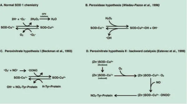
O
XIDATIVE STRESSConsidering oxidative stress as a causative factor of ALS was a reasonable assumption, since the main function of SOD1 is superoxide scavenging (Figure 2.1-a). Initially it was believed that a decrease in SOD1 activity facilitates the reaction between superoxide and nitric oxide producing peroxynitrite (-ONOO). Peroxynitrite was one of the first “non-physiological” substrates thought to bind to mutant SOD1 (mSOD1) (Figure 2.1-c) inducing deleterious nitration of tyrosine protein residues (Beckman et al., 1993). However, as it was mentioned earlier, it became quickly clear that things are not that simple, since several mSOD1 maintain there scavenging activity. This theory was developed and modified over the years, revealing mechanisms that are a lot more complex than the original simplistic idea of “just” oxidative stress (Beckman et al., 2001; Cleveland & Rothstein, 2001).
The discovery that some mutations induce a looser folding of SOD1 protein (Deng et al., 1993) led to the hypothesis that this is the basis of the SOD1 toxicity. The improper folding of mSOD1 affects the binding specificity for the metal ions essential for its activity (Cu2+ and Zn2+), the misfolded mutant SOD1 showing low copper-to-zinc ratios (Goto et al., 2000). Copper ions seem to facilitate the toxic activity of mSOD1 augmenting the catalytic oxidation of substrates by peroxide H2O2 (Fig. 2.1-b) and cupper-chelators
efficiently inhibit apoptosis in neurons expressing mSOD1 (Wiedau-Pazos et al., 1996). In the same time, Zn2+ ion deficiency is toxic selectively to motor neurons increasing tyrosine nitration (Crow et al., 1997). In favor of the low metal binding capacity, when replete with both Cu2+ and Zn2+ mSOD1 protects neurons even in the absences of trophic factors and does not induce apoptosis (Est vez et al., 1999). Estévez and colleagues showed that Zn-deficient SOD facilitates the production of peroxynitrite through the intermediary of nitric oxide (Figure 2.1-d).
Figure 2.1. Chemistry of mSOD1 toxicity (adapted from Cleveland and Rothstein, 2001). A. SOD1 enzymatic
function is that of scavenging superoxide free radicals, by dismutating O2- to O2 and peroxide. B-D. Point
mutations of SOD1 gene, associated with ALS, change the chemistry of SOD1, conferring new toxic properties. Several chemical mechanisms of SOD1 toxicity have been proposed that arise from use of aberrant substrates, having as a result an increased in oxidative stress, rather than ROS scavenging.
All these proposed mechanisms revolve around the accumulation of peroxynitrite (Bruijn et al., 1997), which is strong oxidant compound that induces apoptosis in neuronal cells. However, deletion of nitric oxide synthase (NOS), aimed at reducing the levels of nitric oxide necessary for peroxynitrite production, has no effects in vivo, suggesting that these might not be the main toxic mechanism of mSOD1 (Facchinetti et al., 1999). Moreover, clinical attempts to reduce oxidative stress have been largely discouraging so far (Orrell et al., 2007).
2.2
P
ROTEIN AGGREGATESThe accumulation of toxic protein aggregates is a common feature for neurodegenerative diseases (AD, HD) and it has been showed that these aggregates are involved in neuronal apoptosis. But whether they are cause or consequence of disease, or even a protective mechanism, remains to be confirmed. This is also the case in ALS.
Protein aggregates positive for mSOD1 have been described in patients with SALS and FALS (Shibata et al., 1996) and animal models (Bruijn et al., 1998; Shibata et al., 1998; Watanabe et al., 2001). These aggregates have been described in motor neurons as well as astrocytes. As Bruijn and colleagues pointed out, the accumulation of SOD1-positive aggregates coincide with disease onset in mice. The authors argued that the aggregates might more easily explain the toxic function of misfolded SOD1, since overexpression of human WT SOD1 does not rescue neuronal death. If it were to be any of the oxidant mechanism described before, the WT would compete with the mutant and reduce erroneous oxidative reactions and the concentration of toxic products, which was not the case. However it has been pointed out that since aggregates do not appear before muscle weakness, it may not be a triggering factor, but on the contrary, that it could be an attempt
to protect the cell from toxic damage issued from the enzymatic activity of misfolded SOD1 (Brown et al., 1998).
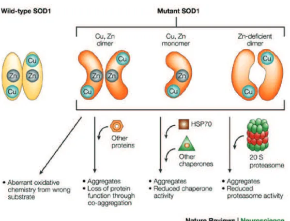
Regardless of this debate, SOD1 aggregates entangle within proteins essential for cell function and protein control (Figure 2.2). For instance, several proteins involved in protein quality control and degradation have been showed to interact with mSOD1 from transgenic mice: proteasome, ubiquitin, Hsc70 (Watanabe et al., 2001) or Hsp 105 (Yamashita et al., 2007). Equally, SOD1 may interact with other members of the heat shock protein family Hsp70 and Hsp40 (Shinder et al., 2001) as it was shown in neuronal cell cultures expressing mSOD1. While most of the Hsp family members that are upregulated in transgenic animal models, some are downregulated. For instance, Hsp70 or Hsp105 (Yamashita et al., 2007) seem to have a protective role, preventing formation of aggregates and increasing cell viability when overexpressed in neuronal cell cultures expressing mSOD1.
SOD1 aggregates also contribute directly/physically to neuronal apoptosis. In fact the WT SOD1 has anti-apoptotic properties while mSOD1 promotes apoptosis, by binding incorrectly to the anti-apoptotic protein Bcl2 and forming aggregates in spinal cord mitochondria from G93A transgenic mice (Pasinelli et al., 2004). Therefore it seems that by sequestering the anti-apoptotic protein Bcl2, mSOD1 promotes apoptosis in motor neurons. In support of this, it is known that overexpressing the protective Bcl2 in transgenic animal considerably extends life span delaying neuronal degeneration (Kostic et al., 1997).
More than 20 years of accumulating evidence suggest that misfolded SOD1 has toxic functions that lead to the same course of disease every time. Independent of the position of the mutation, the result is invariably the same: destabilization of the tertiary structure of SOD1. This is reinforced by the fact that even non-genetic alterations of WT SOD1 leading to protein misfolding (oxidation, metal depletion) give toxic properties similar to those of mSOD1 that are selectively detrimental to motor neurons (Kabashi et al., 2007;
Figure 2.2. Mechanisms of misfolded SOD1 toxicity (from Cleveland ans Rothstein, 2001). SOD1 point
mutations associated with ALS alter the structural conformation of mSOD1. The loosely folded mSOD1 is prone to form aggregates, that entangle within several essential enzymes of the protein degradation pathway.
Rotunno & Bosco, 2013). Despite all efforts however, we have not yet been able to identify a clear toxic mechanism that is common to all mutant variants of SOD1 and that could trigger key pathogenic events in FALS and explain disease etiology.
2.3
M
ITOCHONDRIAL PATHOLOGYSeveral pathological modifications in mitochondria from ALS patients have been described: ultrastructural changes like vacuolization and fragmentation, but also Ca2+ handling, and axonal transport (Cozzolino and Carrì, 2012). Transferring mitochondrial DNA (mtDNA) from patients into mtDNA-depleted neuroblastoma cell line was enough to recapitulate ALS mitochondrial pathology (Swerdlow et al., 1998). Mitochondrial alterations are also very well documented in various ALS mouse models (Bendotti et al., 2001; Kong et al., 1998; Wong et al., 1995).
The ultrastructural changes (vacuolization and fragmentation) described, are related to alterations of mitochondrial fusion-fission dynamics, processes essential for mitochondrial renewal. An initial increase in both fusion and fission proteins is replaced by a decreased expression of fusion proteins in the pre-symptomatic stage of mSOD1 mice (Liu et al., 2013). The co-expression mSOD1 with a dominant negative of dynamin related protein 1 (DRP1), one of the fission proteins, prevented mitochondrial fragmentation and neuronal apoptosis in vitro (Song et al., 2013). The same effect was observed when co-expressing mSOD1 with mitochondrial metabolic regulators, Sirtuin 3 and Peroxisome Proliferator Activated Receptor γ Coactivator 1α (PGC-1α).
The ultrastructural modifications are accompanied by alterations in mitochondrial function. Altered function of the respiratory electron chain, reduced ATP production together and increased ROS generation have been
described in patients and mouse models. Decreased activity of complex IV of the respiratory electron chain has been described in ventral horn motor neurons (Fujita et al., 1996) and muscle (Echaniz-Laguna et al., 2006; Echaniz-Laguna et al., 2002; Vielhaber, 2000) of SALS patients.
decreased activity of complexes II and IV, and increased sensitivity to inhibition of glycolysis (Menzies, 2002). Detailed analysis of isolated mitochondrial from spinal cord of an ALS rat model revealed increased resting oxygen consumption correlating with increased ROS production, while state 3 respiration (oxidative phosphorylation) was significantly impaired (Panov et al., 2011).
Ca2+ is essential signaling molecule for mitochondrial function, coupling ATP demand with synthesis (Jouaville et al., 1999). Dysfunction in mitochondrial Ca2+ handling was proposed to increase intracellular Ca2+ levels and to induce depolarization of mitochondrial membrane (Kawamata and Manfredi, 2010). Depolarized mitochondria are found at the level of neuromuscular junction, in nerve terminals (Nguyen et al., 2009) and in muscle fibers (Zhou et al., 2010). In mSOD1 mice increasing mitochondrial Ca2+ buffering capacity improved the ATP synthesis and preserved mitochondrial function in spinal cord. Moreover, this was accompanied by a decrease of astrogliosis, and significant reduction of motor neuron death (Parone et al., 2013). However, the improvement of mitochondrial Ca2+ buffering capacity did not prevent axonal degeneration and denervation, thus eliminated SOD1-mediated loss of Ca2+ buffering capacity as a primary contributor to ALS pathogenesis in these animal models.
The connection between mSOD1 and mitochondrial pathology are still under investigation, but it seems to be related to the localization, at least to some extent, of mSOD1 into the mitochondria (Carrì and Cozzolino, 2011). Increasing mSOD1 solubility in mitochondria, but not in cytoplasm, prevented mitochondrial fragmentation and neuronal apoptosis in vitro. Zinc seems to play an important role in mSOD1 mitochondrial toxicity. Expression of a zinc-Overexpression of mSOD1 in cultured motor neuron-like cells (NSC34) lead to
deficient SOD1 in Drosophila melanogaster induces vacuolization associated with inefficient ATP production and alterations in motor behavior (Bahadorani et al., 2013).
2.4
G
LUTAMATE EXCITOTOXICITYGlutamate is the major excitatory neurotransmitter in the CNS, with approximately 80-90% of synapses in the brain being glutamatergic. The post-synaptic cells are equipped with three main classes of inotropic glutamate receptors (NMDA, AMPA and kainate receptors) and three main classes of metabotropic glutamate receptors (I, II and III). In the 70’s, a series of experiments revealed that glutamate could act as a potent neurotoxin inducing neurodegeneration (Hassel and Dingledine, 2012). Since then, glutamate excitotoxicity has been associated with many pathological conditions like epilepsy, ischemic brain injury and several neurodegenerative disorders including ALS. The first report evidencing glutamate excitotoxicity in ALS, described an increase of 100 to 200% of glutamate and aspartate in the cerebrospinal fluid (CSF) of 18 ALS patients (Rothstein et al., 1990). This explained the potent toxic effect that the CSF from patients has on healthy neuronal cell cultures (Couratier et al., 1993).
This neurotoxic action of glutamate on neurons was explained by the deleterious effect that calcium ions exert when in high concentration. The glutamate excitotoxicity theory posits that under certain circumstances,
and a promising therapeutic target for several reasons. Firstly, it is known that
22 stimulation of the glutamate receptors on the post-synaptic membrane.
glutamate is found in excess in the synaptic cleft leading to an over-stimulation
Specififically, AMPA and NMDA receptors, when over-stimulated, facilitate large quantities of Ca2+ ions to enter into the cytoplasm.
motor neurons have low calcium buffering capacity. This makes them particularly vulnerable to calcium-mediated glutamate toxicity. Secondly, motor neurons express mainly glutamate receptors that are highly permeable to Ca2+. Indeed, motor neurons are particularly vulnerable to AMPA-induced calcium toxicity. The experiments conducted by Couratier and colleagues in the early 90’s clearly showed that AMPA but not NMDA antagonists could block the toxic effect of CSF from ALS patients on cultured motor neurons (Couratier et al., 1993). AMPA receptors are composed of 4 subunits (GluR1-4), out of which the GluR2, impermeable to Ca2+ ions and the most frequent in other neuronal populations in the CNS, is little expressed in human motor neurons (Kawahara et al., 2003; Williams et al., 1997). Mice lacking the GluR2 AMPA subunit do not present with motor neuron disease (Jia et al., 1996), however deleting the GluR2 subunit in a mouse line expressing the mutated G93A SOD1 accelerated the progression of the disease (Van Damme et al., 2005). Taken all together, these data indicate that glutamate excitotoxicity is a selective pathologic mechanism for motor neuron degeneration.
But what could explain the excess of glutamate present in the synaptic cleft in ALS? The fate of glutamate is under the control of astrocytes responsible for its clearance through glutamate transporters, critical for maintaining low concentrations of glutamate in the synaptic cleft. The absence of glutamate transporters, specifically Glut1/EAAT2 and GLAST leads to neurodegeneration and progressive paralysis (Rothstein et al., 1996). ALS patients present a dramatic loss of GLT-1/EAAT2 transporters compared to healthy patients, a decrease visible in both motor cortex and spinal cord (Rothstein et al., 1995). However, the overexpression of EAAT2 in transgenic SOD1 mice revealed a delay in motor neuron death but did not delay disease onset nor did it increase the life span of transgenic animals (Guo et al., 2003). Moreover, Riluzole, the only therapeutic molecule used as treatment for ALS, has been a major disappointment. Riluzole targets glutamate excitotoxicity and has been used with some success in transgenic models (Gurney et al., 1996; Gurney et al., 1998) but its administration extends patient life span for
about three months (Miller et al., 2012). While this approach did not give the expected results, it brought attention on one essential aspect: the involvement of astrocytes in pathological events in ALS.
As it is outlined is this section multiple mechanisms have been associated with SOD1 mutations but so far a clear-cut pathogenic mechanism is missing. Although it is evident that all these alterations, from oxidative stress to glutamate excitotoxicity are detrimental for neurons, some with a degree of selectivity for motor neurons, they do not stand out as causative factors, at least not by themselves. Blocking either one of them did not stop ALS in transgenic mice. For this reason the therapeutic approaches had extremely limited results. Understanding the chemistry of mSOD1 will probably be a big step in our understanding of mSOD1 related pathologic mechanisms and bring us closer to an efficient therapeutic approach.
3 M
ULTICELLULAR PATHOLOGY INALS
As it was originally defined, ALS is solely a disease of motor neurons!, and for the last two decades scientists tried to understand why the disease is so selective for this neuronal population. Several theories have been postulated, as it was described in the previous section. Interestingly, some of the molecular and ultrastructural mSOD1-related modifications described above, were also found in cells other than motor neurons, like astrocytes and microglia. Moreover independent studies overexpressing different variants of mSOD1 specifically in motor neurons have demonstrated that motor neurons are not the sole participants to the pathogenesis and progression of ALS. Two initial studies showed that expression of mSOD1 in motor neurons does not induce motor neuron death, or an ALS-like pathology (Lino et al., 2002; Pramatarova et al., 2001). However, a subsequent study that used a different promoter reported a late onset ALS, with motor neuron degeneration and aggregates associate with muscular atrophy (Jaarsma et al., 2008). This late onset of disease in animal expressing mSOD1 specifically in motor neurons indicated that other cellular population might accelerate disease onset and/or progression, supporting the theory of non-cell autonomous mechanisms involved in ALS.
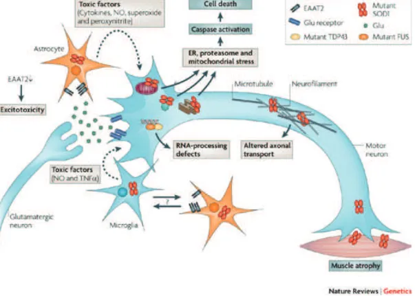
Nevertheless, the question of selectivity did not become obsolete, just more complicated. As Boillée et al. defined it, ALS is a disease of motor neurons and their non-neuronal neighbors (Boill"e et al., 2006a). Different cellular populations surround the motor neuron and each of them has distinct roles in maintaining its integrity, like astrocytes and microglia. Astrocytes have an important metabolic function, supplying the substrate (lactate) for neuronal energy production. They form complex networks around neurons aimed at coupling the blood flow with the local energetic demands (Allaman et al., 2011). Microglia are the macrophages of the CNS and they play a major role in neurodegenerative diseases. They eliminate pathogens, dead and dying neurons, but also neuronal processes and live neurons that show sign of
Figure 3.1. Cell autonomous and non-cell autonomous pathogenic mechanisms in ALS (from Dion et al.,
2009). The mSOD1 pathology in ALS is the best characterized so far. Several cellular types present with
mSOD1 aggregates: motor neurons, astrocytes, microglia and muscle. The mSOD1-related pathology in each of these cells contribute to motor neuron degeneration and disease progression.
stress (Brown et al., 2014). At the same time, muscle, so far considered as a simple victim of denervation, emerges as a target of mSOD1 toxicity (Dobrowolny et al., 2008). Muscle tissue pathology in ALS and its relevance for disease progression is probably the least understood so far. In order to design new efficient therapies for ALS we have to understand the participation of each of these cell types to the progressive deterioration and subsequent selective death of motor neurons (Figure 3.1).
3.1
A
STROCYTESThe astrocyte net is extremely important in maintaining extracellular and intracellular energy homeostasis in neurons. It controls pH and ion buffering, supplies metabolic substrates and clears waste products such as excess glutamate (B"langer and Magistretti, 2009). It is noticeable that in spinal cord, astrogliosis is one of the hallmarks of ALS in patients and mouse models. As mentioned previously, in ALS, astrocytes came into the spotlight mainly due to their crucial role in protecting neurons from glutamate excitotoxicity through the astrocyte-specific glutamate transporter EAAT2. The expression of mSOD1 exclusively in this cell type proved to be detrimental to astrocytes, inducing hypertrophy and astrocytosis but did not induce motor neuron loss nor muscle atrophy (Gong et al., 2000). While not excluding a role for astroglia in ALS, the logical conclusion was that they do not initiate the disease. Reducing mutant SOD1 expression specifically in astrocytes did not affect disease onset, but significantly slowed disease progression and microglial activation (Yamanaka et al., 2008).
A series of elegant in vitro studies showed that astrocytes carrying SOD1 mutations induce apoptosis exclusively in motor neurons (when compared to other neuronal and non-neuronal lines) through soluble toxic factors (Cassina et al., 2005; Marchetto et al., 2008; Nagai et al., 2007). In these studies, the apoptotic mechanism seemed to be independent of
glutamate excitotoxicity, but rather dependent on the production of nitric oxide. Importantly, astrocyte toxicity seems to be a conserved mechanism in ALS, independent of the gene mutation involved. Astrocytes derived from patients with both FALS and SALS without SOD1 mutations had the same selective apoptotic effect on motor neurons (Haidet-Phillips et al., 2011). Interestingly, deleting WT SOD1 from these astrocytes prevented the neuronal death in this experimental setting.
In line of this evidence it became clear that astrocytes play an important part in neuronal degeneration. This role goes beyond glutamate excitotoxicity as other factors seem to be the crucial mediators of astrocyte-dependent neuronal damage.
3.2
M
ICROGLIAMicroglia are the macrophages of the nervous system. They become activated following neuronal injury, secreting several toxic molecules like reactive oxygen, nitrogen species and cytokines that are damaging for the surrounding cells. Inflammation accompanies neuronal injury in several degenerative diseases. It has also been described in ALS patients in affected areas (motor cortex, brainstem, corticospinal tract and spinal cord) (Kawamata et al., 1992). In animal models inflammation correlates with disease progression (Alexianu et al., 2001).
Several molecules that inhibit microglial activation were shown to have a beneficial effect on disease progression in animal models. Best results were obtained with the antibiotic and anti-inflammatory minocycline that delayed disease onset and extended survival by approximately 16% (Van Den Bosch et al., 2002; Zhu et al., 2002). The beneficial effect of minocycline seems to be mediated by the inhibition of cytochrome c release from the mitochondria, a potent activator of caspases 3 and 9. In order to completely isolate the role of microglia in the progression of the disease a transgenic mouse, expressing a
removable mSOD1, was created (Boill"e et al., 2006b). By diminishing mSOD1 expression specifically in microglia disease onset was not significantly delayed. However, the mean survival was extended by more than 30%, suggesting that microglia are not pathogenic but they considerably modulate disease progression. It is important to note that in the experiments performed by Boillée and colleagues the activation of microglia and astrocytes was not reduced nor delayed, suggesting that factors other than inflammation-related molecules may be involved.
3.3
M
USCLEThe observation that the first event in disease progression is the retraction of nerve terminals and loss of neuromuscular synapses (Frey et al., 2000; Pun et al., 2006; Vinsant et al., 2013) turned the attention towards the distal end of the motor axis (Figure 3.2). From this perspective, ALS was proposed to be a distal axonopathy, with the dismantlement of the neuromuscular junction as a premier pathologic event, preceding denervation and any sign of motor neuron death (Fischer et al., 2004). Several series of experiments followed in support of this hypothesis attesting the fact that the neuronal cellular body is a late event and that rescuing the neuronal body is not enough to salvage the NMJ and prevent muscular atrophy (Gould et al., 2006; Rouaux et al., 2007).
These observations encouraged investigations of muscle pathology in ALS. The role of muscle tissue in disease pathogenesis and/or progression is probably the most debated. Muscle is considered a simple bystander merely reflecting the effects of denervation. For this reason mSOD1 pathology in muscle cells has not been extensively explored. Several mitochondrial defects have been described in muscles from patients (Echaniz-Laguna et al., 2006; Echaniz-Laguna et al., 2002) and animal models (Luo et al., 2013; Zhou et al., 2010). In animal models this defects consisting in reduced mitochondrial
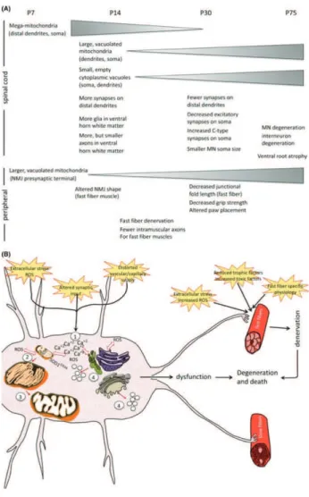
Figure 3.2. Timeline of physiopathological changes in the SOD1G93A ALS mouse model (from Vinsant et
al., 2013). Several pathological alterations have been associated with mSOD1. One way to understand the
pathogenesis of ALS is to identify the sequence of events leading to motor neuron degeneration. In the ALS mouse model expressing mSOD1G93A these alterations are measurable starting after the first week of life, both in spinal cord and at the level of NMJ.
dynamics, fragmentation and membrane depolarization that appear before disease onset. Moreover, targeting mSOD1 specifically to muscle mitochondria in healthy animals, induces the same type of mitochondrial defects (Luo et al., 2013). This suggests that these alterations are more likely related to mSOD1 toxicity than to the retraction of nerve endings. Equally, increased ROS production in non-symptomatic muscle of SOD1 mice has been shown to induce the over-expression of Ras-related associated with diabetes (Rad), a potent inhibitor of voltage-dependent calcium channels. Rad up-regulation correlates with disease severity in patients diagnosed with sporadic ALS (Halter et al., 2010). As it was mentioned earlier, protein quality control is altered due to SOD1 aggregates. While inclusions are not visible in muscle tissue, a robust activation of the protein quality control system and induction of autophagic response was evidenced in muscle from SOD1 mouse model. The activation was more important than in motor neurons (Crippa et al., 2013). This can be explained by differences in SOD1 chemistry according to cellular environment. In the muscle cell line C2C12 mSOD1 remains soluble without forming aggregates that affect the proteasome function. That allows a better clearance of misfolded mSOD1 than in the motor neuron cell line NSC34 (Onesto et al., 2011).
By applying the same strategy used to underpin the role of astrocytes and microglia, muscle came into the spotlight as a premiere target of SOD 1 toxicity. Two independent studies have showed that restricted expression of mSOD1 induces muscle atrophy and oxidative stress (Dobrowolny et al., 2008; Wong and Martin, 2010). The expression of mSOD1 specifically in muscle tissue recapitulates many aspects of the disease, including motor neuron death and glial activation (Wong and Martin, 2010).
Several lines of evidence suggest that muscle can play an active role in motor neuron degeneration. For instance the neurite outgrowth inhibitor Nogo-A was found to be upregulated in muscle tissue of patients and in pre-symptomatic SOD1 mouse model, in strong correlation with disease severity. For this reason Nogo-A was proposed to participate to the destabilization of
the NMJ and retraction of nerve terminals. The ablation of Nogo A in muscle of SOD1 mouse model extends survival by 10%, while overexpression in WT mice induces atrophy and denervation (Jokic et al., 2006). Altered muscle metabolism is another potential mechanism of muscle-induced denervation. Inducing a hypermetabolism in muscle by uncoupling mitochondrial respiration is enough to affect NMJ stability and function and to induce distal degeneration of motor neurons (Dupuis et al., 2009). Since mitochondrial defects come out as the most evident SOD1-related muscle defect in ALS, protection of mitochondria was another potential target. However, it has been shown that by rescuing muscle mitochondria through the upregulation of PGC-1α atrophy is prevented but is not enough to prevent neuronal death in an ALS mouse model (Da Cruz et al., 2012).
The evidence available so far indicates an early muscle pathology that parallels the neuronal pathology. It is noticeable that targeting muscle opens therapeutic possibilities aimed at preventing muscle denervation and atrophy. It seems however that the mechanisms involved in mSOD1-related muscle pathology are different from those in neurons, since the chemical behavior of mSOD1 is different in the two cell types. Further characterization of muscle pathology in ALS is imperatively needed. Equally the cause-consequence relationship remains a matter of controversy. The experiments presented here proposed several potential pathways of muscle-nerve signaling that could participate to the destabilization of the NMJ. However, the extent to which muscle tissue may participate to disease progression and the potential mechanisms involved are less clear, the scientific literature remaining poor and inconclusive.
4 M
USCLE,
METABOLISM ANDALS
Skeletal muscle is perfectly shaped to fulfill its function of transforming chemical energy into mechanic energy and movement. In order to do so continuous amounts of energy are needed, therefore it is not surprising that the muscular system consumes 30% of the overall body energetic supply at rest (Zurlo et al., 1990). In the same time, muscle fibers adapt perfectly to variations in energetic demands and nutrient supply just by tuning their metabolism. For instance, during periods of nutrient scarcity, muscle metabolism is tuned down in order to preserve energy for essential life supporting processes. Due to its intrinsic plasticity, muscle ultrastructure and metabolic profile are shaped by changes in activity, aging and disease. From this perspective muscle is a metabolic organ of high importance for whole body energy homeostasis.
4.1
M
USCLE STRUCTURE AND PHYSIOLOGYFrom standing to running, from writing to boxing, skeletal muscles are designed to produce a diversity of movements with different energetic demands. Muscle fibers have the capacity of instantly providing high amounts of ATP for intense/acute activities, but also continuously providing ATP for longer periods of enduring physical activities. This is possible due to the structural and metabolic diversity of muscle fibers (Schiaffino and Reggiani, 2011).
Every muscle fiber is comprised of a multitude of protein complexes called myofibrils that are strings of smaller units called sarcomeres. The main sarcomeric proteins are the thick myosin filaments interposed by thin, shorter, actin filaments. The actin filaments slide during contraction over the thick filaments, toward the center of the sarcomere shortening the length of the
myofibrils and generating muscle contraction (Huxley, 1957). The number of aligned sarcomeres per area unit gives the force of one muscle.
The hexameric thick filament is a molecular motor. A thick filament consists of two myosin heavy chains of 220 kd (MyHCs), two myosin light chains of 20kd (MyLC20) and two myosin light chains of 17kd (MyLC17). The
myosin heavy chain has an enzymatic function that consists in transforming the chemical energy stored in ATP molecules into mechanical energy through ATP hydrolysis. The mechanical function of myosin relies on conformational changes and binding of actin filaments through so-called cross-bridges or myosin heads. In fact a contraction is given by cyclic tightening and loosening of actin binding onto the myosin chain and this interaction requires ATP hydrolysis (Rayment et al., 1993). The MyLC17 is involved in maintaining the
structural stability of the cross-bridges, while the MyLC20 has a regulatory
function that is not well understood.
Muscle fibers are heterogeneous, and that is visible to the naked eye. The different muscle types can be distinguished by their color. The fact that muscle can be categorized into white and red is a known fact since the 19th century (Schiaffino and Reggiani, 2011). Advances in histochemical methods allowed the classification of muscle fibers using their enzymatic activity profile. The red slow-twitch muscles are rich in oxidative enzymes such as succinate dehydrogenase (SDH) and cyclooxygenase (COX), as opposed to white fast-twitch muscles that depend on glycolysis for fast ATP production (Burke et al., 1971; Edstr m and Kugelberg, 1968). Histochemical determination of enzymatic activities of such proteins is today a well-accepted method for fiber type characterization (SDH, COX or ATP-ases). Moreover, differences in size and mitochondrial content have equally been thoroughly characterized. The SDH positive fibers are generally smaller and have higher mitochondrial content than the bigger SDH-negative fibers (Schiaffino et al., 1970).
Today this classification has become more complex with the discovery of different myosin heavy chain (MyHC) isoforms that give the specificity of muscle fiber type. There are four main types of MHC isoforms: I, IIa, IIx and
IIb. The type I corresponds to slow oxidative fibers (SO or IA fibers), IIa and IIx for fast oxidative glycolytic (FOG or IIA and IIX fibers) and IIb for purely glycolytic fibers (FG or IIB fibers). A minority of fibers is hybrid, expressing more than one MyHC isoform. The ATP-ase activity differs between the different MyHC isoforms and that is reflected in the contractile properties of muscles. The rate of ATP hydrolysis is slower in type I fibers but the energetic cost of contraction is also lower (Bottinelli et al., 1994). That is why type I fibers are more suited for long duration but moderate activity. The glycolytic fibers are recruited for high intensity efforts but they fatigue quicker than the oxidative fibers because the capacity of providing enough energy at a sufficient rate is limited. That is due in part to the depletion glycogen stores, which are the main source of glucose during high intensity exercise. In the same time glycolytic metabolism induces acidosis through the accumulation of lactic acid, which seems to negatively affect the activity of several key enzymes involved in glycolytic ATP production (Sahlin et al., 1998).
From this rough characterization of muscle fiber types, it is obvious that between the MyHC isoforms present in one fiber, resistance to fatigue and fiber metabolism there is a strict correlation. This specialization is extended at the level of the entire motor unit, including the nerve terminals and the neuromuscular junction (Deschenes et al., 1994). Motor units are therefore classified according to their physiological properties in fast-fatiguable (corresponding to FG fibers), fast fatigue resistant (corresponding to FOG fibers) and slow fatigue resistant (corresponding to SO fibers) (Burke et al., 1971; Dum and Kennedy, 1980).
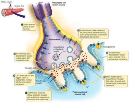
The neuromuscular junction is the synapse between a motor neuron and the muscle. One motor neuron branches and innervates a group of muscle fibers of the same type constituting a motor unit. The main neurotransmitter is acetylcholine, and specialized acetylcholine receptors (AChRs) coupled to ion channels are present on the sarcolemma of muscle fibers. At the synaptic level the muscle forms junctional folds, in order to enlarge the surface of the sarcolemma and enrich the expression of AChRs and voltage gated Na2+
Figure 4.1. Structure of neuromuscular junction (from Sadava, 2010). The neuromuscular junction is the
synapse between a motor neuron and the muscle. The sarcolemma of the muscle fibers present invagination named junctional folds, that increase the surface of interaction. The main neurotransmitter is acetylcholine, and specialized acetylcholine receptors (AChRs) coupled to ion channels are present on the sarcolemma of muscle fibers. Voltage gated Na+ channels present in the junctional folds help spreading the action potential along the postsynaptic membrane.
channels (Figure 4.1). Between the different types of fibers variations in the surface of the NMJ area but also density of AChRs (Sterz et al., 1983; Waerhaug and L!mo, 1994) and ion conductance (Milton et al., 1992) have been described. The NMJs of fast-twitch muscle necessitate a higher depolarization than the slow-twitch. This is accomplished through a higher degree of folding and higher density of AChRs and voltage gated Na2+ channels is higher (Hughes et al., 2006). Motor neurons innervating the different fiber types also present variations. Neurons innervating fast-twitch muscles are thicker and with bigger cell bodies and a complex dendritic ramification, which correlates with a higher conductance velocity (Henneman et al., 1965; Mendell, 2005).
4.2
M
ETABOLIC REGULATORS OF MUSCLE FUNCTIONAND FIBER TYPE
Physical exercise has been used as a tool in order to understand the dynamics between glucose and fat metabolism during exercise and the long-term effects of training on muscle metabolism. It was firstly observed that changes in intensity and duration of exercise employ differently the energy source for ATP production and mobilization of energy stores (Figure 4.2). During high intensity exercise less than half of energy comes from lipid breakdown. Carbohydrate metabolism prevails and inhibits fatty acids transport into the mitochondria (Holloszy et al., 1998; Wolfe, 1998). Lipolysis in the white adipose tissue and fatty acid uptake in muscle decrease with increased exercise intensity, while fatty acid oxidation is maximal during moderate prolonged exercise, and lower during low and high intensity exercise (Romijn et al., 1993).
During contraction changes in energetic homeostasis act as a signal in order to activate important pathways involved in preserving energy stores and ensuring substrate availability. Exercise increases translocation of
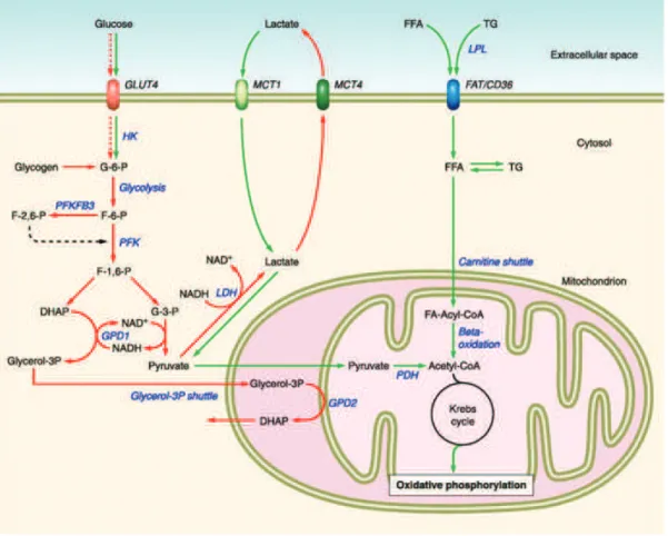
Figure 4.2. Metabolic profile of the different types of muscle fibers (from Schiaffino & Reggiani, 2011).
According to intensity and duration of exercise, muscle fibers employ different energy sources for ATP production and mobilization of energy stores. Muscle fiber types are specialized in order to respond to different energetic demands. Fast glycolytic and and fast oxidative glycolytic muscle fibers depend on glucose metabolization (in red) to produce ATP. Slow oxidative muscle fibers depend on lipid oxidation to produce the energy necessary for muscle contraction. Abbreviations: GLUT4-glucose transporter4; HK-hexokinase; G-6-P-glucose 6 phosphate; F-6-P-fructose 6 phosphate; F-2,6 (1,6)-P-fructose 2,6 (1,6) biphosphate; PFK-phosphofructokinase; DHAP-dihydroxyacetone phosphat; G-3-P-glyceraldehyde 3 phosphate; MCT (1,4)-monocarboxylate transport protein (1,4); LDH-lactate dehydrogenase; FFA-free fatty acid; TG-triglyceride; LPL-lipoprotein lipase; FAT/CD36-fatty acid transporter CD36; PDH-pyruvate dehydrogenase; GPD2-glycerol-3-phosphate dehydrogenase 2
sarcoplasmic glucose transporter GLUT4, fatty acids (FAs) transporter FAT/CD36 and induces substrate uptake and utilization. Several key metabolic regulators participate in concert to modulate these processes and ensure efficient energy production.
Contraction induces the opening of calcium pumps on the membrane of the sarcoplasmic reticulum and the release of calcium into the cytoplasm (Smith et al., 2013). Calcium signaling plays a central role in regulating muscle metabolism during contraction by activating calmodulin-dependent protein kinase (CaMK) in an intensity dependent manner (Egan et al., 2010). CaMK regulates muscle plasticity and activates mitochondrial biogenesis (Wu et al., 2002). It also upregulates glucose transport by enhancing GLUT4 exocytosis (Li et al., 2014) and lipid uptake and oxidation (Raney and Turcotte, 2008).
Increased ATP turnover (increased AMP/ATP ratio) activates AMP-activated protein kinase (AMPK). AMPK acts as a gatekeeper for metabolic equilibrium. During energy-deprived states, such as physical exercise, it switches off anabolic energy-consuming pathways and enhance substrate breakdown. Specifically, the primary role of AMPK during exercise is to reduce protein synthesis. Also it phosphorylates and inhibits glycogen synthase activity preventing glucose to be diverted to glycogen stores. Pharmacological up-regulation of AMPK with AICAR has also been shown to increase glucose uptake (Merrill et al., 1997) and lipid uptake and oxidation at rest (Raney et al., 2005). However its role in glucose and lipid utilization during exercise is not well understood (J!rgensen et al., 2006). It has been shown that FAT/CD36 and lipid uptake is not dependent on AMPK activity during contraction (Jeppesen et al., 2011). More likely it acts as a facilitator, by releasing inhibition exerted over lipid uptake pathway at rest (Catoire et al., 2014). Knock down of muscle AMPK in vitro reduces GLUT4 translocation in response to Ca2+ entry following nervous stimulation (Li et al., 2014). However in vivo inactivation of this enzyme only partially reduced contraction-stimulated glucose uptake (Mu et al., 2001). Another important role of AMPK is the
activation of several other metabolic regulators essential for muscle activity and plasticity like PGC-1α, class II histone deacetylases (class II HDACs) and class III histone deacetylases (sirtuins).
Physical activity modulates muscle fiber function, inducing changes in fiber-type specific contractile proteins, changes in enzymatic activity and mitochondrial content (Egan and Zierath, 2013). Training has long lasting effects on muscle function, reducing its capacity to use glucose for ATP production and increasing its respiratory capacity (Spina et al., 1996). This is reflected by an increased mitochondrial biogenesis (Hood et al., 1994) and increased activities of mitochondrial enzymes like citrate synthase, cytochrome c oxidase (Gollnick and Saltin, 1982; Murakami et al., 1994).
The most important intrinsic regulator of muscle function is Peroxisome Proliferator Activated Receptor γ Coactivator 1α (PGC-1α). Initially PGC-1α was characterized as a key element of adaptive thermogenesis, an important component of energy homeostasis. Upon exposure to cold PGC-1α was found to up-regulate key genes encoding mitochondrial enzymes involved in respiration (ATP-synthase, cytochrome c oxidase, uncoupling protein 1) and induced mitochondrial biogenesis in brown adipose tissue (Puigserver et al., 1998). The role of PGC-1α in muscle physiology was outlined later by overexpressing Pgc-1α specifically in type II muscle fibers. The result was a potent induction of oxidative metabolic program together with a switch in fiber-specific protein content and increased fatigue resistance, white muscles becoming red (Lin et al., 2002). An essential aspect of this program of muscle fiber switch is that the induction of Pgc-1α is activity- dependent. Physical activity was shown to induce its mRNA expression in muscle (Goto et al., 2000). Transcription and activity of Pgc-1α is ensured by the upstream activity-sensitive elements CaMK and AMPK. The promoter region of Pgc-1α includes myocyte enhancer 2 (MEF2) and c-AMP responsive element (CRE) binding regions. CaMK and AMPK activate MEF2 and CREB that in turn activate Pgc-1α transcription (Egan et al., 2010). AMPK also regulates PGC-1α activity through direct phosphorylation (J ger et al., 2007). The regulation
Figure 4.3. Remodeling of skeletal muscle phenotype (adapted from Egan & Zierath, 2013). Physical training
induces metabolic and ultrastructural changes in muscle fibers, inducing a switch in muscle fiber type, from fast glycolytic to slow oxidative. This remodeling of skeletal muscle fibers is highly complex and necessitates the coordination of several pathways within the cell. The master regulator of skeletal muscle phenotype is PGC-1α, that upregulates the oxidative metabolism, associated with increased mitochondrial biogenesis and respiration. Abbreviations: HDAC-hystone de-acetylase; PGC1α-peroxisome proliferator-activated receptor γ coactivator α; MEF2-myocyte enhancer factor2; CREB-cAMP response element binding protein; GEF-Glut4 enhancer factor; FOXO-forkhead transcription factor, O box subfamily; ERRα-estrogen related receptor α; NRF1/2-nuclear respiratory factor 1&2; PPARα-peroxisome proliferator-activated receptor α; mtDNA-mitochondiral DNA; TFAM-mitochondrial transcription factor A; P-phospho; Ac-acetyl