RESEARCH OUTPUTS / RÉSULTATS DE RECHERCHE
Author(s) - Auteur(s) :
Publication date - Date de publication :
Permanent link - Permalien :
Rights / License - Licence de droit d’auteur :
Institutional Repository - Research Portal
Dépôt Institutionnel - Portail de la Recherche
researchportal.unamur.be
University of Namur
p38 MAPK-regulated EGFR internalization takes place in keratinocytes monolayer during stress conditions
Lambert, Sylviane; Frankart, Aurélie; Poumay, Yves
Published in:
Archives of Dermatological Research
Publication date:
2010
Document Version
Peer reviewed version
Link to publication
Citation for pulished version (HARVARD):
Lambert, S, Frankart, A & Poumay, Y 2010, 'p38 MAPK-regulated EGFR internalization takes place in
keratinocytes monolayer during stress conditions', Archives of Dermatological Research, vol. 302, pp. 229-233. <http://www.springerlink.com/openurl.asp?genre=article&id=doi:10.1007/s00403-009-1020-0>
General rights
Copyright and moral rights for the publications made accessible in the public portal are retained by the authors and/or other copyright owners and it is a condition of accessing publications that users recognise and abide by the legal requirements associated with these rights. • Users may download and print one copy of any publication from the public portal for the purpose of private study or research. • You may not further distribute the material or use it for any profit-making activity or commercial gain
• You may freely distribute the URL identifying the publication in the public portal ? Take down policy
If you believe that this document breaches copyright please contact us providing details, and we will remove access to the work immediately and investigate your claim.
1
p38 MAPK-regulated EGFR internalization takes place in
keratinocyte monolayer during stress conditions
Sylviane Lambert, Aurélie Frankart and Yves Poumay*
Cell and Tissue Laboratory, URPHYM, University of Namur (FUNDP), Namur, Belgium
*Corresponding author: Yves Poumay
Cell and Tissue Laboratory, URPHYM, University of Namur (FUNDP), 61, rue de Bruxelles, B-5000 Namur, Belgium
Tel: +32 81 724257 Fax: +32 81 724261
Abstract
The epidermis is the outermost protection of the organism. As so, defence program has to be initiated in stress situation in order to protect keratinocytes. The EGF receptor (EGFR) controls cell proliferation and migration in keratinocytes, being a major regulator of keratinocyte homeostasis within the epidermis. The EGFR is known to be internalized without addition of ligand under the control of p38 MAPK during stress conditions in HeLa cells, but also following lipid rafts disruption in keratinocytes. This could represent an alternative internalization process that removes the EGFR from cell surface. Here, we investigated whether other stress conditions such as scratch wounding keratinocyte monolayer or incubation with a sensitizer chemical (i.e. DNFB), could also induce this peculiar mechanism of EGFR internalization. Our results show that both stressing conditions induce p38 MAPK activation concomitantly with EGFR internalization, independently of ligand-binding to the EGFR. Inhibition of p38 MAPK activity during scratch wound blocks EGFR internalization at the margin of the wound while cell migration is impeded. Our results show thus that the p38 MAPK-dependent EGFR internalization is a process shared by keratinocytes when submitted to challenging conditions.
3
Introduction
The epidermis is the most external layer of the skin and represents the most important barrier against external physical or chemical stresses. In this regard, the regulation of the keratinocyte homeostasis in epidermis has to be well-balanced between proliferation and differentiation. This regulation can be obtained and controlled by signalling through the EGF receptor. This member of the Receptor Tyrosine Kinase family controls cell proliferation by activation of the ERK1/2 MAPK pathway, but also controls cell migration, for instance by its interaction with integrin 64 [5], and cell survival.
It has been shown that during stress conditions such as UV exposure and treatment with
TNF- or anisomycin, HeLa cells were able to induce a particular non-typical internalization of EGFR (independent of ligand addition) under the control of p38 MAPK [9, 10]. In this regard, we have recently demonstrated that this non-typical process also takes place during recovery period following lipid rafts disruption in normal human epidermal keratinocytes [3]. Actually, disruption of the lipid rafts signalisation platforms produced by cholesterol depletion leads to activation of several signalling pathways, including the EGFR [4] and the p38 MAPK [2] pathways in keratinocytes. Following recovery periods after cholesterol depletion, the activation of p38 MAPK and EGFR are sustained and the growth factor HB-EGF is produced [6]. The EGFR internalization induced during these recovery periods depends on p38 MAPK activity as it is blocked by inhibition of p38 MAPK [3]. Here, we examined whether other stress conditions that potentially affect epidermal keratinocytes could also trigger this ligand-independent internalization of EGFR under p38 MAPK control. For this purpose, we tested the effect of scratch wound or contact with the chemical sensitizer dinitrofluorobenzene (DNFB), on p38 activation as well as on the cellular localization of EGFR in keratinocytes.
Materiel and methods
Normal human keratinocytes were obtained and cultured as described in [2]. For scratch wounding, a linear scratch wound was made with a 200 µl pipette tip in confluent culture of kératinocytes (if cultures were assigned for Western blot analysis, 16 scratch wounds were realized in a 30 mm Petri dish). Cultures were then washed to remove cellular debris, transferred to fresh medium (Epilife, Cascade Biologics) without any added serum or growth factors and allowed to recover for 30 min or 1 hour. Other keratinocytes cultures were incubated in presence of DNFB (0.08 g/ml, Sigma) for 30 min or 1 hour. Cell lysates were extracted and subjected to SDS-PAGE analysis and Western blotting for p38 and its phosphorylated form using specific antibodies: p38 and pp38 (Thr 180/Tyr 182)(Cell Signalling Technology, Boston, MA). Optical densities were obtained and measured using the ImageQuant 350 device and software from GE Healthcare. Culture media were tested for AR using ELISA test (BD Pharmingen) according to manufacturer’s protocol. Results from three independent experiments were analysed using Student’s t-test.
For p38 MAPK and MEK inhibition, cells were pre-treated for 30 min with the specific p38 MAPK inhibitors PD169316 (15 µM) or SB202190 (10 µM) or MEK inhibitor PD98059 (2 µM; all from Calbiochem VWR, Leuven). Keratinocytes were then incubated with DNFB (0.08 g/ml, Sigma) for 1 hour or scratched as described above, allowed to recover for 1 hour. Cells were fixed with 4% paraformaldehyde in PBS and immuno-stained for EGFR using the LA1 antibody (Millipore, Molsheim), followed by antibody directed against mouse IgG and coupled with AlexaFluor 488 (Invitrogen, Merelbeke). Cells were observed by confocal microscopy (Leica) using a 63X objective. Cells on four images from three independent experiments were quantified as described in [3] and statistical analysis was performed using Student’s t-test.
Alternatively, cells were allowed to recover and migrate for 24 hours before being fixed with 4% paraformaldehyde in PBS and stained with toluidin blue (0.05% in 1% Borax). Cells were then observed with bright field light microscopy (Olympus) using a 4X objective.
For EGFR ligand-binding site neutralization, cells were pre-treated with the EGFR neutralizing antibody LA1 (10 µg/ ml, Millipore) or the amphiregulin neutralizing antibody (10 µg/ml, BD Pharmingen) for 30 min. Cells were then either scratched and allow to recover for 30 min or incubated with DNFB (0.08 µg/ml) for one hour in presence or absence of the
5
neutralizing antibody. Keratinocytes were then fixed and stained for the EGFR as described above.
Results and discussion
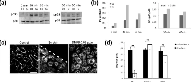
Confluent autocrine cultures of keratinocytes produced as monolayers were scratched and allowed to recover in autocrine culture medium (without growth factors) or treated with the sensitizer DNFB before cell lysis and Western blot analysis for p38 MAPK phosphorylation. 30 min and 1 hour after the scratch wound or after incubation with DNFB, p38 MAPK is found activated (Fig. 1a and 1b). Indeed quantification of the Western blot performed in Fig. 1a shows a strong induction of p38 phosphorylation in keratinocyte’s cultures subjected to scratch wounding and allowed to recover for 30 min (Fig. 1b). After DNFB treatment, p38 was found slightly phosphorylated as shown by Western blot (Fig. 1a) and optical densities measurements (Fig. 1b). Moreover, using confocal microscopy, the cellular localization of the EGFR in the margin of the scratch wound, or in DNFB-treated cells, is found inside the cell cytoplasm despite no ligand has been (Fig. 1c). Measurement of the pixel intensities at the cell periphery or in the cell cytoplasm show significant differences in the control cells but not in cells located at the margin of the scratch wound or treated with DNFB (Fig. 1d).
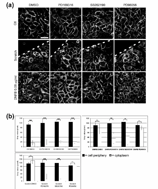
We next examined whether this internalization of the EGFR could be under the control of p38 MAPK, as we had previously reported in keratinocytes after cholesterol depletion [3]. Indeed, when p38 MAPK activity was blocked by specific inhibitors such as PD169316 or SB202190 before identical treatments, the EGFR was mainly found at the cell periphery despite the stressing conditions, indicating that this EGFR internalization process is p38 MAPK-mediated (Fig. 2a). Moreover, quantification of the pictures obtained by microscopy and analysed as previously described [3] demonstrated a significantly increased localization of the EGFR at the cell periphery at the margin of the scratch wound, as well as in cells treated with DNFB when p38 MAPK is inhibited (Fig. 2b). This effect depends specifically on p38 MAPK activity since the use of PD98059, inhibiting MEK activity, responsible for ERK1/2 activation, does not impede EGFR internalization after scratch wounding or chemical stress (Fig. 2).
As in our conditions, the EGFR internalization is concomitant with cell migration appearing following scratch in cell cultures, we suggest that this particular processing of EGFR dependently of p38 MAPK activation might be involved in the setting up of cell migration process. Indeed, when p38 MAPK is inhibited in challenged keratinocytes, the ligand-independent EGFR internalization does not occur (Fig 2. a) and the process of migration is blocked [1, 8] (and data not shown).
7
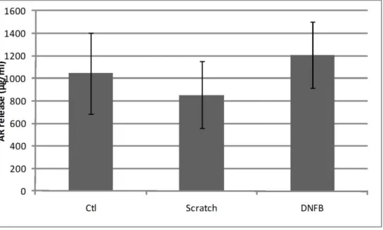
Since the EGFR may be activated and internalized due to ligand-binding, we tested whether stress conditions would induce cleavage of EGFR proligands from cell membrane. As keratinocytes are known to depend on AR liberation to induce autocrine growth [7] and because we found that AR was secreted following cholesterol depletion (Lambert and Poumay, unpublished results), we tested whether scratch wound or DNFB treatment would induce AR release. As shown in Fig. 3, no significant increase of the AR concentration was measured following either scratch wound or DNFB treatment. Moreover, the neutralization of the ligand-binding pocket of the EGFR with the LA1 antibody did not induce changes in the internalization of the EGFR in these conditions (data not shown), suggesting that the EGFR internalization is independent of any ligand binding to the receptor.
In summary, a ligand-independent but p38-dependent EGFR internalization process is involved in the control of keratinocyte physiology during the challenging situations that correspond to tissue repair or chemical stress as we reported here. This common atypical EGFR processing strongly suggest that a temporary re-localization of the EGFR can be part of the mechanisms involved during the aetiology of epidermal disease.
Acknowledgements:
The authors are grateful to Valérie Deglas and Françoise Herphelin for excellent technical assistance and to Noëlle Ninane and Catherine Demazy for help with confocal microscopy. SL holds a fellowship from the Fonds de la Recherche dans l’Industrie et l’Agriculture, Belgium. This work was financially supported by Fonds de la Recherche Fondamentale Collective grant 2.4506.01 and Fonds National de la Recherche Scientifique grant 1.5.033.06F to YP.
9
Figures:
Figure 1: p38 MAPK is activated in DNFB-treated or scratch-wounded keratinocytes
(a): Cells were left as control (C), or a linear scratch wound was made (Sc) and cells were allowed to recover for 30 min or 1 hour, or cells were treated with DNFB (D, 0.08 µg/ml) for 30 min or 1 hour. Cell lysates were then analysed by Western blotting for p38 MAPK phosphorylation.
(b): Optical density measurement of the Western blot presented in (a).
(c): Keratinocytes were treated as in (a) and prepared for confocal microscopy. Cells were fixed and stained for the EGFR before being observed by confocal microscopy (scale bar: 40 µm).
(d): Histogram showing the repartition of the EGFR between the cell periphery (black column) and the cell cytoplasm (white column) after analysis of the images obtained in (b) by confocal analysis using the ImageJ software. Data are means +/- SD of 3 independent experiments. ns= non significant, ***=p<0,005.
Figure 2: EGFR internalization depends on p38 MAPK activation in stress situation
(a): Keratinocytes were treated as in Figure 1a in presence of p38 MAPK inhibitors PD169316 (15 µM) or SB202190 (10 µM) or MEK inhibitor PD98059 (2 µM). After one hour, cells were fixed with paraformaldehyde 4% in PBS and stained for EGFR using the LA1 antibody followed by antibody directed against mouse IgG coupled with Alexa Fluor 488. Cells were observed by confocal microscopy (Leica) using a 63X objective (scale bar: 40 µm) and images were quantified as described in [3].
11
(b): The images in (a) were analysed using the ImageJ software and histogram represents means +/- SD of 3 independent experiments. ns= non significant, **=p<0,01 and ***=p<0,005.
Figure 3: No amphiregulin secretion during stress conditions
Cells were either let as control (Ctl), scratched and allowed to recover for 30 min or treated for one hour with DNFB (0.08 µg/ml). Culture medium was then collected and tested for AR using ELISA test.
[1] Fitsialos G, Chassot AA, Turchi L, Dayem MA, LeBrigand K, Moreilhon C, et al. Transcriptional signature of epidermal keratinocytes subjected to in vitro scratch wounding reveals selective roles for ERK1/2, p38, and phosphatidylinositol 3-kinase signaling pathways. J Biol Chem 2007;282:15090-102.
[2] Jans R, Atanasova G, Jadot M, Poumay Y. Cholesterol depletion upregulates involucrin expression in epidermal keratinocytes through activation of p38. J Invest Dermatol 2004;123:564-73.
[3] Lambert S, Ameels H, Gniadecki R, Herin M, Poumay Y. Internalization of EGF receptor following lipid rafts disruption in keratinocytes is delayed and dependent on p38 MAPK activation. J Cell Physiol 2008;217:834-45.
[4] Lambert S, Vind-Kezunovic D, Karvinen S, Gniadecki R. Ligand-independent activation of the EGFR by lipid raft disruption. J Invest Dermatol 2006;126:954-62.
[5] Mainiero F, Pepe A, Yeon M, Ren Y, Giancotti FG. The intracellular functions of alpha6beta4 integrin are regulated by EGF. J Cell Biol 1996;134:241-53.
[6] Mathay C, Giltaire S, Minner F, Bera E, Herin M, Poumay Y. Heparin-binding EGF-like growth factor is induced by disruption of lipid rafts and oxidative stress in keratinocytes and participates in the epidermal response to cutaneous wounds. J Invest Dermatol 2008;128:717-27.
[7] Pittelkow MR, Cook PW, Shipley GD, Derynck R, Coffey RJ, Jr. Autonomous growth of human keratinocytes requires epidermal growth factor receptor occupancy. Cell Growth Differ 1993;4:513-21.
[8] Stoll SW, Kansra S, Elder JT. Keratinocyte outgrowth from human skin explant cultures is dependent upon p38 signaling. Wound Repair Regen 2003;11:346-53.
[9] Vergarajauregui S, San Miguel A, Puertollano R. Activation of p38 mitogen-activated protein kinase promotes epidermal growth factor receptor internalization. Traffic 2006;7:686-98.
[10] Zwang Y, Yarden Y. p38 MAP kinase mediates stress-induced internalization of EGFR: implications for cancer chemotherapy. EMBO J 2006;25:4195-206.