ORIGINAL PAPER
Clinical, laboratory and pathological findings in cats
experimentally infected with Aelurostrongylus abstrusus
M. Schnyder&A. Di Cesare&W. Basso&F. Guscetti&B. Riond&T. Glaus&P. Crisi&P. Deplazes
Received: 19 December 2013 / Accepted: 10 January 2014 / Published online: 7 February 2014 # Springer-Verlag Berlin Heidelberg 2014
Abstract Aelurostrongylus abstrusus parasitizes the respira-tory tract and can heavily affect the breathing and general condition of cats. Experimental infections of six cats were initiated by intragastric administration with 100 or 800 third-stage larvae (L3) obtained from the terrestrial snail Helix aspersa. First-stage larvae were isolated from faecal samples after 35–41 days post infection (dpi) in five animals and until end of study (84 dpi) in two cats. Cough and respiratory sounds were observed starting from 28 to 41 dpi and dyspnoea and panting starting from 52 dpi. All cats had enlarged lymph nodes and, starting from 56 dpi, reduced body weight, and four cats showed intermittent reduced general condition with apathia and anorexia. Eosinophilia and leucocytosis partially with massive lymphocytosis, and occasional basophilia and monocytosis were observed. Mild anaemia was present in five cats, while alterations in coagulation parameters suggested stimulation of the coagulation cascade with increased consump-tion of coagulaconsump-tion factors (delayed PT, hypofibrinogenemia).
Adult A. abstrusus specimens were isolated from the five patent cats at necropsy and all six cats showed pathological changes in the lungs, including disseminated inflammatory cell infiltrates, often associated with incorporated larvae and eggs. There was some degree of overlap between the severity and the inocula-tion doses. Infecinocula-tions starting from 100 L3 of A. abstrusus had an impact on the lung tissues and on the health of the cats, despite the presence of only mild haematological abnormalities. Due to the worldwide occurrence of feline lung worms, para-sitic infections should be considered in the differential diagnosis of lung diseases regardless of the presence of clinical signs and larval excretion.
Introduction
Aelurostrongylus abstrusus is a metastrongylid nematode that resides as adult stage in the lung parenchyma in cats and other felines all over the world. Snails and slugs are obligatory intermediate hosts. Cats become infected by ingesting intermediate or paratenic hosts such as rodents, birds, amphibians and reptiles (Hamilton and McCaw 1967; Hobmaier and Hobmaier 1935; Scott 1973). Clinical manifestations of A. abstrusus infection in cats vary from asymptomatic to fatal. Most affected cats are asymptomatic (Hamilton1963; Payo-Puente et al.2005) or present with prevailing respiratory signs including a chron-ic cough with gradually increasing dyspnoea (Grandi et al. 2005) and/or other respiratory (wheezing, sneezing, nasal discharge) and non-specific (apathia, anorexia, fever) signs (Scott 1973; Traversa et al. 2008a). In very severe infec-tions or decreased resistance of the animals, the infection occasionally results in death (Ellis et al. 2010; Hamilton 1963). Young and free-ranging cats are considered to be at risk for clinical A. abstrusus infection (Traversa et al. 2008c).
M. Schnyder (*)
:
W. Basso:
P. DeplazesInstitute of Parasitology, Vetsuisse Faculty, University of Zürich, Winterthurerstrasse 266a, 8057 Zurich, Switzerland
e-mail: manuela.schnyder@uzh.ch
A. Di Cesare
:
P. CrisiDepartment of Comparative Biomedical Sciences, Faculty of Veterinary Medicine, Piazza Aldo Moro 45, 64100 Teramo, Italy F. Guscetti
Institute of Veterinary Pathology, Vetsuisse Faculty, University of Zurich, Winterthurerstrasse 268, 8057 Zurich, Switzerland B. Riond
Clinical Laboratory, Vetsuisse Faculty, University of Zurich, Winterthurerstrasse 260, 8057 Zurich, Switzerland T. Glaus
Clinic for Small Internal Medicine, University of Zurich, Winterthurerstrasse 260, 8057 Zurich, Switzerland
The diagnosis is mainly based on the detection of first-stage larvae (L1) in faecal samples. Direct faecal smears, flotation methods or bronchoalveolar lavage fluid examination may be positive, but larval migration methods followed by microscopic identification of the isolated larvae are more sensitive (Lacorcia et al.2009; Traversa et al.2008a). Nevertheless, potential false negative results during prepatency and due to irregularity of larval excretion, in particular, after re-infections (Hamilton 1969; Ribeiro and Lima Dos Santos2001), have to be consid-ered. Alternatively, biomolecular assays performed with pharyn-geal swabs show high specificity and sensitivity (Traversa et al. 2008b) and can be useful when morphological differentiation is challenging, as recently shown by the (re)discovery of other feline lungworm parasites, i.e., Troglostrongylus spp., having both highly morphological similar features of their L1 (Annoscia et al. 2014; Brianti et al. 2012; Di Cesare et al. 2013; Gerichter1949; Vevers1923).
As summarised by Scott in 1972, physical examination, haematology, diagnostic imaging, gross pathology and histo-pathology may also assist in diagnosing aelurostrongylosis in cats. Changes in serum proteins were mild and showed to be not useful for diagnosing aelurostrongylosis (Barsanti and Hubbell1980), while eosinophilia was observed in several case reports of cats with clinical signs and also in an asymp-tomatic cat (Grandi et al.2005; Hamilton1963). In a further case report with four naturally infected cats, all were eosino-philic and, additionally, blood gas analysis suggested hypoventilation and respiratory acidosis (Yildiz et al.2011). Diagnostic imaging describing the characteristic changes in cats infected with Aelurostrongylus abstrusus has been per-formed by radiology (Grandi et al. 2005; Losonsky et al. 1983) and, more recently, by computed tomography (Dennler et al.2013; Payo-Puente et al.2005). Alterations in gross pathology and histopathology were also previously described (Hamilton1963,1966; Scott1973).
In the context of the establishment of A. abstrusus infec-tions in cats for improving future clinical, diagnostic and especially chemotherapeutic and prophylactic strategies, six cats were experimentally inoculated with two different doses of third-stage larvae (L3) of A. abstrusus and subjected to intensive diagnostic and clinical monitoring. Results of diag-nostic imaging have been described elsewhere (Dennler et al. 2013), while the development of clinical signs and accompa-nying haematological and coproscopic findings, completed by post mortem analysis, are herein presented.
Materials and methods
Study design, experimental inoculation
The study was carried out at the experimental units of the Vetsuisse Faculty at the University of Zurich after approval by
the Cantonal Veterinary Office of Zurich (permission number 21/2011). The activities on experimentally infected snails were performed at the Faculty of Veterinary Medicine of Teramo after approval of“Comitato di Etica Interateneo per la Sperimentazione animale” (CEISA); approval number: 33/ 2010/CEISA/COM.
Six healthy facility-born European short-hair cats, five female and one male (all neutered), with a body weight of 2.4 to 3.6 kg, aged from 8 to 12 months, were orally inocu-lated via stomach tube with infectious L3 of A. abstrusus. L3 were isolated from experimentally infected snails (Helix aspersa) by cutting the foot of the snails, mincing the snail tissue with a scalpel and digestion of the material for 10– 20 min in 0.7 ml HCl 37 % and 0.6 g pepsin (molecular weight 35 kDa) mixed with tap water at 37 °C. The digested material was passed through gauze and centrifuged at 2,000 rpm for 5 min before the supernatant was discarded. The sediment with larvae was washed 2–3 times with tap water. The number of larvae in a sub sample was counted under a stereomicro-scope and individual infection doses for each cat were pre-pared. Cats were randomly divided into two groups. Three animals (A1, A2, A3) were infected with 100 L3 (low dose) and three (B1, B2, B3) with 800 L3 (high dose), correspond-ing to cats 1–6 in the previously mentioned study (Dennler et al. 2013). Before inoculation, the cats received metoclopramide (Paspertin®, Abbot) in a dosage of 0.3 mg/kg BW i.m. in order to prevent vomiting/regurgitation. The cats were anaesthetised for inoculation with ketamine (Narketan®, 10 mg/kg BW, Vétoquinol), midazolam (Dormicum®, 0.1 mg/kg BW, Roche), propofol (Propofol®, 0.7 ml/kg BW, Fresenius), morphasol (Morphasol-4®, 0.2 mg/kg BW, Dr. E. Graeub) and acepromazine (Prequillan®, 30 μg/kg BW, Arovet). All drugs were applied intravenously. After inocula-tion, the cats were observed for vomiting/regurgitation for up to 1 h. Two cats of group B vomited 10 (B2) and 23 min (B3) post inoculation; cat B2 was reinfected with an additional dose of 100 L3. All animals were humanely sacrificed 84–92 days post inoculation (dpi) by an overdose of pentobarbital after sedation with acepromazine.
Clinical follow-up, haematology, biochemistry and coagulation analysis
All cats underwent physical examination and weighing by a veterinarian 2 days before inoculation and then approximately biweekly until end of study on 14, 28, 41, 56, 69 and 82 dpi. Venous blood samples were contemporaneously collected from all cats during the study and immediately forwarded to the clinical laboratory of the Vetsuisse Faculty, University of Zurich, for haematology, chemistry and coagulation analysis. A complete blood cell count (CBC) was performed from EDTA-anti-coagulated blood using a veterinary haematology analyzer (Sysmex XT-2000iV, Sysmex Corp., Kobe, Japan)
previously validated for its use in the feline species (Weissenbacher et al.2011) and a manual white blood cell differential was carried out. Sodium citrate anti-coagulated plasma was immediately processed after collection for coag-ulation testing and used to determine prothrombin time (PT), thrombin time (TT), and activated partial thromboplastin time (aPTT) by automated analysis (Start 4, Roche Diagnostics, Rotkreuz, Switzerland). Furthermore, fibrinogen concentra-tion has been determined using the Claus method (STA Fib, Roche Diagnostics AG, Switzerland) on a semi-automated bench top analyzer (Start 4, Roche, Diagnostics, Switzerland). For quality control, two commercial control specimens with normal and high concentrations were analysed on a daily basis (PreciClot I and PreciClot II, Roche Diagnostics, Switzerland). Serum samples were used to determine substrates (total bilirubin, total glucose, urea, creatinine, total protein, albumin, cholesterol and triglycer-ides), enzymes (alkaline phosphatase, amylase, lipase, aspar-tate transaminase and alanine transaminase) and electrolytes (sodium, potassium, chloride, calcium and phosphorous) by automated analysis (Cobas Integra 800, Rotkreuz, Switzerland).
Coproscopic examination
A faecal examination from each cat was performed before inoculation using combined sedimentation/flotation and the Baermann-Wetzel larval migration technique (Deplazes et al. 2013) in order to exclude intestinal and respiratory parasite infestations. Starting from 26 dpi, individual faecal samples were collected approximately twice a week from each cat and examined for shedding of L1 of A. abstrusus by Baermann-Wetzel technique and determination of the number of L1 per gramme of faeces (LPG) in 10 g of faeces.
Post mortem examination
The macroscopic extent and severity of pneumonic change was assessed semi-quantitatively using the categories 0 (none), 1 (mild), 2 (moderate), 3 (strong) and 4 (massive). Lung tissue comprising the whole accessory lobe and one tracheobronchial lymph node were fixed in 10 % neutral-buffered formalin for 24 h. Thereafter, three to four lung tissue slices per animal (resulting in an approximately equal amount of tissue for each cat) and a cross-section of the lymph node were embedded in paraffin wax by routine procedures. Histopathological changes were assessed using sections rou-tinely stained with hematoxylin and eosin and the approxima-tive percentage of tissue involved was estimated assessing pulmonary parenchyma and bronchi, separately.
Adult worm burdens were determined using worm isolat-ing and countisolat-ing procedures differisolat-ing for each cat. Airway and lung tissue dissection and lung tissue digestion were
tested. Trachea, bronchi and bronchioli were opened and checked for macroscopically visible parasites. Parts of the lung lobes were directly cut in pieces and checked for lung worms under the stereo microscope by dissecting piece by piece with scalpel and forceps. For closer inspections, the optical microscope was used. For lung tissue digestion, parts of the lung lobes were cut in pieces and digested with HCl and pepsin, as described for snail digestion. The digested material was passed through a 180-μm sieve. Afterwards, the lung pieces were dissected piece by piece. The liquid of the digested material was passed through a 300-μm sieve and the sieve was then inverted and flushed in a glass petri dish. All collected liquid was centrifuged, the supernatant was eliminated and the sediment was checked for larvae (optical microscope) and adult parasites (stereo microscope). Worm burden analysis was prior-ranking and comprised all lung lobes except for the accessory lobe, which was used for histological examinations and half of the lung of cat B3.
Results
Clinical follow-up
The heart rate of the cats did not significantly change during the study and ranged from 92 to 210/min. Intermittently elevated rectal temperature up to 39.9 °C was observed in five of six cats.
Before inoculation, the mean respiratory rate in the six cats was 38 (range 24–44), while at end of study, it was 52 (range 36–68), but differences were not significant: the rates highly varied between examination days and cats. Forced respiration was observed in one cat of each group (A1 and B2) starting from 69 dpi, while respiratory sounds were auscultated in four cats: in two cats of group A (A1, A3) starting from 41 dpi, and in two cats of group B (B2, B3) starting from 28 dpi. These sounds were inspiratory and varied from slight rustling to harsh vesicular noise and stertor or stridor. Mandibular lymph nodes were enlarged in all cats, starting from 41 dpi at the earliest; cat B3 also had enlarged prescapulary and popliteal lymph nodes. Overall condition was reduced in the four cats with respiratory sounds, starting from 56 dpi, and this change was associated with depression or apathy. Spontaneous coughing was observed in cat B2 (56 dpi). All cats lost weight until the end of the experiment, in average 100–300 g in group A and 300–400 g in group B.
Haematology, biochemistry and coagulation analysis All cats developed eosinophilia starting from 14 dpi (four cats) or 28 dpi (two cats: A1, B1). Occasional basophilia (two cats, A3, B3), monocytosis (four cats: all except A1 and B3), lymphocytosis (five cats: all except B2) and
leucocytosis (all cats) were observed. In particular, two cats (A1 and B1) developed massive lymphocytosis at the end of the experiment (36.6 and 34.0×10³lymphocytes/μl, respec-tively). Mild anaemia was present in five cats starting from 14 (one cat), 69 (three cats) or 81 (one cat)dpi. Coagulation parameter values were irregularly out of reference ranges: PT was increased in one cat of each group (A2 and B3) on 81 and 56 dpi, respectively, while aPTT was reduced in all cats, mostly starting from 41 dpi. TT was reduced on several occasions in four cats (A1, A3, B2, B3). Fibrinogen values were below reference ranges in all cats in at least one occasion, mostly starting from 41 dpi; on 81 dpi, fibrinogen values of four cats (A2, A3, B1, B2) were reduced. Concerning chem-istry, no trends were observed.
Coproscopic examination
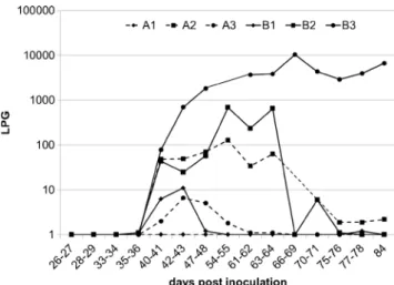
Faecal examination before experimental inoculation was neg-ative. Larval shedding started from 35 dpi (cat B2), 36 dpi (cats A3, B1), 40 dpi (cat B3) and 41 dpi (A1), while Baermann analysis of faeces from cat A2 remained negative during the whole study. Patency lasted until 48–84 dpi, with two cats excreting larvae until end of study (Fig.1, Table1). After an initial increase of the number of larvae excreted, the intensity of larval shedding decreased continuously by trend, except for cat B3. This cat and cat B2 excreted the highest daily number of larvae, reaching 10,457 and 687 LPG, respectively.
Post mortem examination
The macroscopic changes consisted of multifocal, nodular to coalescing, irregularly shaped areas of consolidation and brownish to greyish colour of variable extent between the cats, randomly distributed over the whole lung and variably interspersed with dark red, hyperaemic areas (Figs.2and3). The nodules partly protruded from the lung surface and meandering pale corridors were observed (Fig.4). The sever-ity of the pulmonary changes varied between cats (Table1), i.e., in cat A2, only single, small dark red slightly consolidated regions were present. Despite some overlap between the groups, the lesions appeared somewhat more pronounced in the high-inoculation dose cats. The lung lymph nodes were consistently enlarged in all cats.
Histological examination of the accessory lobe illustrated the presence of nodular to coalescing areas of densely packed inflammatory cells randomly distributed in the lung parenchy-ma and variably comprising parenchy-macrophages, epitheloid cells and multinucleated giant cells, sometimes forming small granulo-mas, as well as eosinophils, neutrophils, lymphocytes and plasma cells (Fig.5a, c). These areas contained a few small scattered necrotic foci in cat B3. The inflammatory infiltrates frequently contained moderate numbers of developing stages
(eggs with larvae, Fig.5b) or rare individual adult parasites (Fig. 5d, Table 1), and completely obliterated the alveolar lumina; they were often associated to bronchial structures showing epithelial hyperplasia. Adjacent to these areas, there were focally extensive regions with alveolar wall thickening with variable numbers of inflammatory cells in the alveolar septa and lumina and associated with pneumocyte prolifera-tion. In these areas, and multifocally, in further regions, there also were variable degrees of hypertrophy of alveolar duct smooth muscles. The approximate extent of accessory lobe tissue area collectively affected by all these microscopic changes is reported for each cat in Table 1. In addition, lymphocytic and plasmacellular infiltrates and the forma-tion of lymph follicles was observed in the peribronchial tissue of the inoculated animals. Intensity of this cellular reaction was mild in cats A2 and B3, moderate in cats A1 and B1, and marked in cats A3 and B2; the percental areas of peribronchial tissue affected by this change are reported in Table 1. In general, the bronchial glands were prominent. Hyperplasia and hypertrophy of the media of large pulmonary arterial vessels was moderate in one cat (B1) and focal in the other animals. There also were focal intimal and subintimal infiltrations of eosinophils and var-iable numbers of lymphocytes, plasma cells and eosino-phils in the adventitia of the arteriae. Altogether, there was some degree of overlap in the severity of the lesions between the high- and low-dose groups although the le-sions shown by cats B2 and B3 appeared as the most severe, and cat A2 showed only mild changes mostly consisting of mild disseminated residual inflammatory in-filtrates and slight focal hypertrophy of alveolar duct smooth musculature. In addition, the tracheobronchial lymph nodes of all cats were enlarged to variable degrees and presented large secondary follicles.
Fig. 1 Detection of first stage larvae per gramme of faeces (LPG) by the Baermann-Wetzel technique in three cats experimentally inoculated with 100 (A1–A3, dashed lines) or 800 (B1–B3, continuous line) third-stage larvae of A. abstrusus
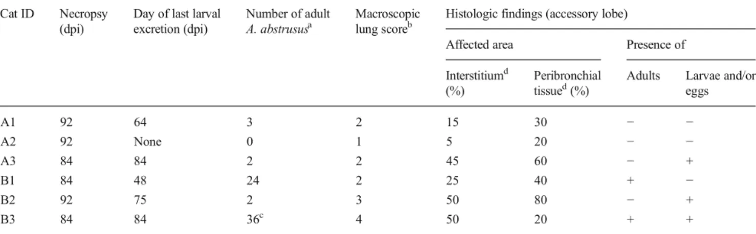
In two cats of group A (A1, A3) and all three cats of group B adult worms were found at necropsy (Table1). In the cat without larval excretion (A2), no adult worm was found. All parasites were identified as A. abstrusus. Numbers of adult worms ranged between 0 and 3 worms in cats of group A and 2–36 worms in cats of group B.
Discussion
This study correlates the successful experimental infection of six cats inoculated with 100 or 800 L3 of A. abstrusus with moderate clinical and haematological and coagulation
changes, by contrast to massive pathological manifestations. In a study performed with kittens inoculated with different numbers of L3 (Hamilton1967), the author concluded that 50 L3 were producing pulmonary lesions, but that at least 100 L3 were probably necessary for a successful infection with larval shedding and clinical signs. In the same study, animals given 1,600 or even 3,200 L3 had obvious clinical respiratory signs and multiple lesions throughout the lobes, as observed in the cats of this study inoculated with 800 L3. Animal numbers in the present experiment were small due to animal welfare reasons and the preliminary character of the study. Thus,
Table 1 Post mortem findings of six cats after experimental inoculation with 100 (group A) or 800 (group B) third-stage larvae of A. abstrusus
Cat ID Necropsy
(dpi)
Day of last larval excretion (dpi)
Number of adult
A. abstrususa
Macroscopic
lung scoreb
Histologic findings (accessory lobe)
Affected area Presence of
Interstitiumd
(%)
Peribronchial
tissued(%)
Adults Larvae and/or
eggs A1 92 64 3 2 15 30 − − A2 92 None 0 1 5 20 − − A3 84 84 2 2 45 60 − + B1 84 48 24 2 25 40 + − B2 92 75 2 3 50 80 − + B3 84 84 36c 4 50 20 + +
Gross and histological lesions in the lungs and adult worms recovered by reverse lung perfusion and dissection of heart and lungs
aDifferent worm isolating and counting procedures were applied for each cat
b
Extent and severity was semi-quantitatively scored ranging from 0 (none), 1 (mild), 2 (moderate), 3 (strong) to 4 (massive)
c
Only approximately half of the lung was dissected for worm counting
d
Data included in a previous report (Dennler et al.2013)
Fig. 2 In situ view of the right lung of cat B3 experimentally inoculated with 800 third-stage larvae of A. abstrusus, presenting with nodular to coalescing, brown to greyish areas of consolidation randomly distributed over all lobes. Nodules partly protrude from the pleural surface
Fig. 3 Lungs of cat A3 experimentally inoculated with 100 third-stage larvae of A. abstrusus, presenting with consolidated, dark red haemorrhagic patches and irregular pleural surfaces and enlarged lymph nodes
differences between groups could not be examined statistical-ly and have to be considered cautiousstatistical-ly and at the utmost to indicate trends.
Respiratory signs are the most common observed clin-ical consequence of A. abstrusus infections (Hamilton 1966; Traversa et al. 2008a). In this presented study, the main common clinical parameter indicative for a respira-tory tract infection was the enlargement of mandibular lymph nodes. Correspondingly, tracheobronchial lymphadenomegaly with significant lymph node enlarge-ment after inoculation has been detected in the same
infected cats by computed tomography (Dennler et al. 2013). Furthermore, respiratory sounds were present in four out of six cats: they were of varying quality, as previously described (Scott 1973), and started by trend earlier in cats with higher inoculation doses. Evident signs of respiratory distress such as dyspnoea and open-mouthed breathing have been previously described in some cases (Traversa et al.2008a) and, similarly, forced respiration was observed in two cats in this study. Important changes for parameters such as heart and respiratory rate and body tem-perature were not pathognomonic for A. abstrusus infection. Further non-specific signs indicative for a reduced overall condition were noted, i.e. all cats lost weight, by trend more in the group with high-inoculation dose.
CBC confirmed eosinophilia for all cats, an alteration regularly present in naturally infected cats (Grandi et al. 2005; Hamilton1963; Yildiz et al.2011). A high degree of eosinophilia caused by endoparasitism is often reported in literature (Center et al.1990). Eosinophilia in the studied cats was not only persistent, but also massive, independent of the inoculation dose. This can be explained by the constant pres-ence of the parasite and therefore of antigen stimulation, to which lymphocytes react with an immune response of the IgE-type leading to the observed massive lymphocytosis, in par-ticular, in two cats (i.e. A1 and B1). High numbers of lym-phocytes were also present in the affected lungs (Fig.5a–d), and, given the lack of other causes for this high inflammatory response in this experimental setting, these reactions can be attributed to individual immunological responses.
Further alterations in CBC were irregularly present and non-specific. Mild normochromic normocyte anaemia,
Fig. 4 Partially meandering pale corridors on the pleural surface of a lung lobe of cat A2 experimentally inoculated with 100 third-stage larvae of A. abstrusus
Fig. 5 Histological sections (hematoxylin and eosin stain) of lung tissue from cats inoculated with third-stage larvae (L3) of A. abstrusus. a Lung of a cat (B2) necropsied 92 days after inoculation with 800 L3: a consolidated area is invaded with masses of inflammatory cells. b Highly affected areas associated with incorporated larvae and eggs (cat B3). c Extract of Fig. 5b showing embryonated eggs containing the first stage larvae, surrounded by macrophages, epithelioid histiocytes and lymphocytes (cat B3). d Transversal section of an adult A. abstrusus nematode in the lung of a cat (B1) inoculated with 800 L3
present in five cats, has also been observed in single cats with natural A. abstrusus infection (Yildiz et al. 2011) and was a s c r i b e d t o t h e c h r o n i c i n f l a m m a t i o n d u e t o aelurostrongylosis. Coagulation parameters were evaluated for the first time, to the authors’ knowledge, in cats infected with A. abstrusus. In two cats, a slightly delayed PT could be observed indicating increased consumption of coagulation factors. Hypofibrinogenemia in the absence of other biochem-ical indications of decreased liver function did support the hypothesis of stimulation of the coagulation cascade with increased consumption of coagulation factors (Stockham and Scott2008). No explanation can be given for the shortened aPTT as it is not a reliable parameter for detecting hypercoag-ulability, and increased fibrinogen concentrations were not observed (Kurata et al.2003).
The prepatency of 35–41 days in this study was in the range (35–48 days) of previous observations (Hamilton 1963; Ribeiro and Lima Dos Santos2001), confirming the successful development of adult mature parasites in five out of six cats. During patency, the number of detected larvae in the faeces was variable and therefore did not allow an estimate of the present worm burden. In experiments performed in the 1960s, during a patent period of approximately 8–13 weeks, cats were shown to excrete up to 17 million of L1, with a peak approximately around 4–5 weeks of patency followed by a gradual diminution in numbers (Hamilton and McCaw1968). This was also observed in the two cats of this study inoculated with 800 L3 and with highest larval excretion. Experimentally infected cats were shown to excrete larvae up to 7 months and occasionally, more (Hamilton1968), in opposition to three cats of this study for which larval excretion stopped much earlier, between 48 and 75 dpi. Individual immunological responses, which were confirmed by individually different haematological responses and post mortem analyses, may be at the origin of these differences, as previously suggested (Hamilton1969).
Cats clinically suspicious for lung worm infection with larval excretion have been regularly described (Foster et al. 2004; Grandi et al.2005; Traversa et al. 2008a). However, larval excretion was also observed in a clinically asymptom-atic stray cat which had been examined in the context of a routine clinical visit before adoption (Grandi et al.2005) and in further two clinically asymptomatic cats which showed significant lesions on CT scanning (Payo-Puente et al. 2005). In an older study performed with apparently healthy cats (but most probably not subjected to detailed clinical examination), two cats were found to have larvae in faeces but were free of macroscopic lesions, while nine other cats were also free of larvae in the faeces but had macroscopic lung lesions and histological changes suggestive of past infections (Dubey and Beverley1968). Further studies performed with experimentally infected cats also showed how irregular larval excretion can be in cats known to be infected with A. abstrusus
(Barsanti and Hubbell 1980; Hamilton 1968; Ribeiro and Lima Dos Santos2001). In one cat of the present study, neither larval excretion nor adult worms were recovered. However, enlarged lymph nodes were observed at necropsy. It can be assumed that this cat halted the parasite development and reproduction at an early stage. Diagnostic imaging performed with the same cats confirms this hypothesis, as all cats showed dose-dependent pulmonary nodular and bronchial changes and lymphadenomegaly in the radiographic study and in the CT examination (Dennler et al. 2013). In summary, larval excretion is an unsteady feature, as previously discussed (Traversa et al.2010), and contributes to an underestimation of prevalence in studies based on larval detection. This has also been recently evidenced by the comparison between prevalence data from Germany obtained through faecal anal-ysis or post mortem examination of different cat populations: highest prevalence rates were observed by post mortem anal-ysis of feral cats, followed by faecal examination of cats with signs of respiratory disease, and lowest values were obtained from animals tested for faecal routine analysis (Barutzki and Schaper2013). Therefore, in addition to biomolecular tools (Traversa et al.2008b), serological methods with detection of specific antibodies or circulating antigens of A. abstrusus, as described for other metastrongyloids, i.e., for Angiostrongylus vasorum in dogs (Schnyder et al.2011; Schucan et al.2012), would be helpful for diagnosis of A. abstrusus infections.
Success of infection was further confirmed by detection of adult A. abstrusus in the lungs of five cats. Different worm isolating and counting procedures were tested for each cat, given that no gold standard procedure for worm recovery has been described yet. Thus, the comparison of the total numbers of detected worms between different animals must be consid-ered with caution. In addition, for one cat in the group with high-inoculation dose, only one half of the lung tissue was examined. Due to the very thin and fragile texture of the up to 1-cm long adult worms, and their deep embedment in the lung parenchyma, their detection and isolation resulted highly chal-lenging. Except for the two extreme cases (cat A2 with neither larval excretion nor adults found at necropsy and cat B3 with high larval excretion and harbouring probably approximately 70 worms), a poor correlation between larval shedding and the number of adult parasites was found. From the practical point of view, accurate dissection with scalpel and forceps under the stereo microscope gave highest recovery results but was labo-rious and extremely time consuming. Histological lesions corresponded to lesions described in naturally (Dubey and Beverley 1968; Ellis et al. 2010) and experimentally (Hamilton1963,1966) infected cats. Differences at necropsy between cats of group A and B were not evident. Aelurostrongylosis is described to have a trend for self-limitation at some point (Scott 1973), and as already men-tioned, potential heterogeneity in the immunological response of each cat may have contributed to individual differences in
outcome. Sequential diagnostic imaging performed with the same cats as in the present study showed that the distribution of pulmonary changes was irregular but affected all lung lobes in both groups A and B, with CT allowing a more precise assessment of the distribution and severity of the pulmonary and bronchial changes than conventional radiography (Dennler et al.2013). Through the increased use of sophisti-cated diagnostic imaging, veterinarians need to consider par-asitic diseases for differentiation from other lung problems such as neoplastic diseases. As an example, Toxocara cati, which is a very common cat parasite, was recently shown to cause lung lesions identified by CT already 11 days after infection and, interestingly, alterations were present indepen-dent from the development of adult parasites and therefore, long before production of eggs that could be detected by coproscopy (Dillon et al.2013). Similarly, A. abstrusus needs to be considered for differential diagnosis, independently from the presence of clinical signs and larval excretion.
Acknowledgments The authors kindly thank the keepers, Armin
Ruedemann and Esther Merz, for their great participation and support throughout the study, Lucia Kohler for intense laboratory tasks and the veterinarian Ruth Fiechter for her help during the trial. The authors also acknowledge Bayer Animal Health GmbH, Germany, for the financial contribution to the study. ADC had a Ph.D. fellowship on cardio-pulmonary nematodes of dogs and cats which was supported by Bayer Animal Health GmbH, Germany.
References
Annoscia G, Latrofa MS, Campbell BE, Giannelli A, Ramos RA, Dantas-Torres F, Brianti E, Otranto D (2014) Simultaneous detection of the feline lungworms Troglostrongylus brevior and Aelurostrongylus abstrusus by a newly developed duplex-PCR. Vet Parasitol 199:
172–178
Barsanti JA, Hubbell J (1980) Serum proteins in normal cats and cats
infected with Aelurostrongylus abstrusus. Am J Vet Res 41:775–778
Barutzki D, Schaper R (2013) Occurrence and regional distribution of Aelurostrongylus abstrusus in cats in Germany. Parasitol Res 112:
855–861
Brianti E, Gaglio G, Giannetto S, Annoscia G, Latrofa MS, Dantas-Torres F, Traversa D, Otranto D (2012) Troglostrongylus brevior and Troglostrongylus subcrenatus (Strongylida: Crenosomatidae) as agents of broncho-pulmonary infestation in domestic cats. Parasit Vectors 5:178
Center SA, Randolph JF, Erb HN, Reiter S (1990) Eosinophilia in the cat: a retrospective study in 312 cats (1975 to 1986). J Am Anim Hosp
Ass 26:349–358
Dennler M, Bass DA, Gutierrez-Crespo B, Schnyder M, Guscetti F, Di Cesare A, Deplazes P, Kircher PR, Glaus TM (2013) Thoracic CT findings in cats experimentally infected with Aelurostrongylus
abstrusus. Vet Radiol Ultrasound 54:459–469
Deplazes P, Eckert J, von Samson-Himmelstjerna G, Zahner H (2013) Lehrbuch der Parasitologie für die Tiermedizin, 3rd edn. Enke Publisher, Stuttgart
Di Cesare A, Frangipane di Regalbono A, Tessarin C, Seghetti M, Iorio R, Simonato G, Traversa D (2013) Mixed infection by Aelurostrongylus abstrusus and Troglostrongylus brevior in kittens
from the same litter in Italy. Parasitol Res. doi:
10.1007/s00436-013-3690-y
Dillon AR, Tillson DM, Hathcock J, Brawner B, Wooldridge A, Cattley R, Welles B, Barney S, Lee-Fowler T, Botzman L, Sermersheim M, Garbarino R (2013) Lung histopathology, radiography, high-resolution computed tomography, and bronchio-alveolar lavage cy-tology are altered by Toxocara cati infection in cats and is indepen-dent of development of adult intestinal parasites. Vet Parasitol 193:
416–426
Dubey JP, Beverley JKA (1968) Lung changes and Aelurostrongylus
abstrusus infestation in English cats. Vet Rec 83:191–194
Ellis AE, Brown CA, Yabsley MJ (2010) Aelurostrongylus abstrusus
larvae in the colon of two cats. J Vet Diagn Invest 22:652–655
Foster SF, Martin P, Allan GS, Barrs VR, Malik R (2004) Lower
respi-ratory tract infections in cats: 21 cases (1995–2000). J Fel Med
Surgery 6:167–180
Gerichter CB (1949) Studies on the nematodes parasitic in the lungs of
Felidae in Palestine. Parasitology 39:251–262
Grandi G, Calvi LE, Venco L, Paratici C, Genchi C, Memmi D, Kramer LH (2005) Aelurostrongylus abstrusus (cat lungworm) infection in five cats from Italy. Vet Parasitol 134:177–182
Hamilton JM (1963) Aelurostrongylus abstrusus infestation of the cat. Vet Rec 75:417–422
Hamilton JM (1966) Experimental lung worm disease of the cat. J Comp Pathology 76:145–157
Hamilton JM (1967) The number of Aelurostrongylus abstrusus larvae required to produce pulmonary disease in the cat. J Comp Pathology 77:343–346
Hamilton JM (1968) Studies on re-infestation of the cat with Aelurostrongylus abstrusus. J Comp Pathology 78:69–72 Hamilton JM (1969) Production of immunity in the cat against lungworm
disease by administration of third-stage larvae. J Comp Pathology 79:161–165
Hamilton JM, McCaw AW (1967) The role of the mouse in the life cycle of Aelurostrongylus abstrusus. J Helminthol 41: 309–312
Hamilton JM, McCaw AW (1968) The output of first stage larvae by cats infested with Aelurostrongylus abstrusus. J Helminthol 42:295–298 Hobmaier M, Hobmaier A (1935) Intermediate hosts of Aelurostrongylus
abstrusus of the cat. Proc Soc Exp Biol Med 32:1641–1647 Kurata M, Sasayama Y, Yamasaki N, Kitazawa I, Hamada Y, Horii I
(2003) Mechanism for shortening PT and APTT in dogs and rats–
effect of fibrinogen on PT and APTT. J Toxicol Sci 28:439–443
Lacorcia L, Gasser RB, Anderson GA, Beveridge I (2009) Comparison of bronchoalveolar lavage fluid examination and other diagnostic tech-niques with the Baermann technique for detection of naturally occurring Aelurostrongylus abstrusus infection in cats. J Am Vet
Med Ass 235:43–49
Losonsky JM, Thrall DE, Prestwood AK (1983) Radiographic evaluation of pulmonary abnormalities after Aelurostrongylus abstrusus
inocu-lation in cats. Am J Vet Res 44:478–482
Payo-Puente P, Diez A, Goonzalo-Orden JM, Kikuyo Notomi M, Rodriguez-Altonaga JA, Rojo-Vazquez FA, Asuncion Orden M (2005) Computed tomography in cats infected by Aelurostrongylus
abstrusus: 2 clinic cases. Intern J Appl Res Vet Med 3:339–343
Ribeiro VM, Lima Dos Santos W (2001) Larval production of cats infected and re-infected with Aelurostrongylus abstrusus
(Nematoda: Protostrongylidae). Revue Méd Vét 152:815–
820
Schnyder M, Tanner I, Webster P, Barutzki D, Deplazes P (2011) An ELISA for sensitive and specific detection of circulating antigen of Angiostrongylus vasorum in serum samples of naturally and
exper-imentally infected dogs. Vet Parasitol 179:152–158
Schucan A, Schnyder M, Tanner I, Barutzki D, Traversa D, Deplazes P (2012) Detection of specific antibodies in dogs infected with
Scott DW (1973) Current knowledge of aelurostrongylosis in the cat.
Literature review and case reports. Cornell Veterinarian 63:483–500
Stockham SL, Scott MAS (2008) Fundamentals of veterinary clinical pathology, 2nd ed, Blackwell Publishing
Traversa D, Di Cesare A, Conboy G (2010) Canine and feline cardiopul-monary parasitic nematodes in Europe: emerging and underestimated. Parasit Vectors 3:62
Traversa D, Di Cesare A, Milillo P, Iorio R, Otranto D (2008a) Aelurostrongylus abstrusus in a feline colony from central Italy: clinical features, diagnostic procedures and molecular
characteriza-tion. Parasitol Res 103:1191–1196
Traversa D, Iorio R, Otranto D (2008b) Diagnostic and clinical implica-tions of a nested PCR specific for ribosomal DNA of the feline lungworm Aelurostrongylus abstrusus (Nematoda, Strongylida). J
Clin Microbiol 46:1811–1817
Traversa D, Lia RP, Iorio R, Boari A, Paradies P, Capelli G, Avolio S, Otranto D (2008c) Diagnosis and risk factors of Aelurostrongylus abstrusus (Nematoda, Strongylida) infection in cats from Italy. Vet
Parasitol 153:182–186
Vevers GM (1923) On the parasitic nematoda collected from mammalian hosts which died in the Gardens of the Zoological Society of
London during the years 1919–1921; with a description of three
new genera and three new species. Proc Zool Soc London 61:901–
919
Weissenbacher S, Riond B, Hofmann-Lehmann R, Lutz H (2011) Evaluation of a novel haematology analyser for use with feline
blood. Vet J 187:381–387
Yildiz K, Duru SY, Gokpinar S (2011) Alteration in blood gases in cats naturally infected with Aelurostrongylus abstrusus. J Small Anim