EFFECT OF A POROUS
COLLAGEN-GLYCOSAMINOGLYCAN COPOLYMER ON EARLY
TENDON HEALING IN A NOVEL ANIMAL MODEL
by
Libby K. Louie
B.S., Materials Science and Engineering University of California, Berkeley, 1990
Submitted to the Department of Materials Science and Engineering in Partial Fulfillment of the Requirements for the
Degree of
Doctor of Philosophy in Polymers at the
Massachusetts Institute of Technology February 1997
© 1997 Massachusetts Institute of Technology All Rights Reserved
Signature of Author
De tment of Materials Science and Engineering
/ January 10, 1997
Certified by
Professor loannis V. Yannas Professor of Polymer Science and Engineering I Thesis Supervisor Accepted by
•.AS .-A,';i-.USEfMS 'NST'-i OF TECHNOLOGY
JUN 16 1997
Professor Linn W. Hobbs John F. Elliott Professor of Materials S Chairman, Departmental Committee on Graduate Students
Science
LIBRAR!ES '
/
y
Professor Ioannis V. Yannas Professor of Polymer Science and Engineering I Thesis Supervisor Acetdb
EFFECT OF A POROUS
COLLAGEN-GLYCOSAMINOGLYCAN COPOLYMER ON EARLY
TENDON HEALING IN A NOVEL ANIMAL MODEL
by
LIBBY K. LOUIE
Submitted to the Department of Materials Science and Engineering on January 10, 1997 in Partial Fulfillment of the Requirements for the Degree of
Doctor of Philosophy in Polymers
ABSTRACT
The effects of a collagen-glycosaminoglycan (CG) copolymer on the early healing of transected Achilles tendon were studied. A novel rabbit model that isolates the tendon defect site from surrounding tissue during healing was developed in order to evaluate the effects of implanting the defect site with analogs of extracellular matrix. The objective of this study was to investigate the effect of resorbable (CG) matrices with different pores diameters on the early healing response of Achilles tendon in this animal model.
The animal model consisted of creating a lesion gap of 11±1 mm and isolating it from the surrounding tissue by a silicone tube into which the tendon stumps were inserted. In one group of animals, the tube was empty and, in the other three, it contained a CG matrix with average pore diameter of 25, 60, or 120 pm. The lesion site was protected from mechanical loading in the early stages of healing. Animals were sacrificed at intervals of 1.5, 3, 6 and 12 weeks. Tissue sections were evaluated histomorphometrically, immunohistochemically and ultrastructurally.
The results showed that the tendon stumps induced synthesis of a tissue cable inside the silicone tube in both the presence and absence of CG matrix. Histologically and ultrastructurally, the tissue cable did not appear similar to normal tendon. At 12 weeks, the new tissue in the empty tubes consisted of a dense aggregate of crimped fibers with a wavelength that was significantly shorter that in normal tendon. The average collagen fibril diameter was significantly smaller for the healing tissue compared to normal.
Tubes filled with CG matrix contained a significantly greater volume of tissue at all time periods. The presence of the CG matrix appeared to modify the kinetics of tendon healing. Unlike tissue in the empty tube, by 12 weeks, tissue in the CG-filled tubes did not have a crimp pattern. CG matrices with the 120 pm pore size appeared to have a greater percentage of fibrous tissue than CG matrices with 25 and 60 pm pore sizes. CG matrices were resorbed by
12 weeks.
Thesis Supervisor: loannis V. Yannas
Acknowledgments
I foremost would like to thank Professor Yannas and Professor Spector for their guidance and advice through this whole experience. Thank you for allowing the space to grow as a scientist while knowing wisely when to rein me in.
I would like to also thank Professor Hobbs for being the third member of my thesis committee. It was a challenge to get you guys together but it was all worth it for your useful critiques and suggestions.
This project would absolutely not been possible without the help of Dr. Hsu with the development of the animal model and the surgical implementation of the model.
Many thanks to Sandra and Pat for taking the time to teach me about histology and TEM usage. I will never see the world the same way again.
Thanks to everyone at the Brigham and Women's Hospital Orthopedic Lab, especially Karen and Michelle who were willing to help in time of need. Thanks to Sonya, Christina, Stefan, Howie, Arun and all other cell culturists who taught me everything about keeping the cells alive and they died anyway.
Thanks to everyone at MIT past and present for all your useful and not so useful suggestions. Thanks for putting up with my whining and I promise to tell you all where everything is located before I leave. Thanks to Debbie, Lila, Diane, Bernie, Mark, Jon and Sunil for putting up with me during this stressful time in my life and despite dire threats of bodily harm kept nagging me about my thesis. Well, it's done. Are you happy now?!
Thanks (I think) to Carol who started me on this odyssey toward a Ph.D. Hurry up and finish yours!
Many thanks to my family who didn't understand why I wanted to go to graduate school, but supported me anyway.
Last, but not least, many many thanks to Desi for just being there. You're a pain in the behind sometimes but I still love ya.
Table of Contents
Abstract... 2 Acknowledgments... 3 Table of Contents ... 4 List of Figures ... 8 List of Tables ... 11 CHAPTER I INTRODUCTION 1.1 Statement of the Problem...121.2 Clinical Significance...12
1.3 Animal models to study healing of tendon injuries...14
1.4 Current treatment for large tendon wound gaps...14
1.5 Rationale for using porous analogs of extracellular matrix...15
1.6 Scope of present research...15
CHAPTER II BACKGROUND 2.1 Review of normal tendon anatomy and function ... 17
2.2 Tendon Injury ... 24
2.3 Spontaneous healing of tendon...26
2.4 Normal tendon versus "scar"...30
2.5 Techniques for treatment of tendon injuries...31
2.6 Animal models used to investigate healing of tendon...35
2.7 Structural and functional techniques for evaluation of tendon healing...38
2.8 Previous studies of tissue regeneration using collagen-GAG copolymers ... 43
CHAPTER III PROCESSING OF CG MATRICES 3.1 Introduction...44
3.2 Theory of fabrication of collagen-glycosaminoglycan matrices ... 47
3.2.1. Dendritic solidification ... 48
3.2.2. Freeze-drying theory ... 48
3.2.3. Heat transfer theory of collagen-glycosaminoglycan solidification ... 49
3.3 Materials and Methods 3.3.1. Manufacturing of collagen-glycosaminoglycan tendon grafts...51
3.4 Results
3.4.1. Average Pore Size...62
3.4.2. Percent Porosity and Aspect Ratio...63
3.4.3. Effect of Cylinder Diameter on Pore Characteristics of the Matrix...65
3.5 Discussion 3.4.1. Scaling-up size of collagen-glycosaminoglycan matrix ... 66
3.4.2. Choice of processing conditions to produce the desired pore morphology...68
3.6 Conclusion...69
CHAPTER IV EVALUATION OF TENDON HEALING IN A NOVEL ANIMAL MODEL 4.1 Introduction...70
4.2 Materials and Methods 4.2.1. Animal model...71
4.2.2. Collagen-glycosaminoglycan (CG) implants ... 73
4.2.3. Experimental grid ... 78
4.2.4. Tissue sampling, fixation and sectioning ... 78
4.2.5. Gross Evaluation...80
4.2.6. Histology 4.2.6.1. Routine histology...81
4.2.6.2. Immunohistochemistry...82
4.2.7. Histomorphometry...84
4.2.8. Transmission Electron Microscopy (TEM) ... 92
4.3 Results 4.3.1. Animal model evaluation...93
4.3.2. Gross morphology 4.3.2.1. Qualitative evaluation...94
4.3.2.2. Volume of tissue...95
4.3.3. Histology Evaluation 4.3.3.1. Qualitative cellular evaluation ... 99
4.3.3.2. Qualitative matrix evaluation ... 100
4.3.3.3. Presence of crimp...101
4.3.3.4. In vivo degradation of CG matrices...102
4.3.4. Immunohistochemistry
4.3.4.1. Collagen staining ... 107
4.3.4.2. Alpha-smooth muscle actin staining...108
4.3.5. Ultrastructural evaluation 4.3.5.1. Collagen fibril diameter distribution... 111
4.3.5.2. Ultrastructural morphology of cells ... 113
4.4 Discussion 4.4.1. Animal model...115
4.4.2. Healing response of tendon in empty tube...118
4.4.3. Effect of collagen-GAG matrices on kinetics of healing ... 121
4.4.4. Class IV tissue: Is it scar? ... .. .. ... .. .. ... .. .. ... .. .. 126
4.5 Conclusions...126
CHAPTER V CELL SEEDING OF CG MATRICES 5.1 Introduction...128
5.2 Materials and Methods 5.2.1. CG matrices for in vitro studies...129
5.2.2. Tenocyte recovery from rabbit Achilles tendon ... 130
5.2.3. Culturing of primary tenocytes...131
5.2.4. Seeding of cultured tenocytes into CG matrices...132
5.2.5. Experimental Grid...133
5.2.6. Characterization of seeded matrices...135
5.3 Results 5.3.1. General observations...141
5.3.2. Shrinkage measurements...141
5.3.3. Efficiency of cell seeding ... 141
5.3.4. Degree of infiltration...146
5.3.5. Celldensity...148
5.3.6. Immunohistochemistry...148
5.4 Discussion 5.4.1. Shrinkage and shape change of matrices...150
5.4.2. Effect of pore size on cellular incorporation into matrices ... 151
5.4.3. Effect of agitation and agarose coating on cellular incorporation...152
CHAPTER VI CONCLUSIONS AND RECOMMENDATIONS FOR FUTURE STUDIES
6.1 Sum m ary of results...155
6.2 Evaluation and conclusions 6.2.1. Processing of CG matrices ... 157
6.2.2. A nim al m odel...158
6.2.3. Kinetics of tendon healing in an empty tube...159
6.2.4. Effect of CG matrix parameters...160
6.3 Potential future studies 6.3.1 A nim al m odel...162
6.3.2 Kinetics of tendon healing ... 163
6.3.2 CG matrix parameters ... 164
Appendices ... 166
A. 1. Chemical formulations of fixation solutions ... 166
A.2. Histological protocols...167
A.3. Immunochemical protocols...174
A.4. Ultrastructural staining protocol...185
List of Figures
Figure 2.1 Figure 2.2 Figure 2.3 Figure 2.4 Figure 2.5 Figure 2.6 Figure Figure Figure Figure 2.7 2.8 3.1 3.2 Figure 3.3 Figure 3.4 Figure Figure Figure Figure 3.5 3.6 3.7 3.8 Figure 3.9 Figure 3.10 Figure 3.11 Figure 3.12 Figure 3.13 Figure 3.14Posterior view of lower extremity in a human...17
Photomicrograph of longitudinal section of normal rabbit Achilles tendon...19
Schematic representation of the hierarchical structure of collagen to form a m icrofibril...19
Schematic representation of the hierarchical architecture of tendon...20
Photomicrograph of normal rabbit Achilles tendon as seen under polarized light ... 21
Schematic of the type of covering surrounding Achilles tendon and flexor tendon ... 22
Schematic of stress-strain curve for tendon...25
Schematic of a Achilles tendon wound site ... 29
Schematic of different geometric configuration of CG matrices...46
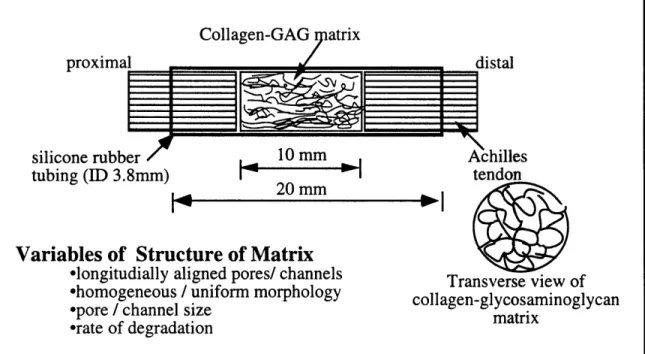
Schematic of a collagen-GAG matrix encased in a silicone rubber tube used in the study of tendon healing...47
Phase diagram of water.. ... 49
Prediction of orientation for typical values of temperature of the freezing bath, T and velocity of immersion, ... 51
Schematic of solidification of CG suspension set-up...54
Schematic of the freezing bath set-up...55
Schematic of frozen CG suspension prior to freezedrying...56
Schematic of collagen-GAG matrix manufacturing steps from beginning (raw material) to end (packaging and storage)...57
Grid of manufacturing conditions investigated...58
Schematic of algorithm of the method of directed secants used in analysis of collagen-GAG matrix pore characteristics...61
Average pore diameter as a function of freezing temperature for three velocities of immersion...62
Average pore diameter as a function of velocity of immersion into freezing bath for three freezing bath temperatures ... 64
ESEM micrographs of a CG matrix with different pore sizes...64
Pore morphology, distribution, and orientation resulting from various combinations of velocity of immersion and coolant bath tem perature.. ... 65
Figure 3.15 Figure 4.1 Figure 4.2 Figure 4.3 Figure 4.4 Figure 4.5 Figure 4.6 Figure 4.7 Figure 4.8 Figure 4.9 Figure 4.10 Figure 4.11 Figure 4.12 Figure 4.13 Figure 4.14 Figure 4.15 Figure 4.16 Figure 4.17 Figure 4.18 Figure 4.19 Figure 4.20
ESEM micrographs of CG matrix with different pore orientation ... 66
Schematic of surgical procedure.. ... 74
Schematic of animal model ... 75
Schematic of implant preparation process...77
Schematic of tissue allocation ... 79
Class I tissue, granulation tissue...87
Class II tissue, loose fibrous tissue...88
Class HI tissue, dense fibrous tissue...89
Class IV tissue, dense fibrous tissue...90
Class V tissue, normal adult tendon...91
Gross morphology of tissue spanning empty tubes at 3 weeks, 6 weeks, and 12 weeks ... 96
Gross morphology of tissue spanning collagen-GAG filled tubes at 3 weeks, 6 weeks, and 12 weeks ... 97
Schematic of the location of tissue mass spanning lesion site for a typical 12 w eek case...98
The average volume of new tissue found in the lesion site as a function of the implant group at the time periods of evaluations ... 98
The average volume of new tissue found in the lesion site as a function of time for all implant groups...99
Percentage of each class filling defect site as a function of time for the em pty control tubes...103
Percentage of each class filling defect site as a function of time for the empty tubes and tubes filled with 60 gm pore diameter CG matrix...104
The percentage of the defect volume filled with granulation tissue (class I) as a function of time for empty tubes and CG-filled (60 tm pore diam eter) tubes...105
The percentage of the defect volume filled with dense fibrous tissue (class II and class IV tissue combined) as a function of time for empty tubes and CG-filled (60 gtm pore diameter) tubes...105
The percentage of the defect volume filled with granulation tissue (class I) as a function of collagen-GAG matrix pore size...106
The percentage of the defect volume filled with dense fibrous tissue (class III and class IV tissue combined) as a function of collagen-G Acollagen-G m atrix pore size...107
Figure 4.21 Light micrograph of normal tendon stained for collagen type I and
collagen type HI ... 108 Figure 4.22 Light micrograph of tissue recovered from two different samples
from empty tubes at 12 weeks, each stained for collagen type I and
collagen type III ... 109 Figure 4.23 Normal adult tendon that stains positive for alpha-smooth muscle
actin ... 110 Figure 4.24 Tissue recovered from empty tubes that stain positive for
alpha-sm ooth m uscle actin... 111 Figure 4.25 Tissue recovered from CG-filled tubes that stain positive for
alpha-sm ooth m uscle actin...112 Figure 4.26 Histogram of collagen fibrils for repair tendon tissue at 12 weeks in
both empty and filled lesion sites ... 113 Figure 4.27 TEM micrographs of normal Achilles tendon and new tendon tissue
at 12 weeks in an empty tube ... 114 Figure 4.28 TEM micrographs of myofibroblasts...114 Figure 5.1 Schematic of cell seeding protocol...134 Figure 5.2 Schematic of allocation of cell seeded collagen-GAG matrix samples
in to different embedding medium...138 Figure 5.3 Schematic of cell density measurement of cell-seeded matrices...139 Figure 5.4 Uniform distribution of tendon cells within a collagen-GAG matrix.
Cells are evenly distributed throughout the matrix and appear
spreadout along the collagen-GAG fibers...142 Figure 5.5 Uneven distribution of tendon cells within a collagen-GAG matrix.
Higher concentration of cells at the surface of the matrix...143 Figure 5.6 Shrinkage of cell-seeded collagen-GAG matrices as a function of
implant incubation time for matrices in culture wells that were
agarose-coated and agitated for the first 20 hours post-seeding...144 Figure 5.7 Light micrographs of culture wells uncoated and coated with a thin
layer of 2% agarose ... 145 Figure 5.8 Number of cells recovered as a function of method of cell seeding
and culturing ... 146 Figure 5.9 Depth of infitration of cells into CG matrices as a function of pore
size and implant seeding and culturing at day one post-seeding...147 Figure 5.10 Light micrographs of collagen-GAG matrices stained for
List of Tables
Table 2.1 Comparison of normal Achilles tendon and healing Achilles tendon (scar)... 32 Table 2.2 Tendon and ligament substitutes...33 Table 2.3 Animal models for study of tendon healing... 37 Table 2.4 Summary of evaluation techniques ... 40-41 Table 3.1 Comparison of average pore diameter for AECM diameters of 3.8 mm
and 1.5 m m ... ... 66 Table 4.1 Table of manufacturing conditions to produce
collagen-glycosaminoglycan matrices of average pore sizes 15 gm, 60 gm , and
120 m ... 76 Table 4.2 Chart of experimental test grid ... 78 Table 4.3 Classification of tissue ... 86 Table 5.1 Table of manufacturing conditions to produce
collagen-glycosaminoglycan matrices of average pore sizes 25 jtm, 60 gm, and
120 tim ... 130 Table 5.2 Cell seeding experimental grid with the cell seeding conditions and the
number of matrices per seeding condition ... 135 Table 5.3 Table of number of cells per square millimeter on the surface of the CG
matrix and in the middle of the matrix as a function of matrix pore size
CHAPTER I: INTRODUCTION
1.1 Statement of the Problem
When tendon is injured with a significant loss of tissue or when a large wound gap is formed, surgical reapposition of the uninjured tendon ends is generally not feasible. If the wound is left untreated either tissue with inferior mechanical properties fills the defect site or, in the worst case, the ends of the tendon are not bridged with any tissue.
1.2 Clinical Significance
Clinically, Achilles tendon injuries can be classified into four groups with increasing severity of the injury:
I Partial to full rupture of the Achilles tendon, II a Gap (0.5 - 1 cm) between the tendon ends,
b Gap (> 1 cm),
III Gap with complications, such as:
a. necrosis of tendon in a chronic situation
b. problems such as additional trauma to surrounding tissue, c. open wound with possibility of infection, and trauma to
surrounding tissue, and IV Absence of tendon.
The work in this thesis is primarily targeted for clinical situations in which the severity of tendon injury is of class IIb and higher, in which surgical apposition of tendon ends is not possible. In clinical situations of class IIb and higher, the use of a graft material, either synthetic or biological in origin, is needed to bridge the gap between the tissue ends. This prosthesis could a) provide a resorbable scaffold in which cells could migrate, proliferate and produce repair tissue that would reconnect the tendon ends [23, 68].or b) serve as a prosthesis to provide a permanent link between to the tendon ends, thus restoring the mechanical integrity of the tissue [105]. Without a prosthesis to bridge the gap between the tendon ends, the tendon ends will not reconnect.
Though the majority of patients with Achilles tendon injury return to their pre-injury level of activity [108], there are several potential complications in Achilles tendon healing. It should be noted, the majority of patients that were able to return to pre-injury activity levels were patients with a less severe injury than those injuries to be addressed in this thesis (tendon injuries of class I or class IIa) [108]. Although there appear to be few clinical statistics on return to activity level of patients with tendon injuries of class lIb and above, it is reasonable to assume that the percentage of patients returning to pre-injury activity levels decreases with severity of the injury.
Complications due to inadequate repair of the tendon can include: tendon ends that do not reconnect or the tissue that bridges the gap is so inferior to the original tissue in mechanical properties as to compromise the function of the tendon. This can lead to either 1) reduced flexibility of the ankle joint or 2) instability of the ankle joint. Since, Achilles tendon is connected to the calcaneous bone (ankle bone) and is one of the tendons responsible for the stability of the ankle joint, the extent of Achilles tendon movement and mechanical integrity of the tendon influences the stability of the ankle joint. Reduced flexibility of the ankle joint can be due to adhesion of the healing tissue to surrounding tissues. This adhesion reduces the movement of the tendon. Reduced flexibility of the ankle joint can also be due to reparative tissue that is contracted, placing the ankle joint in an overly extended position. There is an "optimal" length-tension relationship for the muscle-tendon-bone complex [91]. When this relationship is violated, for example when the tendon is "too short", there is increased tension (referred to as over-tensioning) on the complex which can lead to reduced flexibility. Instability of the ankle joint can be due to either a lack of sufficient tissue bridging the gap or healing tissue with under-tensioning which allows for over extension/ flexion of the ankle joints which leads to instability of the ankle joint. In all cases, the patient's quality of life,
1.3 Animal models for the study of healing of tendon injuries
Most animal models that are used to study the spontaneous healing of tendon injuries involve either partial or full transection of the tendon. Transection results in a spontaneous retraction of the tendon ends and a gap is produced. There have been very few if any studies that investigated the ability of tendon to bridge a large wound gap (i.e., larger than that formed by retraction of the cut ends). Moreover, most models are not capable of studying the intrinsic capability of the tendon to bridge a large defect because the wound site in current models are in contact with the environment surrounding the tendon. An exception is a model developed by Gonzalez [57] in the 1940s in which a polyethylene tube was placed around the lesion site of a flexor tendon in a dog. However, the surgical wound was not a gap. In Gonzalez's model the tendon was transected but the tendon ends were surgically reapposed before the tubing was placed around the wound area. There was, in effect, no gap between the tendon ends.
1.4 Current treatment for large tendon wound gaps
Current clinical treatment of large tendon wounds in which surgical reapposition of the tendon ends is not feasible involve use of a replacement tendon. Tendon grafts are of either biological [56, 68, 69, 126] or synthetic [23, 54, 62, 63, 105] origins. These prostheses are, in general, designed to mimic the mechanical properties of the tendon thereby allowing for earlier remobilization of the patient. Most prostheses are generally comprised of close-packed aligned fibers, either resorbable[23, 56, 68, 126] or permanent [54, 62, 63, 105].
Results from the use of these prostheses have been mixed. At one extreme, Kato et al [68] found a very favorable healing response of the tendon to a collagen-based fiber tow. Tissue that infiltrated and replaced the collagen tow had many morphological and biochemical similarities to normal tendon. In contrast, Amis et al [2] comparing carbon and polyester based tows, found an adverse response to the carbon implant.
1.5 Rationale for using porous analogs of extracellular matrix
The rationale for our use of analogs of extracellular matrix (AECM) to facilitate healing of tendon is based, in part, on the success in implementing such analogs for the regeneration of dermis in animals [129, 130, 132] and human subjects [11, 83, 117] and the reconnection of axons across large gaps in transected peripheral nerves in rats [16, 129]. Moreover, there is an indication that such an approach has also been successful in the regeneration of the knee meniscus [119]. Left untreated, defects of the size treated in these tissues do not heal by regeneration.
1.6 Scope of present research
The effect of an AECM on early tendon healing in a novel animal model was investigated. This AECM was a porous graft copolymer of collagen-chondroitin 6-sulfate (CG). Although the chemistry of the CG copolymer used in this study was similar to CG copolymer grafts used in dermal [123, 130, 132-134, 136] and sciatic nerve [15, 76, 129, 137] studies, CG copolymer implants of a size and configuration required for treating tendon lesions had not been previously fabricated. A protocol for the manufacturing and characterization of AECM implants for tendon treatment was developed.
A new animal model for tendon healing was developed in order to evaluate the CG implants. Criteria for selection of this model included: 1) isolation of lesion site from factors external to the injured tendon and 2) accommodation of the porous CG matrices. That the primary interest of the study was the influence of the CG matrices on the intrinsic capability of the tendon to bridge a wound gap, is why an approach was taken to minimize the effect of external influences. Previous animal models used to study Achilles tendon healing did not satisfy the criterion of isolating the lesion site from external influences.
The criteria for the animal model led to the design of an implant consisting of a CG matrix within a silicone rubber tube. The tendon ends were inserted into the ends of the
tubing. The tube formed a barrier between the lesion site and the external environment. This was the first study of tendon gap healing in a tubular construct.
In this thesis, focus was placed on the early stages of tendon healing, .i.e., the first 12 weeks after injury. Time periods of evaluation were chosen to correspond to the various phases of spontaneous tendon healing as reviewed in section 2.3. Time periods were chosen to coincide with the inflammation phase, the repair phase, and the beginnings of the remodeling phase in an animal model of spontaneous healing [8, 32, 99, 107].
As a first approach, the effect of the CG matrices' pore channel diameter on the extent of tendon healing was chosen for investigation. The extent of dermal [133-135] and sciatic nerve [15, 129, 137] regeneration was found to be a strong function of pore channel diameter of the collagen-GAG AECM.
Techniques were developed to analyze the tissue resulting from the healing process. Quantitative evaluation of the extent of healing was based primarily on the morphological appearances of the cellular and matrix components. Histomorphometric techniques were used to determine the percentage of selected types of tissue present in the lesion site at sacrifice. Immunohistological techniques and transmission electron microscopy techniques were also utilized to evaluate the cellular and matrix contents in the reparative tissue.
Lastly, preliminary studies were undertaken to determine the feasibility of tenocyte cell seeding of the CG matrices. Although seeding of chondrocytes and keratinocytes onto CG matrices of similar chemistry have been documented, seeding of tenocytes (tendon fibroblasts) have not been previously attempted.
CHAPTER II: BACKGROUND
2.1 Review of normal tendon anatomy and function
Anatomy. Tendon is a specialized dense connective tissue that links bone to muscle and allows for the transmission of muscle contraction forces to the bone. Achilles tendon links the triceps surae muscle, the grouping of the gastrocnemius, the soleus and the plantaris muscle, to the calcaneous bone. Tendon consists of three parts: the muscle attachment, the substance of the tendon itself, and the bone attachment (Figure 2.1).
Composition. Morphologically, tendon is composed of highly aligned collagen fibers sparsely interspersed with spindle-shaped cells (fibroblasts) aligned in rows along the direction of the collagen fibers, as seen in a longitudinally derived tissue section (Figure 2.2). When
Gastrocnemius
muscle '
Soleus
muscle
Calcaneous
bone
Achilles
tendon
Figure 2.1. Posterior view of lower extremity in a human. The Achilles tendon connects the soleus and gastrocnemius muscles to the calcaneous bone.
viewed transversely, the cells appear as star-shaped figures among bundles of collagen.
Matrix. The major constituent of tendon is Type I collagen (approximately 86-90% dry
weight), with the rest of the constituents comprised of Type Ill collagen, elastin, proteoglycans and glycosaminoglycans. The collagenous component of tendon is arranged in a hierarchical microstructure. The primary structure of collagen is the sequence of amino acids that make up the collagen chain. The collagen chain possess a high concentration of glycine (33%), proline (15%), and hydroxyproline (15%). The secondary structure relates to the left-hand arrangement of each primary chain and the tertiary structure describes the right-handed triple helical configuration formed from hydrogen and covalent bonding of three collagen chains into a "tropocollagen" molecule (Figure 2.3). For Type I collagen, two of the three chains are identical, called 1(I), and one is slightly different in primary structure, referred to as 2(I).
The quaternary structure encompasses several sublevels. Adjacent collagen molecules (tropocollagens) are arranged in a quarter stagger such that oppositely charged segments are aligned. Five collagen molecules in the staggered configuration form a microfibril. Collagen bundles are arranged in closely packed parallel bundles, oriented in a distinct longitudinal pattern to form ordered units of subfibrils, fibrils, and fascicles. At the fibril unit, a characteristic 64 nm periodic banding is seen by transmission electron microscopy (Figure 2.4).
Adult tendons in many species have a bimodal distribution of collagen fibril diameters [31, 65, 93, 95]. Collagen fibrils in adult human tendon have mean diameters of 60 and 175 nm [31]. Ultrastructural comparison [65] of collagen fibril diameters in various ages of rabbit tendon found that with aging, collagen fibril diameters increased. Collagen fibrils in newborn rabbit tendons were uniform in size and ranged between 18 to 55 nm in diameter with an average diameter of 37 nm; while the collagen fibrils of mature rabbit tendon ranged between 18 to 166 nm with the two most frequently encountered populations of fibril diameters measuring 55 and 92 nm.
2.2. Photomicrograph of longitudinal section of normal rabbit Achilles tendon
showing the spindle-shaped fibroblasts. Hematoxylin and eosin stain. Bar: 10 ýtm. (• rl 7Zone 0.4 0 N4&icvfibrI. B -- llI A !- 11 Ck,"01.iO. C laitlgibn Z - -h •'m-'"104 A• (n IS U CTrip.. "all.
Typica Gline Hydro.yprOlin
s.q.... l I i
Chalin6
41-- - --- -
--Figure 2.3. Schematic representation of the hierarchical structure
microfibril. of collagen to form a
I I I I
Figure 2.4. Schematic representation of the hierarchical architecture of tendon. (Modified from Kastekic et al (1978) [67].
A collection of fibrils bundled together form a fascicle. Fascicles are bound together by loose connective tissue that allows the fascicles to slide past one another. This loose connective tissue region, called the interfascicular membrane or endotenon, also supports the nervous tissue, blood vessels and lymphatics.
Tendons have a crimped, waveform appearance when seen under polarized light microscope (Figure 2.5a). Crimp has been observed at the level of the optical microscope. Diamant et al [22, 27] has shown that the crimp is a planar zigzag pattern which unfolds during initial loading of collagen. The periodicity of the alternating light and dark bands of crimped fibers can be used to determine the crimp angle, crimp length and crimp wavelength. [27, 68]. The crimp is frequently assumed to be a planar zig-zag, although other investigators have also modeled crimp as a sinusoidal waveform [20]. With these assumptions, the crimp wavelength (c) and crimp length (1) can be determined directly under polarized light using a calibrated eyepiece. The crimp angle (0) is calculated as the inverse cosine of the crimp period
divided by twice the length of the crimp. Figure 2.5b illustrates the calculation of crimp parameters from a polarized light image of normal tendon.
15A 35 100200 A 500- 5000 A so- 300p
SIZE SCALE
! I
I I
Figure 2.5.
Figure 2.5.
a) Photomicrograph ot normal rabbit Achilles tendon as seen under polarized light showing the crimp in the collagen fibers of the tendon. The alternating light and dark bands represent the collagen fibers folding into a planar pattern. Bar: 50 im.
b) Diagram illustrating the calculation of crimp parameter. Crimp period (lc) and crimp length (1) are determined directed from the polarized light image. Crimp angle (0) is calculated from the. crimp wavelength and crimp length. Nomenclature adopted from Kato et al [68].
Tendons that generally move in a straight direction, such as Achilles tendon, have a loose areolar connective tissue, the paratenon, which is continuous with the tendon (Figure 2.6). The paratenon stretches several centimeters and recoils without tearing or disrupting tendon blood supply. An interlacing mesh work of thin collagen fibrils, elastic fibers and glycosaminoglycans gives the paratenon this elasticity and extensibility. On the other hand, tendons which bend sharply, such as the flexor tendons of the hand, are enclosed by a tendon sheath (Figure 2.6). The sheath helps to direct the path of tendon movement by acting like a pulley and allows low friction movement between tendon and the adjacent bones and joints. The sliding of these tendons through the sheath is assisted by the presence of synovial fluid between the outer wall of the tendon and the inner wall of the tendon sheath.
Cross-section of Achilles and Flexor Tendon
loose paratenon
tendon
tendon
Achilles tendon is covered by
a loose filmy areolar tissue, the
paratenon. The filmy tissue is
continuous with the tendon
Flexor tendon is surrounded
by a tendon sheath. In general
the tendon is separated from the
sheath by a synovial filled space
Figure 2.6. Schematic of the type of covering surrounding Achilles tendon and flexor tendon. Achilles tendon is surrounded by a loose areolar tissue, paratenon, while the flexor tendon has a tendon sheath. The tendon sheath is separated from the tendon by a synovial filled space.
Cells. Fibroblasts (also called tenocytes) form the predominant cell type in tendon.
Endothelial cells and nerve processes form only a small part of the of the cell population. Ippolito et al [64] has shown that there is a subpopulation of myofibroblast-like cells present in normal tissue. These myofibroblast-like cells are speculated to be involved in a contractile activity [4, 50, 58, 64, 83, 114] This subpopulation of cells has been speculated to be involved in either the "tensioning" of the tendon and/or in the modulation contraction-relaxation of the muscle-tendon complex [64].
Blood supply and innervation. Tendons receive their blood supply from vessels in the
perimysium, the periosteal insertion, and from the surrounding tissue via vessels in the paratenon or mesotenon. In the Achilles tendon and other tendons which are surrounded by a paratenon, vessels enter from many points on the periphery and anastomose with a longitudinal system of capillaries. Blood flow is low, averaging between 0.5 to 1.5 ml per 100 grams of tissue per minute [9, 61] and concentrated mainly on the circumferential surface of the tendon with decreasing blood supply in the interior of the tendon unit. For these reasons, tendon is considered to be a tissue of low vascularity.
Tendon has a complex set of sensory nerve receptors on its surface and throughout the tendon proper. Nerve endings function as pain receptors, vasomotor efferents, and mechanoreceptors. Mechanoreceptors sense joint position, muscle tension, and loads applied to the tendon. The stretch or deformation of a tendon can trigger a reflex muscle contraction or an adjustment in muscle tension. This reflex muscle contraction may play a role in stabilizing and protecting joints from potentially injurious movements.
Mechanical Properties. The main function of a tendon is to transmit tensile forces during muscle contraction. Because its collagen fiber structure is highly aligned in the direction of the tensile force and because collagen is one of the strongest fibrous protein in the body, tendon has one of the highest tensile strengths of any of the soft connective tissues in the body. The stress-strain curve begins with a region call the "toe region" in which the tendon deforms easily without much tensile force (Figure 2.7). This is due to the "uncrimping" of the
crimp structure and the alignment of collagen fibers in the direction of applied stress. The toe region is followed by a linear region in which the slope of the region is referred to as the elastic stiffness, or Young's modulus, of the tendon. At large strains, plastic deformation of the tendon occurs and leads to eventual failure of the tendon. The ultimate tensile strength, the maximum stress level obtained, of tendons varies greatly from 5 to 7 MPa for mouse rat tail tendon [127, 128] to over 100 MPa for human tendon [12]. The ultimate strains, stain at which failure occurs, ranges from 9% to 35% for Achilles tendon.[12]. Achilles tendon is strained between 10 to 12% during normal activities such as walking [12, 127]. Mechanical properties measurements vary greatly with the age of donor, location, species, donor history, and testing procedures.
The mechanical properties of tendon have been specutlated and modeled as a function of the morphology and hierachical structure of the collagen fibers [20, 27, 41, 44, 93, 95] Although there have been many studies investigating the relationship between morphology and structure of collagen fibers in physiological tendon, few studies have investigated the relationship between morphology and return of mechanical properties in healing tendon [26, 41, 44] Frank et al [41] have investigated the possible presence of a correlation between collagen fibril diameter in healing medial collateral ligaments in a rabbit with the mechanical properties of these healing ligaments.
2.2 Tendon injury
Tendon injury occurs as a result of direct trauma, such as laceration or contusion, or indirect trauma such as tensile overload. Direct trauma frequently occurs from accidents involving sharp instruments. Although direct trauma of tendons can occur in any tendon, this is most prevalent in the tendons of the hand. Often, direct trauma of the tendon involves more than just a tendon injury; there can be accompanying nerve, soft tissue and bone damage. The complexity of the injury site complicates treatment.
Ultimate tensile strength
(UTS)
stress
(a)
yield and failure region
I \I/
I 'I~
%/o
strain
aE)
Figure 2.7. Schematic of stress-strain curve for tendon. The toe region represents the uncrimping and alignment of the collagen matrix in the direction of stress. The linear region represents the response of collagen matrix to further elongation. The curve appears linear throughout this region. The beginning of the yield and failure region is the start of matrix failure. Once maximum stress is reached, total failure in rapid.
The extent of tendon healing depends on many factors such as the anatomical location, vascularity, skeletal maturity as well as the magnitude of the mechanical forces applied. In general, failure of a muscle-tendon-bone complex occurs at the weakest link, which in most cases is the junction of the muscle-tendon complex. Indirect trauma to the tendon, itself, usually requires the presence of pre-existing pathology during mechanical overload. The classical case involves a middle-aged, typically male "weekend athlete" engaged in strenuous activities such as basketball or volleyball. The Achilles tendon rupture is often abrupt and accompanied by a popping sensation. Frequently the patient has no prior history of Achilles tendon injury or discomfort. In patients who had no prior history of Achilles tendon discomfort, there were reports of the presence of degenerative tendon tissue [19, 103, 108]
toe region
lear region
present prior to tendon rupture. Microscopic studies of Achilles tendons that have spontaneously ruptured have shown tendon tissue distant from the site of rupture to have undergone a degenerative process. There is generally fragmentation and fraying of collagen bundles and edema of the tendon [19] and a reduction of cell popluation. In some cases, there appears to be a transformation from tenocytes to chondrocyte cells with an accompanying increase in the concentration of matrix mucopolysaccharides [103].
2.3 Spontaneous healing of tendon
Spontaneous healing of tendon has been studied extensively in both Achilles tendon and flexor tendons of the hand. Tendon healing involves the formation of scar, which is different morphologically, biochemically, and biomechanically from physiological tissue. With time, scar tissue assumes some of the characteristics of tendon; however, complete regeneration does not appear to occur. Although both Achilles and flexor tendons respond to injury by forming scar tissue, scarring in the flexor tendon appears to have a more detrimental effect on the function of the tissue. Flexor tendons need the ability to glide within their sheath to function properly and formation of adhesion sites to the sheath during tendon healing interferes with this gliding function.
Healing of a tendon covered with a paratenon (Achilles tendon). The response of Achilles tendon's response to injury follows a sequence similar to that found in other connective tissues such as ligament and skin. This sequence is generally considered to consist of four overlapping phases: injury, inflammation, repair, and remodeling.
Following tendon injury by full transection, there is a spontaneous retraction of the cut tendon ends. This retraction has also been reported to occur in a full-transection rabbit animal model for the medial collateral ligament. In the medial collateral ligament, the retraction of the cut ligament ends produced a gap as large as 2-4 mm [40]. In the Achilles tendon of both a rat and rabbit animal model, the gap formed by the retraction of ends, with the joints held in
neutral position, was observed to be approximately 9-12 mm [8]. Additional retraction of the ends can occur with movement of calcaneous and knee joints.
The response of Achilles tendon to injury has been reviewed in several articles [1, 9, 77, 127] Below is a summary of the response of tendon to a lesion produced by a full transection of tendon as reported by several authors [1, 8, 9, 32, 33, 77, 127].
Injury to the tendon triggers a cascade of events. Immediately after injury, the collagenous matrix is disrupted and tendon and blood cells die [32, 36, 37]. A hemorrhagic exudate fills the lesion site and within minutes, a fibrin clot forms and seals the wound [8]. The clot appears to contains inflammatory products (fibrin, platelets, red cells, and nuclear and matrix debris) [8, 99]. This clot has little tensile strength. These events occur within the first few hours after tendon injury.
The inflammatory stage generally starts within hours of injury and can take from 3 to 10 days to complete. This stage is associated with "clean-up" of the lesion site. Polymorphonuclear neutrophils and lymphocytes, acute inflammatory cells, invade and populate the wound site as early as hours after tendon injury. Monocytes and macrophages appear soon after the appearance of the polymorphonuclear neutrophils and lymphcytes.
The period of dramatic fibroblast migration and proliferation and matrix synthesis, the repair phase, start as early as 10 days and can take up 2 to 5 weeks to complete [8, 57]. Undifferentiated and disorganized fibroblasts containing well-developed endoplasmic reticulum from the wound edge and paratenon begin to proliferate and migrate into the wound site along the fibrin mesh of the clot. Simultaneously, endothelial cells of surrounding vessels enlarge and proliferate forming capillary buds that follow the migrating fibroblasts. Together the fibroblasts, macrophages and capillaries form the granulation tissue in the wound site [32]. This early stage of the repair phase is characterized by increased cellularity. Fibroblasts that have migrated to the wound site continue to proliferate. The fibrin clot is replaced by a collagen bridge. The collagen initially deposited in the wound site is predominantly collagen type III collagen. The collagen fibers are smaller in diameter than the collagen type I to be
deposited in the next stage of healing and are referred to as reticular fibers because of the network-like pattern of their deposition; the collagen type III fibers do not aggregate in a preferential direction the collagen type I fibers. Both collagen production and fibroblast proliferation peak during this phase (characterized as loosely organized fibrous tissue), and subsequently decrease over the next several months [37].
The remodeling phase, which can begin as early as 3 weeks and last for over 1 to 2 years, is marked by a reduction in the production of type Ill collagen and reorganization of the type I collagen fibers [37, 84, 99]. During the remodeling stage, the matrix fibers reorient themselves along the long direction of the tendon. This direction coincides with the direction of tensile stress in the tendon [37, 84, 99]. Figure 2.8 depicts, schematically, the re-orientation of the matrix fibers during the remodeling stage. The remodeling stage is also marked by a decrease in the number of fibroblasts present in the tissue.and a decrease in the overall volume of the scar tissue. The tensile strength of the tendon increases through this period of remodeling even though the total volume is decreasing. This increase has been explained by the reorganization of the collagen fibers, which has been observed to occur during the same period. However, Frank et al [37, 38, 42, 43, 84, 99] have shown that the tensile strength of healing ligament did not appear to return to normal levels even after a year. These data, appropriate to the case of the ligament, can probably be extended to the case of the tendon.
In short, the response of mature Achilles tendon to a injury involving a full transection of the tendon, results in reparative fibrous tissue that lacks the structure of normal tendon. A comparison of normal Achilles tendon and this fibrous "scar" tissue is detailed in section 2.4. There is a process of remodeling of the matrix fibers oriented in random directions to matrix fibers oriented in the longitudinal direction of the tendon, as well as a decrease in the fibroblast cell density , and an approach of the composition to normal biochemical levels. However, tendon scar does not appear to have regenerated to mature adult Achilles tendon [8, 99].
Matrix of I tissu connects to gastrocnemius muscle tendon rund
Matrix alignment at early period of healing (< 4 weeks) Random orientat n of matrix fibers.
calcaneous bone
Matrix alignment at later period of healing (> 4 weeks). Longitudinal orientation of matrix fibers.
Figure 2.8. Schematic of a Achilles tendon wound site (left figure). An enlargement of the wound site at an early time period of healing in which the matrix fibers are oriented randomly (upper right figure). The same wound site at a later time period of healing (remodeling stage) in which the matrix fibers are aligned in the longitudinally direction of the original tendon. This longitudinal direction is also the same direction of the stress experienced by the tendon. (lower right figure).Based on structural data reported in Flynn (1965) and McGaw (1986) [37, 84]. Proximal line of stress Distal
Remodeling
\\
IX
line of stressSource of collagen producing cells. There is controversy surrounding the identity and location of the cells responsible for collagen synthesis during tendon repair, particularly flexor tendon repair. On one side of the controversy is the concept that tendon has the necessary cells to produce collagenous tissue [51, 78, 81], while on the other side, there is the belief that the source of collagen-producing cells is outside of the tendon (i.e., an extrinsic source such as the surrounding tissues or from the tendon sheath) [101, 102]. Others believe that both intrinsic and extrinsic sources of collagen-producing cells contribute to the healing process [80, 106].
In vitro studies [78, 79, 81] have shown that in response to tendon injury, cells within
the tendon had the ability migrated and proliferate into the wound site. In these studies, by six weeks, the injury site appeared to be filled with collagen. In vivo data appears to parallel tthe in vitro studies. Matthews and Richards [53, 82] showed that cells within the rabbit flexor tendon particpated in the wound healing process when the synovial sheath was both not violated and the tendon was mobilized with early controlled passive motion. However, if the tendon sheath was compromised and the tendon was immobilized, cells from external sources (e.g., tendon sheath, blood vessels, and other neighboring tissue) migrated and proliferated into the wound site. Tendon repair, in that case, involved the participation of all surrounding tissue in the healing of the entire wound.
2.4 Normal tendon versus "scar"
Regeneration of healing tissue results in a tissue that is indistinguishable from the original tissue i.e. the new tissue is morphologically, ultrastructurally, biochemically, biomechanically, and functionally indistinguishable from the original tissue. Repair, in the classical use of the term, results in a fibrocollageneous tissue that is distinguishable from the original tissue, and generally referred to as "scar". Many studies have claimed regeneration of tendon [68, 99], but close examination of the studies shows that the tissue in question may appear to fulfill the criteria of regeneration in one area but not in another. In the field of tendon healing, regeneration and repair have, in general, not been clearly distinguished in the
literature. There is accordingly, a lack of consensus on the degree of functional recovery which can be considered acceptable to restore function.
There are many similiarities between normal adult Achilles tendon and tissue formed in a tendon wound site (Table 2.1). Both tissue tend to have highly aligned matrix fibers and a relatively low density of fibroblast cells present in the tissue. Morphologically, differences between these two tissue appear to be the crimp pattern and the average fibril diameter and distribution of the tissue. One year post-injury, Kato et al [68] found the crimp length of the healing tendon in their animal model, was smaller than that of normal tendon. Collagen fibril diameters were also significantly smaller than that of normal tendon [65, 99]. Biomechanically, mechanical properties of tissue formed in a tendon wound site appears to be 40-60% of normal tendon levels [8, 90].
2.5 Techniques for treatment of tendon injuries
Several studies investigated different techniques to facilitate the healing of tendon. Studies have ranged from investigation of the effect of suture technique [72, 124] to investigation of the effect of mechanical loading [8, 34, 40, 53, 62, 86, 89, 120]. on tendon healing. In the cases in which the tendon is absent or the wound site is too large to allow for reapposition of the ends , a tendon replacement is necessary. Several studies have focused on the use of biological replacements [3, 85, 121]., permanent replacements [2, 54, 63, 87, 105, 110]., or bioresorbable replacements[23, 56, 68, 69, 110] (Table 2.2). In this section, I will briefly review some of the current techniques being explored for facilitating tendon healing.
Surgical apposition of tendon ends. Considerable attention has been directed toward methods of suturing tendon lacerations and the effects of continuous passive motion on healing of this tissue. Several techniques of suturing have been proposed to increase the immediate strength of the repair, and to facilitate subsequent healing [72, 124]. Most modern techniques of suturing are variation of a technique devised by Kessler [70]. The challenge in obtaining a
Normal Tendon versus "Scar"
Criteria for
Post-operative
Comparisa
for
tiNraof"scar"time of "scar"
comparison Parameter measured Normal "scar" measurement Reference
Morphological
cells density of cells 1.6 cells / mm^2 2.4 tol2 cells/mm^2 1 year Kato (1991)
alignment/shape of cells "spindle-shaped" "aligned, thin"
matrix alignment of fibers highly aligned random to highly aligned 1 year
crimp* crimp angle: 28°± 50 crimp angle: 25 to 35 ° 1 year Kato (1991)
crimp length: 65 ± 10 pm crimp length: 8 to 12 pm
Ultrastructural
cells presence of es yes (staining more 30 weeks Ippolito (1980)
ces myofibroblasts intense than control) Postacchinni (1978)
range: 180 to 1600nm range: 200 to 400nm 30 weeks Ippolito (1980) matrix collagen fibril diameter distribution: bimodal distribution: unimodal Postacchinni (1978)
Biochemical
total collagen 86.8% dry wt NR
collagen type I > 95% NR
collagen type III <5% NR
GAG content 2.75 mg hexosamine/g
GAG content
dry wt NR
Biomechanical*
Kato (1991)
ultimate tensile strength 36.7 ± 9.4 MPa 40-60% of normaltt 1 year Buck (1953)
Kato (1991)
elastic modulus 184
±
45 MPa Buck(1953)
* parameters defined in section 2.1
tt values for animal models such as rat model NR: no values reported for healing Achilles tendon
Tendon and Ligament Substitutes
Material Autografts Allografts (fixed in glutaraldehyde) Xenograft (fixed in glutaraldehyde) Dacron PTFE* Carbon fiber Woven nylon PGAt fibers Collagen fibers Expanded polytetratiuoroe t Polyglycolic acid. Advantages Physiological structure Initial strength Physiological structure Initial strength Physiological structure Initial strengthVersatility of weaves and shape Low toxicity
Initial strength
Versatility of shape, structure Low toxicity Initial strength Ingrowth potential Initial strength Biocompatibility Initial strength Low toxicity Biocompatibility Initial strength Bioresorption Biocompatibility Initial strength Bioresorption
nyiene (uore- I ex)
Disadvantages Additional morbidity of graft Limited availabilility
Limited availability
Reactivity (glutaraldehyde leaching) Possible immunogenicity
Immunogenicity
Reactivity (glutaraldehyde leaching) Limited ingrowth of new tissue
Limited ingrowth of new tissue
Limited ingrowth fixation strength Reactivity of fibers
Limited ingrowth fixation
Slight reactivity to degradation products
Fibrous encapsulation if degradation rate of prosthesis not controlled
References Tauro (1991); McMaster (1976); Ellingsworth (1986) Jaeger (1987); Park (1985) Bolton (1985); Dunlap (1990) Gleason (1984) Rueger (1986); Shieh (1990) Howard (1985) Kato (1991)
Table
2.2.
Tendon and ligament substitutes. List of materials investigated for use in tendon and ligament substitues.
( Modified from Amadio PC, et al: Tendon and Ligament. In Cohen IK, et al (eds): Wound Healing, 1992. pp 384-395.)
-M
u
suture repair of a tendon injury increases significantly the greater the segmental loss of tissue. Suturing techniques alone are not adequate for the treatment of tendon injuries with a large defect area resulting from segmental loss of tendon tissue.
Mechanical Loading during Tendon Healing. There have been many studies investigating the effect of post-operative mobilization on the healing of tendon injuries [8, 34, 40, 53, 62, 86, 89, 120]. The majority of studies investigated the mobiliation of healing flexor tendon. There is added concern of tendon scar tissue attaching surrounding tissue during healing which limits the gliding ability of the tendon. Limitation in gliding of Achilles tendon is generally not as large a concern.
Prostheses to Replace Tendons. Tendon Grafts. The performance of soft tissue autografts, allografts and xenografts used in treating tendon injuries have been evaluted in both human [10, 74, 109, 122] and animal [3, 85, 121]. Questions relate to the source of the donor graft, antigenicity of grafts, methods of harvesting and preserving grafts for surgery, methods of attachment to the residual tendon fragments, and proper tensioning. These grafts are generally used in situations where there is a desire to have immediate postoperative mobilization. Kato et al [68] used an autogenous tendon graft as a control in their study found that although the autogeneous tendon graft "had been completely filled with neotendon that was characterized by crimped, aligned collagen fibers", the neotendon was not identical to normal Achilles tendon. The "neotendon's" mechanical properties were approximately 60% of normal at 1 year and the histological morphology of the "neotendon" was not identical to normal, particularly, the crimp length was only 20% of normal at 1 year.
Permanent tendon prostheses. The search for a prosthesis to replace tendons and
ligaments has been prompted by the desire to obtain immediate load bearing capability. Most of the efforts have focused on the replacement of ligaments, due to the greater number of overall ligament injuries that occur annually. Of the 33,000 tendon and ligament injuries that occur annually, two thirds were ligament injuries [115]. Prostheses fabricated from
polytetrafluorethylene, polypropylene, and carbon fibers have been investigated in animal and human trials [2, 54, 63, 87, 105, 110]. Problems generally relate to the insertion of the device into bone, proper tensioning, and issues related to the abrasion of the prosthesis against bone at sites at which the prosthesis exits a tunnel through bone. While encouraging results have been reported in the short term [54, 105, 110 Park, 1985 #149], questions about the long-term performance of these devices have limited their use [75].
Bioresorbable tendon implants. Types of bioresorbable prosthesis currently being investigated to facilitate tendon healing include: collagen fibers tows [56, 68, 69] , resorbable fibers tows of dimethyltrimethylene carbonate -trimethylene carbonate copolymer [110], and a composite artificial tendon of poly (2-hydroxyethylmethacrylate)/ poly (caprolactone) blend hydrogel matrix and poly (lactic acid) fibers [23].
Kato and associates [56, 68, 69] have reported the use of a carbodiimide-crosslinked and a glutaraldehyde-crosslinked collagen-fiber prosthesis for the Achilles tendon of rabbits. They found that the healing response of the tendon was affected by the rate of implant degradation. The slower degrading implant (glutaraldehyde-crosslinked) was surrounded by a "capsule of collagenous connective tissue" at twenty weeks and both capsule and implant were still present at one year. Repair tissue infiltrated into the glutaraldehyde-crosslinked implant but the tissue was "not as developed" (was not as aligned and was not crimped) as the carbodiimide-crosslinked implant. The carbodiimide-crosslinked implant was resorbed by 10 weeks and was replaced with "neotendon". This "neotendon" was characterized by "aligned, crimped collagen fiber bundles" as early as at 20 weeks. However, it should be noted that Kato's study also stated that the neotendon produced was "similar, but not identical, to normal tendon", one year after implantation of the prosthesis.
2.6 Animal models used to investigate healing of tendon
Animal models are developed to study the in vivo response of a particular tissue subjected to various conditions. Table 2.3 summarizes some of the types of animals models
have been used to study Achilles and flexor tendon healing. The animal models listed for flexor tendon healing were limited to studies that claimed to have differentiated between intrinsic and extrinsic healing (as defined in section 2.3).
Animals models developed to investigate tendon healing involved a lesion produced either by a simple transverse incision or an excision of the tendon tissue. Models have ranged from simple transections of the tendon to excision of the whole tendon. Animal species used to study tendon healing have included the rat, sheep, rabbit, dog and chicken.
Various immobilization techniques have been employed in the animal model. Techniques include placing a plaster cast on the operated leg [62, 85] or removal of the sciatic nerve[8]. However, in many cases, since a desire to obtain immediate load bearing is an important consideration to many clinicians and patients, immobilization in many of the animal models was not utilized.
None of the tendon animal models, including those used to study intrinsic and extrinsic healing, appeared to be capable of studying tendon healing in isolation from effects due to the presence of other tissues adjacent to the injury (e.g., overlying dermal tissue, tendon sheath, neighboring blood vessels). Although these models may have accurately portrayed the clinical situation, as a model system in which to study the effects of particular test devices on the healing capability of tendon, itself (i.e. the intrinsic healing response), these models did not appear to conclusively differentiate between healing response due to the test implants and the response due to external influences. A better animal model may be one that could physically isolate the lesion site from the external environment. This would allow for a controlled, isolated environment in which to test the response of the tendon to (almost) solely the input devices. An introduction of external factors could, in the future, be introduced by choosing an appropriate lesion barrier that would allow for selected factors to move into and out of the lesion site.
Animal M~odels for Study of Tendon Healing
I.Achilles tendon
Flexor tendon
Animal
rabbit
sheep
rat
dog
chicken
Lesion (size of lesion)
transection
excision of proximal third excision of 1.5 cm excision of 2.0 cm midsection
excision of 3 cm excision of (2.5 to 4 cm)
transection
transection (sheath & tendon) transection (sheath & tendon)
50% of tissue removed
Immobilization method
(time)
None ? (45 days) None Nonelong plaster cast (6 weeks) None
plaster (6 weeks) none ? (7 days) or
removal of sciatic nerve or removal of tibia and fibula external fixation knee & ankle
(12 days)
shoulder -spica cast or controlled passive motion
none ? (throughout post-op)
Reference
Postacchinni (1978) Davis (1991) Aragona (1981); Shieh (1990) Tauro (1991) McMaster (1976) Kato (1991) Howard (1985) Rueger (1986) Buck (1953) Murrell (1994) Gelberman (1983) Garner (1989) Joyce (1992)Table 2.3.
The various types of animal models used to investigate tendon healing.
Under immobilization method,
"?"
means that the author indicated that
occur but did not state the method of immobilization.
immobilization did
AnialModlsforStdvof enon eain
2.7 Structural and functional techniques for evaluation of tendon healing
Evaluation techniques utilized to study tendon healing can be classified into: structural and functional techniques. Structural techniques have included those at the morphological, ultrastructural and biochemical level. "Functional" techniques have included biomechanical testing and gait analysis. Table 2.4 summarizes the types of evaluation techniques employed in tendon healing and the parameters that were evaluated. Below are descriptions of the aforementioned techniques.
Structural Techniques. Structural evaluation of healing tendon, particularly morphological/ histological evaluation, is the oldest and most widely used technique to evaluate the extent of tendon healing. Knowledge of histological and morphological parameters, whether qualitative or quantitative, provides investigators with information to allow them to extrapolate the progression and extent of healing. Morphological evaluation is primarily histology-based (i.e. hematoxylin and eosin, trichrome staining), although new techniques in immunohistochemistry are being developed and adapted for tendon studies. Ultrastructural evaluation allows observation at the cellular and subcellular level. Since to a certain extent, the morphology/ function of the cells and matrix at the ultrastructural level is a determinant of ultimate "gross" tissue properties and morphologies, ultrastructural evaluation is an important tool. Biochemical analysis provides quantitative information about the chemical composition of the tissue.
Morphological evaluation. Historically, the extent of tendon healing was evaluated
qualitatively by comparing histological sections of repair tissue to normal tendon [8, 52, 53, 68, 71, 99, 116]. This subjective method of evaluation provides useful information on the qualitative histomorphology of the cellular and matrix components in the tissue. The major drawback of a qualitative evaluation is it subjectiveness. One investigator's criteria are not necessarily another's. This leads to a technique that lacks objectivity and reproducibility.
Histomorphometry, the term used to describe the variety of methods for measuring morphological features in histological images, is steadily being adapted for use, particularly in