REVIEWS OF INFECTIOUS DISEASES • VOL. 3, NO. I • JANUARY-FEBRUARY 1981 ©1981 by the University of Chicago 0162-0886/81/0301-0011$01.09
Intermittent or Continuous Therapy of Experimental Meningitis Due to
Streptococcus pneumoniae
in Rabbits: Preliminary Observations on the
Postantibiotic Effect in Vivo
Merle A. Sande,*Oksana M. Korzeniowski,t George M. Allegro, Robert O. Brennan,t Otokar Zak,§and W. Michael ScheId
From the Department of Internal Medicine, University of Virginia School of Medicine, Charlottesville, Virginia; and Ciba-Geigy, Basel, Switzerland The relative effectiveness of bolus vs. constant intravenous administration of equivalent
doses of penicillin G in killing bacteria in vivo was studied in a rabbit model of meningi-tis due toStreptococcus pneumoniae. Samples of cerebrospinal fluid (CSF) and serum
were obtained from 30 rabbits at intervals of~8hr after treatment for determination of antibiotic concentrations and titers of viable bacteria in the CSF. When penicillin G was given by continuous infusion(105 units/hr after an initial lOS-unit loading dose), con-centrations of drug in serum and CSF reached a steady state in 1 hr. With intermittent bolus administration of 4 x 10⁵units every 4 hr, higher peak and lower trough concen-trations were achieved, and these concenconcen-trations paralleled those in the CSF. Although an initial acceleration in bactericidal rate was observed with the bolus infusion between the first and second hour of therapy, after the second hour the rate of bacterial killing was identical for the two methods of administration. The duration of therapy required for sterilization of the CSF was dependent only on the bacterial count before treatment and not on the mode of drug administration. The effect of single bolus intravenous ad-ministration of ampicillin was examined in experimental pneumococcal meningitis. Am-picillin was given at various dosages (3.25-62.5 mg/kg), and frequent samples of CSF were obtained for determination of concentrations of pneumococci and ampicillin. A long postantibiotic effect was observed in the CSF of all animals, and this effect consis-tently was longer than that observed in vitro.
The question of the most effective mode of admin-istration of (3-lactam antibiotics for treatment of severe bacterial infections is controversial. Com-monly used regimens are largely empirical and are based on pharmacokinetic parameters (often only available from studies done with normal volun-teers), knowledge of susceptibility characteristics of the suspected pathogen, and tradition. Paren-teral therapy is preferred, but unpredictable ab-sorption (e.g., shock) and clotting abnormalities may make the im route unreliable. Continuous iv administration produces a relatively fixed concen-tration of the antibiotic over time. Intermittent, or bolus, administration produces high initial
con-Please address requests for reprints to Dr. W. Michael Scheid, Box 385, Division of Infectious Diseases, University of Virginia Medical Center, Charlottesville, Virginia 22908.
·Present address: Department of Medicine, University of California, San Francisco, San Francisco General Hospital, Room 4-H22, San Francisco, California 94101.
tPresent address: Department of Internal Medicine, Medical College of Pennsylvania, Philadelphia, Pennsylvania 19104.
tPresent address: Department of Internal Medicine, Johns Hopkins School of Medicine, Baltimore, Maryland 21233.
§Present addess: Ciba-Geigy, Basel, Switzerland.
98
centrations in serum that are followed by a rapid fall in levels of drug. Both intermittent and contin-ous modes of administration have their advocates, whose opinions usually are based on studies that examined the effect of the mode of antibiotic ad-ministration on survival of animals with experi-mental infections [1-3]. Other studies have ap-proached this problem by examining the dynamics of penetration of antibiotics into restricted sites as a function of the mode of drug administration [4, 5]. None of these investigations have examined the kinetics of bacterial stasis and killing, factors that might influence the rate of response to therapy.
Both types of regimens usually are adjusted to achieve concentrations of drug in serum above the MIC for the duration of therapy. However, clini-cal experience suggests that administration of these large doses may not be necessary. Pneumo-coccal pneumonia can be cured with only 8
x
103 to 1.2 X 10⁴ units of penicillin G given every 12 hr[6], despite concentrations of drug in tissue below the MIC. Cure of scarlet fever has occurred when penicillin was detectable in the serum for only 6 hr per day [7]. Eagle et al. [1, 2, 8] eradicated
experi-levels in tissue below the MIC during each treat-ment interval; they ascribed this result to a persis-tent effect of the drug on the organism, a phenom-enon described by Parker et al. more than 30 years ago [9, 10]. The postantibiotic effect has been clarified further by in vitro studies [11, 12], in
which short-term exposure of Staphylococcus
aureusorEscherichia colito various antibiotics at or above the MIC produced a continued postanti-biotic effect (decreased or stable bacterial counts) for various periods of time after removal of the antibiotic. Even subinhibitory levels of antibiotics can alter the growth rate and morphology of
bac-teria [13] and may modify important host defense
mechanisms [14, 15]. Previous exposure ofE. coli
to sub-MIC levels of chloramphenicol resulted in a prolonged decline in bacterial concentrations after exposure of the culture to leukocytes, antibody, and complement in vitro [12]. Thus, in vivo, con-tact of the organism with subtherapeutic concen-trations of drug may potentiate host defenses and playa role in outcome of therapy. However, these kinetic concepts have not as yet been demon-strated in vivo.
We have used a rabbit model of pneumococcal meningitis to study these problems. The purposes
of our investigations were (1) to compare the
ef-fect of continuous vs. intermittent iv administra-tion of penicillin on the kinetics of drug concen-tration and the rate of bacterial killing in the CSF
in vivo, and(2)to examine the sequence and
dura-tion of bactericidal and bacteriostatic effects in vivo produced by a single bolus infusion of ampi-cillin.
Materials and Methods
Streptococcus pneumoniae. A strain of S.
pneumoniae type 3 originally isolated from the blood and CSF of an adult with meningitis and previously characterized in our laboratory [16] was used. The strain was grown (from the same stock culture on glass beads) in trypticase soy broth (TSB; Difco Laboratories, Detroit, Mich.)
supplemented with 5070 defibrinated sheep blood
for 18 hr at 37 C in an atmosphere of 10070
COr 90OJo air. Before inoculation the culture was centrifuged at 200 g. for 5 min for removal of erythrocytes, centrifuged again at 2,000 g for 15
min, washed twice in0.9070 NaCI, and suspended
an inoculum of 107 cfu, which was verified for
each experiment by quantitative plating on tryp-ticase soy agar (TSA) pour plates.
In vitro studies The MICs and MBCs of peni-cillin and ampipeni-cillin were determined by a micro-titer method in heart infusion broth with inocula of 104 or 106 cfu [16, 17].
In addition, the effects of in vitro exposure of the organism to ampicillin for short periods were examined by quantitative "time-kill" methods. S.
pneumoniae type 3 was grown for 18 hr in TSB
plus 5070 sheep blood at 37 C in 10070 CO2-90OJo
air. This culture was diluted 10-4 , transferred to fresh media, and incubated under identical condi-tions for 5 hr until the culture reached log phase. A 10-1dilution (1.0 ml) of this culture was divided
into five 25-ml cotton-stoppered flasks, each con-taining 10 ml of fresh media. After incubation for 1 hr, quantitative titers of bacteria were measured by 10-fold dilutions on TSA pour plates supple-mented with sheep blood. Standardized ampicillin powder (Wyeth Laboratories, Philadelphia, Pa.)
was dissolved in 0.9070 NaCI and added to the
flasks; the final concentrations of drug were 0.01, 0.1, 1.0, and 10 IAg/ml. One flask contained no an-tibiotic and served as a control. After administra-tion of ampicillin, all flasks were incubated at 37 C in 10070 COr 90OJo air with continuous shaking for 2 hr. Antibiotic was then removed by
cen-trifugation (2,000 g for 10 min); the pelleted
bacteria were washed three times with 10 ml of
0.9070 NaCI and suspended in fresh TSB contain-ing sheep blood. Bacterial concentrations were counted with use of quantitative pour plates at this time, then every hour for 3 hr, and every 2 hr for 18 hr.
Rabbit model of meningitis. Pneumococcal meningitis was produced in New Zealand white rabbits or Russian hares weighing 2-3 kg as de-scribed previously [16, 17]. Anesthesia was in-duced with 30 mg of sodium pentobartital (Barber Veterinary Supply Co., Richmond, Va.) adminis-tered iv, and a dental acrylic "helmet" was at-tached to the animal's skull to facilitate immobil-ization in a stereotaxic frame. Several days later, the animals were anesthetized again, placed in the stereotaxic frame, and inoculated intracisternally
with 107 cfu of S. pneumoniae via a Quincke
spinal needle (25 gauge
x
3 Y2 in) held in a geared electrode introducer. All therapeutic experiments100
were performed 16-18 hr after inoculation when the rabbits had meningitis, as manifested by fever
(temperature, >39.6 C), lethargy, and other
neurologic signs: cerebrospinal fluid (CSF)
pleocytosis (~95070 polymorphonuclear leukocytes
[PMNs]) and bacterial titers in CSF that ranged from 104 to >108 cfu/ml. Only animals with
con-centrations of bacteria in CSF of >104 cfu/ml
(~95OJoof all inoculated rabbits) were included for analysis. Ifuntreated, this degree of meningitis is uniformly fatal within 24-36 hr.
Experimental design: intermittent vs. continu-ous penicillin therapy. After reinduction of light anesthesia with sodium pentobarbital, indwelling femoral arterial and venous catheters (Intra-medic® polyethyene tubing 7420, Clay-Adams, Parsippany, N.J.) were placed, and the animals positioned in the stereotaxic frame. Two groups of 15 animals each received iv penicillin G (Bristol Laboratories, Syracuse, N. Y.) by the intermittent or continuous mode of administration. The
inter-mittent group received 4
x
105 units given as abolus over 2 min; two such doses were given, 4 hr apart. The animals in the continuously treated group were treated iv with an initial loading dose of 105 units, followed by 105 units/hr given via a
syringe infusion pump (model 352; Sage Instru-ments, White Plains, N.Y.) for the 8 hr of obser-vation. Samples of serum and CSF were taken si-multaneously at 0, 1, 2, 4, 5, 6, and 8 hr of ther-apy. CSF was withdrawn in 0.3-ml aliquots, of which 0.2 ml was used for quantitative culture and 0.1 ml for determination of the penicillin concen-tration. Samples for culture were treated with penicillinase (5,000 units/ml of CSF) for inactiva-tion of the residual penicillin and either were plated directly without dilution (0.1 ml) or were serially diluted and quantitatively titrated on TSA blood agar pour plates, which were incubated in 10070 CO r 90OJo air at 37 C for 24 hr. Serum samples, in 1.0-ml aliquots from the femoral ar-tery catheter, were used for determinations of the penicillin concentration.
Single-dose ampicillin therapy. Procedures identical to those described above were followed in these experiments, except that ampicillin was used instead of penicillin and the drug was given as a single iv bolus in one of the following dos-ages: 3.25, 6.5, 12.5, 25, 37.5, or 62.5 mg/kg. Samples of CSF and serum were withdrawn for determination of quantitative bacterial counts and
Sandeet al.
ampicillin concentrations at intervals of 0.25, 0.5, 0.75, 1, 1.5,2,2.5,3,4,6, and 8 hr after adminis-tration of ampicillin and at frequent intervals thereafter for up to 24 hr or until the animal died, whichever occurred first.
Antibiotic assay. Concentrations of penicillin and ampicillin were determined by an agar well diffusion technique [18]. Assay plates (diameter,
105 mm) were prepared by the addition of a
Bacil-lus subtilisspore suspension (0.2 ml) to 100 ml of
antibiotic medium no. 1 (Difco). Wells of rvO.6
x
0.6 cm were cut and filled with rvO.03 ml of serum or CSF. All samples and standards were tested in triplicate. Serum standards consisted of known concentrations of penicillin or ampicillin dissolved in pooled rabbit serum. CSF standards were
prepared in0.9070 NaCl since identical zones were
observed for standards prepared in uninfected
rabbit CSF, infected rabbit CSF, or 0.9070 NaCI.
Infected rabbit CSF did not produce a zone of in-hibition in this assay. The smallest concentration of drug that could be measured accurately was 0.1 jAg/mI.
Results
In vitro susceptibility. All MICs and MBCs of penicillin were 0.06 jAg/ml for the test strain at
both inocula examined, i.e., 104 or 106 cfu/mI.
Corresponding MICs and MBCs of ampicillin
were 0.06-0.08 jAg/ml at 104 cfu/ml and 0.1-0.125
jAg/ml at 106 cfu/mI.
(1) Intermittent vs. continuous penicillin ther-apy. Serum and CSF penicillin concentrations.
The concentrations of penicillin in the serum and CSF clearly reflect the mode of drug administra-tion (figure 1). With continuous iv infusion, a rel-atively steady-state concentration of penicillin in the serum was achieved 1 hr after therapy began.
The mean(± SD) levels of penicillin were 12.8 ±
1.8 jAg/ml at 2 hr, 13.9± 1.4 jAg/ml at 4 hr, 12.5 ± 2.0 jAg/ml at 6 hr, and 7.9± 1.7 jAg/ml at 8 hr; the overall mean was 11.8 jAg/mI. Administration of penicillin by intermittent (bolus) infusion
pro-duced mean peak levels of drug in serum of 9.9±
2.5 jAg/ml and 19.6± 4.1 jAg/ml at 1 and 5 hr,
re-spectively, and mean trough levels of 0.7 ± 0.2
jAg/ml and 1.8 ± 0.6 jAg/ml at 4 and 8 hr,
respec-tively.
The concentration of penicillin in CSF par-alleled the concentration in serum (figure 1). A
rel-20 OQl-_..._...L.._...r-_...:lI: - - - ' ' - - _ . l . . - _..._ - ' CEREBROSPINAL FLUID MEAN CONCENTRATION 10 (ug/ml) 2 1
h..
.'.
...•... ca_----L_---JL.-_J..._....L..-_...L.._...r- ----L_---' MEAN CONCENTRATION (Ug/ml )Figure 1. Mean concentration of penicillin in the serum and cerebro-spinal fluid of rabbits with pneumo-coccal meningitis during therapy with equivalent doses of penicillin by con-tinuous (0 - - - -0) or intermittent
(. .) iv infusion. Bars
repre-sentSE.
o
2 3 4 5 6 7 8DURATION OF THERAPY (HRS.) ative steady-state concentration of penicillin was
seen in CSF of animals receiving continuous infu-sions. The mean levels of penicillin were as fol-lows: 0.6±0.2IAg/ml at 2 hr, 0.4±0.07 IAg/ml at4 hr, 0.3 ± 0.07 IAg/ml at 6 hr, and 0.4 ± 0.08 IAg/ml at 8 hr; the overall mean was 0.4 IAg/ml. The peak CSF concentrations of penicillin achieved after bolus infusion exceeded the highest levels obtained with continuous infusion. At 1 and 5 hr of treat-ment, the mean levels of penicillin in CSF were
0.96±0.20 IAg/ml and 1.79 ± 0.67 IAg/ml,
respec-tively. Mean trough concentrations of penicillin in
the CSF were 0.12 ± O.OllAg/ml at4hr and 0.23 ±
0.07 IAg/ml at 8 hr. Eighteen of 28 individual sam-ples of CSF from animals receiving bolus infu-sions had trough levels of penicillin of <0.06 IAg/ml as compared with none of 29 samples taken at the same time period from rabbits receiv-ing the continuous infusion. Thus, higher peak but lower trough levels of penicillin were seen in the CSF of bolus-treated animals.
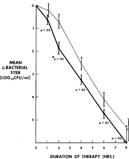
Bactericidal activit)'. in vivo. The mean change in titer of bacteria in CSF of animals receiving either continuous or intermittent infusions of
penicillin were examined at 1- and 2-hr intervals (figure 2). Before the start of therapy, the mean bacterial titers in CSF did not differ between ex-perimental groups. Mean concentrations of 105.25
± 1.06 cfu/ml and 105.27 ± 1.21 cfu/ml were
found for the continuous and intermittent groups, respectively(P>0.1). With the initiation of ther-apy, a rapid bactericidal effect was seen in each group. This bactericidal effect was equivalent at all time periods except for the interval between the first and second hour of treatment. In the first hour of treatment, the mean decline in bacterial titers in CSF was 10°·20 ± 0.29 cfu/ml for the
con-tinuous group and 10°· 06 ± 0.17 for the
intermit-tent group(P>0.1). However, between 1 and 2 hr of treatment, the mean bacterial titer of the
con-tinuous group had decreased by only 10°·48 ±0.31
cfu/ml, whereas the mean titer of the intermittent
or bolus group was reduced by 101.25 ± 0.26
cfu/ml(P< 0.05). After the second hour of treat-ment, the magnitude of bacterial killing was the same for both modes of drug administration. In each group, mean titers of bacteria in CSF de-creased by 1"\.11.5 logs during every 2-hr interval.
102
o
2 MEAN f).BACTERIAL TITER 3 (LOG1oCFU/ml) 4 5 Sondeet01.Figure 2. Magnitude of bacterial killing in the cerebrospinal fluid of rabbits with pneumococcal menin-gitis during therapy with equiva-lent doses of penicillin by continuous (0----0)or intermittent (. . )
iv infusion.
o
2 3 4 5 6 7 8DURATION OF THERAPY (HRS.)
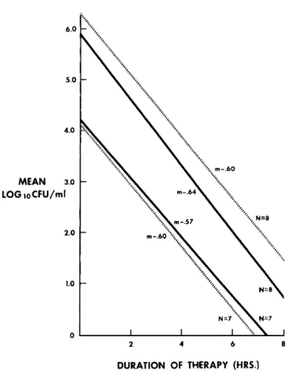
Rate of bacterial killing. The rate of bacterial killing in the CSF for the two experimental groups was compared by least-squares regression analysis (figure 3). Both experimental groups showed a logarithmic first-order diminution in bacterial titer. The slopes of the lines representing continu-ous and intermittent modes of treatment were
- 0.613 and - 0.612, respectively, and thus nearly
were identical(P
>
0.05).The effect of the initial (before therapy) bac-terial titer in the CSF on the rate of bacbac-terial kill-ing was examined (figure 4). The 30 infected ex-perimental animals were divided arbitrarily into two groups: those with initial titers of~105cfu/ml and those with titers of <105 cfu/ml. The rates of bacterial killing produced by the intermittent or continuous mode of penicillin administration were compared for each group by least-squares regres-sion analysis. Sixteen animals had initial titers of
~105cfu/ml. Eight received continuous and eight,
intermittent penicillin therapy. Fourteen animals
had bacterial titers in CSF of <105 cfu/ml, and seven received either continuous or intermittent infusion, respectively. Again, a logarithmic first-order diminution in titer was seen in all four
groups, and the slopes (m) of these lines did not
differ statistically. Thus, the initial bacterial titer did not influence the rate of bacterial killing in this experimental model.
It is also evident that, whatever the mode of
drug administration, 8 hr of therapy was ade-quate to sterilize the CSF of animals with initial bacterial titers of <105 cfu/ml. At equivalent rates of killing, animals with higher titers of bacteria in the CSF would therefore require a longer duration of treatment (extrapolated to a mean of 9-10 hr) in order to achieve sterility. This fact was con-firmed when the therapeutic outcome was ex-amined.
Outcome of therapy. Administration of penicillin for 8 hr by either the continuous or in-termittent mode resulted in similar outcomes
8 7 6 5 4 3 2 o 1.0 2.0 3.0 4.0 5.0 ..•••••.••••••.•••••..••. '" '" '"" '..,'... ... " ...•...•... "" " ... '. "" N=15 ... ..•.•,... ". MEAN LOG 10CFU/ml
Figure 3. Rate of bacterial killing in the cerebrospinal fluid of rabbits with pneumococcal meningitis during therapy with equivalent doses of pen-icillin by continuous (0----0)or in-termittent (. .) iv infusion.
Values were obtained by least-squares regression analysis.
DURATION OF THERAPY (HRS.)
(table 1). Ten of 15 rabbits receiving continuous infusions of penicillin as well as 11 of 15 rabbits treated with intermittent boluses of penicillin had sterile CSF after 8 hr of treatment. Within these groups all the animals with CSF bacterial titers of <105 cfu/ml before treatment had a sterile CSF after 8 hr. In contrast, of those animals with CSF titers ~105 cfu/ml, only three of eight receiving continuous therapy and four of eight receiving in-termittent treatment had sterile CSF after 8 hr of treatment. Thus, the initial bacterial titer, rather than the mode of drug administration, determined the outcome of therapy within the experimental period.
Four partially treated animals (e.g, continuous administration of penicillin for 4 hr; 105 units/hr) were allowed to relapse and succumbed to infec-tion within 24 hr. Thus, no spontaneous clearing of meningitis occurred.
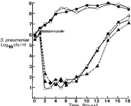
(2) Single-dose ampicillin therapy. Quantita-tive "time-kill" experiments in vitro. Exposure of
S. pneumoniae to superinhibitory concentrations of ampicillin (0.1, 1.0, or 10 IAg/ml) for 2 hr resulted in a reproducible postantibiotic effect (figure 5). This effect was reflected by the lag phase after removal of ampicillin from the flasks by centrifugation during which time no bacterial multiplication was demonstrated. This interval, defined as the time necessary to reach a bacterial concentration 1 log/ml greater than that observed at the end of centrifugation was 6.5, 6.7, and 7.6 hr for 0.1, 1.0, and 10 lAg of ampicillin/ml, respec-tively. A maximal effect was apparent at ampicil-lin concentrations that exceeded the MIC. In con-trast, when the organism was exposed to 0.01 lAg of ampicillin/ml for 2 hr, a concentration well below the MIC of 0.08IAg/ml, bacterial concentra-tions increased steadily and were indistinguishable from those observed in control flasks without an-tibiotic (figure 5). In the presence of 0.01 lAg of ampicillin/ml, bacterial titers increased by 2-3 logs in the first 4-6 hr and then remained stable at
104 6.0 5.0 4.0 MEAN 3.0 LOG loCFU/ml 2.0 1.0 Sandeet al.
Figure 4. Rate of bacterial killing in CSF of rabbits with pneumococcal meningitis during therapy with equiv-alent doses of penicillin by con-tinuous (0----0) or intermittent ( • .) iv infusion.
Experimen-tal animals were grouped by pretreat-ment bacterial titers in CSF of >10' cfu/ml (mean for continuous group, 106
.0' ± 0.97 cfu/ml; mean for
in-termittent group, 1Os.86 ± 1.12 cfu/ml) or of <lOS cfu/ml (mean for continuous group, 104.37 ± 0.77; mean for intermittent group, 104.56 ±
0.27 cfu/ml).
O'-- ... --JL..- ..._ - ' - - . . a . . . ~
2 4 6 8
DURATION OF THERAPY (HRS.)
Table 1. Results of penicillin treatment (8 hr) of ex-perimental meningitis due to Streptococcus pneumoniae in rabbits: comparison of intermittent vs. continuous administration.
tion of bacteria in the CSF increased in all control (untreated) animals; mean increases of 0.16 and 2.11 logs were observed after 8 and 24 hr, respec-tively.
A postantibiotic effect, defined as a continued cidal and static period after the concentration of
NOTE. CSF = cerebrospinal fluid.
108 and 109 cfu/ml for the duration of the ex-periments.
Despite this prolonged postantibiotic effect, all flasks demonstrated an increase in bacterial con-centrations and were equivalent to controls by 10-12 hr after removal of the drug. In contrast, when flasks containing S. pneumoniae were con-tinuously incubated with stable superinhibitory concentrations of ampicillin (0.1, 1.0, or 10 IAg/ml), all cultures were sterile by 18 hr.
In vivo. After induction of pneumococcal meningitis in rabbits by procedures identical with those utilized for the penicillin experiments, a series of animals were treated with a single iv bolus of ampicillin at dosages that varied over a wide range (3.25-62.5 mg/kg). These dosages were chosen to achieve measurable peak concentrations of ampicillin in CSF that were below, approxi-mately equal to, and well above the MIC for the test strain ("-'0.1 IAg/ml). Bacterial titers declined in all treated rabbits. In contrast, the
concentra-Rabbits
No. with sterile CSF after treatment/total no. No. with sterile CSF after
treatment and Initial bacterial titer
<10' cfu/ml Initial bacterial titer
~1O'cfu/ml Continuous 10/15 7/7 3/8 Intermittent 11/15 7/7 4/8
Figure 5. Concentration (loglocful
ml) ofStreptococcus pneumoniae vs.
time before, during, and after ex-posure to ampicillin in vitro. Ex-posure interval (1-3 hr) is indicated by the cross-hatched bar(upper left).
Concentrations of ampicillin(j..tg/ml)
are as follows: (0---0)
=
control (no ampicillin);(e e)=
0.01; (e ----e)=
0.1;(.&----.&)=
1.0; and(~) = 10. 8 7 6 S. pneumoniae LOQ10cfu/ml 5 4 1o
2 4 6 8 10 12 14 16 18 Time (hours)ampicillin in CSF had fallen below 0.1 Jig/ml, was observed in all animals (figure 6). A shorter modi-fied effect (total time, 6-8 hr after infusion) also was observed in two animals in which all measured ampicillin concentrations were~0.1 Jig/ml (figure 6). Thus even subinhibitory concentrations of am-picillin produced a clear cidal-static effect in vivo. The duration of this postantibiotic effect was variable. An initial rapid decline in bacterial titer was observed in all animals; the decline was fol-lowed by a static phase prior to the resumption of bacterial multiplication (figure 6). For purposes of comparison, the postantibiotic effect was defined as the interval from the time when concentrations of ampicillin in CSF first dropped below 0.1 Jig/ml (the MIC of the test strain) until bacterial titers in CSF were loglo 1.0 cfu/ml higher than the bacterial concentration achieved at the nadir of the curve. The duration of the initial decline in bacterial titer was variable (1-6 hr) and did not correlate with peak concentrations of ampicillin in CSF. The second, static phase also varied so that the total duration of the postantibiotic effect was
6-12 hr. Two of three animals that received the highest dose of ampicillin(62.5 mg/kg) had sterile CSF after4-6 hr and were presumed "cured".
Discussion
This study examined two areas of potential
impor-tance in the therapy of bacterial meningitis:(1)the influence of mode of drug administration (con-tinuous vs. intermittent bolus) on antibiotic phar-macokinetics within the CSF, bactericidal activity, kinetics of bacterial killing in vivo, and therapeu-tic outcome in experimental pneumococcal men-ingitis; and (2) the influence of a short duration of exposure to ampicillin of bacteria in the CSF on the development of a postantibiotic effect in vivo. When equivalent therapeutic doses of penicillin
S. pneu- 5 moniae Log10 cfu/ml 3 2
Figure 6. Counts of bacteria (ordinate) in two repre-sentative animals with experimental pneumococcal men-ingitis treated with a single dose of ampicillin plotted vs. time after injection (abscissa). The area under the curve for concentration of ampicillin in cerebrospinal fluid varied;(0---0) =
<
0.1 j..tg/ml·hr;( e - - e ) = 0.7106
were employed, the results in this experimental model were essentially identical for both continu-ous and intermittent therapy. A postantibiotic ef-fect in vivo was demonstrated when the single bolus of ampicillin was given; this effect also was present after exposure to near or subinhibitory concentrations of ampicillin.
The choice of the most effective mode of anti-biotic administration in serious infections has been a subject of continuous debate. In studies of mice with streptococcal peritoneal infections, where survival was used as a measure of treatment outcome, the avoidance of long periods of subin-hibitory concentrations of penicillin in the serum appeared critical [8]. Although the actual mode of drug administration was irrelevant in this study, the findings tended to support advocates of con-tinuous drug administration.
Arguments also have been made in favor of in-termittent iv injections of antibiotic, especially in the treatment of bacterial endocarditis [19]. The resultant higher levels of drug achieved in blood with this mode of administration theoretically would allow increased diffusion of the antimicro-bial agent into an infected vegetation. Recently, Barza et al. observed an increased accumulation of ampicillin within sc fibrin clots in rabbits after intermittent as compared to continuous administra-tion [4]. In addiadministra-tion, it has been postulated that brief periods of low concentrations of antibiotic might permit bacterial multiplication and, there-by, enhance the susceptibility of the remaining bacteria to antimicrobial action [20, 21].
This controversy has particular relevance in the treatment of meningitis. The blood-brain barrier limits the entry of most antimicrobial agents into the CSF [22]. Even in the presence of inflamed meninges, when a generalized nonspecific increase in membrane permeability allows enhanced drug penetration [23], the therapeutic margin may be small.
Penetration of penicillin into the CSF and other foci of infection has been studied since the intro-duction of this antimicrobial agent into clinical use [24]. The chemical characteristics of penicillin are major factors limiting its entry into the CSF [25]. Protein binding, acidity, ionization, and lack of a transport system result in slow diffusion across the leptomeninges. Concentration of pen-icillin in the CSF is further limited by the exis-tence of an active transport process that pumps
Sandeet a/.
penicillin out of the CSF and into the serum [26]. Thus the cornerstone of treatment of infections in the CSF has been the parenteral administration of large doses of antibiotic to achieve maximal serum concentrations that enhance diffusion into the in-fected site [27].
Debate has centered, however, on which mode of administration (continuous infusion or inter-mittent bolus injection) is most effective in pro-ducing high concentrations of antibiotic in CSF and a rapid bactericidal effect in vivo. Plorde et al. [5] examined the kinetics of penicillin pene-tration through uninflamed and inflamed men-inges (aseptic meningitis) of dogs with use of dif-ferent modes of iv injection of antibiotic. Early in therapy, high levels of penicillin were detected in the CSF only after bolus infusion; however, after several hours of therapy, the levels achieved with continuous infusions were as high as, or even slightly higher than, those produced by bolus infu-sion. These studies did not include the effect of these differences on the elimination of bacteria from the CSF.
We examined the kinetics of bacterial killing in the CSF in experimental pneumococcal meningitis as a function of the mode of administration of equivalent doses of penicillin. The therapeutic reg-imens chosen represent those in common clinical usage and achieved serum levels equivalent to those achieved in humans. As in Plorde's studies [5], higher concentrations of penicillin in CSF were detected earlier after intermittent bolus administra-tion than after continuous iv infusion. Despite these differences in concentration, only slight dif-ferences in bacterial killing were seen. An en-hanced bactericidal effect was seen with intermit-tent infusion of penicillin early in the treatment period (only between the first and second hours of therapy). This effect probably reflected the higher initial levels of penicillin found in the CSF after bolus administration and thus the earlier initiation of bacterial killing. Over the remaining 6 hr of treatment, the magnitude of bacterial killing (change in log titer) was equivalent for groups of animals receiving both intermittent and continu-ous dosages.
Despite the low (sub-MIC in 18 of 28 animals) trough levels of penicillin in CSF of animals re-ceiving penicillin by bolus every 4 hr, no rebound in bacterial titers was noted during the 1-2 hr when levels of drug were low. This finding is consistent
rial growth noted in vitro after short periods of ex-posure of test cultures to penicillin (postantibiotic effect) [11].
The rate of bacterial killing did not differ with the modes of therapy. Both continuous and inter-mittent administration of penicillin resulted in a
straight-line logarithmic decrease in bacterial
counts, and the decrease was independent of the titer of bacteria in the CSF before treatment. Thus, neither the quantity of bacteria in the initial sample of CSF nor the manner of penicillin ad-ministration influenced the rate at which bacteria were killed.
In contrast, the duration of therapy required to sterilize the CSF correlated directly with the initial bacterial titer of the CSF. An 8-hr period of treat-ment reduced the bacterial count by a mean of 5 logs and therefore was adequate to sterilize the CSF of rabbits with initial bacterial titers of <105
cfu/ml. At an equivalent rate of bacterial killing, 500/0 of animals with higher initial titers (>105
cfu/ml) still had detectable bacterial counts in the CSF at the end of the experiment. A similar obser-vation on the role of initial bacterial titers was made by Feldman [28], who studied humans with meningitis. A higher rate of complications and a slower response to therapy was seen in patients with meningitis whose bacterial titers before
treat-ment exceeded 107 cfu/ml as compared with the
response of patients who had lower CSF titers. These initial experiments resulted in levels of penicillin in CSF that were at or above the MIC for the test strain for virtually the entire 8 hr of observation. Thus, they are equivalent to in vitro "time-kill curves" with continuous exposure to stable levels of antibiotic. Under these conditions,
titers 'Of S. pneumoniae continually decline until
sterility is achieved during exposure to 0.1, 1.0, or
IOllgof ampicillin/ml. In this situation, and in the experiments with penicillin in this study, a postan-tibiotic effect cannot be discerned. If, however, the bacteria are exposed to ampicillin for a rela-tively short period of time, a postantibiotic effect is observed in vitro. In this study, when similar conditions were designed for exposure to ampicil-lin in the CSF in rabbits with experimental pneu-mococcal meningitis, a postantibiotic effect was demonstrated in vivo.. This phenomenon was ob-served in all animals.
The effect of ampicillin in experimental
men-sistent effect of antibiotics on bacteria has been demonstrated in vitro for a number of different species: S. aureus[11-13],S. pneumoniae, Strepto-coccus[29], andE. coli[12]. The results are quali-tatively similar for each of these organisms. A postantibiotic effect was observed with several
an-tibiotics including penicillins, cephalosporins,
macrolides, chloramphenicol, and vancomycin [11, 12]. However, the duration of the effect varies (generally 1.5 to 5 hr after a 2-hr exposure) and is not demonstrable for some agents (e.g.,
gentamicin). A maximal postantibiotic effect
could be achieved by an increase in the duration of exposure to the drug, and the effect was prolonged slightly if the test concentration of drug was in-creased from one to 10 times the MIC. Similar re-sults were found in this study. A near maximal
postantibiotic effect was seen when log-phase S.
pneumoniaewas exposed to ampicillin at the MIC (0.1 Ilg/ml) for the test strain (figure 5). Further increases in concentration of drug (e.g., 1 or 10
~g/ml) did not appreciably augment the duration of the postantibiotic effect.
This study demonstrates that a postantibiotic effect also occurs in vivo (figure 6). This pheno-menon could reflect either nonlethal damage done by the drug or continued persistence at the bac-terial binding site. Qualitatively, the postantibiotic effect in the CSF was similar to that observed in the parallel in vitro experiments but differed in
two important respects: (1) it occurred after
ex-posure to near- or subinhibitory concentrations of ampicillin; and (2) the maximal effect lasted con-siderably longer.
The in vivo model is obviously more complex than the in vitro situation. Variables such as growth rate of bacteria in the CSF, pharmaco-kinetics of entry and persistence of drug in the CSF, and various host factors must be considered. In our model the bacterial titer in untreated ani-mals remains essentially unchanged during the first 8 hr of observation. In addition, if the organ-isms are removed and placed in either fresh broth or sterile CSF, there is a long latent period before titers begin to increase [30]. Therefore, the rate of growth of the bacteria in the CSF in the early stages of disease in this model appears to be slow. However, this phenomenon would be expected to decrease rather than increase the duration of a postantibiotic effect, since the antibacterial
activ-108
ity of all f3-lactam antibiotics is greatest in rapidly growing cells.
In this study exposure of S.pneumoniaein vivo
to near- or subinhibitory concentrations of ampi-cillin in the CSF «0.1 IAg/ml) for short periods «1-3 hr) still resulted in an antibacterial effect of
6-7 hr. Some effects of subinhibitory
concentra-tions of antibiotics on bacterial morphology in vitro are well known [13]. In our model, scanning electron microscopy of specimens of CSF aspir-ated 3 hr after exposure to ampicillin revealed large and aberrant pneumococcal forms. Potential-ly even more important was the effect of sub-MIC concentrations of antibiotic on the host defense
mechanism. WhenE. coliis exposed to
chloram-phenicol in vitro, an enhancement in bactericidal activity of leukocytes is seen. This postantibiotic effect was dependent on active factors in serum (e.g., antibody and complement) and did not oc-cur at sub-MIC concentrations of chlorampheni-col; however, very short exposure times (10 min) were used [12]. These effects may be due to pertur-bation of the outer membrane and a change to serum sensitivity [31]. Antibiotics also may alter lysozyme function [32], antibody and complement [15], or phagocytic ability [11, 33, 34]. In addi-tion, pretreatment of staphylococci or streptococ-ci with sub-MICs of penistreptococ-cillin fastreptococ-cilitates phagocyt-ic ingestion and killing [35, 36]. These effects may explain, in part, why the postantibiotic effect was observed in vivo in our experiments at near to sub-MIC levels of ampicillin in the CSF when similar results have not been apparent in other in vitro ex-periments [11, 12]. The fact that the postantibiotic effect is of longer duration in vivo and is achieved with relatively short, low-level exposure to ampi-cillin may reflect the contribution of host defense mechanisms.
The results outlined in these studies may have important clinical relevance. The highest immedi-ate concentrations of penicillin in the CSF were achieved after the injection of a bolus, and this correlated with a significant, but small, enhance-ment in early bactericidal effect. Thus, in a criti-cally ill adult patient (with normal renal function) with pneumonococcal meningitis, a therapeutic advantage might be gained by the initial admini-stration of penicillin by iv bolus (1-2
x
106units) followed by either mode of drug delivery for a total of 2.0x
107 units daily. The clinical rele-vance of the postantibiotic effect observed in vivoSandeet al.
is not known at this time. However, even sub-MIC levels of drug in the CSF produced this pheno-menon, a fact that may be of importance in the
results of diagnostic procedures in partially
treated outpatients receiving orally administered drugs and in the therapeutic results obtained when antibiotics are administered intermittently in full doses. The results of a study of single, large-dose penicillin therapy of meningococcal meningitis in Africa tend to support this concept [37].
References
1. Eagle, H., Fleischman, R., Musselman, A. D. The effec-tive concentrations of penicillin in vitro and in vivo for streptococci, pneumococci, and Treponema pallidum.
J. Bacteriol. 59:625-643, 1950.
2. Eagle, H., Fleischman, R., Musselman, A. D. Effect of schedule of administration on the therapeutic efficacy of penicillin: importance of the aggregate time penicillin re-mains at effectively bactericidal levels. Am. J. Med. 9:280-299, 1950.
3. Schmidt, L. H., Walley, A. The influence of the dosage regimen on the therapeutic effectiveness of penicillin G in experimental lobar pneumonia. J. Pharmacol. Exp. Ther. 103:479-488, 1951.
4. Barza, M., Brusch, J., Bergeron, M. G. Weinstein, L. Penetration of antibiotics into firbrin loci in vivo. III. Intermittent vs. continuous infusion and the effects of probenecid. J. Infect. Dis. 129:73-78, 1974.
5. Plorde,J. J.,Garcia, M., Petersdorf, R. G. Studies on the pathogenesis of meningitis. IV. Penicillin levels in the cerebrospinal fluid in experimental meningitis. J. Lab. Clin. Med. 64:960-969, 1964.
6. Meads, M., Harris, H. W., Finland, M. Treatment of pneumococcal pneumonia with penicillin. N. Engl. J. Med. 232:747-755, 1945.
7. Weinstein, L., Daikos, G. The treatment of scarlet fever with crystalline penicillin G administered orally or parenterally twice a day. Am. Pract. Digest Treatment 2:60-64, 1951.
8. Eagle, H., Fleischman, R., Levy, M. "Continuous" vs. "discontinuous" therapy with penicillin. N. Engl. J. Med. 248:481-488, 1953.
9. Parker, R. F., Marsh, H. C. Action of penicillin on
Staphylococcus. J. Bacteriol. 51: 181-186, 1946. 10. Parker, R. F., Luse, S. The action of penicillin on
Staphylococcus: further observations on the effect of a short exposure. J. Bacteriol. 56:75-81, 1948.
11. McDonald, P. J.,Craig, W. A., Kunin, C. M. Persistent effect of antibiotics onStaphylococcus aureusafter ex-posure for limited periods of time. J. Infect. Dis. 135:217-223, 1977.
12. Pruul, H., McDonald, P. J. Enhancement of leukocyte ac-tivity against Escherichia coli after brief exposure to chloramphenicol. Antimicrob. Agents. Chemother. 16:695-700, 1979.
1046-1055, 1975.
14. Friedman, H.,Warren, G. H. Enhanced susceptibility of penicillin-resistant staphylococci to phagocytosis after in vitro incubation with low doses of nafcillin. Proc. Soc. Exp. BioI. Med. 146:707-711, 1974.
15. Friedman,H.,Warren, G.H.Antibody-mediated bacteri-olysis: enhanced killing of cyclacillin-treated bacteria. Proc. Soc. Exp. BioI. Med. 153:301-304, 1976. 16. Sherertz, R. J., Dacey, R., Sande, M. A. Cefamandole in
the therapy of experimental pneumococcal meningitis. J. Antimicrob. Chemother. 2:159-165, 1976.
17. Sande, M. A., Sherertz, R. J., Zak, 0., Strausbaugh, L. J. Cephalosporin antibiotics in therapy of experimental
Streptococcus pneumoniaeandHaemophilus influenzae
meningitis in rabbits. J. Infect. Dis. (Suppl.) 137:S161-S168, 1978.
18. Bennett, J. V., Brodie, J. L., Benner, E. J., Kirby, W. M. M. Simplified accurate method for antibiotic assay of clinical specimens. Applied Microbiology 14:170-177,1966.
19. Gerber, I.E., Shwartzman, G., Baehr, G. Penetration of penicillin into foci of infection. J.A.M.A. 130:761-764, 1946.
20. Bigger, J. W. Treatment of staphylococcal infections with penicillin by intermittent sterilization. Lancet 2:497-500, 1944.
21. McDermott, W. Microbial persistence. Yale J. BioI. Med. 30:257-291, 1958.
22. Barlow,C. R. Clinical aspects of the blood-brain barrier. Annu. Rev. Med. 15:187-202, 1964.
23. Ruedy, J. The concentrations of penicillins in the cerebrospinal fluid and brain of rabbits with experimen-tal meningitis. Can. J. Physiol. Pharmacol. 43 :763-772, 1965.
24. Dumoff-Stanley, E., Dowling, H. F., Sweet, L. K. The ab-sorption into and distribution of penicillin in cerebrospina,1 fluid. J. Clin. Invest. 25:87-93, 1946. 25. Schanker, L. S. Passage of drugs across body membranes.
Pharmacol. Rev. 14:501-530, 1962.
26. Fishman, R. A. Blood brain and CSF barriers to peni-cillin and related organic acids. Arch. Neurol. 15:113-124, 1966.
W., Hirsh, H. L. The treatment of pneumococcal men-ingitis with massive doses of systemic penicillin. Am. J. Med. Sci. 217:149-156, 1949.
28. Feldman, W. E. Relation of concentrations of bacteria and bacterial antigen in cerebrospinal fluid to prognosis in patients with bacterial meningitis. N. Eng!. J. Med. 296:433-435, 1977.
29. Eagle, H., Musselman, A. D. The slow recovery of bacteria from the toxic effects of penicillin. J. Bacteriol. 58:475-490, 1949.
30. Strausbaugh,L. J.,Sande, M. A. Factors influencing the therapy of experimentalProteus mirabilismeningitis in rabbits. J. Infect. Dis. 137:251-260, 1978.
31. Reynolds, B. L., Pruul, H. Sensitization of complement-resistant smooth gram-negative bacterial srains. Infect. Immun. 3:365-372, 1971.
32. Warren, G. H., Gray, J. Effect of sublethal concentrations of penicillins on the lysis of bacteria by lysozyme and trypsin. Proc. Soc. Exp. BioI. Med. 120:504-511, 1965. 33. Adam, D., Schaffert, W., Marget, W. Enhanced in vitro phagocytosis of Listeria monocytogenes by human monocytes in the presence of ampicillin, tetracycline and chloramphenicol. Infect. Immun. 9:811-814, 1974. 34. Nishida, M., Mine, Y., Nonoyama,5.,Yokota, Y. Effect
of antibiotics on the phagocytosis and killing of
Pseudomonas aeruginosaby rabbit polymorphonuclear leukocytes. Chemotherapy 22:203-210, 1976.
35. Yourtee, E., Metcalf, J. A., Root, R. K. Augmented kill-ing of penicillin pretreated S. aureusby human neutro-phils: role of complement [abstract]. Clin. Res. 28: 383A, 1980.
36. Isturiz, R., Metcalf, J. A., Root, R. K. Augmented killing of streptococci by human neutrophils after penicillin pretreatment [abstract]. Clin. Res. 28:512A, 1980. 37. MacFarlane, J. T., Anjorin, F.I.,Cleland, P. G.,
Hassan-King, M., Tor-Agbidye, 5., Wali, S. S., Weir, W. R. C., Whittle, H. c., Yahaya, H. N., Greenwood, B. M. Single injection treatment of meningococcal meningitis. 1. Long-acting penicillin. Trans. R. Soc. Trop. Med. Hyg. 73:693-697, 1979.