Biodegradable polylactide/hydroxyapatite nanocomposite foam scaffolds for bone tissue engineering applications
15
0
0
Texte intégral
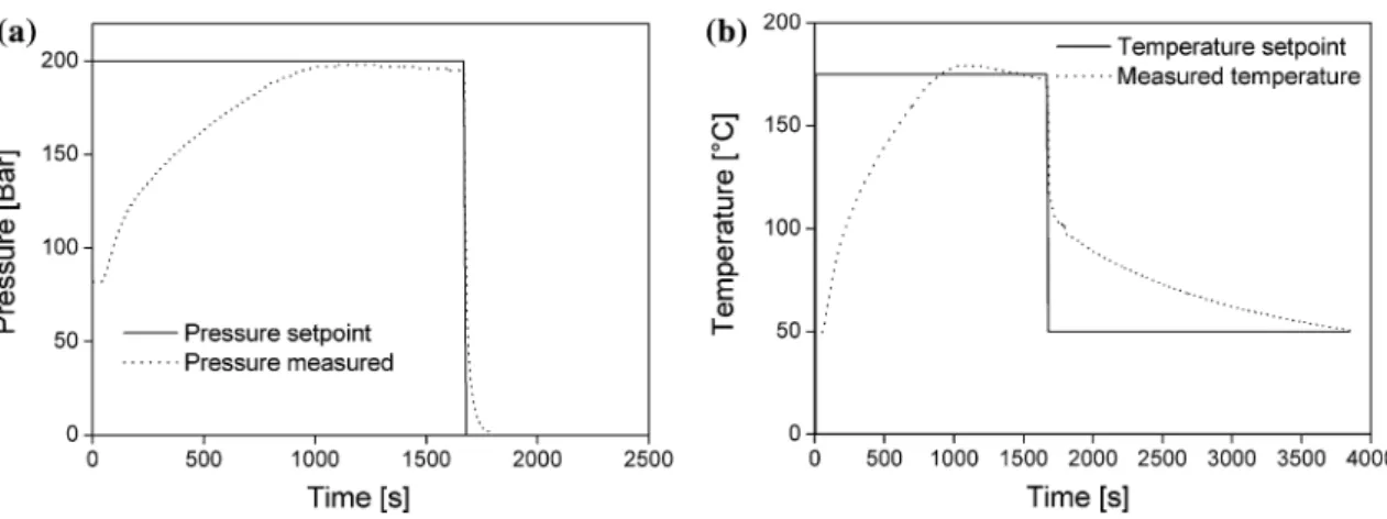
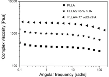
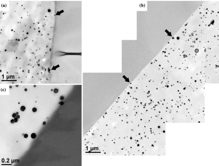
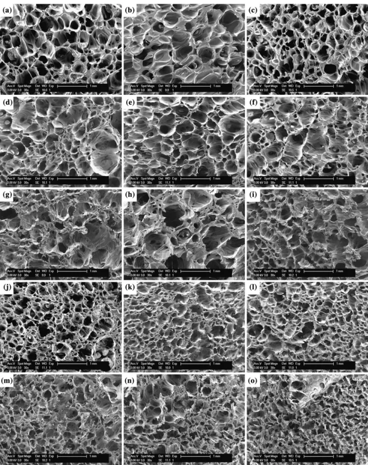
Figure
+6
Documents relatifs