0021-9193/06/$08.00⫹0 doi:10.1128/JB.00334-06
Copyright © 2006, American Society for Microbiology. All Rights Reserved.
PBP5 Complementation of a PBP3 Deficiency in Enterococcus hirae†
S. Leimanis,
1N. Hoyez,
1S. Hubert,
1M. Laschet,
1Eric Sauvage,
1R. Brasseur,
2and J. Coyette
1*
Centre d’Inge´nierie des Prote´ines, Universite´ de Lie`ge, Institut de Chimie, B6, B-4000 Lie`ge, Belgium,1and Centre de Biophysique Mole´culaire Nume´rique, Faculte´ Universitaire des Sciences Agronomiques, Gembloux, Belgium2
Received 7 March 2006/Accepted 9 June 2006
The low susceptibility of enterococci to-lactams is due to the activity of the low-affinity penicillin-binding protein 5 (PBP5). One important feature of PBP5 is its ability to substitute for most, if not all, penicillin-binding proteins when they are inhibited. That substitution activity was analyzed in Enterococcus hirae SL2, a mutant whose pbp5 gene was interrupted by the nisRK genes and whose PBP3 synthesis was submitted to nisin induction. Noninduced SL2 cells were unable to divide except when plasmid-borne pbp5 genes were present, provided that the PBP5 active site was functional. Potential protein-protein interaction sites of the PBP5 N-terminal module were mutagenized by site-directed mutagenesis. The T167-L184region (designated site D)
appeared to be an essential intramolecular site needed for the stability of the protein. Mutations made in the two globular domains present in the N-terminal module indicated that they were needed for the suppletive activity. The P197-N209segment (site E) in one of these domains seemed to be particularly important, as single
and double mutations reduced or almost completely abolished, respectively, the action of PBP5.
Cell morphology in eubacteria is determined mainly by the peptidoglycan layer that is synthesized by a battery of cytoplas-mic and membrane-bound enzymes, among which are theDD -peptidases (26, 42, 61). These -peptidases, commonly desig-nated penicillin-binding proteins (PBPs), are serine-active enzymes that belong to a large family of penicillin-recognizing proteins whose active sites contain three conserved motifs. Generally, the PBPs lose theirDD-peptidase activity by forming
a long-lived covalent bond with penicillins (26).
The PBPs are separated into two large groups on the basis of their sizes. The low-molecular-mass proteins are essentially monomodularDD-peptidases that have been shown to play a
morphogenetic role in Escherichia coli as well as in
Streptococ-cus pneumoniae cells (11, 12, 41, 43, 66). In contrast, the
high-molecular-mass PBPs are essential bimodular enzymes that bind penicillins on their C-terminal module. They are further subdivided into two classes. Class A PBPs have an N-terminal module acting as a glycosyltransferase and are characterized by the presence of five typical conserved amino acid motifs. Class B PBPs possess three conserved amino acid motifs in their N-terminal modules that have no detectable enzymatic activity but seem, however, to be essential for cell morphogenesis (26). Different observations suggest that rod-shaped bacteria pos-sess two morphogenetic systems, one responsible for cell elon-gation and the other responsible for cytokinesis (50, 52). For example, separate inactivation of E. coli class B PBP2 and PBP3 either by-lactam antibiotics or by mutagenesis shows that the first is specifically required for cell elongation and that the second is required for cell division (4, 59). In addition, two protein complexes containing different PBPs could be isolated from Haemophilus influenzae and Bacillus subtilis cells (1, 58).
Consistent with their function, PBP3 of E. coli (38, 63, 65) or PBP2b and PBP1 of Bacillus subtilis (54) localize in the area of developing septa and of completed poles. These PBPs are part of a multiprotein-synthesizing machinery, designated the divi-some (6, 21, 42, 54), whose assembly has been studied by different approaches (16, 31, 38, 45, 63).
The composition of the multiprotein complex needed for cell elongation is not yet well documented, but it is thought to be structurally analogous to the divisome (30). In addition to pbpA, encoding PBP2, other different genes such as rodA (59, 60) and
mreBCD (33, 54, 60) are required for elongation in E. coli.
Morphogenesis of cocci was thought to be relatively simple, with most of the cell wall synthesis taking place at the site of septum formation (25, 28). However, on the basis of the inhibition of cell division by antibiotics or by thermosensitive mutations, it was proposed that they be separated into two groups: those that possess a site for septum formation only and those that in addi-tion have a site for lateral wall elongaaddi-tion that would be less effective than that in rod-shaped cells (35, 52). Staphylococcus
aureus is probably a member of the first group, as cell wall
syn-thesis appears to occur only at the division site (48).
The low susceptibility of enterococci to-lactams is due to the activity of a low-affinity class B PBP, designated PBP5 (23, 64). In the presence of antibiotic concentrations that inhibit all the PBPs except PBP5, enterococci continue to multiply (22). Under these conditions, even in highly resistant strains, PBP5 maintains sufficient transpeptidase activity to synthesize a nor-mal peptidoglycan in Enterococcus faecium and Enterococcus
hirae (7, 55, 67). When grown in the presence of sub-MIC
concentrations, the penicillin-resistant Enterococcus faecalis strain 56R has a slightly less cross-linked peptidoglycan with fewer oligomers but more dimers than the wild type (56). Note that when PBP5 is the only active DD-transpeptidase, these
cells also need the help of an active glycosyltransferase to synthesize peptidoglycan, as class B PBPs were shown to be unable to polymerize glycan strands (26, 49).
Different morphological observations of -lactam-treated enterococcal cells indicate that they should have two cell
wall-* Corresponding author. Mailing address: Centre d’Inge´nierie des Prote´ines, Universite´ de Lie`ge, Institut de Chimie, B6, B-4000 Lie`ge, Belgium. Phone: 32 4 3663399. Fax: 32 4 3663364. E-mail: jcoyette @ulg.ac.be.
† Supplemental material for this article may be found at http://jb .asm.org/.
synthesizing machineries (52). Treatment of E. hirae ATCC 9790 with ampicillin or cephalotin concentrations that have little effect on the increase in cell growth but that result in a relatively strong inhibition of cell division lead to a doubling of average volumes and lengths of the cells during a 60-min pe-riod (29). Under these conditions, on the basis of 50% inhib-itory concentrations, all the PBPs except PBP5 should be sat-urated (9).
Exponential-phase cells of E. hirae ATCC 9790 treated with low concentrations of cefotaxime (ⱕ0.25 ⫻ MIC) form bloated rods in which septa are initiated but not formed as a result of the specific inhibition of class B PBP2 and PBP3 (10). It was also shown that when thermosensitive PBP3 E. hirae mutants constitutively overproduce PBP5, they grow normally at the nonpermissive temperature (8). This again indicates that PBP5 can compensate for the lack of PBP3 that is determined by a gene located in the dcw cluster devoted to cell division (17). Presumably, like in other bacteria (42, 62), when PBP3 is specifically inhibited, the divisome is inactivated, and the cells continue to build their peptidoglycan with their peripheral wall-synthesizing complex. One may suppose that to be able to fulfill its suppletive activity in E. hirae, PBP5 is integrated into the divisome with PBP3 or into a multiprotein biosynthetic machinery that has similar functions.
In this study, we modified E. hirae cells to place PBP3 syn-thesis under the control of the nisAp promoter that is strongly repressed in the “nisin-controlled expression” (NICE) system that was successfully used to analyze the expression of different genes in various gram-positive bacteria (15, 46). Our results indicate that for several hours, noninduced recombinant cells do not produce PBP3, stop dividing, and elongate. However, if PBP5 is synthesized, these cells divide normally in spite of the absence of PBP3. In parallel, examination of the N-terminal-domain sequence of PBP5 with a predictive method that was used to establish the functionality of different regions of E. coli PBP3 and FtsW led to the identification of potential protein-protein interacting segments (24, 38, 47). Some residues in these segments were modified by site-directed mutagenesis. One residue appeared to be crucial to maintain the stability of PBP5, and another seemed to be more specifically required for its suppletive activity in noninduced cells.
MATERIALS AND METHODS
Bacterial strains, media, growth conditions, and MIC determinations.The E.
coli and E. hirae strains used in this work are listed in Table 1. E. coli strains were
grown in Luria broth (LB) at 37°C. E. hirae strains were grown in either M17 medium to prepare electrocompetent cells (51) or brain heart infusion (BHI) broth at 37°C or at 28° and 42°C when homologous recombinants were isolated. Growth curves were established by measuring the absorbance at 550 nm (A550)
or by determining cell numbers on plates at regular intervals. The media were supplemented with ampicillin (100g/ml) (Bristol-Myers Squibb, Brussels, Bel-gium), kanamycin (100g/ml) (Calbiochem, Bierges, Belgium), chloramphenicol (10g/ml), erythromycin (5 or 10 g/ml) (both from Fluka, Bornem, Belgium), and benzylpenicillin (PenG) (0.1g/ml) (Aventis, Lyon, France) as indicated. Other selective additives such as 5-bromo-4-chloro-3-indolyl--D -galactopyrano-side (X-Gal) (40g/ml), isopropyl--D-thiogalactopyranoside (IPTG) (7g/ml) (both from Immunosource, Zoersel-Halle, Belgium), and 5-bromo-4-chloro-3-indolyl-glucuronide (X-Gluc) (0.5 mM) (Research Organics Inc., Cleveland, Ohio) were used or applied to the media as required. MICs were determined in 1 ml of BHI broth in 24-well culture plates, in at least three independent determinations, essentially as previously described (51).
Plasmid constructions.Common E. coli plasmids (Table 1) were used for cloning purposes. Gene disruption or replacement experiments were carried out by using the pER924 shuttle vector as previously described (51).
(i) pDML1615.A 2.2-kb pbp3 fragment was amplified by PCR by using the DNA of ATCC 9790 as a template and primers SELpbp3BamHI and SELpbp3SacI (Table 2). These oligonucleotides were designed to place a BamHI site just upstream of the pbp3 ATG codon and a SacI site just downstream of the stop codon, respectively. The pbp3 fragment was cloned into pUC18, which was used as an intermediate, since for unknown reasons, direct cloning into pNZ8020 was unsuccessful. The pbp3 gene was then transferred into pNZ8020 under the dependence of the nisAp promoter (nisAp-pbp3 fusion) to generate pDML1615. (ii) pDML1621.A 3.6-kb chromosomal DNA fragment of E. hirae SL1, which contained the entire pbp3 gene as well as the upstream mraW and ftsL and down-stream mraY genes, was amplified by PCR by using primers MFPMupBamHI and MFPMlowEcoRI (Table 2) and cloned into BamHI- and EcoRI-digested pUC18 to obtain pDML1616 (see the supplemental material).
By digestion with BamHI and KpnI (the KpnI site is present in the last quarter of pbp3), the pDML1616 insert was reduced to 2.6 kb and cloned into pUC18 to generate pDML1617, where it was mutagenized to introduce an NcoI restriction site at the level of the starting ATG codon of pbp3 (NcoIup and NcoIlow primers) (Table 2) by site-directed mutagenesis. That new plasmid, containing both the mraW and ftsL genes as well as a large part of pbp3 (designated below as pbp3a), was named pDML1618.
In parallel, the 0.8-kb kanamycin resistance cassette (aph3⬘ or Kmrgene) present
in pDG792 was amplified by PCR by using primers KANup-NcoI and KANlow-KpnI (Table 2) (the NcoI site overlapped the Kmrgene starting ATG codon), cloned
into the pGEM-T Easy vector, digested by the NcoI and KpnI enzymes, and finally substituted into pDML1618 for the 1.6-kb pbp3a fragment. The resulting plasmid, pDML1619, was then digested by the BamHI and KpnI restriction enzymes to isolate the 1.8-kb mraW-ftsL-Kmrfragment that was introduced into pDML1616 as
a substitute for the 2.6-kb mraW-ftsL-pbp3a fragment. The resulting plasmid, pDML1620, thus contained the mraW-ftsL-Kmr-pbp3b-mraY genes (pbp3b
corre-sponds to the terminal end of pbp3) that were finally excised by BamHI and EcoRI digestion and cloned into the thermosensitive shuttle vector pER924 to obtain pDML1621 (see the supplemental material).
(iii) pDML1626 and pDML1627 to pDML1633.The 2.6-kb pbp5 insert of pDML546 was cloned into pBR322 at the SalI and EcoRI sites to yield pDML1626. When fused at the EcoRI site of pIL253, it produced the E. coli/E.
hirae shuttle vector pDML1627. Plasmids pDML1628 to pDML1633 were the
result of similar fusions with the different pDML1626 mutants.
(iv) pDML2230 and pDML2231.Two plasmids carrying the pbp5 gene of E.
faecium (pbp5Efm) with its promoter and terminator sequences were constructed with the 2.2-kb PCR fragment amplified from pDML517 with primers ON1 and ON2*, which were designed to introduce the EcoRI and BamHI sites at each end, respectively. That 2.2-kb PCR fragment was cloned into pBR322 to yield pDML1699, which was then fused to pIL253 to generate pDML2330. It was also inserted into vector pUC18del that was obtained after the removal of AatII-XbaI from pUC18. Plasmid pDML1698, thereby obtained, was used to mutagenize the Ser422 residue and then construct the pDML2231 shuttle vector.
Construction of the E. hirae pbp5::nisRK SL1 strain.A 2.4-kb blunt-ended HpaII-PstI DNA fragment that contained the complete nisRK operon was iso-lated from plasmid pNZ9500 and inserted into the blunt-ended SstI site (position 1144 from the pbp5 ATG codon) inside the 1.6-kb pbp5 fragment of pDML1600 to generate pDML539. The 4-kb EcoRI-BamHI pbp5::nisRK fragment isolated from pDML539 was cloned into the thermosensitive pER924 shuttle vector to construct the integrative plasmid pDML1614, which was used to electroporate E.
hirae ATCC 9790 cells and to isolate, as previously described (51), pbp5-negative
double recombinants appearing on plates as penicillin-sensitive pbp5-negative colonies.
The susceptibility of these pbp5-negative clones, verified by measuring the penicillin MIC, as expected, was 40 times higher (MIC⫽ 0.015 g/ml) than that of the parental strain (0.6g/ml) (51). Integration of the nisRK gene cluster into
pbp5 was also verified by PCR by using both pbp5 4546 and pbp5 B4 primers
(Table 2), which hybridized upstream and downstream, respectively, of the in-sertion point in pbp5. The PCR fragments obtained from the recombinants were 2.4 kb longer (4.7 kb) than those obtained from ATCC 9790 (2.3 kb), indicating that the nisRK fragment had been inserted (results not shown). Membranes of a randomly picked pbp5::nisRK recombinant, designated SL1, were prepared, la-beled with [14C]PenG, and compared with those of ATCC 9790 and the
peni-cillin-resistant strain R40, which overproduces PBP5. As expected, SL1 did not synthesize PBP5 (Fig. 1A).
General DNA methods.Standard DNA methodologies were used (2). DNA fragments, PCR products, plasmids, and the genomic DNA of Enterococcus cells
(collected at the end of the exponential phase) were purified as previously reported (36, 51). E. hirae plasmids were isolated from protoplasts prepared from cells at the end of the exponential phase that were lysed for 30 min at 37°C with lysozyme (3 mg/ml) in a solution containing 10 mM Tris, 1 mM EDTA, and 27%
(wt/vol) sucrose (pH 8) and collected by low-speed centrifugation (3,000 rpm for 3 min). Nucleotide sequencing was performed as previously described (51), with Cy5 primers identical to some of those shown in Table 2. Restriction endonucle-ases from Promega (Leiden, The Netherlands), Life Technologies (Merelbeke,
TABLE 1. Strains and plasmids used in this study
Strain or plasmid Genotype and/or phenotypea Source or reference
Strains
E. coli
Top10F⬘ F⬘ 关lacIqTn10 (Tetr)兴 mcrA ⌬(mrr-hsdRMS-mcrBC) 80 lacZ⌬M15 ⌬lacX74 deoR recA1 araD139⌬(ara-leu)7697 galU galK rpsL(Strr) endA1 nupG
Invitrogen
DH5␣ F⫺endA1 hsdR17(rK⫺mK⫹) glnV44 thi-1 recA1 gyrA(NaIr) relA1⌬(lacIZYA-argF)U169 deoR
80dlacZ⌬(lacZ)M15 Invitrogen
E. hirae
ATCC 9790 Wild-type strain (MIC of PenG, 0.6g/ml) ATCCbcollection
R40 Penrmutant derived from ATCC 9790 (MIC of PenG, 60g/ml) 22
SL1 ATCC 9790 pbp5::nisRK (MIC of PenG, 0.015g/ml) This study
SL2 SL1 pbp3 negative/pDML1615 This study
Plasmids
pBR322 E. coli cloning vector Amersham Pharmacia
Biotech pDG792 E. coli vector carrying the 1.5-kb aph3⬘ (Kmr) gene cassette 27
pER924 E. coli/gram-positive shuttle vector (Ts ori⫹) 3
pGEM-T Easy E. coli cloning vector for PCR products Promega
pIL253 Ermrhigh-copy-number gram-positive cloning vector 57
pSL1190 lacZ E. coli phagemid Amersham Pharmacia
Biotech
pUC18 lacZ E. coli cloning vector Amersham Pharmacia
Biotech pNZ8008 CmrpNZ273 carrying the nisAp-gusA reporter gene, gram-positive/gram-negative origin 13
pNZ8020 CmrpNZ8008 without gusA 14
pNZ9500 pUC19ery carrying the 3⬘ end of nisP and nisRK genes of Lactococcus lactis NZ9700 (2.7-kb DNA fragment)
32
pDML517 pBR322 carrying the pbp5Efmgene of E. faecium D63
r(7.7-kb EcoRI fragment) 67
pDML539 pDML1600 carrying the pbp5::nisRK genes This study
pDML540 pBR322 carrying the ftsW psr pbp5 genes of E. hirae R40 (6.9-kb EcoRI fragment) 20 pDML541 pBR322 carrying the pbp5 gene (2.6-kb EcoRI/PvuI fragment derived from pDML540) 40 pDML545 pET22b⫹(Kmr) carrying the truncated NcoI-NotI PCR fragment of pbp5 (94 to 2,036 bp) of E.
hirae R40
44
pDML546 pUC18 carrying the pbp5 gene (2.6-kb EcoRI-SalI fragment derived from pDML541) 40
pDML1600 pUC18 carrying the pbp5 gene (1.6-kb PCR fragment) 51
pDML1614 pER924 carrying the pbp5::nisRK genes (EcoRI/BamHI insert of pDML539) This study
pDML1615 pNZ8020 carrying the nisAp-pbp3 ( pbp3i) gene This study
pDML1616 pUC18 carrying the mraW-ftsL-pbp3-mraY genes (3.6-kb BamHI-EcoRI fragment of ATCC 9790)
This study
pDML1617 pUC18 carrying the mraW-ftsL-pbp3a genes (BamHI-KpnI fragment of pDML1616) This study pDML1618 pDML1617 with an additional NcoI restriction site at the pbp3a starting codon This study
pDML1619 pDML1617 carrying the mraW-ftsL-Kmrgenes This study
pDML1620 pDML1616 carrying the mraW-ftsL-Kmr-pbp3b-mraY fragment This study
pDML1621 pER924 carrying the mraW-ftsL-Kmr-pbp3b-mraY fragment of pDML1621 This study
pDML1623 pUC18 carrying the 270-bp HindIII-PstI pbp5 fragment encoding the D and E sites This study pDML1626 pBR322 carrying the wild-type or mutated pbp5 genes (EcoRI-SalI fragment of pDML541) This study
pDML1627 pIL253/pDML1626 fusion carrying pbp5 This study
pDML1628 pDML1627 carrying the pbp5(K172N/R173N) gene (M1 mutant) This study pDML1629 pDML1627 carrying the pbp5(K199Q/K203Q) gene (M2 mutant) This study
pDML1630 pDML1627 carrying the pbp5(K203Q) gene (M3 mutant) This study
pDML1631 pDML1627 carrying the pbp5(5K-S) gene (M4 mutant) This study
pDML1632 pDML1627 carrying the pbp5(R173N) gene (M5 mutant) This study
pDML1633 pDML1627 carrying the pbp5(K199Q) gene (M6 mutant) This study
pDML1697 pSL1190 carrying the 5⬘-end 390-bp NcoI-SpeI fragment of pbp5 isolated from pDML545 This study pDML1698 pUC18 derived, carrying the pbp5Efmgene (2.2-kb EcoRI-BamHI fragment of E. faecium D63r) This study
pDML1699 pBR322 carrying the pbp5Efmgene (2.2-kb EcoRI-BamHI fragment of E. faecium D63
r) This study
pDML2330 pIL253/pDML1699 fusion carrying pbp5Efm This study
pDML2331 pDML2330 carrying pbp5Efm(S422A) This study
aCmr, chloramphenicol resistant; Ermr, erythromycin resistant; Kmr, kanamycin resistant; Penr, penicillin resistant. bATCC, American Type Culture Collection.
Belgium), and Fermentas (St. Leon-Rot, Germany); calf intestinal alkaline phos-phatase from Roche Diagnostics (Brussels, Belgium); T4 DNA polymerase from Life Technologies (Merelbeke, Belgium); and T4 DNA ligase from Boehringer Ingelheim Bioproducts (Verviers, Belgium) were used as recommended by the suppliers.
PCR.PCR amplifications were performed in a TRIO-Thermoblock Biometra thermocycler (Eurogentec, Lie`ge, Belgium) with primers obtained from either Amersham Pharmacia Biotech or Eurogentec (Table 2). The Taq, Pwo, Vent, and Biotools DNA polymerases from Amersham Pharmacia Biotech, Euro-gentec, New England Biolabs (Westburg, Leusden, The Netherlands), and
Lab-system (Brussels, Belgium), respectively, were used according to the manufac-turers’ recommendations. Amplifications were usually carried out as previously described (51).
Directed mutagenesis.The site-directed mutagenesis of pbp5 was performed with the QuickChange site-directed mutagenesis kit (Stratagene Europe, Am-sterdam, The Netherlands) on a 270-bp fragment encoding the S125-A215peptide
of PBP5 that was excised from pDML541 by a double HindIII and PstI digestion and cloned into pUC18 to generate pDML1623. Primer pairs O5M1a/O5M1b, O5M2a/O5M2b, O5M3a/O5M3b, O5M5a/O5M5b, and O5M6a/O5M6b (Table 2) were used to produce the K172N/R173N (M1 mutant), K199Q/K203Q (M2
mutant), K203Q (M3 mutant), R173N (M5 mutant), and K199Q (M6 mutant)
substitutions, respectively. The resulting plasmids were named pDML1623M1, pDML1623M2, pDML1623M3, pDML1623M5, and pDML1623M6, respectively. The HindIII-PstI fragments carrying the different mutations were then exchanged with the corresponding fragment of pDML1626 to obtain pDML1626M1, pDML1626M2, pDML1626M3, pDML1626M5, and pDML1626M6, respec-tively.
To introduce the five K-to-S substitutions in the N-terminal end of PBP5, a 390-bp NcoI-SpeI fragment was isolated from pDML545 and cloned into pSL1190 to generate pDML1697. The construct was then mutagenized in three successive steps with three pairs of primers, O1K75K76/O1K75K76*, O2K108K109/O2K108K109*, and O3K146/O3K146* (Table 2). The final mu-tated fragment was then exchanged with the corresponding wild-type fragment of pDML541 by a double ScaI-SpeI digestion. The ScaI site is present 60 bp downstream of the NcoI site. Finally, the mutated pbp5 gene was introduced into pDML1626 by exchange of the EcoRI-SalI fragment to generate pDML1626M4. The S422A substitution of PBP5Efmwas made in pDML1698 by site-directed
mutagenesis with the ON6 and ON7* primers. The AccI fragment isolated from the S422A pbp5Efm[ pbp5Efm(S422A)] gene was then exchanged with that of
pDML1699 to generate pDML1699S422A, which was fused to pIL253 at the
EcoRI site to yield pDML2331.
Transformation of E. hirae cells and isolation of recombinants.E. coli
com-petent cells and E. hirae electrocomcom-petent cells were transformed as previously described (2, 51). Gene disruptions or deletions in E. hirae cells were achieved by
TABLE 2. Oligonucleotides used in this study
Oligonucleotideb Sequencea Gene
pbp5 4546 5⬘-TAAGAAAATGTGAGCGGAGGG-3⬘ pbp5
pbp5 B4* 5⬘-TCAGGATTCACAGCAAATAGGAAGC-3⬘ pbp5
SELpbp3BamHI 5⬘-GGGGGATCCAAATGAGTCTTAAAAATA-3⬘ pbp3
SELpbp3Sacl* 5⬘-GAAGAGCTCTCTCTCCCAATTCTATTCTTA-3⬘ pbp3
MFPMupBamHI 5⬘-GATATGCGTATGGATCCATCAGCACCTCTG-3⬘ mraY-ftsL-pbp3-mraW
MFPMlowEcoRI 5⬘-CGTACCAGGATAGAATTCTGAACGATAGAC-3⬘ mraY-ftsL-pbp3-mraW
KANAupNcol 5⬘-GAAATAATCCATGGCTAAAATGAGAATATC-3⬘ aph3⬘
KANAlowKpnl* 5⬘-CTCCTCGGTACCACTAAAACAATTCATCCAGTAAAATATAATA-3⬘ aph3⬘
Ncol-up 5⬘-GAAAGTGAAGTCCATGGGTCTTAAAAATAAATTC-3⬘ pbp3 Ncol-low* 5⬘-GAATTTATTTTTAAGACCCATGGACTTCACTTTC-3⬘ pbp3 O5M1a 5⬘-ACTAGTGAGGAAGCT-AAC-AAC-GGCGATATCCTT-3⬘ pbp5 O5M1b* 5⬘-AAGGATATCGCC-GTT-GTT-AGCTTCCTCACTAGT-3⬘ pbp5 O5M2a 5⬘-ATCGTTCCTAGA-CAG-CTGGGAGAA-CAA-GAAGAGAAAACA-3⬘ pbp5 O5M2b* 5⬘-TGTTTTCTCTTC-TTG-TTCTCCCAG-CTG-TCTAGGAACGAT-3⬘ pbp5 O5M3a 5⬘-AAGCTGGGAGAA-CAA-GAAGAGAAAACA-3⬘ pbp5 O5M3b* 5⬘-TGTTTTCTCTTC-TTG-TTCTCCCAGCTT-3⬘ pbp5 O5M5a 5⬘-GAGGAAGCTAAA-AAC-GGCGATATCCTT-3⬘ pbp5 O5M5b* 5⬘-AAGGATATCGCC-GTT-TTTAGCTTCCTC-3⬘ pbp5 O5M6a 5⬘-ATCGTTCCTAGA-CAG-CTGGGAGAAAAA-3⬘ pbp5 O5M6b* 5⬘-TTTTTCTCCCAG-CTG-TCTAGGAACGAT-3⬘ pbp5 ON6 5⬘-TATGCCCCTGGAGCGACGTTTAAAATGATC-3⬘ pbp5 ON7* 5⬘-GATCATTTTAAACGTCGCTCCAGGGGCATA-3⬘ pbp5 O1K75K76 5⬘-CATTCACAGACTAGCAGCACAATTTCTGAA-3⬘ pbp5 O1K75K76* 5⬘-TTCAGAAATTGTGCTGCTAGTCTGTGAATG-3⬘ pbp5 O2K108K109 5⬘-ATTTAAAAGTGACGAGCAGCGATAGTGAAACCTATT-3⬘ pbp5 O2K108K109* 5⬘-AATAGGTTTCACTATCGCTGCTCGTCACTTTTAAAT-3⬘ pbp5 O3K146 5⬘-AATGATCAGATCAGCATCAACTGGCAG-3⬘ pbp5 O3K146* 5⬘-CTGCCAGTTGATGCTGATCTGATCATT-3⬘ pbp5
UP 5⬘-GTAAAACGACGGCCAGT-3⬘ M13 universal sequencing
RP 5⬘-CAGGAAACAGCTATGAC-3⬘ M13 reverse sequencing
aBoldface type indicates the BamHI, SacI, EcoRI, KpnI, and NcoI restrictions sites, and underlined sequences correspond to modified codons. b*, complementary.
FIG. 1. SDS-PAGE and fluorogram of the membrane-bound PBPs of different E. hirae strains. (A) Membranes were labeled for 60 min with 100M [14C]PenG. Lane 1, purified soluble PBP5; lane 2, R40;
lane 3, ATCC 9790; lane 4, SL1. (B) Membranes (pretreated for 15 min with 10M nonradioactive PenG) were incubated for 60 min with 100M [14C]PenG for specific labeling of PBP5
Eh. Lane 1, purified
soluble PBP5Efm; lane 2, SL1/pbp5Eh; lane 3, SL1/pbp5(K172N/ R173N); lane 4, SL1/pbp5(K199Q/K203Q); lane 5, SL1/pbp5(K203Q);
lane 6, SL1/pbp5(5K-S); lane 7, SL1/pbp5(R173N); lane 8, SL1/
double recombination of electroporated integration vectors as previously de-scribed (5, 51).
PBP detection by radioactive labeling and immunoblotting.PBP profiles were established with cells that were collected at an A550 of 1.0. Preparation of
membranes of the different E. hirae clones, estimation of protein contents, [14
C]PenG labeling, sodium dodecyl sulfate-polyacrylamide gel electrophoresis (SDS-PAGE), and fluorography of PBPs were performed as described elsewhere previously (67). Except when stated otherwise, 200-g protein samples were used. Modification of the fluorography procedure, detection of labeled PBPs, and Western blot analysis with anti-PBP5 rabbit polyclonal antibodies were previously described (51). The truncated soluble PBP5Efmused as a control in
some of these procedures was obtained as previously described (53). -Glucuronidase activity. Nisin was obtained from Sigma as a 2.5% solution in sodium chloride containing denatured milk proteins. Stock solutions of nisin were made by suspending 10 mg of the nisin mixture per ml of 0.05% acetic acid and then diluting the preparation (10-fold) with dimethyl sulfoxide. These stock solutions were stored at⫺20°C. Further dilutions were made with water and were used immediately.
For quantitative-glucuronidase assay, E. hirae cells were grown up to an A550
of 0.5, induced with different nisin concentrations, grown for another 90 min, and harvested. After being resuspended in 50 mM NaHPO4buffer (pH 7) to a final A600of 2.0, these cells were permeabilized and used immediately for the
gluc-uronidase assay in the presence of 100 mM para-nitrophenyl--D-glucuronic acid
(Clontech Laboratories, Inc., Palo Alto, Calif.) as previously described (34). Prediction of interaction sites.In the method used here, a 7- or 11-amino-acid segment is moved along the primary structure of a protein one residue at a time. For the central residue of each segment, a mean hydrophobicity,⬍H⬎, value and a mean hydrophobic moment,⬍⬎, value were calculated. Both values characterizing each residue were plotted (18, 19, 24). It has been determined that different putative sites possess one or several residues for which⬍H⬎ values are smaller than⫺0.5 and ⬍⬎ values are greater than 0.5. Replacements of one residue that in principle do not modify the secondary and tertiary structures may affect the predicted amphiphilicity. Those that have the strongest influence on that property could greatly change the putative interacting capacity of a site.
RESULTS
Testing of the NICE system and the trans expression of pbp5 and pbp3 in E. hirae SL1.To be fully functional, the NICE system requires the presence and activity of the nisRK genes that signal the presence of nisin (the inducer) and activate genes placed under the control of the nisA promoter (nisAp). The two nisRK genes were thus first inserted into the pbp5 gene that was shown to be dispensable in E. hirae ATCC 9790 (51). The thereby constructed PBP5-deficient clone, SL1, was also needed to assess the activity of PBP5 variants.
As expected, in the presence of nisin, SL1/pNZ8008 transfor-mants appeared as blue colonies on plates containing X-Gluc but remained uncolored if noninduced. Induction of the glucuroni-dase activity by the nisAp-gusA gene was also followed by mea-suring the rate of hydrolysis of the p-nitrophenyl--D-glucuronide substrate in permeabilized cells. The activity was detectable at very low concentrations of nisin (10 ng/ml) and presented, as previously shown (34), a good linear dose-response relationship up to concentrations just below the MIC (nisin MIC⫽ 5 g/ml). The activity of noninduced SL1/pNZ8008 cells remained under the limits of detection. The NICE system was thus functional in E.
hirae. It is important that in spite of the presence of nisin (0.25
g/ml), the growth rates of either ATCC 9790 and SL1 or SL1/ pNZ8008 (⬃29-min and ⬃32-min generation times, respectively) were not modified.
Expression of pbp5Eh.The 11-kb pDML1627 shuttle vector
carrying wild-type E. hirae pbp5 ( pbp5Eh) was used to test
the trans expression of pbp5Eh. As shown by SDS-PAGE and
fluorography, radioactively labeled membranes of SL1/ pDML1627 clones designated below as SL1/pbp5Eh, grown in
the presence of erythromycin, synthesized relatively large amounts of active PBP5Eh(Fig. 1B, lane 2). As expected, the
resistance of SL1/pbp5Eh cells to PenG was much increased
(MIC⫽ 20 g/ml) compared to that of the parental SL1 strain. The PBP5Ehproduced in SL1 was thus functional.
Expression of the nisAp-pbp3 fusion.To assess the ability of native and mutant forms of PBP5Ehto substitute for PBP3, we
needed a strain whose PBP3 synthesis could be controlled. We chose to inactivate the chromosomal gene in the presence of the inducible nisAp-pbp3 gene fusion carried on the pDML1615 plas-mid. However, the expression of that fusion had to first be tested.
Induced PBP3 synthesis in SL1/pDML1615 transformants, designated below as SL1/pbp3i, progressively increased with increasing nisin concentrations of up to 2g/ml (Fig. 2). Syn-thesis of PBP3 in noninduced SL1/pbp3i remained weak and was most probably due to the chromosomal gene that is still present. It is important that neither the growth nor the mor-phology appeared to be changed in E. hirae SL1/pbp3i cells producing large amounts of PBP3.
Isolation of SL2, a pbp3-conditional derivative of SL1. In-activation of the chromosomal pbp3 gene was performed in SL1/pbp3i cells transformed with the integrative pDML1621 vector. Nisin (0.5g/ml) and chloramphenicol (10 g/ml) were added to the medium at all steps to produce enough PBP3 in the absence of the chromosomal pbp3 and to maintain pDML1615, respectively. However, no kanamycin-resistant colonies were found. Double pbp3-deficient recombinants could nevertheless be differentiated, as in the absence of nisin, they formed colonies much later (minimum of 2 days) than parental SL1/pbp3i cells. One randomly picked pbp3-deficient SL1/pbp3i recombinant was named SL2.
PCRs were used to verify the integration of the 0.8-kb kana-mycin cassette. It was amplified by using primers KANup-NcoI and KANlow-KpnI (Table 2) from SL2 cells but not from SL1 cells. In addition, PCR fragments obtained from pDML1616 and SL1 (two pbp3 controls) were 0.8 kb longer than those (2.8 kb) obtained from pDML1621 and SL2 when primers MFPMupBamHI and MFPMlowEcoRI (Table 2), which hybridized at both ends of the mraW-ftsL-pbp3-mraY locus, were used (data not shown). That difference was in good accordance with the replacement of the PBP3a frag-ment by the Kmrcassette.
One could explain the lack of kanamycin resistance of the recombinants by either a lack of activity of the Kmrgene or, more
probably, an insufficient expression of the Kmrgene due to a pbp3
promoter that was too weak. As the recombinants grew normally,
FIG. 2. SDS-PAGE and fluorogram of the 77-kDa membrane-bound PBP3 of nisin-induced SL1/pbp3i cells. Lane 1, noninduced cells; lanes 2 to 5, nisin-induced cells (10, 20, 40, and 80 ng/ml, respec-tively). Membranes (500g) were labeled for 10 min with 100 M [14C]PenG. Part of the gel shows PBPs 2 to 4. Under these conditions,
one may conclude that the recombination events had no polar effect on the genes placed downstream of pbp3.
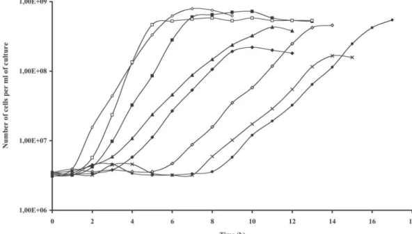
Induced SL2 cells (0.25g of nisin/ml) grown overnight in BHI broth supplemented with chloramphenicol were collected by centrifugation, resuspended in nisin-free medium, and used as inoculum (1%). As can be seen in Fig. 3, after a short lag period (⬍1 h), induced SL2 cells could multiply almost as fast as SL1 cells (⬃37-min and ⬃32-min generation times, respec-tively). Noninduced SL2 cells, unable to synthesize both PBP5 and PBP3, did not divide for about 8 h before they resumed their growth at a slower rate (⬃70-min generation time). In Fig. 3, all the samples were plated in the presence of nisin, but when they were plated onto nisin-free medium, during about the first 7 h of the lag, the cells of the inoculum (⬃5 ⫻ 106
cells/ml) needed at least 5 days to form colonies. The number of cells capable to produce colonies after only 1 day then progressively increased (results not shown). It is important that when subcultured in nisin-free liquid medium, cells collected after regrowth in the absence of nisin grew without going through a long lag period.
Complementation of the PBP3 deficiency in SL2 by PBP5. (i) Complementation by PBP5 of E. hirae.SL2 cells were trans-formed with pDML1627 bearing the pbp5Ehgene to assess the
complementation activity of PBP5 when PBP3 was not pro-duced in noninpro-duced SL2 cells. Chloramphenicol and erythro-mycin were always present in the medium, and noninduced SL2/pDML1627 cells designated below as SL2/pbp5Eh(culture
grown overnight) were used as inoculum (1%). As can be seen in Fig. 3, noninduced and induced SL2/pbp5Ehcells had slightly
longer lag periods than induced SL2 cells. However, induced SL2/pbp5Eh cells multiplied more rapidly than noninduced
SL2/pbp5Eh and induced SL2 cells (⬃27-min and ⬃38-min
generation times, respectively). Thus, by producing PBP5, in-duced (producing PBP3) or noninin-duced (not producing PBP3)
SL2/pbp5Ehcells behaved much more like SL1 and ATCC 9790
cells than like noninduced SL2 (PBP3-deficient) cells. PBP profiles of radioactively labeled membranes of nonin-duced SL2 and noninnonin-duced and innonin-duced SL2/pbp5Ehcells were
compared with those of membranes of the wild-type strain, the PenG-resistant R40 strain, and the SL1 and induced SL2 strains. It is worth noticing that the noninduced SL2 cells, collected during the growth phase that followed the 8-h lag period, produced as much PBP3 as the ATCC 9790, R40, SL1, and SL2/pbp5Ehcells used as controls (Fig. 4A). Obviously, in
those noninduced SL2 cells, expression of the pbp3 gene was, for unknown reasons, made possible again.
It is important that noninduced SL2/pbp5Ehcells produced
PBP5 but not PBP3 (Fig. 4A and B). When nisin was present, the same cells produced PBP3 at the same time as PBP5. In
FIG. 3. Growth curves of SL2 cells (cell counts) transformed with wild-type and mutated pbp5Ehgenes. Mean values of four to six independent
cultures were used. Shown are induced (E) and noninduced (F) SL2, induced (䊐) and noninduced (■) SL2/pbp5Eh, noninduced SL2/pbp5(K199Q/ K203Q) (⫻), SL2/pbp5(K203Q) (〫), SL2/pbp5(5K-S) (}), and SL2/pbp5(K199Q) (Œ).
FIG. 4. SDS-PAGE and fluorogram of the membrane-bound PBP5Eh of E. hirae SL2 cells expressing pbp5Ehvariants. (A)
Mem-branes were labeled for 60 min with 100 M [14C]PenG. Lane 1,
noninduced SL2; lane 2, induced SL2; lane 3, noninduced SL2/pbp5Eh;
lane 4, induced SL2/pbp5Eh. (B) Membranes were labeled for 10 min
with 10M [14C]PenG. Lane 1, noninduced SL2/pbp5
Eh; lane 2,
in-duced SL2/pbp5Eh. Under these conditions, labeling of PBP5Ehis
both cases, the amount of PBP5 was apparently similar. One can conclude that, in contrast to the noninduced SL2 cells collected after the 8-h latent period, in the SL2/pbp5Ehcells,
the nisAp promoter was not activated. One may thus conclude that when PBP5 is produced in these cells, it is able to replace PBP3.
(ii) Complementation by PBP5 of E. faecium.As the PBP5 of
E. faecium (PBP5Efm) was shown to be closely related (80%
identity) to that of E. hirae (PBP5Eh) (67), both proteins were
compared for their complementation activity in noninduced SL2 cells. An S422A PBP5Efm[PBP5Efm(S422A)] mutant,
un-able to bind [14C]PenG and used as a control in binding
ex-periments, was included in the assay to confirm that theDD -peptidase activity was essential for complementation.
Two shuttle plasmids, pDML2330 ( pbp5Efm) and
pDML2331 [pbp5Efm(S422A)], analogous to those described
above to produce PBP5, were used to complement SL2 cells. Production of the PBP5Efm variants was verified by Western
blot analysis (data not shown). Nisin-induced SL2/pbp5Efmand
SL2/pbp5Efm(S422A) cells grew at almost the same rate (
⬃40-min generation time) but slower than induced SL2/pbp5Ehcells
(⬃27-min generation time). PBP5Efm could complement the
PBP3 deficiency but apparently not to the same extent as PBP5Eh. Noninduced SL2/pbp5Efmcells indeed multiplied at a
slower rate (⬃60-min generation time) than SL2/pbp5Ehcells
(⬃38-min generation time). The PBP5Efmmutant synthesized
in those noninduced cells did not help them to recover their division processes. Their growth curves were not different from those of noninduced SL2 cells with an⬃8-h latent phase pre-ceding slow growth.
In conclusion, PBP5Efmis able to rescue cells that do not
synthesize PBP3 but apparently with a lower efficiency than its
ortholog. It is also clear that for that compensatory activity, PBP5Efmneeds to have a functionalDD-peptidase module.
Identification of putative protein-protein interaction sites in the N-terminal module.Because of its very high identity with PBP5Efm(67), PBP5Ehshould have a three-dimensional
struc-ture very similar to that established for PBP5Ef(Fig. 5) (53).
Calculation of⬍H⬎ and ⬍⬎ values based on the primary structure of PBP5Ehled to the identification of six segments
that were positioned in the N-terminal module made of a long and narrow-sheet formed by three strands and bearing two small globular domains (Fig. 5). Site A (H71SQTKKTISEKE
ALEK86 segment), site B (K105VTKKDSETY114 segment),
and site C (L138TNKNDQK146segment) are part of the
glob-ular N-terminal domain extending from residues 39 to 162. Site D (T167SEEAKRGDILDRNGKKL184segment), with the
im-portant E170-I176 and N180-K182 residues, overlaps the first
conserved motif (motif I) characterizing class B PBPs. It ex-tends over a large part of one of the-strands and a turn in close contact to the C-terminal module. Site E (segment P197
RKLGEKEEKTAN209), placed about 15 amino acids
down-stream of motif I, is part of the globular domain at positions 190 to 260 in the center of the N-terminal module. Finally, site F (T306YDKELRGTNGGK318 segment) starts immediately
downstream of the conserved motif III and extends over an-other one of the three-strands.
On the basis of the predictive method, sites D and E ap-peared to be most important, and the K172N and R173N single
and double substitutions were expected to drastically reduce the potential interacting activity of site D. A similar conclusion was reached for the K199Q and K203Q modifications in site E.
Expression of PBP5Ehmutants in E. hirae SL1.The
differ-ent variants of pDML1626, carrying pbp5Eh or the pbp5Eh
FIG. 5. Model presenting the principal putative protein-protein interaction sites identified in the N-terminal module of the PBP5 structure extending from residues 39 to 353 (53). Conserved motifs of class B PBPs are shown as the thickened gray segments (roman numerals). The sequences and the positions of the predicted interaction sites are represented by letters and black segments. The numbers and positions of substituted amino acids are indicated by black circles. Nt and Ct, N and C termini.
mutants, were fused to pIL253 to obtain E. coli/E. hirae shuttle vectors that were named pDML1627 (pbp5Eh), pDML1628
[pbp5(K172N/R173N)], pDML1629 [pbp5(K199Q/K203Q)], pDML1630 [ pbp5(K203Q)], pDML1632 [ pbp5(R173N)], and pDML1633 [ pbp5(K199Q)]. The additional pDML1631 [pbp5(5K-S)] construct included in this study was previously prepared for other purposes (N. Hoyez, unpublished results). It carried a mutated pbp5Ehgene that encoded the five K75S,
K76S, K108S, K109S, and K146S substitutions located in the
three first putative protein-protein interaction sites identified in PBP5Eh(Fig. 5). The effects of these five substitutions on the
amphiphilicity of sites A, B, and C were not estimated by the predictive method.
SL1 cells were transformed separately by these different shuttle vectors and grown in BHI in the presence of erythromycin. Iso-lated membranes of the different SL1 transformants were labeled with [14C]PenG and examined by fluorography and Western
blot-ting. SL1 transformants harboring pDML1629 [pbp5(K199Q/
K203Q)], pDML1630 [pbp5(K203Q)], pDML1631 [pbp5(5K-S)],
and pDML1633 [pbp5(K199Q)] produced as much active PBP5Eh
as SL1/pbp5Ehcells (Fig. 1B). These mutations had apparently no
structural impact on PBP5Eh, as the binding activities appeared to
be normal. In contrast, SL1/pDML1628 and SL1/pDML1632 transformants bearing the pbp5(K172N/R173N) and pbp5 (R173N) mutations, respectively, did not synthesize any PBP5, as it could not be detected by either radioactive labeling or Western blot analysis with anti-PBP5 antibodies. As both plas-mids were reisolated from these transformants and were shown to have the expected sizes and restriction sites, one can con-clude that the lack of PBP5Ehwas not due to visible plasmid
modifications. However, the sequences of these pbp5Eh
mu-tants were not verified.
Complementation of PBP3-deficient SL2 by the mutated PBP5s.The capacity of the different PBP5Ehvariants to
comple-ment the PBP3 deficiency in noninduced SL2 cells was analyzed in several separate experiments by growing the different transfor-mants in liquid medium as described above. The pDML1628 [pbp5(K172N/R173N)] and pDML1632 [pbp5(R173N)] plasmids were not included in these assays. The growth curves were estab-lished by monitoring the absorbance or the cell counts and re-mained similar for each clone in the different experiments. Those shown in Fig. 3 were based on cells counts made on plates con-taining nisin (0.25g/ml).
The four pbp5(K199Q/K203Q), pbp5(K203Q), pbp5(5K-S), and pbp5(K199Q) mutants multiplied at rates similar to that of noninduced SL2 cells (⬃70 min), but they differed greatly in their lag periods. In addition, some of them reached the stationary phase much earlier than the controls. The pbp5 (K199Q) and pbp5(5K-S) cells needed about 3 h to reach the exponential phase. However, the pbp5(5K-S) cells stopped growing earlier than pbp5(K199Q) cells. The pbp5(K203Q) mu-tant and the pbp5(K199Q/K203Q) double mumu-tant had to wait more than 5 h and about 7 h respectively, before they could grow regularly. In addition, pbp5(K199Q/K203Q) cells reached the stationary phase earlier than the pbp5(K203Q) cells.
On the basis of these experiments, one has to conclude that these mutations reduced, sometimes to a large extent, the complementation activity of PBP5Eh.
DISCUSSION
The natural low susceptibility of enterococci to-lactams is attributed to the protective effect of the low-affinity PBP5 that takes over most functions of the other PBPs when they are not produced or inhibited by penicillin (22, 23, 64). This means that when the essential cell division of PBP3 is inhibited by sub-MIC concentrations of PenG and PBP5 is present, the cells continue to divide (22, 23). E. coli and B. subtilis orthologs of E. hirae PBP3 (10, 17) interact with other membrane pro-teins to form the divisome, a multiprotein synthetic machinery (21, 37, 42, 62), through specific protein-protein interactions that were recently analyzed in E. coli (6, 16, 31, 38, 39, 45, 47, 65). For an efficient suppletive activity, the enterococcal PBP5 is probably integrated into the divisome or in a multiprotein complex with similar functions, and it should interact with one or several other proteins there.
The substitution activity of PBP5 was first specifically exam-ined with PBP3-conditional E. hirae mutants that were unable to multiply at the nonpermissive temperature unless PBP5 was produced in large quantities (8). Unfortunately, those mutants were generally obtained by chemical mutagenesis and were unstable (P. Canepari, personal communication).
Determination of the principal structural elements needed for the suppletive activity of PBP5 should be done in a known and stable genetic background. A relatively long genetic engi-neering process was thus undertaken to first insert the nisRK genes by homologous recombination into the dispensable
pbp5Ehgene (SL1 strain) (23, 51) and then inactivate the
es-sential pbp3 gene of E. hirae SL1 in the presence of plasmidic copies of pbp3 placed under the control of the nisA promoter (nisAp-pbp3 gene fusion).
Examination of the SL2 strain thereby obtained revealed that under inducing conditions, it did not behave not differently than the ATCC 9790 and pbp5::nisRK SL1 strains used as controls. As expected, noninduced SL2 cells did not multiply, but after about 8 h, they regained the capacity to multiply at a slower rate than induced cells. That long lag period appeared to be the time they needed to bypass, by a unknown mechanism, the control imposed by the lack of inducer. Although the amount of PBP3 produced after that lag was comparable to that found in control cells, it was insufficient to restore a normal growth rate. The presence of mutants able to produce PBP3 among the SL2 cells of the inoc-ulum is very unlikely. From the beginning and during most of the lag period, all the cells needed at least 5 days to form colonies. This indicated that each cell of the inoculum could survive on the plates and, after a certain time, was capable of multiplying in the absence of the inducer.
The SL2 strain was nonetheless a good tool to assess the complementation activity of PBP5Eh. When PBP5Ehwas
synthe-sized, the noninduced SL2 cells grew normally in spite of the absence of PBP3. They resumed their growth as fast as the control ATCC 9790, SL1, and induced SL2 cells. If they produced native PBP5Efm, noninduced SL2 cells grew almost as if they were
complemented with its PBP5Ehortholog. But if they
synthe-sized the PBP5Efm(S422A) variant, lacking its DD
-transpepti-dase activity, noninduced SL2 cells were not complemented. This confirmed that, as indicated by previous results (8, 22, 23, 51, 55, 64, 67), to complement PBP-deficient cells, PBP5Eh
Predictive methods used here led to the identification of six amphiphilic peptide segments that could act as protein-protein interaction sites in the structure of PBP5 of E. hirae. Like the other low-affinity PBPs forming subgroup B1 PBPs, PBP5Eh
has a 110-amino-acid polypeptide insert that appears as a glob-ular domain at the end of the N-terminal module of PBP5Efm
(Fig. 5) (26, 53). It contains the three first sites, sites A (H71
-K86segment), B (K105-Y114 segment), and C (L138-K146
seg-ment). Site D is a relatively long peptide (T167-L184segment)
that possesses two critical E170-I176and N180-K182subsites. It
overlaps the class B PBP conserved motif I, located on one of the strands of the-sheet connecting the N-terminal globular domain to the C-terminal module. Site E (P197-N209segment)
was positioned between conserved motifs I and II in a region appearing as a globular domain near the center of the N-terminal module. Finally, site F (T306-K318segment) is found
immediately downstream of motif III on another strand of the -sheet (26, 53).
Sites D and E, which presented the strongest interaction potential, were mutagenized to introduce single or double sub-stitutions that were proposed to reduce or suppress protein-protein interactions. Two double mutants, K172N/R173N (M1
mutant) and K199Q/K203Q (M2 mutant), and three single
mu-tants, K203Q (M3 mutant), R173N (M5 mutant), and K199Q
(M6 mutant), were prepared. A sixth mutant (M4 mutant) was added to this list, as it possessed five lysine-to-serine replace-ments in the three sites identified in the globular domain at the N-terminal end of PBP5. K75S and K76S mutations were
intro-duced into site A, K108S and K109S substitutions were made in
site B, and K146S replacement concerned the end of site C.
The N-terminal globular domain.The Lys-to-Ser substitu-tions made in sites A, B, and C [ pbp5(5K-S)] did not seem to interfere much with the folding of the mutated PBP5Ehthat
was overproduced in SL1 cells, as the binding of [14C]PenG
appeared to be similar to that of PBP5Eh. However, these
substitutions modified the complementation activity of the pro-tein, because SL2/pbp5(5K-S) cells presented a longer lag phase and a reduced growth rate and reached the stationary phase at a much lower cell density than SL2/pbp5Ehcells
pro-ducing PBP5Eh. These results tended to indicate that the
glob-ular domain at the extremity of the N-terminal module is impli-cated in the suppletive action of PBP5Eh.
Site D.The single R173N and double K172N/R173N
replace-ments made in site D in motif I had a very drastic impact, as the PBP5Ehvariants were not found in E. coli and E. hirae cells
grown at 37°C. One may suspect that the K172 and R173 residues are essential for the stability of the protein. The E76N
mutation made in conserved motif I of E. coli PBP3 (variant 10) had a milder effect in reducing the thermostability of the protein, although in this case, the hydrophobic moment plot of the I74-L82segment was not modified (38). The information
gained with these mutations are in good agreement with struc-tural observations suggesting that conserved motif I in class B PBPs has a critical stabilization role in the connection between the N-terminal and C-terminal modules (53). However, as both mutants, prepared separately, were not totally resequenced, one cannot totally exclude the presence of mutations that would interfere with their transcription or transduction.
The central globular domain. Our results demonstrated that site E, predicted to be an important interaction site in
the central globular domain, was indeed essential for the compensatory activity of PBP5Eh. Contrary to PBP5Eh, the
PBP5(K199Q/K203Q) variant was unable to complement PBP3-deficient SL2 cells. Lysine 203 seemed to be much more important. Its replacement by a glutamine reduced the com-pensatory activity to a large extent. SL2/pbp5(K203Q) cells were delayed in their growth but not as much as SL2/
pbp5(K199Q/K203Q) and noninduced SL2 cells. Lysine 199
appeared to be of less importance. Its replacement (K199Q) had a lower impact on the complementation activity than the K203Q substitution. Thus, both residues seem to be required for a normal activity of PBP5Eh, and when the two are
re-placed, PBP5Ehloses its suppletive activity.
In conclusion, this work definitively demonstrates that the low-affinity PBP5Ehalone takes over the function of PBP3 in
cell division when E. hirae cells are submitted to -lactam treatment. To carry out that activity, PBP5Eh, and probably
PBP5Efm, needs the two small globular domains that are
con-nected to the-sheet of its N-terminal module and must have a functional C-terminalDD-transpeptidase module.
ACKNOWLEDGMENTS
We are grateful to NIZO Food Research (Ede, The Netherlands) for the generous gift of the different pNZ vectors used in this research. This work was supported in part by the Belgian Program of Inter-university Poles of Attraction (PAI grant P5/33), the Fonds de la Recherche Fondamentale Collective (contract 2.4521.01), the Actions de Recherche Concerte´es (grant 03/08-297), and European Commis-sion grants (COBRA contract no. LSHM-CT-2003-503335 and EUR-INTAFAR contract no. FP6-512138). M.L., S.H., and S.L. were fellows of the Fonds pour la Formation a` la Recherche dans l’Industrie et l’Agriculture (FRIA). R.B. is a research director of the Fonds National de la Recherche Scientifique (FNRS).
REFERENCES
1. Alaedini, A., and R. A. Day. 1999. Identification of two penicillin-binding multienzyme complexes in Haemophilus influenzae. Biochem. Biophys. Res. Commun. 264:191–195.
2. Ausubel, F. M., R. Brant, R. E. Kingston, D. D. Moore, J. G. Seidman, J. A. Smith, and K. Struhl.2001. Current protocols in molecular biology. John Wiley & Sons, New York, N.Y.
3. Bayles, K. W., E. W. Brunskill, J. J. Iandolo, L. L. Hruska, S. Huang, P. A. Pattee, B. K. Smiley, and R. E. Yasbin.1994. A genetic and molecular characterization of the recA gene from Staphylococcus aureus. Gene 147: 13–20.
4. Begg, K. J., and W. D. Donachie. 1985. Cell shape and division in Escherichia
coli: experiments with shape and division mutants. J. Bacteriol. 163:615–622.
5. Biswas, I., A. Gruss, S. D. Ehrlich, and E. Maguin. 1993. High-efficiency gene inactivation and replacement system for gram-positive bacteria. J. Bac-teriol. 175:3628–3635.
6. Buddelmeijer, N., and J. Beckwith. 2002. Assembly of cell division proteins at the E. coli cell center. Curr. Opin. Microbiol. 5:553–557.
7. Canepari, P., M. M. Lleo, R. Fontana, and G. Satta. 1986. In Streptococcus
faecium penicillin-binding protein 5 alone is sufficient for growth at
submaxi-mal but not at maxisubmaxi-mal rate. J. Gen. Microbiol. 132:625–631.
8. Canepari, P., M. M. Lleo, R. Fontana, and G. Satta. 1987. Streptococcus
faecium mutants that are temperature sensitive for cell growth and show
alterations in penicillin-binding proteins. J. Bacteriol. 169:2432–2439. 9. Coyette, J., J. M. Ghuysen, and R. Fontana. 1980. The penicillin-binding
proteins in Streptococcus faecalis ATCC 9790. Eur. J. Biochem. 110:445–456. 10. Coyette, J., A. Somze´, J. J. Biquet, and J. M. Ghuysen. 1983. Function of penicillin-binding protein 3 in Streptococcus faecium, p. 523–529. In R. Hakenbeck, J. V. Ho¨ltje, and H. Labischinski (ed.), The target of peni-cillin. Walter de Gruyter and Co., Berlin, Germany.
11. de Pedro, M. A., K. D. Young, J. V. Ho¨ltje, and H. Schwarz.2003. Branching of Escherichia coli cells arises from multiple sites of inert peptidoglycan. J. Bacteriol. 185:1147–1152.
12. de Pedro, M. A., C. G. Gru¨nfelder, and H. Schwarz.2004. Restricted mobility of cell surface proteins in the polar regions of Escherichia coli. J. Bacteriol. 186:2594–2602.
and W. M. de Vos.1996. Functional analysis of promoters in the nisin gene cluster of Lactococcus lactis. J. Bacteriol. 178:3434–3439.
14. de Ruyter, P. G., O. P. Kuipers, and W. M. de Vos. 1996. Controlled gene expression systems in Lactococcus lactis with the food-grade inducer nisin. Appl. Environ. Microbiol. 62:3662–3667.
15. de Vos, W. M. 1999. Gene expression systems for lactic acid bacteria. Curr. Opin. Microbiol. 2:289–295.
16. Di Lallo, G., M. Fagioli, D. Barionovi, P. Ghelardini, and L. Paolozzi. 2003. Use of a two-hybrid assay to study the assembly of a complex multicompo-nent protein machinery: bacterial septosome differentiation. Microbiology 149:3353–3359.
17. Duez, C., I. Thamm, F. Sapunaric, J. Coyette, and J. M. Ghuysen. 1998. The division and cell wall cluster of Enterococcus hirae S185. DNA Seq. 9:149– 161.
18. Eisenberg, D., R. M. Weiss, and T. C. Terwilliger. 1982. The helical hydro-phobic moment: a measure of amphiphilicity of a helix. Nature 299:371–374. 19. Eisenberg, D., E. Schwarz, M. Komaromy, and R. Wall. 1984. Analysis of membrane and surface protein sequences with the hydrophobic moment plot. J. Mol. Biol. 179:125–142.
20. El Kharroubi, A., P. Jacques, G. Piras, J. Van Beeumen, J. Coyette, and J. M. Ghuysen.1991. The Enterococcus hirae R40 penicillin-binding protein 5 and the methicillin-resistant Staphylococcus aureus penicillin-binding protein 2⬘ are similar. Biochem. J. 280:463–469.
21. Errington, J., R. A. Daniel, and D. J. Scheffers. 2003. Cytokinesis in bacteria. Microbiol. Mol. Biol. Rev. 67:52–65.
22. Fontana, R., R. Cerini, P. Longoni, A. Grossato, and P. Canepari. 1983. Identification of a streptococcal penicillin-binding protein that reacts very slowly with penicillin. J. Bacteriol. 155:1343–1350.
23. Fontana, R., A. Grossato, L. Rossi, Y. R. Chang, and G. Satta. 1985. Tran-sition from resistance to hypersusceptibility to-lactam antibiotics associ-ated with loss of a low-affinity penicillin-binding protein in a Streptococcus
faecium mutant highly resistant to penicillin. Antimicrob. Agents
Che-mother. 28:678–683.
24. Gallet, X., B. Charloteaux, A. Thomas, and R. Brasseur. 2000. A fast method to predict protein interaction sites from sequences. J. Mol. Biol. 302:917– 926.
25. Giesbrecht, P., J. Wecke, and B. Reinicke. 1976. On the morphogenesis of the cell wall of staphylococci. Int. Rev. Cytol. 44:225–318.
26. Goffin, C., and J. M. Ghuysen. 1998. Multimodular penicillin-binding pro-teins: an enigmatic family of orthologs and paralogs. Microbiol. Mol. Biol. Rev. 62:1079–1093.
27. Guerout-Fleury, A. M., K. Shazand, N. Frandsen, and P. Stragier. 1995. Antibiotic-resistance cassettes for Bacillus subtilis. Gene 167:335–336. 28. Higgins, M. L., and G. D. Shockman. 1970. Model of cell wall growth of
Streptococcus faecalis. J. Bacteriol. 101:643–648.
29. Higgins, M. L., M. Ferrero, and L. Daneo-Moore. 1986. Relationship of shape to initiation of new sites of envelope growth in Streptococcus faecium cells treated with-lactam antibiotics. J. Bacteriol. 167:562–569. 30. Ho¨ltje, J.1996. A hypothetical holoenzyme involved in the replication of the
murein sacculus of Escherichia coli. Microbiology 142:1911–1918. 31. Karimova, G., N. Dautin, and D. Ladant. 2005. Interaction network among
Escherichia coli membrane proteins involved in cell division as revealed by
bacterial two-hybrid analysis. J. Bacteriol. 187:2233–2243.
32. Kleerebezem, M., M. M. Beerthuyzen, E. E. Vaughan, W. M. de Vos, and O. P. Kuipers.1997. Controlled gene expression systems for lactic acid bacteria: transferable nisin-inducible expression cassettes for Lactococcus,
Leuconostoc, and Lactobacillus spp. Appl. Environ. Microbiol. 63:4581–4584.
33. Kruse, T., J. Bork-Jensen, and K. Gerdes. 2005. The morphogenetic MreBCD proteins of Escherichia coli form an essential membrane-bound complex. Mol. Microbiol. 55:79–89.
34. Kuipers, O. P., M. M. Beerthuyzen, P. G. de Ruyter, E. Luesink, and W. M. de Vos.1995. Autoregulation of nisin biosynthesis in Lactococcus lactis by signal transduction. J. Biol. Chem. 270:27299–27304.
35. Lleo, M. M., P. Canepari, and G. Satta. 1990. Bacterial cell shape regulation: testing of additional predictions unique to the two-competing-sites models for peptidoglycan assembly and isolation of conditional rod-shaped mutants from some wild-type cocci. J. Bacteriol. 172:3758–3771.
36. Loureiro Dos Santos, A. L., and A. Chopin. 1987. Shotgun cloning in
Strep-tococcus lactis. FEMS Microbiol. Lett. 166:355–360.
37. Margolin, W. 2000. Themes and variations in prokaryotic cell division. FEMS Microbiol. Rev. 24:531–548.
38. Marrec-Fairley, M., A. Piette, X. Gallet, R. Brasseur, H. Hara, C. Fraipont, J. M. Ghuysen, and M. Nguyen-Diste`che.2000. Differential functionalities of amphiphilic peptide segments of the cell-septation penicillin-binding protein 3 of Escherichia coli. Mol. Microbiol. 37:1019–1031.
39. Mercer, K. L., and D. S. Weiss. 2002. The Escherichia coli cell division protein FtsW is required to recruit its cognate transpeptidase, FtsI (PBP3), to the division site. J. Bacteriol. 184:904–912.
40. Mollerach, M. E., P. Partoune, J. Coyette, and J.-M. Ghuysen. 1996. Impor-tance of the E-46–D-160 polypeptide segment of the non-penicillin-binding module for the folding of the low-affinity, multimodular class B penicillin-binding protein 5 of Enterococcus hirae. J. Bacteriol. 178:1774–1775.
41. Morlot, C., M. Noirclerc-Savoye, A. Zapun, O. Dideberg, and T. Vernet. 2004. The DD-carboxypeptidase PBP3 organizes the division process of
Streptococcus pneumoniae. Mol. Microbiol. 51:1641–1648.
42. Nanninga, N. 1998. Morphogenesis of Escherichia coli. Microbiol. Mol. Biol. Rev. 62:110–129.
43. Nilsen, T., A. S. Ghosh, M. B. Goldberg, and K. D. Young. 2004. Branching sites and morphological abnormalities behave as ectopic poles in shape-defective Escherichia coli. Mol. Microbiol. 52:1045–1054.
44. Partoune, P. 1999. PhD thesis. University of Liege, Liege, Belgium. 45. Pastoret, S., C. Fraipont, T. Den Blaauwen, B. Wolf, M. E. G. Aarsman, A.
Piette, A. Thomas, R. Brasseur, and M. Nguyen-Diste`che.2004. Functional analysis of the cell division FtsW of Escherichia coli. J. Bacteriol. 186:8370– 8379.
46. Pavan, S., P. Hols, J. Delcour, M. C. Geoffroy, C. Grangette, M. Kleerebezem, and A. Mercenier.2000. Adaptation of the nisin-controlled expression sys-tem in Lactobacillus plantarum: a tool to study in vivo biological effects. Appl. Environ. Microbiol. 66:4427–4432.
47. Piette, A., C. Fraipont, T. den Blaauwen, M. E. G. Aarsman, S. Pastoret, and M. Nguyen-Diste`che.2004. Structural determinants required to target pen-icillin-binding protein 3 to the septum of Escherichia coli. J. Bacteriol. 186: 6110–6117.
48. Pinho, M. G., and J. Errington. 2003. Dispersed mode of Staphylococcus
aureus cell wall synthesis in the absence of the division machinery. Mol.
Microbiol. 50:871–881.
49. Pinho, M. G., S. R. Filipe, H. de Lancaster, and A. Tomasz. 2001. Comple-mentation of the essential peptidoglycan transpeptidase function of penicil-lin-binding protein 2 (PBP2) by the drug resistance protein PBP2a in
Staph-ylococcus aureus. J. Bacteriol. 183:6525–6531.
50. Popham, D. L., and K. D. Young. 2003. Role of penicillin-binding proteins in bacterial cell morphogenesis. Curr. Opin. Microbiol. 6:594–599.
51. Sapunaric, F., C. Franssen, P. Stefanic, A. Amoroso, O. Dardenne, and J. Coyette.2003. Redefining the role of psr in-lactam resistance and cell autolysis of Enterococcus hirae. J. Bacteriol. 185:5925–5935.
52. Satta, G., R. Fontana, and P. Canepari. 1994. The two-competing site (TCS) model of cell shape regulation in bacteria: the envelope as an integration point for the regulatory circuits of essential physiological events. Adv. Mi-crob. Phys. 36:181–245.
53. Sauvage, E., F. Kerff, E. Fonze´, R. Herman, B. Schoot, J. P. Marquette, Y. Taburet, D. Prevost, J. Dumas, G. Le´onard, P. Stefanic, J. Coyette, and P. Charlier.2002. The 2.4-Å crystal structure of the penicillin-resistant peni-cillin-binding protein PBP5fm from Enterococcus faecium in complex with benzylpenicillin. Cell. Mol. Life Sci. 59:1223–1232.
54. Scheffers, D. J., and M. G. Pinho. 2005. Bacterial cell wall synthesis: new insights from localization studies. Microbiol. Mol. Biol. Rev. 69:585–607. 55. Sifaoui, F., M. Arthur, L. Rice, and L. Gutmann. 2001. Role of
penicillin-binding protein 5 in expression of ampicillin resistance and peptidoglycan structure in Enterococcus faecium. Antimicrob. Agents Chemother. 45:2594– 2597.
56. Signoretto, C., M. Boaretti, and P. Canepari. 1998. Peptidoglycan synthesis by Enterococcus faecalis penicillin-binding protein 5. Arch. Microbiol. 170: 185–190.
57. Simon, D., and A. Chopin. 1988. Construction of a vector plasmid family and its use for molecular cloning in Streptococcus lactis. Biochimie 70:559–566. 58. Simon, M. J., and R. A. Day. 2000. Improved resolution of hydrophobic
penicillin-binding proteins and their covalently linked complexes on a mod-ified C18 reversed phase column. Anal. Lett. 33:861–867.
59. Spratt, B. G. 1975. Distinct penicillin-binding proteins involved in the divi-sion elongation and shape of Escherichia coli K12. Proc. Natl. Acad. Sci. USA 72:2999–3003.
60. Tamaki, S., H. Matsuzawa, and M. Matsuhashi. 1980. Clusters of mrdA and
mrdB genes responsible for the rod shape and mecillinam sensitivity of Escherichia coli. J. Bacteriol. 141:52–57.
61. van Heijenoort, J. 2001. Formation of the glycan chains in the synthesis of bacterial peptidoglycan. Glycobiology 11:25R–30R.
62. Vollmer, W., and J. V. Ho¨ltje.2001. Morphogenesis of Escherichia coli. Curr. Opin. Microbiol. 4:625–633.
63. Weiss, D. S., K. Pogliano, M. Carson, L. M. Guzman, C. Fraipont, M. Nguyen-Diste`che, R. Losick, and J. Beckwith. 1997. Localization of the
Escherichia coli cell division protein FtsI (PBP3) to the division site and cell
pole. Mol. Microbiol. 25:671–681.
64. Williamson, R., C. Le Bougue´nec, L. Gutmann, and T. Horaud. 1985. One or two low affinity penicillin-binding proteins may be responsible for the range of susceptibility of Enterococcus faecium to benzylpenicillin. J. Gen. Micro-biol. 131:1933–1940.
65. Wissel, M. C., and D. S. Weiss. 2004. Genetic analysis of the cell division protein FtsI (PBP3): amino acid substitutions that impair septal localization of FtsI and recruitment of FtsN. J. Bacteriol. 186:490–502.
66. Young, K. D. 2003. Bacterial shape. Mol. Microbiol. 49:571–580. 67. Zorzi, W., X. Y. Zhou, O. Dardenne, J. Lamotte, D. Raze, J. Pierre, L.
Gutmann, and J. Coyette.1996. Structure of the low-affinity penicillin-bind-ing protein 5 PBP5fm in wild-type and highly penicillin-resistant strains of